0022-538X/09/$08.00⫹0 doi:10.1128/JVI.01048-09
Copyright © 2009, American Society for Microbiology. All Rights Reserved.
Mutational Analysis of the Herpes Simplex Virus Type 1 DNA
Packaging Protein UL33
䌤
Frauke Beilstein, Martin R. Higgs,† and Nigel D. Stow*
MRC Virology Unit, University of Glasgow, Church Street, Glasgow G11 5JR, United Kingdom
Received 22 May 2009/Accepted 15 June 2009
The UL33 protein of herpes simplex virus type 1 (HSV-1) is thought to be a component of the terminase complex that mediates the cleavage and packaging of viral DNA. In this study we describe the generation and characterization of a series of 15 UL33 mutants containing insertions of five amino acids located randomly throughout the 130-residue protein. Of these mutants, seven were unable to complement the growth of the UL33-null virusdlUL33 in transient assays and also failed to support the cleavage and packaging of replicated amplicon DNA into capsids. The insertions in these mutants were clustered between residues 51 and 74 and between 104 and 116, within the most highly conserved regions of the protein. The ability of the mutants to interact with the UL28 component of the terminase was assessed in immunoprecipitation and immunofluo-rescence assays. All four mutants with insertions between amino acids 51 and 74 were impaired in this interaction, whereas two of the three mutants in the second region (with insertions at positions 111 and 116) were not affected. These data indicate that the ability of UL33 to interact with UL28 is probably necessary, but not sufficient, to support viral growth and DNA packaging.
During the packaging of the double-stranded DNA genome of herpes simplex virus type 1 (HSV-1), the cleavage of repli-cated concatemeric viral DNA into single-genome lengths is tightly coupled to its insertion into preassembled spherical procapsids. Upon genome insertion, the internal scaffold pro-tein of the procapsid is lost, and the capsid shell angularizes. Genetic analysis has revealed that successful packaging re-quires acis-acting DNA sequence (thea sequence) together with seven proteins, encoded by the UL6, UL15, UL17, UL25, UL28, UL32, and UL33 genes (6, 10). By analogy with double-stranded bacteriophage, the encapsidation of HSV-1 DNA is thought to be mediated by a heteromultimeric terminase en-zyme. It is envisaged that the terminase is involved in the recognition of packaging signals present in the concatemers and the association with procapsids via an interaction with the capsid portal protein. Terminase initiates packaging by cleav-ing at anasequence present between adjacent genomes within concatemers and subsequently provides energy for genome insertion through the hydrolysis of ATP. Packaging is termi-nated by a second cleavage event at the next similarly orien-tated a sequence, resulting in the encapsidation of a unit-length genome.
An accumulating body of evidence suggests that the HSV-1 terminase is comprised of the UL15, UL28, and UL33 gene products. Viruses lacking a functional version of any of these three proteins are unable to initiate DNA packaging, and un-cleaved concatemers and abortive B-capsids (angularized forms containing scaffold but no DNA) accumulate in the nuclei of infected cells (2, 4, 5, 11, 25, 27, 30, 36, 38). Protein
sequence comparisons revealed a distant relationship between UL15 and the large subunit of bacteriophage T4 terminase, gp17, including the presence of Walker A and B box motifs characteristic of ATP binding proteins (13). Subsequent exper-iments demonstrated that point mutations affecting several of the most highly conserved residues abolished the ability of the resulting mutant viruses to cleave and package viral DNA (26, 39). The UL28 component has been reported to interact with the viral DNA packaging signal (3), a property shared with the homologous protein of human cytomegalovirus (CMV), UL56 (9). Furthermore, both UL15 and UL28 are able to interact with UL6 (33, 37), which form a dodecameric portal complex through which DNA is inserted into the capsid (22, 23, 31). Within the terminase complex, strong interactions have previ-ously been reported between UL15 and UL28 and between UL28 and UL33 (1, 7, 17, 19, 34). Evidence also suggests that UL15 and UL33 may be able to interact directly, albeit more weakly than UL28 and UL33 (7, 15). Temperature-sensitive (ts) lesions in UL33 or UL15 reduced both the interaction of the thermolabile protein with the other members of the ter-minase complex and viral growth at the nonpermissive tem-perature (36). Recent evidence suggests that the terminase complex assembles in the cytoplasm and is imported into the nucleus via a mechanism involving a nuclear localization signal within UL15 (35). UL15 is also necessary for the localization of the terminase to nuclear sites of DNA replication and pack-aging (15). At present, the enzymatic activities necessary for DNA packaging have not been demonstrated for either the complex or individual subunits of the HSV-1 terminase.
This study concerns the UL33 protein, which, at 130 resi-dues, is the smallest subunit of the presumptive terminase (7, 27). No specific role in terminase activity has yet been ascribed to UL33, but several possibilities have been proposed including (i) ensuring correct folding or assembly of the complex, (ii) regulating the functions of the other subunits, (iii) performing an essential enzymatic role per se, and (iv) ensuring correct
* Corresponding author. Mailing address: MRC Virology Unit, Uni-versity of Glasgow, Church Street, Glasgow G11 5JR, United King-dom. Phone: 44 (0) 141 330 4640. Fax: 44 (0) 141 330 2236. E-mail: [email protected].
† Present address: Department of Virology, Hoˆpital Henri Mondor, 51 avenue du Mare´chal de Lattre de Tassigny, 94010 Cre´teil, France.
䌤Published ahead of print on 24 June 2009.
8938
on November 8, 2019 by guest
http://jvi.asm.org/
UL28.
MATERIALS AND METHODS
Cells.Baby hamster kidney 21 clone 13 (BHK) cells andSpodoptera frugiperda strain IPLB-21 (Sf) cells were grown as previously described (1). The HSV-1 UL33 null mutantdlUL33 (originally referred to as UL33⫺) was propagated on the BHK cell-derived complementing cell line A20 (12, 15). Following transfec-tion or infectransfec-tion, BHK cells were maintained in Glasgow minimal essential medium containing 5% newborn calf serum, 100 units of penicillin, and 100g streptomycin per ml (EC5).
Plasmids. The vector pAT153 and its derivate pSA1, which contains the HSV-1 oriSDNA replication origin and a minimal DNA packaging signal, have
been described previously (16, 32). Nucleotides 69110 to 69576 of the HSV-1 genome, containing the UL33 gene, were amplified by PCR and inserted into the BamHI site of the mammalian expression vector pCMV10 (29) to generate pUL33, in which UL33 is expressed under the control of the human CMV major immediate-early promoter. UL33 insertion mutants were generated using the bacteriophage Mu transposon-based Mutation Generation System (Finnzymes). Briefly, after in vitro transposition, pUL33 clones containing the “entrancepo-son” encoding chloramphenicol resistance (M1-Camr
; Finnzymes) were selected. Plasmid DNA was isolated, and clones containing insertions within the UL33 gene were identified by digestion with BamHI. The transposon was removed by digestion with NotI and subsequent religation, and the products were used to transform electrocompetentEscherichia colistrain DH10 cells. This procedure results in the net insertion of 15 bp into the target gene. The exact positions of these insertions and the effects on the encoded protein were determined by DNA sequencing.
The pCMV10 derivative pUL28-c-Myc, encoding a functional c-Myc-tagged UL28 protein (UL28-Myc), has been previously described (33). Plasmid pUL28GFP, encoding UL28 tagged at its N terminus with green fluorescent protein (UL28-GFP), was made by inserting the EcoRI-BamHI fragment con-taining the UL28 gene from pUL28 (1) between the corresponding sites of pEGFP-C1 (Clontech). The fusion protein retained functionality as assessed by the ability of pUL28GFP to complement the growth of the UL28 null mutant virus, gCB (30).
Recombinant baculoviruses.Recombinant baculoviruses expressing wild-type (wt) and mutated UL33 proteins under the control of the polyhedrin promoter were created using the Bac-to-Bac baculovirus expression system (Invitrogen) following the manufacturer’s protocol. Briefly, UL33-containing fragments were transferred from pCMV10 into the transfer plasmid pFastBac1, and the resulting plasmids were transformed into DH10BacE. colicells, which harbor a cloned baculovirus genome (bacmid). White colonies, in which transposition of the UL33 fragment into the polyhedrin locus had occurred, were identified on selective agar plates containing X-Gal (5-bromo-4-chloro-3-indolyl- -D-galacto-pyranoside). The resulting recombinant bacmid DNAs were isolated and trans-fected into Sf cells. Progeny baculoviruses were screened for UL33 expression, and their identities were confirmed by PCR and DNA sequencing prior to the preparation of high-titer stocks. The parental virus used for the construction of the AcUL28Myc baculovirus was AcPAK6 (8). The fragment from pUL28-c-Myc (33) encoding the tagged protein was cloned into the SmaI site of the transfer vector pAcCL29-1 (20) in the correct orientation downstream of the polyhedrin gene promoter. Recombinants were isolated through recombination with Bsu36I-cleaved AcPAK6 DNA in cotransfected Sf cells essentially as described by Kitts et al. (18).
Antibodies.Rabbit polyclonal antiserum R148, reactive with UL33, was de-scribed previously (15). Mouse monoclonal antibody 9E10 (Sigma) was used to detect UL28-Myc. The secondary antibodies used were Alexa Fluor 555-conju-gated goat anti-rabbit immunoglobulin G (IgG) (Molecular Probes), horseradish peroxidase-conjugated goat anti-mouse IgG (Sigma), and horseradish peroxi-dase-conjugated goat anti-rabbit IgG (Sigma).
monolayer were resuspended in Tris-buffered saline and divided into two equal samples which were used to prepare total and encapsidated (DNase-resistant) DNA. Samples of DNA were digested with EcoRI and DpnI (or PstI and DpnI), and the resulting fragments were resolved by agarose gel electrophoresis and transferred to a Hybond-XL membrane (Amersham). Replicated (DpnI resis-tant) pSA1 was detected by hybridization to a probe prepared from32
P-labeled pAT153.
Immunoprecipitation.Immunoprecipitation experiments were carried out as described previously (33). Monolayers of Sf cells were infected with 3 PFU per cell of the appropriate recombinant baculoviruses. At 48 h postinfection the cells were lysed in EZ buffer (100 mM Tris-HCl, pH 8, 100 mM KCl, 0.5% sodium deoxycholate, 10M zinc acetate, 1% NP-40, 10% glycerol), and insoluble protein was removed by centrifugation at 30,000 rpm for 20 min at 4°C in a Beckman TLA100.2 rotor. Soluble extracts were incubated with the appropriate antiserum, and immune complexes were collected on protein A-Sepharose (Sigma). After extensive washing, the proteins were resolved on 10% or 15% sodium dodecyl sulfate-polyacrylamide gels and transferred electrophoretically to Hybond ECL membrane (Amersham). Blots were blocked for 30 min with 5% dried milk powder in 20 mM Tris-HCl, pH 8, 0.15 M NaCl, and 1% Tween 20 (TBS-Tween) and incubated overnight at 4°C with appropriate antibodies di-luted in TBS-Tween containing 1% dried milk. Immunodetection was by en-hanced chemiluminescence using horseradish peroxidase-conjugated secondary antibodies.
Immunofluorescence.Glass coverslips (22-mm diameter) were seeded with BHK cells 1 day before use. Cells were transfected with 0.5g of the indicated plasmid by lipofection (Lipofectamine 2000, Invitrogen) for 24 h, fixed with 4% paraformaldehyde for 10 min at room temperature, and, after two washes with phosphate-buffered saline (PBS), permeabilized with 0.1% Triton X-100 in PBS for 10 min at room temperature and blocked with 10% human serum for 10 min. After incubation with the appropriate primary antibody for 1 h at room temper-ature, the coverslips were washed twice with PBS and incubated with the sec-ondary antibody for an additional hour. After two further washes with PBS, they were mounted in 2.5% 1,4-diazabicyclo-2,2,2-octane (Sigma) in Mowiol (Harco) containing 1 g/ml 4⬘,6-diamidino-2-phenylindole dihydrochloride (DAPI; Sigma). The samples were examined using a Zeiss LSM 510 Meta confocal microscope.
RESULTS
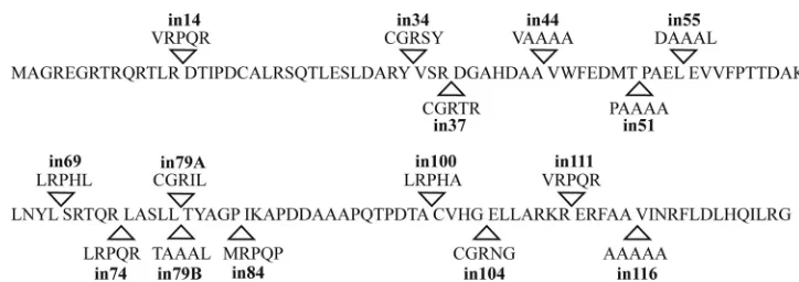
Generation of UL33 insertional mutants. Following trans-poson-mediated mutagenesis of pUL33, 15 clonally distinct insertion mutants were obtained. These encode polypeptides with insertions of five amino acids at 14 separate sites distrib-uted throughout the protein. The names of the mutants and the sites and sequences of the insertions are documented in Fig. 1. Two mutants, in79A and in79B, contained 15-bp inser-tions within the same codon but differed in the identity of the inserted amino acids. Western blotting and immunofluores-cence experiments demonstrated that following transfection each of the mutated plasmids expressed comparable levels of UL33 to the parental plasmid, pUL33 (data not shown).
Transient complementation ofdlUL33.To analyze the effect of the mutations on viral replication, the mutated plasmids were tested for their ability to complement the HSV-1 UL33 null mutant, dlUL33. Nonpermissive BHK cells were trans-fected with plasmids expressing either wt or mutated UL33
on November 8, 2019 by guest
http://jvi.asm.org/
proteins, incubated at 37°C for 24 h, and infected with 5 PFU ofdlUL33 per cell. The plates were harvested, and viral yields were determined on permissive 20A cells. Figure 2 shows the yield obtained for each insertion mutant in comparison with wt and empty vector controls.
Plasmid pUL33 encoding the wt protein supported an al-most 10,000-fold increase in replication compared to cells that received either no plasmid or the vector pCMV10. Seven of the 15 insertion mutants (in34, in37, in44, in79A, in79B, in84, and in100) were able to complementdlUL33 growth to an extent similar to that observed with pUL33. An eighth mutant (in14) also complementeddlUL33 but with an approximately 10-fold reduced efficiency. The remaining seven mutants (in51, in55, in69, in74, in104, in111, and in116) were all seriously impaired in their ability to complementdlUL33, replication, with virus yields being reduced more than 1,000-fold in comparison to pUL33.
These data (Fig. 2) identify four regions of UL33. Two regions (amino acids 14 to 44 and 79 to 100) appear quite
tolerant of insertions of five amino acids, whereas insertions within the other two regions (amino acids 51 to 74 and 104 to 116) significantly impair an essential function of the protein.
Ability of mutants to support DNA packaging.The mutants were next tested for their ability to support DNA packaging in a transient amplicon packaging assay (1, 16). BHK cells were cotransfected with the amplicon pSA1 and plasmids encoding wt or mutated UL33 proteins or the empty vector pCMV10. Helper functions were provided by superinfection with
dlUL33. Packaged (DNase resistant) and total DNA samples were prepared and digested with EcoRI and DpnI, and repli-cated amplicon was detected by Southern blotting and hybrid-ization.
[image:3.585.107.469.64.192.2]Figure 3a shows that, as expected, pSA1 was replicated at similar levels in each instance. In agreement with the known packaging defect of UL33 mutants (4, 36), packaged amplicon was not detectable in cells cotransfected with pSA1 and the vector pCMV10 (Fig. 3b, lane 1). In contrast, efficient DNA packaging was supported in the presence of pUL33 encoding
[image:3.585.136.450.480.677.2]FIG. 1. Position and identity of UL33 insertion mutants. The amino acid sequence of HSV-1 UL33 (GenBank accession no. ACM62256) is shown with arrowheads indicating the sites of insertions. The mutants are named according to the amino acid preceding the insertion site, and the five amino acids inserted at each position are indicated.
FIG. 2. Ability of mutated UL33 proteins to support viral growth. BHK cells were transfected with no plasmid (⫺), empty vector pCMV10 (ⴱ), or pCMV10 derivatives expressing wt UL33 (wt) or UL33 insertion mutants as indicated (14, in14; other designations follow the same pattern). The cells were superinfected for 24 h withdlUL33 virus, and the titer of progeny virus was determined on complementing 20A cells. The graph shows the mean (⫾standard deviation) of the titers obtained in three independent experiments. The horizontal bars group the mutants into four regions according to whether the ability to complementdlUL33 was lost (regions 2 and 4) or retained (regions 1 and 3).
on November 8, 2019 by guest
http://jvi.asm.org/
the wt protein (lane 2). The ability of the plasmids encoding mutated UL33 proteins to complement the DNA packaging defect of dlUL33 exactly paralleled their ability to support virus growth (Fig. 2). Plasmids expressing the wt, in14, in34, in37, in44, in79A, in79B, in84, or in100 proteins all allowed DNA packaging to proceed (lanes 2 to 6 and 11 to 14), al-though with slightly reduced efficiency in the case of in14. In contrast, DNA packaging was undetectable in cells that re-ceived the remaining UL33 insertion mutants (lanes 7 to 10 and 15 to 17).
It has been reported that certain mutations or inhibitory compounds may result in a phenotype in which herpesvirus DNA is cleaved and packaged but is then either lost from capsids or retained in capsids that do not protect the DNA from digestion with DNase. Under such circumstances termi-nal fragments produced by cleavage at the packaging sigtermi-nal can be observed in samples of total DNA in the absence of detect-able DNase-resistant DNA (21, 24). Previous studies demon-strated that UL33 ts mutants fail to generate terminal frag-ments at the nonpermissive temperature, indicating that they are defective in the initiation of the cleavage-packaging pro-cess (4, 36). To determine whetherdlUL33 and the nonfunc-tional mutants were similarly impaired, samples of total DNA were also analyzed following digestion with PstI and DpnI. In contrast to EcoRI, PstI cleaves pSA1 at a site some distance from the packaging signal, generating terminal fragments that can be resolved from monomeric units (4.4 kbp) in an agarose gel. The larger terminal fragment (3.5 kbp) contains sequences required for initiating the cleavage of concatemeric DNA and corresponds to the end that is inserted first into the capsid (16, 28). Figure 3c shows the presence of a major band correspond-ing to pSA1 monomers in all the samples. Because only a small minority of packaging sites are cleaved during the encapsida-tion of replicated amplicon molecules, the 3.5-kbp terminal fragment, where present, is in much lower abundance than the monomer. The terminal fragment is not apparent in cells in-fected with dlUL33 in the absence of any UL33-expressing plasmid (Fig. 3c, lane 1), confirming that the null mutant can-not initiate packaging. As expected, all of the UL33 plasmids
that supported the generation of DNase-resistant DNA gave rise to detectable terminal fragment, and again in14 exhibited a slightly reduced efficiency (lanes 2 to 6 and 11 to 14). The remaining mutants, all of which are impaired in DNA encap-sidation, are additionally deficient in the generation of the terminal fragment (lanes 7 to 10 and 15 to 17), indicating that each is affected in the initiation of the cleavage-packaging process.
Coimmunoprecipitation of mutated UL33 proteins with UL28.Several previous studies have identified a direct inter-action between UL33 and UL28 that appears necessary for DNA packaging (7, 17, 35, 36). It was conceivable, therefore, that the failure of certain mutants to support DNA packaging might result from an impaired ability to interact with UL28.
[image:4.585.112.481.71.224.2]Soluble extracts were prepared from Sf cells coinfected with recombinant baculoviruses expressing wt (AcUL33 virus) or mutated (Acin14 to Acin116) forms of UL33 and with AcUL28Myc, expressing a c-Myc-tagged form of UL28. Pro-teins were precipitated with the anti-UL33 antibody R148 and analyzed by sodium dodecyl sulfate-polyacrylamide gel elec-trophoresis and Western blotting. The results are shown in Fig. 4. Panel c shows that UL28-Myc and UL33 were detected in the starting cell extracts as expected. Following immunopre-cipitation of the extracts with R148, UL28-Myc was not de-tected in cells infected with AcUL33 or AcUL28Myc alone but was readily detected in cells coinfected with the two viruses (Fig. 4a, lanes 1 to 3) demonstrating that precipitation of UL28-Myc by R148 is specifically dependent upon the pres-ence of UL33. Ten of the 15 insertion mutants (in14, in34, in37, in44, in79A, in79B, in84, in100, in111, and in116) simi-larly facilitated coprecipitation of UL28-Myc by R148, whereas the five remaining mutants (in51, in55, in69, in74, and in104) failed to do so. To exclude the possibility that the failure of these mutated proteins to coprecipitate UL28-Myc was be-cause they could not be recognized by R148, the immunopre-cipitates were also examined for the presence of UL33-related proteins. As shown in Fig. 4b, all of the mutated UL33 proteins were precipitated by R148. The data therefore indicate that the five mutants are specifically impaired in their ability to interact
FIG. 3. Ability of mutated UL33 proteins to support packaging of amplicon DNA. BHK cells were transfected with the empty vector, pCMV10 (ⴱ), or pCMV10 derivatives expressing wild-type UL33 (wt) or UL33 insertion mutants as indicated (14, in14; other designations follow the same pattern), and superinfected withdlUL33. At 20 h postinfection total (a and c) and DNase-resistant (b) DNAs were prepared, digested with EcoRI and DpnI (a and b) or PstI and DpnI (c), and analyzed as described in Materials and Methods. M indicates the positions of pSA1 monomers (4.4 kbp), and T indicates the position of the 3.5 kbp terminal fragment.
on November 8, 2019 by guest
http://jvi.asm.org/
with UL28. Comparison with the results from the complemen-tation assays indicates that all of the mutants that support growth and DNA packaging interact with UL28 and that those mutants unable to interact fail to complement. Interestingly, two of the mutants which did not support growth and DNA packaging (in111 and in116) retained the ability to interact with UL28, and these both had insertions near the C terminus of the protein.
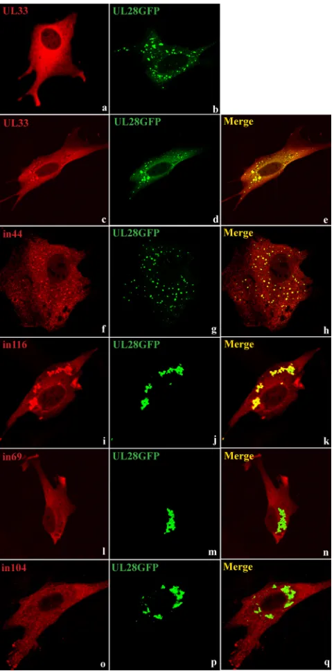
Immunofluorescence assay for the interaction of UL33 and UL28-GFP.Recent data suggest that UL33 and UL28 initially interact in the cytoplasm and that the complete terminase complex is transported into the nucleus by a mechanism utilizing a nuclear localization signal within UL15 (35). We therefore sought to determine whether the results of the im-munoprecipitation experiments could be supported by immu-nofluorescence data.
Coverslips were seeded with BHK cells and cotransfected with plasmids expressing wt or mutated forms of UL33 and a GFP-tagged version of UL28. After permeabilization, UL33 was detected by incubation with R148 and an Alexa Fluor 555-conjugated goat anti-rabbit IgG antibody, and its location relative to the fluorescent UL28 protein was determined by confocal microscopy. Representative images are shown in Fig. 5. Panels a and b show the patterns observed in cells expressing wt UL33 or UL28-GFP alone. UL33 is dispersed throughout the cytoplasm, whereas the tagged UL28 protein tends to be present predominantly in distinct cytoplasmic foci, probably due to its poor solubility. Each of the UL33 mutants exhibited a diffuse staining pattern similar to wt UL33 when expressed alone (data not shown). In cells cotransfected with pUL33 and pUL28GFP, a portion of the UL33 protein was redistributed into foci which colocalized with the UL28 foci, suggesting an interaction between the two proteins (Fig. 5c to e). When the plasmids encoding the mutated forms of UL33 were examined in the presence of pUL28GFP, two distinct phenotypes were observed. In the first, the behavior of the mutated UL33 teins was essentially indistinguishable from that of the wt pro-tein, and foci containing colocalized UL33 and UL28-GFP were readily apparent. This behavior was exhibited by all 10 mutants that coprecipitated with UL28 (i.e., in14, in34, in37,
in44, in 79A, in79B, in84, in100, in111, and in116). Typical data for two mutants belonging to this group, in44 and in116, are shown in Fig. 5f to h and i to k, respectively. In contrast, the remaining five mutants (in51, in55, in69, in74, and in104) ex-hibited diffuse cytoplasmic staining and no obvious colocaliza-tion of UL33 with the UL28 foci. Fig. 5l to n and o to q show data for in69 and in104, respectively, as examples of this group. The ability of the mutated UL33 proteins to be relocalized into foci containing UL28-GFP in BHK cells therefore correlates completely with their ability to be coprecipitated with UL28 from insect cells infected with recombinant baculoviruses. Taken together, the data provide compelling evidence that the 5-amino-acid insertions in in51, in55, in69, in74, and in104 disrupt the interactions of the mutated proteins with UL28.
DISCUSSION
As described in the introduction, previous studies have sug-gested that the UL33 protein is probably an integral compo-nent of a terminase enzyme that mediates the cleavage and packaging of HSV-1 DNA. Here, we report the generation and characterization of a panel of 15 UL33 mutants containing 5-amino-acid insertions following transposon-mediated mu-tagenesis. Insertion of the transposon occurred within 14 sep-arate codons scattered throughout the protein. The two mu-tants affected in the same codon (in79A and in79B) specified distinct polypeptides which behaved indistinguishably in the various assays.
Seven mutants (in51, in55, in69, in74, in104, in111, and in116) were unable to complement the growth of the UL33-null virusdlUL33 in transient assays and also failed to support cleavage and packaging of replicated amplicon DNA (Fig. 2 and 3). In contrast, seven of the remaining mutants (in34, in37, in44, in79A, in79B, in84, and in100) functioned with efficiency similar to that of the wt UL33 in these assays, while the final mutant, in14, exhibited a small impairment. These results pro-vide further confirmation that UL33 plays a key role in the initiation of viral DNA encapsidation and argue against its being independently essential at a later stage of virion assem-bly (e.g., for completion of DNA packaging or during
post-FIG. 4. Coimmunoprecipitation of UL28-Myc with mutated forms of UL33. Sf cells were infected with baculoviruses expressing wild-type UL33 (wt) or UL33 insertion mutants as indicated in the top line (14, in14; other designations follow the same pattern) in either the absence (⫺) or presence (⫹) of AcUL28Myc (second line). Cell extracts were prepared and immunoprecipitated (IP) with R148 and analyzed by Western blotting (WB) with 9E10 (Myc) or R148 antibodies (a and b, respectively). Cell extracts were also screened for the presence of UL28-Myc or UL33 proteins by probing Western blots with antibodies 9E10 (Myc) or R148, respectively (c). The positions of UL28-Myc, UL33, and Ig are indicated.
on November 8, 2019 by guest
http://jvi.asm.org/
packaging maturation), in which case a class of mutant capable of cleaving and packaging replicated DNA but not supporting growth of dlUL33 might be expected. Such a phenotype is exemplified by null mutants with lesions affecting UL25, a protein required for the stable completion of DNA packaging (21, 28).
The HSV-1 UL33 protein belongs to the set of
approxi-mately 40 core genes that are conserved throughout the
Her-pesviridae(14). We performed amino acid sequence alignments
for the HSV-1 UL33 protein and homologues encoded by 17 members of the subfamily Alphaherpesvirinae present in the RefSeq database, and a similarity trace was constructed using PlotSimilarity (GCG). The positions of the insertion mutations and of alpha helices predicted by PSIPRED (http://bioinf.cs .ucl.ac.uk/psipred/) are superimposed on this trace in Fig. 6.
Based upon the similarity plot, UL33 can be broadly divided into four regions. Residues 46 to 77 and 101 to 130 (regions 2 and 4) represent the most-well-conserved portions of the pro-tein and are similarly prominent when alignments of UL33 homologues encoded by all three subfamilies are performed (data not shown). Regions 1 and 3, comprising residues 1 to 45 and 78 to 100, respectively, exhibit lower sequence similarity, which drops to baseline levels in region 1 when sequences from beta- and gammaherpesviruses are included in the alignment (data not shown). It is apparent that the disabling mutations tend to lie within regions of UL33 displaying greatest sequence similarity and that the tolerated mutations affect less-well-conserved regions. The two most-highly-less-well-conserved regions are predicted to have a predominantly␣-helical secondary struc-ture including two long helices (residues 61 to 81 and 102 to 128) which contain five of the seven insertions that abolish function. Previously described ts mutants ts1233 and ts8-22 contain amino acid substitutions at positions 17 and 61, respec-tively (4, 36). The mutation in ts1233 is located only three codons from the insertion in the slightly impaired mutant in14 but affects one of the most conserved residues within region 1, while residue 61 lies in conserved region 2 and is flanked by the sites of four disabling insertions. Further sequence compari-sons and structural predications failed to identify any obvious counterpart of UL33 outside the herpesviruses.
[image:6.585.303.540.67.215.2]Two independent approaches were used to investigate the interaction of mutated forms of UL33 with UL28. In an im-munoprecipitation assay 10 UL33 insertion mutants (in14,
FIG. 5. Immunofluorescence assay of mutated forms of UL33 and UL28-GFP. BHK cells were transfected with pUL33 or pUL28GFP alone (a and b, respectively) or cotransfected with pUL28GFP (ex-pressing a GFP-tagged version of UL28) and plasmids ex(ex-pressing wt (UL33) or UL33 insertion mutants in44, in116, in69, or in104, as indicated (c to q). UL33 was detected with R148 and Alexa Fluor 555-conjugated goat anti-rabbit IgG (red), and UL28 was detected by virtue of its fluorescent tag (green).
FIG. 6. Features of the UL33 protein. The graph shows the Plot-Similarity trace (window: 4) for the aligned sequences of UL33 homo-logues encoded by 18 alphaherpesviruses, and the similarity values are indicated on the vertical axis. Regions of relatively high (2 and 4) or low (1 and 3) sequence similarity are indicated at the top, with pre-dicted␣-helices (PSIPRED) shown by gray bars. The horizontal axis indicates amino acid positions in the HSV-1 protein, and the sites of the insertion mutations are marked by arrows. Open arrows indicate mutants able to complementdlUL33, and filled arrows indicate mu-tants that fail to complement.
on November 8, 2019 by guest
http://jvi.asm.org/
[image:6.585.44.285.68.552.2]in34, in37, in44, in79A, in79B, in84, in100, in111, and in116) retained the ability to interact with the terminase component UL28. In addition to the mutants able to support viral growth and DNA packaging, this group includes two nonfunctional mutants, in111 and in116. All of the remaining five noncomple-menting mutants (in51, in55, in69, in74, and in104) contained insertions that abolished the interaction between the two pro-teins. These findings were supported by the results of an im-munofluorescence assay, in which it was demonstrated that the presence of a GFP-tagged version of UL28 influenced the intracellular distribution of the former but not the latter group of mutated UL33 proteins. Although the interacting UL33 proteins were directed to cytoplasmic foci where they colocal-ized with UL28, it is not known whether these structures are representative of events occurring during normal assembly of the terminase holoenzyme when UL15 is also present. Our results therefore suggest that the ability of UL33 to interact with UL28 is necessary, but not sufficient, for viral growth and DNA packaging. Several UL28 mutants with lesions in the C-terminal region of the protein that abolish interaction with UL33 have likewise been shown to be defective in the cleavage of DNA concatemers (17).
All four mutants with lesions in the internal conserved re-gion were unable to interact with UL28, suggesting that a key function of this region of UL33 may be to mediate binding. This conclusion is supported by the observation that the UL33 protein encoded by ts8-22, which has an amino acid substitu-tion at posisubstitu-tion 61, fails to interact with UL28 at the nonper-missive temperature. Of the three noncomplementing mutants with insertions in the C-terminal region, only in104 was defec-tive in its interaction with UL28. The C-terminal region of UL33 may therefore also contribute to the protein-protein interaction although we cannot exclude the possibility that the insertion in this mutant may have a long-range effect on the conformation of the conserved central region. In contrast, the two other mutants in the C-terminal conserved region (in111 and in116) retained the ability to bind UL28, indicating that they are likely to be impaired in some other essential function of the protein.
Previous studies indicate that UL33 is not required for the localization of the other two terminase components to repli-cation compartments and that it is unlikely to be involved in the interaction of terminase with the capsid portal (15, 37). It is likely, therefore, that its essential role is related to the ability of terminase to perform the initial cleavage of concatemeric DNA and assembly of a functional packaging complex. The development of cell-free assays for these processes will prob-ably be required in order to fully explain the inability of mutants in111 and in116 to support viral growth and DNA packaging.
ACKNOWLEDGMENTS
M.R.H. was the recipient of a Medical Research Council Student-ship.
We are grateful to Duncan McGeoch and Valerie Preston for help-ful comments on the manuscript.
REFERENCES
1.Abbotts, A. P., V. G. Preston, M. Hughes, A. H. Patel, and N. D. Stow.2000. Interaction of the herpes simplex virus type 1 packaging protein UL15 with full-length and deleted forms of the UL28 protein. J. Gen. Virol.81:2999– 3009.
2.Addison, C., F. J. Rixon, and V. G. Preston.1990. Herpes simplex virus type 1 UL28 gene product is important for the formation of mature capsids. J. Gen. Virol.71:2377–2384.
3.Adelman, K., B. Salmon, and J. D. Baines.2001. Herpes simplex virus DNA packaging sequences adopt novel structures that are specifically recognized by a component of the cleavage and packaging machinery. Proc. Natl. Acad. Sci. USA98:3086–3091.
4.al-Kobaisi, M. F., F. J. Rixon, I. McDougall, and V. G. Preston.1991. The herpes simplex virus UL33 gene product is required for the assembly of full capsids. Virology180:380–388.
5.Baines, J. D., C. Cunningham, D. Nalwanga, and A. Davison.1997. The UL15 gene of herpes simplex virus type 1 contains within its second exon a novel open reading frame that is translated in frame with the UL15 gene product. J. Virol.71:2666–2673.
6.Baines, J. D., and S. K. Weller.2005. Cleavage and packaging of herpes simplex virus 1 DNA, p. 135–150.InC. E. Catalano (ed.), Viral genome packaging machines: genetics, structure and mechanism. Kluwer Academic/ Plenum Publishers, New York, NY.
7.Beard, P. M., N. S. Taus, and J. D. Baines.2002. DNA cleavage and packaging proteins encoded by genes UL28, UL15, and UL33 of herpes simplex virus type 1 form a complex in infected cells. J. Virol.76:4785–4791. 8.Bishop, D. H. L.1992. Baculovirus expression vectors. Semin. Virol.3:253–
264.
9.Bogner, E., K. Radsak, and M. F. Stinski.1998. The gene product of human cytomegalovirus open reading frame UL56 binds the pac motif and has specific nuclease activity. J. Virol.72:2259–2264.
10.Brown, J. C., M. A. McVoy, and F. L. Homa.2002. Packaging DNA into herpesvirus capsids, p. 111–153.InA. Holzenburg and E. Bogner (ed.), Structure-function relationships of human pathogenic viruses. Kluwer Aca-demic/Plenum Publishers, New York, NY.
11.Cavalcoli, J. D., A. Baghian, F. L. Homa, and K. G. Kousoulas.1993. Resolution of genotypic and phenotypic properties of herpes simplex virus type 1 temperature-sensitive mutant (KOS) tsZ47: evidence for allelic complementation in the UL28 gene. Virology197:23–34.
12.Cunningham, C., and A. J. Davison.1993. A cosmid-based system for con-structing mutants of herpes simplex virus type 1. Virology197:116–124. 13.Davison, A. J.1992. Channel catfish virus: a new type of herpesvirus.
Virol-ogy186:9–14.
14.Davison, A. J., D. J. Dargan, and N. D. Stow. 2002. Fundamental and accessory systems in herpesviruses. Antivir. Res.56:1–11.
15.Higgs, M. R., V. G. Preston, and N. D. Stow.2008. The UL15 protein of herpes simplex virus type 1 is necessary for the localization of the UL28 and UL33 proteins to viral DNA replication centres. J. Gen. Virol.89:1709–1715. 16.Hodge, P. D., and N. D. Stow.2001. Effects of mutations within the herpes simplex virus type 1 DNA encapsidation signal on packaging efficiency. J. Virol.75:8977–8986.
17.Jacobson, J. G., K. Yang, J. D. Baines, and F. L. Homa. 2006. Linker insertion mutations in the herpes simplex virus type 1 UL28 gene: effects on UL28 interaction with UL15 and UL33 and identification of a second-site mutation in the UL15 gene that suppresses a lethal UL28 mutation. J. Virol. 80:12312–12323.
18.Kitts, P. A., M. D. Ayres, and R. D. Possee.1990. Linearization of baculovirus DNA enhances the recovery of recombinant virus expression vectors. Nu-cleic Acids Res.18:5667–5672.
19.Koslowski, K. M., P. R. Shaver, J. T. Casey, I. I., T. Wilson, G. Yamanaka, A. K. Sheaffer, D. J. Tenney, and N. E. Pederson.1999. Physical and func-tional interactions between the herpes simplex virus UL15 and UL28 DNA cleavage and packaging proteins. J. Virol.73:1704–1707.
20.Livingstone, C., and I. Jones.1989. Baculovirus expression vectors with single strand capability. Nucleic Acids Res.17:2366.
21.McNab, A. R., P. Desai, S. Person, L. L. Roof, D. R. Thomsen, W. W. Newcomb, J. C. Brown, and F. L. Homa.1998. The product of the herpes simplex virus type 1 UL25 gene is required for encapsidation but not for cleavage of replicated viral DNA. J. Virol.72:1060–1070.
22.Nellissery, J. K., R. Szczepaniak, C. Lamberti, and S. K. Weller.2007. A putative leucine zipper within the herpes simplex virus type 1 UL6 protein is required for portal ring formation. J. Virol.81:8868–8877.
23.Newcomb, W. W., R. M. Juhas, D. R. Thomsen, F. L. Homa, A. D. Burch, S. K. Weller, and J. C. Brown.2001. The UL6 gene product forms the portal for entry of DNA into the herpes simplex virus capsid. J. Virol.75:10923– 10932.
24.Nixon, D. E., and M. A. McVoy.2004. Dramatic effects of 2-bromo-5,6-dichloro-1--D-ribofuranosyl benzimidazole riboside on the genome struc-ture, packaging and egress of guinea pig cytomegalovirus. J. Virol.78:1623– 1635.
25.Poon, A. P. W., and B. Roizman.1993. Characterization of a temperature-sensitive mutant of the UL15 open reading frame of herpes simplex virus 1. J. Virol.67:4497–4503.
26.Przech, A. J., D. Yu, and S. K. Weller.2003. Point mutations in exon I of the herpes simplex virus putative terminase subunit, UL15, indicate that the most conserved residues are essential for cleavage and packaging. J. Virol. 77:9613–9621.
on November 8, 2019 by guest
http://jvi.asm.org/
32.Twigg, A. J., and D. Sherratt.1980. Trans-complementable copy-number mutants of plasmid ColE1. Nature283:216–218.
33.White, C. A., N. D. Stow, A. H. Patel, M. Hughes, and V. G. Preston.2003. Herpes simplex virus type 1 portal protein UL6 interacts with the putative terminase subunits UL15 and UL28. J. Virol.77:6351–6358.
34.Yang, K., and J. D. Baines.2006. The putative terminase subunit of herpes
83:4557–4564.
38.Yu, D., A. K. Sheaffer, D. J. Tenney, and S. K. Weller.1997. Characterization of ICP6::lacZinsertion mutants of the UL15 gene of herpes simplex virus type 1 reveals the translation of two proteins. J. Virol.71:2656–2665. 39.Yu, D., and S. K. Weller.1998. Genetic analysis of the UL 15 gene locus for
the putative terminase of herpes simplex virus type 1. Virology243:32–44.