0022-538X/11/$12.00
doi:10.1128/JVI.02688-10
Copyright © 2011, American Society for Microbiology. All Rights Reserved.
Structure of
Bombyx mori
Densovirus 1, a Silkworm Pathogen
䌤
‡
Ba
¨rbel Kaufmann,
1Mohamed El-Far,
2Pavel Plevka,
1Valorie D. Bowman,
1Yi Li,
2†
Peter Tijssen,
2and Michael G. Rossmann
1*
Department of Biological Sciences, Purdue University, 240 S. Martin Jischke Drive, West Lafayette, Indiana 47907-2032,
1and
INRS-Institut Armand-Frappier, Universite
´ du Que
´bec, 531 Boul. des Prairies, Laval, Que
´bec H7V 1B7, Canada
2Received 29 December 2010/Accepted 21 February 2011
Bombyx mori
densovirus 1 (BmDNV-1), a major pathogen of silkworms, causes significant losses to the silk
industry. The structure of the recombinant BmDNV-1 virus-like particle has been determined at 3.1-Å
reso-lution using X-ray crystallography. It is the first near-atomic-resoreso-lution structure of a virus-like particle within
the genus
Iteravirus
. The particles consist of 60 copies of the 55-kDa VP3 coat protein. The capsid protein has
a

-barrel “jelly roll” fold similar to that found in many diverse icosahedral viruses, including archaeal,
bacterial, plant, and animal viruses, as well as other parvoviruses. Most of the surface loops have little
structural resemblance to other known parvovirus capsid proteins. In contrast to vertebrate parvoviruses, the
N-terminal

-strand of BmDNV-1 VP3 is positioned relative to the neighboring 2-fold related subunit in a
“domain-swapped” conformation, similar to findings for other invertebrate parvoviruses, suggesting domain
swapping is an evolutionarily conserved structural feature of the
Densovirinae
.
Parvoviruses are among the smallest viral pathogens. They
form nonenveloped icosahedral particles with a maximum
di-ameter of about 280 Å and have single-stranded DNA
ge-nomes. The parvovirus family has been divided into two
sub-families, the
Parvovirinae
, which infect vertebrates, and the
Densovirinae
, which infect invertebrates.
Bombyx mori
denso-virus 1 (BmDNV-1) belongs to the
Densovirinae
subfamily
(genus
Iteravirus
) (17) and causes potentially lethal flacherie
disease in silkworms (28), thereby posing a major threat to silk
production. So far, only CeDNV (from
Casphalia extranea
),
DpDNV (from
Dendrolimus punctatus
), and BmDNV-1 belong
to the genus
Iteravirus
, whereas BmDNV-2 and -3 have a
bi-partite genome with a total length of about 12.5 kb and have
been excluded from the
Parvoviridae
and reassigned to a new
family (
Bidnaviridae
) (31).
The 5.1-kb genome of BmDNV-1 has two overlapping genes
for the nonstructural proteins in its 5
⬘
half and a single open
reading frame (ORF) that encodes the structural viral proteins
(VPs) in the 3
⬘
portion (17). The VPs are translated using
probably four of five initiator codons in the ORF by a leaky
scanning mechanism, resulting in four coat protein variants
(VP1, 74.9 kb; VP2, 64.3 kb; major capsid protein VP3, 54.9
kb; and VP4, 51.6 kb) (17). The N-terminal part of the largest
capsid protein, VP1, contains phospholipase A2 activity that is
required for successful infection (7, 10, 11, 17, 36).
Parvovi-ruses form T
⫽
1 icosahedral capsids that are assembled out of
60 nearly identical VP subunits. Each subunit consists of an
eight-stranded antiparallel

-barrel known as a “jelly roll” fold
(1, 13, 14, 18, 29, 30, 33–35). The same fold is utilized by the
major capsid protein of many other viruses, ranging from
ar-chaeal to bacterial, plant, and animal viruses (4, 20, 27).
How-ever, parvoviruses have large insertions in loops connecting the

-strands (1, 13, 14, 18, 29, 30, 33–35). These insertions define
the external surface of the virus, creating features that govern
antigenicity, receptor binding, and most of the intersubunit
contacts. The loops also form the structural basis of the
dif-ferences between related parvoviruses. Common features of
parvovirus capsids are protrusions at or around icosahedral
3-fold axes, depressions around 2-fold axes, and canyons
sur-rounding a cylindrical pore at each 5-fold vertex. In full virions
of the members of the
Parvovirus
genus, the 5-fold pores are
occupied by a glycine-rich motif from the N-terminal region of
the capsid protein (33, 35). Such glycine-rich motifs are absent
in densovirus capsid proteins.
VP3 is the dominant protein among the capsid proteins of
BmDNV-1 (17). Here, we determined the three-dimensional
crystal structure of recombinant, empty VP3 virus-like
parti-cles (VLPs) of the silkworm parvovirus BmDNV-1 at 3.1-Å
resolution. The N-terminal

-strands of 2-fold related VP3
subunits are in positions similar to those in
Galleria mellonella
densovirus (GmDNV) (29) and
Penaeus stylirostris
densovi-rus (PstDNV) (13). Their positions are swapped relative to
the N-terminal

-strands in vertebrate parvoviruses. The
BmDNV-1 structure will facilitate studies of its
structure-func-tion relastructure-func-tionships and provide the tools for rastructure-func-tional
site-di-rected mutagenesis and for studying its morphogenesis and
tropism. Moreover, this structure might provide a basis for the
design of capsid-binding antiviral compounds (25) to protect
silkworms against BmDNV-1 infections.
MATERIALS AND METHODS
Preparation of VLPs and crystallization.The complete coding sequence of the BmDNV-1 viral coat protein VP3 was amplified by PCR using the primers BmVP3 (5⬘-GCTCTAGAAC AATGTCTGAA GATATAC-3⬘) and Bm482 (5⬘ -CCGCTCGAGG TACGTGACTT AATGTACG-3⬘) and cloned into the pFastBac1 vector (Invitrogen) using the XbaI and XhoI restriction sites. The insert was sequenced and found to be identical to GenBank entry AY033435. The
* Corresponding author. Mailing address: Department of Biological
Sciences, Purdue University, 240 S. Martin Jischke Drive, West
Lafayette, IN 47907-2032. Phone: (765) 494-4911. Fax: (765)
496-1189. E-mail: [email protected].
† Present address: Huazhong Normal University, 430079 Wuhan,
People’s Republic of China.
‡ Supplemental material for this article may be found at http://jvi
.asm.org/.
䌤
Published ahead of print on 2 March 2011.
4691
on November 7, 2019 by guest
http://jvi.asm.org/
recombinant plasmid was then transfected into Sf9 insect cells to obtain recom-binant baculovirus for the heterologous expression of BmDNV-1 VP3 (494 amino acids, 55 kDa).
Self-assembled, recombinant, empty VLPs were purified from High-Five in-sect cells overexpressing BmDNV-1 VP3 by gradient density centrifugation. Capsids were concentrated from the cell culture supernatant by centrifugation (4.5 h, 150,000⫻g, 10°C) through a CsCl cushion (0.6 g/ml in 50 mM Tris-HCl, pH 8.7, 25 mM EDTA, 0.5% Triton X-100). The particles were further purified by sucrose gradient centrifugation (10 to 25% in 10 mM Tris-HCl, pH 7.5; 2 h, 100,000⫻g, 10°C). The purity and quality of the final virus preparation were estimated by SDS-PAGE with Coomassie blue staining and cryo-electron mi-croscopy (see Fig. S1 in the supplemental material). Two protein bands were observed. The major protein band corresponded to a molecular mass,⬃52 kDa, that was slightly smaller than the expected 55 kDa (minor band), probably due to N- or C-terminal proteolytic processing.
The purified protein capsids were crystallized using the hanging drop vapor diffusion technique. Aliquots (1.5l) of sample in 10 mM Tris-HCl, pH 7.5, 100 mM NaCl, 1 mM MgCl2, and 1 mM CaCl2(protein concentration, 10 mg/ml) were mixed with an equal volume of reservoir solution (2% polyethylene glycol [PEG] 8000 in 10 mM Tris-HCl, pH 7.5, containing 30% glycerol), and the droplets were equilibrated against 1 ml reservoir solution at 20°C. Crystals were flash-frozen directly from the drop for X-ray data collection.
X-ray structure determination.X-ray diffraction data were collected from a single frozen crystal (rectangular plate of about 0.3 by 0.1 by 0.02 mm) at the Advanced Photon Source (beamline BioCARS 14-ID-B; Argonne National Lab-oratory) (Table 1). The crystal diffracted to a 3.1-Å resolution. The diffraction data were integrated and scaled, assuming the space group to be P1 using the HKL data processing package (21) (Table 1). Only the first 900 of 1,100 frames were further processed for structure determination, reducing the multiplicity of observed reflections but perhaps avoiding radiation damage, to ensure high-quality data. Assuming one VP3 VLP per unit cell, the Matthews coefficient
(VM) was 3.6 Å
3
/Da, corresponding to a solvent content of 65.5%. The particle orientation was determined with a self-rotation function calculated with the software program GLRF (32) using data between 5- and 3.5-Å resolution. The radius of integration was set to 150 Å. The results suggested a particle orientation given by the polar angles ⫽49.0°, ⫽45.35°, and ⫽ 90.25° relative to the standard icosahedral orientation as defined by Arnold et al. (2) (XYK polar angle convention [32] and axial orthogonalization as defined by Rossmann and Blow [26].
The coordinates of GmDNV (PDB accession code 1DNV) were used as an initial phasing model for molecular replacement. The model structure was ori-ented and positioned in the unit cell, and using 60 noncrystallographic symmetry (NCS) matrices, the initial phases were calculated with the CNS software pro-gram package (version 1.3) (5, 6). Averaging was performed with the software program AVE (RAVE package; Uppsala Software Factory [http://xray.bmc.uu .se/usf/] [15]) within an initial mask generated from the coordinates of GmDNV (converted to polyalanine) using a 7-Å radius around each atom. The phases from the model were extended from 15 to 3.1 Å in steps of one reciprocal lattice point (1/c), with 3 cycles of 60-fold NCS averaging per extension step, during which calculated structure factors were used for unobserved reflections. The software program SigmaA (23) of the CCP4 package (8) was used to include weights in the electron density calculations. Additional 10 cycles of density, averaging at 3.1-Å resolution, were performed to achieve convergence between observed and calculated structure factors. The molecular mask was improved during alternated averaging rounds and model building. The position and ori-entation of the protein subunits were also subjected to rigid body refinement using the CNS program. The particle position was refined by varying it in steps of 0.1° and checking for highest correlation between observed and calculated structure factors after four cycles of phase refinement using the program AVE. The final orientation of the particle was given by the polar angles ⫽48.96°, ⫽ 45.35°, and ⫽90.20° relative to the standard icosahedral orientation. The unit cell dimensions and angles were subjected to a similar refinement procedure using a step size of 0.3 Å or 0.2°, respectively, resulting in no change to the unit cell parameters.
The BmDNV-1 protein structure was built into the electron density map using the software program Coot (9), starting with the polyalanine model obtained from the GmDNV coordinates. Subsequently, residues were mutated and re-numbered to those of BmDNV-1 (residues 43 to 454 of a total of 494 residues) (Fig. 1). The initialRfactor (calculated with the program CNS) for the manually built model was 31.5% (43.9% in the highest-resolution shell between 3.24 and 3.10 Å). Atomic positions were refined against the observed structure amplitudes for data between 45.0- and 3.1-Å resolution using the CNS program while applying strict icosahedral NCS constraints. The model was refined by manual rebuilding, alternating with coordinate and B-factor refinement in the program CNS. No water molecules were added due to the low resolution of the data. The final R factor for the model was 20.9% (30.1% in the highest-resolution shell) (Table 1). Structure analysis with the software program PROCHECK (12, 16) showed 82.5% of 360 nonglycine, nonproline residues within the most favored regions, 17.2% within allowed regions, and one residue in a disallowed region of the Ramachandran plot. The root mean square deviations (RMSD) of bond lengths and angles from idealized values were 0.0047 Å and 1.258°, respectively. Secondary structure elements were designated as defined in the program PROCHECK (Fig. 1; see also Table S1 in the supplemental material).
Structure comparison.The structure of BmDNV-1 was compared with those of the invertebrate parvoviruses GmDNV (PDB accession code 1DNV; 3.7-Å resolution), a pathogen of the greater wax moth, and PstDNV (PDB accession code 3N7X; 2.5-Å resolution), a shrimp parvovirus. Also, the mammalian par-voviruses canine parvovirus (CPV) (PDB accession code 4DPV; 2.9-Å resolu-tion), adeno-associated virus 2 (AAV-2) (PDB accession code 1LP3; 3.0-Å res-olution), and human parvovirus B19 (B19) (PDB accession code 1S58; 3.5-Å resolution) were used for comparison. Secondary structure elements were des-ignated as defined in the program PROCHECK (12, 16) (see Table S1 in the supplemental material).
Protein structure accession number.The atomic coordinates and structure factors have been deposited in the Protein Data Bank under PDB accession number 3P0S.
RESULTS AND DISCUSSION
[image:2.585.40.284.78.386.2]The crystal structure of VP3 VLPs of BmDNV-1, the first
near-atomic structure of an
Iteravirus
, has been solved using
X-ray diffraction data to 3.1-Å resolution (Fig. 1 and Table 1;
see also Fig. S2 in the supplemental material). Most of the 494
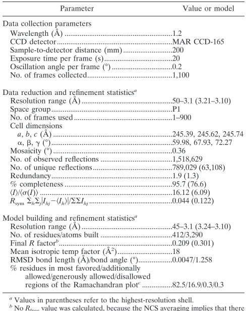
TABLE 1. X-ray data collection and refinement statistics
Parameter Value or model
Data collection parameters
Wavelength (Å) ...1.2
CCD detector ...MAR CCD-165 Sample-to-detector distance (mm) ...200
Exposure time per frame (s)...20 Oscillation angle per frame (°) ...0.2 No. of frames collected...1,100
Data reduction and refinement statisticsa
Resolution range (Å) ...50–3.1 (3.21–3.10) Space group...P1
No. of frames used ...1–900 Cell dimensions
a,b,c(Å) ...245.39, 245.62, 245.74
␣,,␥(°)...59.98, 67.93, 72.27 Mosaicity (°) ...0.36
No. of observed reflections ...1,518,629 No. of unique reflections ...789,029 (63,108) Redundancy...1.9 (1.3) % completeness ...95.7 (76.6)
具I典/具(I)典...16.12 (6.09) Rsym⌺h⌺j兩Ihj⫺具Ih典兩/⌺⌺Ihj...0.044 (0.122)
Model building and refinement statisticsa
Resolution range (Å) ...45–3.1 (3.24–3.10) No. of residues/atoms built ...412/3,290 FinalRfactorb...0.209 (0.301)
Mean isotropic temp factor (Å2)...18 RMSD bond length (Å)/bond angle (°)...0.0047/1.258 % residues in most favored/additionally
allowed/generously allowed/disallowed
regions of the Ramachandran plotc...82.5/16.9/0.3/0.3 a
Values in parentheses refer to the highest-resolution shell.
b
NoRfreevalue was calculated, because the NCS averaging implies that there is little difference betweenRworkingandRfree.Because of the NCS symmetry, an independent subset of structure factors cannot be selected.
c
Percentage of a total of 360 nonglycine, nonproline residues as defined in the program PROCHECK (12, 16).
4692
KAUFMANN ET AL.
J. V
IROL.
on November 7, 2019 by guest
http://jvi.asm.org/
residues of VP3 were traceable in the icosahedrally averaged
electron density, except for 42 N-terminal and 40 C-terminal
residues. The lack of ordered structure in the N-terminal
re-gion of the major coat proteins has been consistently observed
for all parvovirus structures to date, most likely because of a
positional variation incompatible with the icosahedral
symme-try averaging imposed during structure determination. The
absence of interpretable density for the C-terminal residues of
VP3 may similarly be due to either positional variation or
partial proteolytic processing (see Fig. S1 in the supplemental
material). The ordered C terminus is located at the exterior
surface of the capsid close to a 3-fold axis of the capsid. In
contrast, the C termini of vertebrate parvoviruses interface
with the neighboring subunits. The last four residues of the C
terminus of the GmDNV coat protein are also at the surface,
near the 2-fold axis, whereas the last residue of the PstDNV
coat protein is located at the inner surface. The absence of the
40 C-terminal residues in the BmDNV-1 structure could
there-fore be due to proteolytic processing at the capsid surface (see
Fig. S1 in the supplemental material). This would also explain
why the estimated molecular mass of the BmDNV-1 capsid
protein was somewhat lower than expected. The electron
den-sity of BmDNV-1 provided no evidence for the presence of
metal ions.
The BmDNV-1 capsid protein contains an eight-stranded
jelly roll motif, similar to that found in many other viral capsid
proteins, consisting of two

-sheets in the BIDG and CHEF
arrangement (for nomenclature, see Rossmann and Johnson
[27]) (Fig. 1 and 2; see also Table S1 in the supplemental
material). The ß-barrel of BmDNV-1 can be superimposed on
the same motif in GmDNV with an RMSD of 1.2 Å for 69
equivalenced C
␣
atoms (81% of C
␣
atoms in the ß-barrel)
and on PstDNV with an RMSD of 1.5 Å (71 C
␣
atoms). The

-barrel motif is in approximately the same position relative
to the icosahedral symmetry axes as in other parvoviruses
(Fig. 2).
In the vertebrate parvoviruses, such as CPV, the

-BIDG
sheet is extended to an antiparallel

-ABIDG sheet by the
FIG. 1. The structure of the BmDNV-1 capsid protein. (A) Schematic diagram showing the positions of secondary structural elements along
the polypeptide. Numbering corresponds to the starting residues of the

-strands (solid arrows);

-strands that are part of the “jelly-roll” fold are
shown in black, and those in connecting loops are shown in color. Helical elements are depicted as open rectangles. Secondary structure elements
are named as defined previously (13, 35). Additional secondary structure elements designated for BmDNV-1 are labeled in italic font (see Table
S1 in the supplemental material). (B) Ribbon diagram of the BmDNV-1 subunit. The BIDG and CHEF sheets of the eight-stranded

-barrel are
shown in light and dark gray, respectively. The surface loops connecting the strands of the

-barrel are colored as follows: BC loop, dark blue; CD,
purple; DE, dark green; EF, cyan; GH, red; and HI, green. The N-terminal region upstream of

B is shown in magenta, and the C-terminal portion
downstream of

I is shown in gold. The position of icosahedral symmetry axes and their direction from the viral center are indicated by arrows.
Hydrogen-bonded

structures that occur only in BmDNV-1 VLPs are marked with an asterisk. Molecular graphic images were produced using
the Chimera software package (22).
on November 7, 2019 by guest
http://jvi.asm.org/
backfolded

-strand A (

A) in the N-terminal portion
up-stream of

B (Fig. 2). In contrast, in the
Densovirus
GmDNV,

B is essentially the linear extension of

A (29), producing a
“domain-swapped” structure (3). Similarly, the N termini of
PstDNV (13) and BmDNV-1 are pointing toward a 5-fold axis
in a domain-swapped fashion relative to those of the vertebrate
parvoviruses. Therefore, domain swapping is a common
struc-tural feature of the coat protein of invertebrate parvoviruses.
Although the functional implications and benefits of this
do-main swapping are unclear, it may be necessary for structural
stability by compensating for the reduced intertwining of the
GH loops around the 3-fold axis (see Fig. S3 in the
supple-mental material). The last few N-terminal ordered residues of
BmDNV-1 are pointing away from the 5-fold channel slightly
toward the interior of the virus particle, similar to VLPs of B19
and PstDNV (13, 14). No density was found in the 5-fold pores
that might be due to an externalization of the N termini, which
may be the result of the absence of VP1 and VP2 in these
BmDNV-1 VP3 VLPs.
In GmDNV,

A engages in hydrogen bonding with

B of a
neighboring 2-fold-related subunit. However, in PstDNV there
are no additional hydrogen bonds with the N-terminal region
upstream of

B, although there are divalent metal ions that
cross-link neighboring subunits. There is significant but
differ-ent complemdiffer-entarity of shape between 3-fold-related subunits
in densoviruses (see Fig. S3 in the supplemental material).
There are additional hydrogen bonds between

GH1.2 and

GH2 of a 3-fold-related subunit of BmDNV-1. Furthermore,
in the N-terminal region of BmDNV-1, the
〈
strand is
an-tiparallel to

F of a neighboring 3-fold-related

-CHEF sheet
rather than to the 2-fold-related

-BIDG as is the case in
GmDNV. In BmDNV-1, there are only two hydrogen bonds
between the side chain of glutamine 56 in the N-terminal arm
and residues 66 and 67 in the

B-strand of the 2-fold-related
subunit.
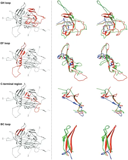
In most parvoviruses, the GH loop (see Fig. 1 for
nomen-clature) interdigitates with neighboring subunits to create
tri-mers, thus probably providing stability (19, 24). The GH loop
forms the characteristically raised structures at and around the
3-fold apices of the mammalian parvovirus capsid (1, 14, 18, 30,
33–35) and a

-annulus-type structure in GmDNV (29). The
structure of the GH loop of BmDNV-1 bears little similarity to
most known parvovirus structures, excepting that of GmDNV
(Fig. 3). In GmDNV, a portion of the GH loop that forms the
tip of the spike at the 3-fold vertices in CPV is entirely missing,
whereas the part of the GH loop that forms the base of the
spike is present but structurally different from that of CPV
(29). In BmDNV-1, the GH loop is more intertwined with its
FIG. 2. The spatial arrangement of the parvovirus core jelly roll and the N-terminal region for the capsid proteins of BmDNV-1, PstDNV,
GmDNV, and CPV illustrate domain-swapping of the N terminus for the three known
Densovirinae
structures. Two 2-fold related symmetry mates
are shown, viewed from the viral center along an icosahedral 2-fold axis. Conserved secondary structure elements of each protein subunit are
colored blue (

-BIDG), green (

-CHEF), and gold (helical elements). The N-terminal region of the capsid protein, upstream of

B, including

A,
is shown in magenta. The positions of icosahedral symmetry axes are indicated by polygonal symbols.
4694
KAUFMANN ET AL.
J. V
IROL.
on November 7, 2019 by guest
http://jvi.asm.org/
3-fold-related neighboring subunit than in GmDNV, but both
are in contrast to PstDNV, for which this loop does not
inter-digitate between neighboring subunits (13) (see Fig. S3 in the
supplemental material).
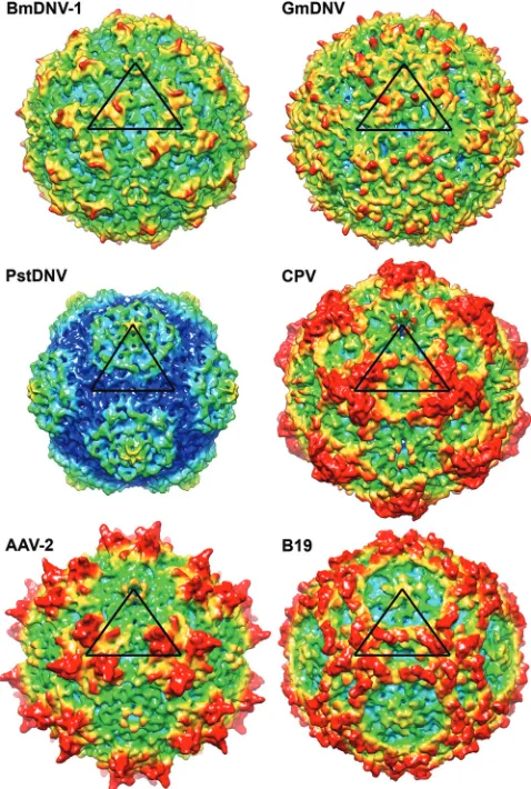
The largest portion in all parvovirus structures is made up of
the loops connecting the

-barrel strands (see Fig. 1 for
[image:5.585.79.503.70.600.2]no-menclature). These loops form most intersubunit contacts and
define the outer surfaces of the different viruses (Fig. 4). The
BmDNV-1 capsid, with a maximum outer diameter of 264 Å,
does not have substantial 3-fold-proximal protrusions as
ob-served in members of the
Parvovirinae
subfamily, such as CPV.
The protrusions on the surfaces of BmDNV-1 and GmDNV
FIG. 3. Structure comparison of loops GH, EF, BC, and the C-terminal region of BmDNV-1 with those of GmDNV and PstDNV. Left, a
ribbon diagram of the BmDNV-1 capsid protein indicates the position of the particular loop (red) relative to the icosahedral symmetry axes. Right,
close-up stereo views of the superpositioned loop regions of BmDNV-1 (red), GmDNV (green), and PstDNV (blue).
on November 7, 2019 by guest
http://jvi.asm.org/
rise only slightly over the general surface, unlike the case for
the
Brevidensovirus
PstDNV (Fig. 4).
The structure and length of the loop regions in BmDNV-1
show some similarity to GmDNV, but they differ greatly
from the vertebrate parvoviruses, as well as the shrimp
par-vovirus PstDNV (Fig. 3), suggesting a smaller divergence in
evolutionary development between BmDNV-1 and GmDNV
than between these and the vertebrate parvoviruses, as well as
PstDNV. The backbone C
␣
atoms of BmDNV-1 superimpose
on PstDNV with an RMSD of 1.9 Å for 140 equivalenced C
␣
atoms (34% of all ordered BmDNV-1 residues and 47% of
ordered PstDNV residues) but on GmDNV with an RMSD of
2.0 Å with 239 C
␣
atoms (58% for BmDNV residues and 55%
of GmDNV residues). Thus, there is a far larger portion of
structure that is similar between BmDNV-1 and GmDNV than
there is between BmDNV-1 and PstDNV. These observations
are consistent with the previous conclusion of a closer
evolu-tionary relationship between the two insect parvoviruses
BmDNV-1 and GmDNV than they have with other
parvovi-ruses.
ACKNOWLEDGMENTS
We are grateful for the support we received from the staff of the
BioCARS beamline. We are thankful to Sheryl Kelly for assistance in
the preparation of this article.
Use of BioCARS Sector 14 was supported by the National Institutes
of Health (NIH), National Center for Research Resources, under
grant no. RR007707. Use of the Advanced Photon Source was
sup-ported by the U.S. Department of Energy, Basic Energy Sciences,
Office of Science, under contract DE-AC02-06CH11357. The work was
supported by NIH grants AI 33468 to Colin R. Parrish (Cornell
Uni-versity) and AI 11219 to M.G.R. and support from the Natural
Sci-ences and Engineering Research Council of Canada to P.T.
REFERENCES
1.Agbandje, M., R. McKenna, M. G. Rossmann, M. L. Strassheim, and C. R. Parrish.1993. Structure determination of feline panleukopenia virus empty particles. Proteins16:155–171.
2.Arnold, E., et al.1984. Virion orientation in cubic crystals of the human common cold virus HRV14. J. Mol. Biol.177:417–430.
3.Bennett, M. J., S. Choe, and D. Eisenberg.1994. Domain swapping: entan-gling alliances between proteins. Proc. Natl. Acad. Sci. U. S. A.91:3127– 3131.
4.Benson, S. D., J. K. Bamford, D. H. Bamford, and R. M. Burnett.2004. Does common architecture reveal a viral lineage spanning all three domains of life? Mol. Cell16:673–685.
5.Bru¨nger, A. T.2007. Version 1.2 of the Crystallography and NMR system. Nat. Protoc.2:2728–2733.
6.Bru¨nger, A. T., et al.1998. Crystallography and NMR system: a new software suite for macromolecular structure determination. Acta Crystallogr. D Biol. Crystallogr.54:905–921.
7.Canaan, S., et al.2004. Interfacial enzymology of parvovirus phospholipases A2. J. Biol. Chem.279:14502–14508.
8.Collaborative Computational Project, Number 4.1994. The CCP4 suite: programs for protein crystallography. Acta Crystallogr. D Biol. Crystallogr. 50:760–763.
9.Emsley, P., and K. Cowtan.2004. Coot: model-building tools for molecular graphics. Acta Crystallogr. D Biol. Crystallogr.60:2126–2132.
10.Farr, G. A., L. G. Zhang, and P. Tattersall.2005. Parvoviral virions deploy a capsid-tethered lipolytic enzyme to breach the endosomal membrane dur-ing cell entry. Proc. Natl. Acad. Sci. U. S. A.102:17148–17153.
11.Girod, A., et al.2002. The VP1 capsid protein of adeno-associated virus type 2 is carrying a phospholipase A2 domain required for virus infectivity. J. Gen. Virol.83:973–978.
12.Kabsch, W., and C. Sander.1983. Dictionary of protein secondary structure: pattern recognition of hydrogen-bonded and geometrical features. Biopoly-mers22:2577–2637.
13.Kaufmann, B., et al.2010. The structure ofPenaeus stylirostrisdensovirus, a shrimp pathogen. J. Virol.84:11289–11296.
14.Kaufmann, B., A. A. Simpson, and M. G. Rossmann.2004. The structure of human parvovirus B19. Proc. Natl. Acad. Sci. U. S. A.101:11628–11633. 15.Kleywegt, G. J., and R. J. Read.1997. Not your average density. Structure
5:1557–1569.
16.Laskowski, R. A., M. W. MacArthur, D. S. Moss, and J. M. Thornton.1993. Procheck—a program to check the stereochemical quality of protein struc-tures. J. Appl. Crystallogr.26:283–291.
17.Li, Y., et al.2001. Genome organization of the densovirus fromBombyx mori (BmDNV-1) and enzyme activity of its capsid. J. Gen. Virol.82:2821–2825. 18.Llamas-Saiz, A. L., et al.1997. Structure determination of minute virus of
mice. Acta Crystallogr. D Biol. Crystallogr.53:93–102.
19.Lombardo, E., J. C. Ramírez, M. Agbandje-McKenna, and J. M. Almendral. 2000. A beta-stranded motif drives capsid protein oligomers of the parvovi-rus minute viparvovi-rus of mice into the nucleus for viral assembly. J. Virol.74: 3804–3814.
20.Nandhagopal, N., et al.2002. The structure and evolution of the major capsid protein of a large, lipid-containing DNA virus. Proc. Natl. Acad. Sci. U. S. A.99:14758–14763.
21.Otwinowski, Z., and W. Minor.1997. Processing of X-ray diffraction data collected in oscillation mode, p. 307–326.InCharles W. Carter, Jr. (ed.), Methods Enzymol., vol. 276. Academic Press, San Diego, CA.
22.Pettersen, E. F., et al.2004. UCSF Chimera—a visualization system for exploratory research and analysis. J. Computat. Chem.25:1605–1612. 23.Read, R. J.1986. Improved Fourier coefficients for maps using phases from
partial structures with errors. Acta Crystallogr. Sect. A42:140–149. 24.Riolobos, L., J. Reguera, M. G. Mateu, and J. M. Almendral.2006. Nuclear
[image:6.585.43.282.71.426.2]transport of trimeric assembly intermediates exerts a morphogenetic control on the icosahedral parvovirus capsid. J. Mol. Biol.357:1026–1038.
FIG. 4. Comparison of the BmDNV-1 protein capsid with those of
other parvoviruses. Surface renderings of three-dimensional electron
density maps of BmDNV-1, GmDNV, PstDNV, CPV, B19, and
AAV-2 generated from atomic coordinates to 8-Å resolution. One
icosahedral asymmetric unit is indicated by a black triangle. The
sur-face is colored according to the distance from the viral center (blue,
100 Å; cyan, 107.5 Å; green, 115 Å; yellow, 122.5 Å; red, 130 Å).
4696
KAUFMANN ET AL.
J. V
IROL.
on November 7, 2019 by guest
http://jvi.asm.org/
25.Rollinger, J. M., and M. Schmidtke.2011. The human rhinovirus: human-pathological impact, mechanisms of antirhinoviral agents, and strategies for their discovery. Med. Res. Rev.31:42–92.
26.Rossmann, M. G., and D. M. Blow.1962. The detection of sub-units within the crystallographic asymmetric unit. Acta Crystallogr.15:24–31. 27.Rossmann, M. G., and J. E. Johnson.1989. Icosahedral RNA virus structure.
Annu. Rev. Biochem.58:533–573.
28.Shimizu, T.1975. Pathogenicity of an infectious flacherie virus of the silk-worm, Bombyx mori, obtained from sericultural farms in the suburbs of Ina city. J. Seric. Sci. Jpn.44:45–48.
29.Simpson, A. A., P. R. Chipman, T. S. Baker, P. Tijssen, and M. G. Ross-mann.1998. The structure of an insect parvovirus (Galleria mellonella denso-virus) at 3.7 Å resolution. Structure6:1355–1367.
30.Simpson, A. A., et al.2002. The structure of porcine parvovirus: comparison with related viruses. J. Mol. Biol.315:1189–1198.
31.Tijssen, P., et al.Parvoviridae.InA. M. Q. King, M. J. Adams, E. Carstens, and E. J. Lefkowitz (ed.), Virus taxonomy: classification and nomenclature of viruses. Ninth report of the International Committee on Taxonomy of Viruses, in press. Elsevier, San Diego, CA.
32.Tong, L. A., and M. G. Rossmann.1990. The locked rotation function. Acta Crystallogr. A46:783–792.
33.Tsao, J., et al.1991. The three-dimensional structure of canine parvovirus and its functional implications. Science251:1456–1464.
34.Xie, Q., et al.2002. The atomic structure of adeno-associated virus (AAV-2), a vector for human gene therapy. Proc. Natl. Acad. Sci. U. S. A.99:10405– 10410.
35.Xie, Q., and M. S. Chapman. 1996. Canine parvovirus capsid structure, analyzed at 2.9 Å resolution. J. Mol. Biol.264:497–520.
36.Zadori, Z., et al.2001. A viral phospholipase A2 is required for parvovirus infectivity. Dev. Cell1:291–302.
on November 7, 2019 by guest
http://jvi.asm.org/