Int. J. Electrochem. Sci., 9 (2014) 3736 - 3745
International Journal of
ELECTROCHEMICAL
SCIENCE
www.electrochemsci.org
Detecting of Benzo[a]pyrene Using a Label-free Amperometric
Immunosensor
Yanhua Zhang1, Qing Su1, Junhui Xu2, Yan Zhang1,*, Songtao Chen1 1
Department of Municipal and Environmental Engineering, Henan University of Urban Construction, Pingdingshan 467044, China
2
China Offshore Environmental Service Ltd, Tianjin 300452, China *
E-mail: [email protected]
Received: 22 February 2014 / Accepted: 27 March 2014 / Published: 14 April 2014
A label-free amperometric immunosensor was manufactured for the fast and sensitive detection of Benzo[a]pyrene. Prussian blue was firstly electrodeposited onto a glass carbon electrode as an electron mediator. Afterwards nano-gold layer was formed on the prussian blue film by electrochemical reduction of HAuCl4 solution. Then, L-cysteine and nano-gold particles were self-assembled layer-by-layer. Finally, anti-benzo[a]pyrene was adsorbed onto the bilayer nano-gold films to construct a sensitive immunosensor. Under the optimal experimental condition, the current response of the immunosensor was inversely proportional to the concentration of benzo[a]pyrene in the range of 0.2~100 ng·mL-1 with a detection limit of 0.08 ng·mL-1. The reported immunosensor exhibited a long-term stability and was easy to be regenerated. This method was applied to the benzo[a]pyrene detection in waste water samples with satisfactory results.
Keywords: Prussian blue, Gold nanoparticles, L-cysteine, Amperometric immunosensor, Benzo[a]pyrene
1. INTRODUCTION
The main methods used for the determination of BaP are fluorometry, gas chromatography and high-performance liquid chromatography (HPLC) [7-10]. These methods exhibit high sensitivity, but need relatively expensive instrument, sample pretreatment and long time to complete the determination process. However, there is a huge demand for rapid, sensitive, low-cost and on-site detection in environmental monitoring.
The electrochemical immunoassay combines successfully the specificity of immuno-chemical systems with the advantages of electrochemical transducer [11,12]. In addition, electrochemical immunoassay is compatible to miniaturization for producing portable device. Therefore, this method has attracted great attention for the on-site screening of environmental pollutants [13,14]. Prussian blue (PB) is a prototype of metal hexacyanoferrates, and has been widely used as a good electron transfer mediator for analytical applications and has found a wide use in the biosensor development [15,16]. Nanometer-size gold (NG) particles have been extensively studied in immobilization biomaterials via the large specific surface area, low electron transfer impedance, desirable biocompatibility and high surface free energy [17-19].
In order to enhance the sensitivity of the immunoassay, a new type of voltammetric immunosensor made of PB and nano-gold NG for analysis Bap was reported. The preparation, characterization, and possible application of the immunosensor were described. Additonally, several parameters, such as pH value of the supporting electrolyte, the incubation time and incubation temperature were optimized. The performance of the resulting immunosensor was investigated in detail for the determination of environmental BaP.
2. EXPERIMENTAL
2.1. Reagents
BaP, Fluoranthene, phenanthrene, naphthalene, anthracene and HAuCl4·4H2O were purchased from Shanghai Chemical Reagent Company. Ovalbumin (OVA, MW45000) was purchased from Sino-American Biotechnology Company. Phosphate buffered solutions (PBS, 0.1 mol·L-1
) at various pH were prepared by using the stock solutions of K2HPO4, KH2PO4 and 0.1 mol·L-1 KCl, then adjusting the pH with 0.1 mol·L-1
NaOH and H3PO4. All reagents used herein were of analytical grade. Doubly distilled water was used throughout.
2.2. Apparatus
2.3 Preparation of the NG particles
[image:3.596.196.400.261.399.2]All glassware used in the following procedure was cleaned in a bath of K2Cr2O7-H2SO4 solution, rinsed thoroughly in twice distilled water and dried in air. NG particles were prepared according to the literature [20]. 2.5 mL 1% (w/w) sodium citrate solution was quickly added to 100 mL boiling aqueous solution containing 1 mL 1% (w/w) HAuCl4 with vigorously stirring. Boiling was continued for an additional 15 min. The heater was removed and the solution was naturally cooled to the room temperature. The color of the NG solution was claret and the average diameter of the NG particles was about 16 nm (Figure 1). The prepared NG solution was stored at 4 °C in dark glass bottles ready for use.
Figure 1. The TEM images of NG particles
2.4 Preparation of BaP polyclonal antibodies
Two male New Zealand white rabbits were immunized with BaP-BSA conjugates. The immune response consisted of an initial primary response followed by a secondary immune response. The initial dose for the primary immune response injection was 0.5mg·kg-1
of the immunogen to the rabbit bodyweight, with complete Freund’s adjuvant. Four weeks after the initial injection, the rabbit received booster injection every 2 weeks, 7-10 days after the third booster injection, the rabbits were bled from the ear vein and the sera were tested for titer by the agar diffusion test and the indirect fluoroimmunoassay. The antiserum from the rabbit was purified by the method of octanoic acid ammonium sulfate two-step precipitation, and the purified antiserum was freeze dried and stored in aliquots at -20 °C.
2.5 Fabrication of the immunosensor
Prior to surface modification, the GCE was polished sequentially with 0.3 and 0.05 μm alumina slurries on the polishing cloth to produce a mirror-like surface. It was then sonicated for 2 min in 1mol·L-1
aqueous solution containing 2.5 mmol·L-1
FeCl3, 2.5 mmol·L-1 K3[Fe(CN)6], 0.1 mol·L-1KCl and 0.1 mol·L-1
HCl [21]. Afterwards the electrode (PB/GCE) was washed carefully with water and electrochemically scanned between -0.05 and 0.35 V (vs. SCE) at a sweep rate of 50 mV·s-1 in 0.1 mol·L-1 KCl + 0.1 mol·L-1
HCl solution for 25 cycles to activate the PB film. After washing with water, the electrode was dried at 100 °C for 1 h and cooled to the room temperature. A layer of NG was deposited on the PB/GCE by applying a constant potential of -0.2 V for 30 s in a 100 mg·L-1 HAuCl4 solution. Afterwards, the modified electrode (NG/PB/GCE) was rinsed with water and immersed in a pH 4.5 PBS containing 0.2 mol·L-1
L-cysteine (LC) for 4h to self-assemble LC. The obtained electrode (LC/NG/PB/GCE) was thoroughly washed with water to remove the physically absorbed LC. Subsequently, the resulting electrode was immersed in NG at 4 °C overnight to form another NG monolayer. Then the modified electrode (NG/LC/NG/PB/GCE) was immersed in a 0.5 ml anti-BaP solution at 4 °C overnight. At last the multilayer modified electrode (anti-BaP /NG/LC/NG/PB/GCE) was incubated in 1% OVA solution for about 2 h at 37 °C to block the remaining active site of the NG layer and to avoid the non-specific adsorption. The finished immunosensor was stored at 4 °C until required. The construction procedure of the immunosensor is shown as Scheme 1.
Fe3+ + Fe(CN)6
3-GCE
S-CH2-CH-NH3+
S-CH2-CH-NH3 +
S-CH2-CH-NH3 + AuCl4 -COOH COOH COOH
SH-CH2-CH-NH2
COOH
S-CH2-CH-NH3+
S-CH2-CH-NH3 +
S-CH2-CH-NH3 +
COOH COOH COOH
S-CH2-CH-NH3 +
S-CH2-CH-NH3 +
S-CH2-CH-NH3 +
COOH COOH COOH
NG particles
PB/GCE NG/PB/GCE LC/NG/PB/GCE
NG/LC/NG/PB/GCE anti-BaP/NG/LC/NG/PB/GCE
electrodeposition potentiostatic reduction
anti-BaP
Scheme 1. Preparation process of the immunoelectrode on the surface of GCE
2.6 Experimental measurements
Before each measurement, the immunosensor was incubated in a PBS (pH7.4) solution containing various amounts of BaP at 35 °C for 20 min. The electrochemical measurements were performed in the potential range of - 0.1 to 0.4 V (vs. SCE) at a scan rate of 100 mV·s-1 in the0.1 mol·L-1 KCl + 0.1mol·L-1
3. RESULTS AND DISCUSSION
3.1 Electrochemical characteristics of the immunosensor
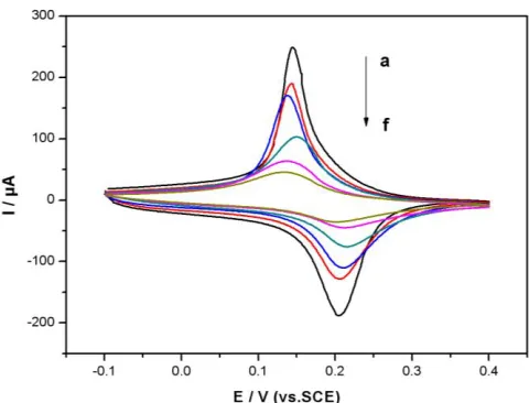
[image:5.596.175.415.498.681.2]The cyclic voltammograms of different modified electrodes in PBS (pH7.0) were showed in Fig. 2. There was no obvious redox peak at the bare GCE due to the lack of electron mediator. When violet-blue PB was electrodeposited onto the GCE surface, a pair of well redox peaks were achieved (Fig. 2a) at 0.20 V and 0.14 V (vs. SCE). The ratio of the redox peak current was 1, indicating that PB was a good electron transfer mediator. After the deposition of Au via electrochemical reduction of HAuCl4, the redox peak potentials remained the same as that of PB/GCE whilst the peak current decreased (Fig. 2b). This phenomenon is due to the fact that NG particles are similarly to a conducting wire or electron-conducting tunnel [22], hence enhance the reversibility of the electrochemical reaction; whilst the porous NG film increased the thickness of the film on the electrode surface and partly blocked direct access for the redox couple to diffuse to the GCE surface. LC owns mercapto- and amino- functional groups and has a pKa value of about 5.07. LC exists as a cationic form at pH sufficiently below the pKa since most of the amino groups are protonated. On top of the deposited NG particles, a thin layer of positively charged LC film is self-assembled via the chemical bonding between their mercapto groups and Au. The self-assembled LC film decreased the peaks current and increased the redox potential peak difference to 72 mV (Fig. 2c). After the modified electrode was immersed in the negatively charged NG colloids [23], NG particles were firmly anchored to the LC/NG/PB/GCE via the electrostatic interaction and the covalent bond between Au and amino groups of the LC. Both the redox current and the peak potential difference decreased (ΔE = 65 mV) (Fig. 2d). When anti-BaP and OVA were immobilized, the medication layer became less conductive and the peak current was reduced furthermore (Fig. 2e).
Figure 2. Cyclic voltammograms of substrate at different electrodes (a) PB/GCE; (b) NG/PB/GCE; (c) LC/NG/PB/GCE; (d) NG/LC/NG/PB/GCE; (e) OVA/anti- BaP/LC/NG/PB/GCE; (f) e treated with the 10ng·mL-1 BaP. Scan rate, 100mV·s-1
After the immunosensor was incubated with 10 ng·mL-1
BaP solution, the amperometric response exhibited dramatic decrease (Fig. 2f). The reason was that the formed immunocomplex acts as an inert layer blocking the electron and mass transfer, which shielded the active center of the mediator and decreased its ability to transfer electron. When a large amount of immunocomplexes were formed, the surface of the electrode was totally blocked by the complexes and the redox peak disappeared.
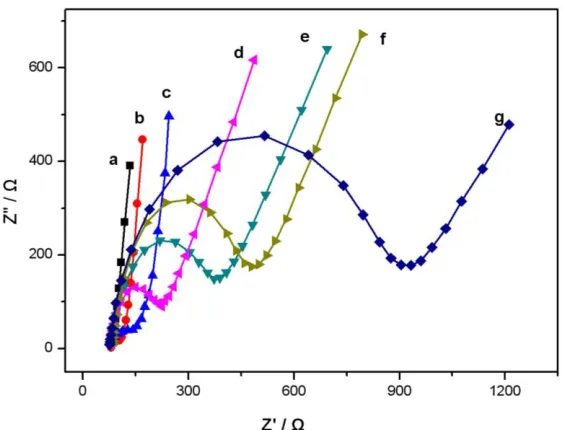
Electrochemical impedance spectroscopy (EIS) is an effective method for probing the interfacial properties of modified electrodes. Figure 3 showed the EIS results of different modified layer in the presence of redox probe [Fe(CN)6]4-/3-. They were measured at the formal potential of the electrochemical probe. It could be seen that the bare electrode exhibited an almost straight line. It indicated a diffusion limiting process of electrochemical process (Fig. 3a). After deposition a layer of PB (Fig. 3b), the EIS of the PB/GCE was similar to that of the bare GCE. This implied that the conductivity of the PB/GCE was essentially equivalent to a bare GCE. When gold nanoparticles were electrodeposited, the semicircle diameter increased (Fig. 3c), indicating that the NG film obstructed electron-transfer of the electrochemical probe. After self-assembled LC (Fig. 3d) and NG film (Fig. 3e), the semi-circle diameter in the impedance spectrum increased gradually. This was attributed to the immobilization of LC and NG on the NG/PB/GCE film that hindered the access of the redox probe to the electrode. With the stepwise assembly of antibody and OVA on the modified electrode, the semicircle diameter surprisingly increased (Fig. 3f), due to the formation of insulating protein layers. The electron transfer resistance increased further after the immunosensor was incubated with BaP solution (Fig. 3g). The results indicated that the anti-BaP and model pollutant were successfully fixed on the modified electrode surface for further work.
[image:6.596.155.437.475.690.2]
3.2 Optimization of the assay conditions 3.2.1 pH value of the supporting electrolyte
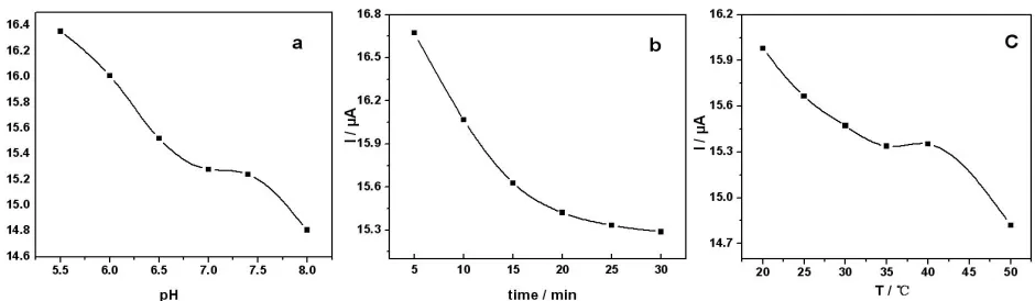
The influence of pH value of the supporting electrolyte on the performance of the immunosensor was studied (Fig. 4a). With the increase of the pH value from 5.5 to 8.0, the cathodic peak potential shifted to the negative potential and the peak current decreased. The reason is the fact that the reduced form of PB can be dissolved at high pH values [24]. Therefore, PB-film modified electrode is not stable at pH value above 7.0. Moreover, the activity of the antibody and antigen are inhibited at a pH value which is either higher or lower than the physiological pH. All the experiments were carried out in pH 7.0 of PBS.
3.2.2 The incubation time
The immunosensor was incubated in pH 7.4 PBS containing 80 ng·mL−1 BaP for different time. It could be seen that the peak current decreased rapidly with time up to 20 min and then leveled off slowly in Fig.4b [25]. Longer incubation time was not able to improve the response. It suggested that the interaction reached the maximum value. The incubation time of 20 min was chosen for the preparation of immunosensor.
[image:7.596.62.532.448.585.2]3.2.3 Incubation temperature
Figure 4. The influence of (a) pH, (b) incubation time and (c) temperature on the current responds of the immunosensor in base solution after incubated in 0.1 mol·L-1 PBS (pH7.4) containing 80 ng·mL−1 BaP.
proteins caused by the high temperature. It is well known that long-time usage in high temperature may damage multilayer film and affect the lifetime of immunosensor [26]. Considering the activity of biomolecule and the sensitivity of the sensor, we employed 35 °C as the incubation temperature.
3.3 Performance of the immunosensor 3.3.1 CV response and calibration curve
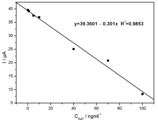
Under the optimum working conditions, the reduction peak current changed in proportion to the concentration of BaP, and thus could be used for quantitative determination of BaP with the proposed immunosensor. The calibration curve for determination of BaP was characterized under the optimized experiment conditions. It could be seen that the cathodic peak current response was inversely proportional to the analyte concentration in the range of 0.2 to 100 ng·mL-1
in figure 5. The linear equation was I (μA) = 39.3601 - 0.301C (ng·mL-1
) (R2 = 0.9853) with a detection limit of 0.08 ng·mL-1
(S/N=3).
Figure 5. Cyclic voltammograms of the immunosensor in 0.1 mol·L-1 PBS (pH7.0) containing 0.1 mol·L-1 KCl after incubated in 0.1 mol·L-1
PBS (pH7.4) containing different concentrations of BaP. CBaP/ ng·mL-1from a to f: 0.2,1,5,10, 40,70, 100. Scan rate: 100 mV·s-1
3.3.2 Selectivity
Under the optimum conditions, the potential interferences of naphthalene, anthracene, fluoranthene and phenanthrene were studied to investigate the selectivity of the immunosensor (Table 1). The current obtained for each interference at a concentration of 60 ng·mL-1 in the presence of 60 ng·mL-1
[image:8.596.160.433.338.545.2]
Table 1. The selectivity of the immunosensor (n=3) 60
ng·mL-1 BaP
each interference at a concentration of 60 ng·mL-1
in the presence of 60 ng·mL-1
BaP
naphthalene anthracene fluoranthene phenanthrene
peak current (A) 21.4 19.5 18.8 20.3 19.7
reduction rate (%) 91 88 95 92
3.3.3 Regeneration and stability
The prepared immunosensor could be regenerated by simple immersing in a stirred regeneration solution consisting of 0.2 mol·L-1 glycine - HCl (pH 2.8) and 0.25 mol·L-1 NaCl for about 5min after each usage. The immunosensor kept 88% of the original amperometric value after reusing for 7 times because the activity of proteins was degraded gradually.
The stability of the successive assays was studied from the amperometric response to 50 ng·mL-1
BaP. The relative standard deviation of 3.5% for 7 times detections was acquired. After one month storage in the refrigerator at 4 °C and measured intermittently (every 6 days), 90% of the initial response current remained. The good stability arises from the fact that BaP nanoparticles not only provide a natural environment for biocompatibility but also can strongly interact with antibody molecules.
3.3.4 Application
The immunosensor was applied to the determination of BaP in water samples with the standard addition method. Waste water from the coke-oven plant was selected as representative samples. Water samples were collected in bottles, filtered and adjusted to pH 7.0 with 0.1mol/L H3PO4 or NaOH. The quantitative recoveries of 90.9 ~ 107.5% were obtained and shown in Table 2.
Table 2. Recovery of spiked sample (n=5)
Initial value (ng·mL-1
)
Added (ng·mL-1
)
Found (ng·mL-1
)
Recovery (%)
6.6 10 17.1 107.5
20 26.4 97.0
30 36.3 95.5
40 46.0 90.9
4. CONCLUSIONS
a detection limit of 0.08 ng·mL-1 (S/N=3) for BaP. The immunosensor exhibited several attractive features such as the simple preparation, fast amperometric response, long-term stability and good reproducibility. The proposed method is promising to be further developed for the device fabrication in monitoring the environmental condition.
ACKNOWLEDGEMENTS
We acknowledge the Program for Science and Technology Innovative Talents of Pingdingshan (2013097) and the Program for Doctoral Innovative Research Foundation in Henan University of Urban Construction.
References
1. S. Boujday, S. Nasri, M. Salmain, C. Pradier, Biosens. Bioelectron., 26(2010) 1750. 2. P. Simko, J Chromatogr. B, 770(2002)3.
3. W. A. Lopes, J. B. de Andrade, Quim. Nova., 19(1996)497.
4. A. D. Pereira Netto, J. C. Moreira, A. E. X. O. Dias, G. Arbilla, L. F. V. Ferreira, A. S. Oliveira, J. Barek, Quim. Nova., 23(2000)765.
5. E. Puglisi, F. Cappa, G. Fragoulis, M. Trevisan, A.M. Del Re, Chemosphere, 67 (2007) 548. 6. M.A. Heitkamp, C.E. Cerniglia, Appl. Envir. Microbiol., 54 (1988) 1612.
7. H. M. Amzad, S. M. Salehuddin, Arab. J Chem., 1(2011) 49-53. 8. B. Janoszka, Food Chem. 126(2011) 1344.
9. S. Gülten, F. Gögüs, S. Fadiloğlu, J Food Qual., 3 (2007)300. 10. H.S. Zhuang, C. Zhou, Anal. Chim. Acta, 633(2009) 278.
11. N. Daud, N. A. Yusof and S. M. M. Nor, Int. J. Electrochem. Sci., 8 (2013) 10086.
12. H.X. Wang, X.Z. Wu, P.T. Dong, C.G. Wang, J.F.Wang, Y.Z. Liu, J. Chen, Int. J. Electrochem. Sci., 9 (2014) 12.
13. M. Badihi-Mossberg, V. Buchner, J. Rishpon, Electroanalysis, 19 (2007) 2015.
14. J. Wu, J.H. Tang, Z. Dai, F. Yan, H.X. Ju, N.E. Murr, Biosens. Bioelectron., 22 (2006) 102.
15. Y.D. Yang, Z.Y. Zhong, H.M. Liu, T.Y. Zhu, J.J. Wu, M.X. Li, D. Wang, Electroanalysis, 20 (2008) 2621.
16. D. Zhang, K. Zhang, Y.L. Yao, X.H. Xia, H.Y. Chen, Langmuir, 20 (2004) 7303. 17. Y. Zhang, S. H. Wei, S. T. Chen. Int. J. Electrochem. Sci., 8( 2013) 6493.
18. Y. Song, Y.Z. Song, A.F. Zhu, H. Zhong, Indian J. Chem. A, 50A (2011) 1006. 19. Y.Z. Song, Y. Song, H. Zhong, Gold Bull., 44 (2011)107.
20. K.C. Grabar, R.G. Freeman, M.B. Hommer, M.J. Natan, Ana1. Chem., 67(1995) 735.
21. Y. Zhao, Principle and application of biochemical technology. Wuhan University Press, Wuhan, 1999.
22. I.L.D. Mattos, L. Gorton, T. Laurell, A. Malinauskas, A.A. Karyakin, Talanta, 52 (2000) 791. 23. J.G. Zhao, R.W. Henkens, J. Stonehuerner, J.P. O'Daly, A.L. Crumbliss, J. Electroanal. Interfac.
Chem., 327 (1992) 109.
24. N.B. Li, J.H. Park, K. Park, S.J. Kwon, H. Shin, J. Kwak, Biosens. Bioelectron., 23 (2008) 1519. 25. R. Chai, R. Yuan, Y.Q. Chai, C.F. Ou, S.R. Cao, X.L. Li, Talanta, 74(2008) 1330.
26. Y. Zhang, H.S. Zhuang, Chin. J. Ana.l Chem., 38(2010) 153.