peripheral lymphoid tissue
A thesis submitted for the degree of
Doctor of Philosophy
of the Australian National University
Wichat Srikusalanukul
Developmental Physiology Group
Division of Molecular Medicine
John Curtin School of Medical Research
The Australian National University
Canberra, Australia
Statement of originality
The work described in this thesis is original and was carried out by myself under the supervision of Dr P J McCullagh in the Division of Molecular Medicine at the John Curtin School of Medical Research, Australian National University. With the exception of the analysis of model parameters in Chapter 6, which were
performed on the basis of a collaborative work with Dr Maria Durisova of the Institute of Experimental Pharmacology, Slovak Academy of Sciences, all of the contents of this thesis are the original work of the author and have neither been presented nor are currently being presented for any other degree.
Acknowledgments
Many people have been involved and contributed to the success of this study. First of all, I would like to express my great gratitude to ANU and JCSMR for providing me an ANU PhD scholarship which allowed me to pursue my
enthusiasm about the model applications in bio-medicine, and to my supervisor, Dr Peter McCullagh, for his kind patience and invaluable support throughout the four years since starting my course.
Also I would like to thank Prof Stjepan Marcelja (Department of Applied
Mathematics, Research School Physical Sciences and Engineering, ANU) for his help at the initial stage of my course, Dr Max Simpson-Morgan for his help in setting up an automated collection system for lymph, Dr Ross Odell (Graduate School of Biomedical Engineering, University of New South Wales) and Dr Goeff Aldis (School of Mathematics and Statistics, University College, Australian
Defence Force Academy) for their advice about compartmental models, and especially Dr Franky De Bruyne (Department of Systems Engineering, Research School Information Sciences and Engineering, ANU) for his advisory role in Chapter 5 (Prediction models), Dr Maria Durisova (Institute of Experimental Pharmacology, Slovak Academy of Sciences) and Dr Ladislav Dedik (Slovak University of Technology) for their supervision of Chapter 6 (Structured models).
I want to thank all of my laboratory colleagues, Mr Bernie Barancewicz, Mrs Karen King, Ms Sandra Veness and all of the staff in Wing F, who were full of help in all matters involved with my experiments, Ms Ruth Hagan and Ms
Michelle Stevens for their help in reading manuscripts, and Mr Geoffrey Osborne and Ms Sabin Gruninger (Flow Cytometry Unit, JCSMR) for their help with the flow cytometry study, Dr Elizabeth Washington and Dr Wayne Kimpton
(Department of Veterinary Preclinical Sciences, University of Melbourne) for their help in monoclonal staining and wonderful hospitality during my visit in 1996.
I owe special thanks and love to my family in Thailand and S.Korea and especially to my wife, Ms Sunkyung Shynn, and my children, for their durable support. Special thanks also goes to my sister, Ms Wiwan Srikusalanukul, who came from Thailand to help us deal with some intense situations during the last few weeks of the preparation of this thesis.
Abstract
Lymphocyte recirculation and migration is one of the most essential components of the immune system as it allows lymphocytes to recognise, to interact with and to respond to antigen in any site of the body. Lymphocyte migration through peripheral lymphoid tissue, plays a greater part in antigen recognition by the immune system than traffic through the major lymphatic vessels, but its regulation is still poorly understood.
To understand the kinetics of lymphocyte migration within lymphoid tissue and to construct suitable models to explain it, a sheep model was chosen for the
present study. The efferent lymphatic ducts draining a peripheral lymph node, either the popliteal or the prescapular lymph node, were cannulated.
Lymphocytes were collected and labelled with a selected fluorescent dye, then injected back into the blood circulation and monitored in both blood and lymph by means of flow cytometry.
Theoretical models have been increasingly recognised by biomedical researchers as having applications to biomedical problems. Three different approaches, namely compartmental models, prediction error models and structured models, have been explored to describe the dynamics of lymphocyte migration within lymphoid tissues under normal conditions. The information obtained from each individual model has shown some degree of variation from the others. This will assist in providing more comprehensive descriptions of the entire complicated events of lymphocyte migration. Even though several
modelling approaches often used in medical and physiological modelling will be introduced a completely true model has yet to be attained.
Table of contents
Statement of originality ii
Acknowledgments iii
Abstract iv
Chapter 1/ Introduction
Part I: Biological backgrounds
1.1 The contribution of lymphocytes to the functioning of the 1 immune system
1.2 Physiology and anatomy of lymph nodes 2 1.2.1 Lymphoid tissues and lymphatic systems 2 1.2.2 Anatomy and organisation of lymphoid tissue in 4 lymph nodes
1.3 Lymphocyte recirculation and its functions 5 1.3.1 Life history of lymphocytes and the discovery of 5 lymphocyte recirculation
1.3.2 The discovery of sites and routes of lymphocyte 6 migration
1.3.3 Role of lymphocyte-endothelial cell interaction 8 1.3.4 The tempo of migration of lymphocytes in lymphoid 10 tissues
1.3.5 The immunological significance of lymphocyte 10 recirculation
1.3.6 The diversity of lymphocytes and the contribution 11 of this to immunological functions
1.4 Objectives of the present study 12
1.5 General review on studies of lymphocyte recirculation 12 1.5.1 Animal models in studies of lymphocyte 12 recirculation
1.5.2 Theoretical models in studies of lymphocyte 14
recirculation
Part II: Modelling fundamentals and methodology
1.7 Systems and signals 18
1.8 Categorisation of systems 19
1.9 Functional and physical identification of systems 21 1.10 An attempt to construct a theoretical model of lymphocyte 23 migration from novel data specially collected for this purpose
1.11 Thesis structure: Multiple approaches 25
Chapter 2/
Experimental procedures
Animals 27
Pre operative preparations and anaesthesia 27 Cannulation of the efferent lymphatic vessel 28 Surgical approach to the efferent vessel of the popliteal 29 lymph node
Surgical approach to the efferent vessel of the prescapular 29 lymph node
Catheterisation of the external jugular vein 30
Post operative maintenance 30
Collection of lymphocytes 31
Fluorochrome labelling of lymphocytes 32
Infusion of labelled lymphocytes 32
Blood sampling 33
Lymph sampling 33
Preparation of lymphocytes 33
Monoclonal staining 34
Flow cytometry study 35
Statistical Analysis 36
Chapter 3/
Experimental results
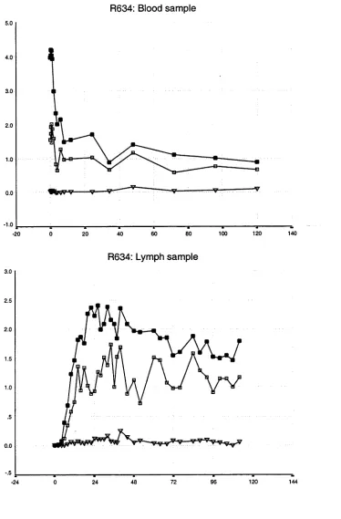
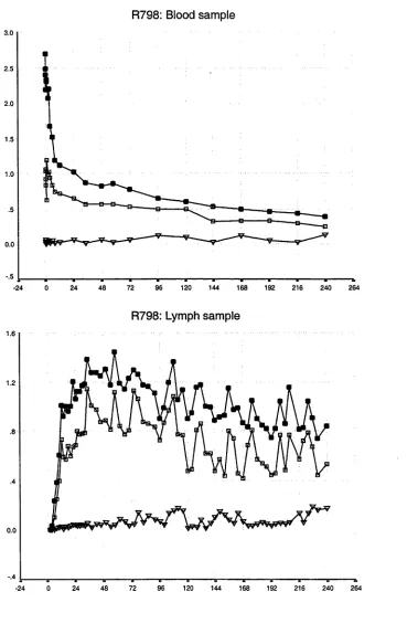
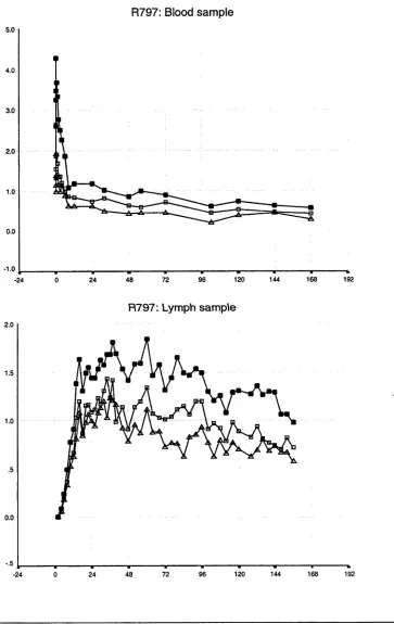
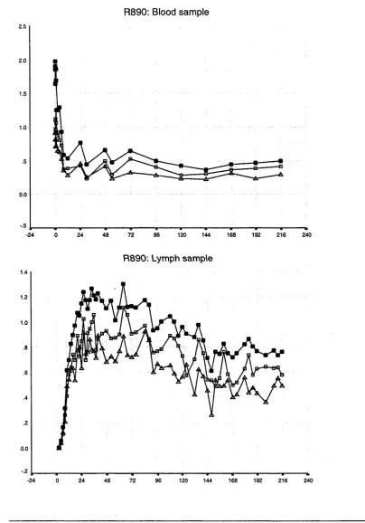
3.1 Introduction 37
3.2 Results of whole population studies 44 3.2.1 The disappearance of labelled lymphocytes 44 from venous blood
Observations from previous studies 50 Observations from the present study 50 3.3 Results of lymphocyte subset studies 76 3.3.1 Distribution of lymphocyte subsets in the blood 76 and efferent lymph in individual animals
3.3.2 The relationship between lymphocyte subsets in 98 the blood and efferent lymph and its interpretation
Chapter 4/
Compartmental models
4.1 Introduction 105
4.2 Compartmental models 106
4.3 Lymphocyte migration: A view of compartmental modelling 108
4.4 Model construction 110
4.4.1 Blood model: a forcing function 110
4.4.2 Lymph node model 112
4.4.3 Experimental model 116
4.5 Experimental data 117
4.6 Model fitting procedures: SAAMII 117
4.7 Results 119
4.8 Discussion 124
4.9 Conclusion 127
Chapter 5/
Prediction error models
5.1 Introduction 129
5.2 Prediction error models 132
5.2.1 Impulse response 132
5.2.2 Transfer functions 134
5.2.3 Generalised prediction error models 136
5.2.4 Candidate models 137
5.3 System identification procedures 140
5.3.1 Lymph node system 140
5.3.2 Experimental data 140
5.3.3 Matlab as a tool 140
5.3.6 Model validation 143 5.4 Results from the system identification procedure 144
5.4.1 System delay 144
5.4.2 Comparison of different model structures 145
5.4.3 Model validation 148
5.4.4 Comparison of the actual outputs and the 149 model outputs
5.4.5 Summary of the estimated parameters 161 5.5 Analysis of the simulated unit impulse response 163
5.5.1 Simulation of unit impulse responses 163 5.5.2 Feature extraction from the simulated unit 176 impulse response
5.6 Discussion 178
5.7 Conclusion 182
Chapter 6/
Structured models
6.1 Introduction 183
6.2 System modelling in the frequency domain 184
6.2.1 System transfer function 184
6.2.2 System frequency response 186
6.3 Fundamental systems 187
6.3.1 Ideal linear dynamic system 187 6.3.2 Proportional linear dynamic system 187 6.3.3 Ideal linear dynamic system with time delay 187 6.3.4 First-order linear dynamic system 188 6.4 Building of a structured model of the lymph node system 189
6.4.1 Experimental data 189
6.4.2 Definition of lymph node system 189 6.4.3 Modelling of the lymph node system in the 190 frequency domain
6.4.4 Modelling of the lymph node system in the time 190 domain
6.4.5 Candidate structured models of the lymph node 192 system
6.4.6 Mathematical descriptions of the parallel model 193
6.5 Results 195
6.5.2 Comparison of outputs of parallel models with 206
different numbers of branches
6.5.3 Comparison between experimental outputs and 207 final structured parallel models
6.5.4 Estimation of parameters in selected parallel models 218 6.5.5 Estimation of mean transit times and percentages of 221 lymphocytes travelling along each path of selected parallel
models
6.6 Discussion 223
6.7 Conclusion 226
Chapter 7/
General discussion
Introduction 227
Modelling 229
Compartmental models 230
Prediction error models 231
Structured models 234
Conclusion 235
Appendix 1/ 237
Data normalisation
Appendix 21 241
Matlab algorithms and commands
Introduction
As this thesis deals with the application of mathematical principles of analysis to a biological problem, its introduction falls into two parts. The first of these gives an account of the history and background to lymphocyte recirculation and of the significance of this phenomenon for the immune response. Essential features of the anatomy and physiology of lymphocyte migration that are relevant to
attempts at its mathematical modelling will be outlined. General reviews of previous attempts to apply theoretical models to describe lymphocyte recirculation and migration will also be included. The second part of the
introduction considers the fundamental principles and methodology of modelling that are relevant to the contents of this thesis. It is unavoidable that both
biological and mathematical aspects of the subject have necessarily to be considered in detail. However it is also necessary that data and calculations be presented in a manner that is accessible to readers from both of these
backgrounds. To facilitate this, terminology relating to both biology and modelling contexts will be clarified when first used.
Part I: Biological backgrounds
1.1 The contribution of lymphocytes to the functioning of the
immune system
In everyday life, the body is continually exposed to many pathogens (ie micro
organisms that are capable of producing diseases), such as viruses, bacteria
and parasites. The consequences for the body of diseases caused by micro organisms can vary from very mild to irreversible damage to parts of, or to the
whole body. Fortunately, the damage to the body produced by infectious diseases is commonly mitigated by the response of the immune system. The
immune system is able to distinguish non-self entities, such as pathogenic
micro-organisms', and to mount responses against them. A variety of
mechanisms underpin immune responses, but in general, they are mediated by a
class of cells known as leucocytes (eg white blood cells, WBC). The most important type of leucocyte for mounting immune responses is the lymphocyte.
These cells play a crucial role in immune responses because they continually move through the body and have the capacity to monitor for signs of the entry of any micro-organisms expressing foreign antigens2.
1.2 Physiology and anatomy of lymph nodes
1.2.1 Lymphoid tissues and lymphatic systems
From an anatomical perspective, the immune system can be considered to comprise two main components. One of these is a circulating (or recirculating) component of mobile cells (eg lymphocytes). Recirculating lymphocytes will be fully described in the next section of this chapter. The other component of the immune system is fixed lymphoid tissue through which migrating cells transit. Lymphoid tissues have been subdivided into primary and secondary types.
The distinguishing feature is that cellular proliferation and differentiation
occurs in primary tissues independently of any exposure to antigen, whilst it is necessary for both processes to be driven by antigen in secondary tissues. Primary lymphoid tissues include the thymus and bone marrow. In these tissues, lymphocytes differentiate from stem cells, and mature before
emigration. The thymus is the primary lymphoid tissue for T lymphocytes in all species that have been studied. B lymphocytes originate from the bone marrow of mice and, in the chick, from a structure unique to avian species, the bursa of
Fabricius. The origin of B lymphocytes in mammalian species other than the
mouse has yet to be formally defined, although evidence is accumulating to suggest that, in the sheep, the gut associated lymphoid tissue functions as an avian bursa equivalent. After emigrating from primary lymphoid tissues, lymphocytes enter the blood and lymph circulation system to reach secondary lymphoid tissues, such as the spleen and lymph nodes.
The lymphatic system is a network comprising lymphatic vessels, lymphoid
tissues and special organs (such as the spleen and lymph nodes). This lymphatic network extends throughout almost all organs and tissues of the body in a
distribution similar to that of the blood circulation network. The term central
lymphatic system will be used to refer to the central parts of the lymphatic
system, especially to the great lymphatic vessels such as the thoracic duct* in mammals. The term peripheral lymphatic system will be used to describe the regional parts of the lymphatic system that includes many minor lymphatic vessels. These vessels are responsible for returning lymph from all parts of the body to the central system.
There are many routes (eg skin, gut and respiratory system) by which potentially pathogenic micro-organisms may enter the body but, irrespective of the route, they will soon encounter lymphocytes in the draining regional lymph nodes. The special structure of (secondary) lymphoid tissues provides an optimal
environment in which continually circulating lymphocytes can interact with each other, with other types of cell such as antigen-presenting cells and with antigens (of micro-organisms) carried via lymphatic vessels. For this reason, lymphocyte recirculation and migration within the peripheral lymphatic system may be expected to provide a more comprehensive description of the
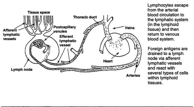
[image:14.518.65.471.453.678.2]participation of lymphatics and lymphoid tissues in the immune response than can be obtained from studies performed in the central lymphatic system. Figure 1.1 demonstrates the route of foreign antigen delivery to the lymph node via afferent lymph vessels. Most immune reactions occur in the lymphoid tissues.
Figure 1.1: The pathway of lymphocyte recirculation_____________________________________
Tissue space
Lymphocytes escape from the arterial blood circulation to the lymphatic system (in the lymphoid tissue) and then return to venous blood system.
Foreign antigens are drained to a lymph node via afferent lymphatic vessels and react with several types of cells within lymphoid tissues.
1.2.2 Anatomy and organisation of lymphoid tissue in lymph nodes
Lymph nodes are the most plentiful type of secondary lymphoid tissue. They are encapsulated round kidney or bean-shaped organs composed of lymphoid tissue. They very often appear in groups or chains rather than singly. In a typical lymph node, the concave side represents the hilum, where arterial vessels and nerves enter and veins leave the organ. Lymph enters the lymph node through
afferent lymphatic vessels, which pass through the convex margin of the
organ. The efferent lymphatic vessels transport lymph away from the node
(Figure 1.2).
[image:15.518.69.460.405.621.2]A highly packed collagen fibre capsule encloses the entire outer surface of the lymph node and is generally thickened at the hilum. A number of collagen fibres, called trabeculae, originate from the inner surface of the capsule and extend to the interior of the lymph node. The trabeculae divide the lymphoid tissue into several segments with each segment consisting of the outer and inner portions termed the cortex and medulla respectively.
Figure 1.2: Cross section of lymph node
Subcapsular sinus
Collagenous capsule Cortex
Paracortex Medulla Hilus
Efferent lymphatic vessel
Artery and vein Medullary cords
Trabecula
Diagram depicting the schematic structure of a lymph node. Full details are given in section 1.2.2.
The cortex contains a number of follicles of aggregated lymphocytes.
Furthermore, there are lymphocyte aggregations that extend to the medullary region and form solid cords termed medullary cords. Within the follicles during certain stages of the immune response, vigorous mitosis4 results in the
production of lymphocytes. In some of the inner areas of the cortex, the
paracortex, numerous postcapillary venules (PCVf can be identified. In some
species, the endothelium of these venules is characterised by a peculiar thick or cuboidal appearance5 6. These special endothelial venules play a most important role in lymphocyte migration by allowing lymphocytes to adhere and start their migration processes from the blood (see more in section 1.3 in this chapter).
Histologically, there are two main components of lymph node tissue. One of these is the scaffolding, formed by a mesh of reticular fibres. This mesh, which is synthesised by reticular cells, supports the whole structure of the lymph node. The second or cellular component in lymph node tissue consists principally of lymphocytes with a small number of macrophages7. The cell component is
considered to be a mobile component through which unbound cells can migrate along the interstitial matrices8 within spaces between this reticular mesh.
Interaction with foreign antigens, which is one of the first components of the defence mechanisms of the immune response, starts as lymphocytes traverse this mesh. Lymphoid tissues also contain a network of blood vessels and sinuses which provide abundant pathways for lymphocytes to recirculate from the blood stream to the lymphatic system.
1.3 Lymphocyte recirculation and its functions
1.3.1 Life history of lymphocytes and the discovery of lymphocyte
recirculation
Studies on the life history of lymphocytes were undertaken as early as the 1920’s. At that time, it was believed that the lymphocytes in blood were short lived cells. Several controversial hypotheses were advanced to explain the short life span that lymphocytes in the blood circulation were considered to have. First, it was postulated that lymphocytes were excreted (by gut) from the blood
circulation (Bunting and Huston, 1921) or destroyed in some tissues or organs such as lymph nodes (Ehrich, 1946) or even skin (Andrew and Andrew, 1949). Second, it was suggested that lymphocytes could transform into other cell types in the blood circulation (Bloom, 1937; Rebuck and Crowley, 1955). Third, the
5 In living tissues, blood is transported along the arterial blood system and passed through the venous blood system via a network of very tiny vessels, the capillary vessels. The postcapillary venules are tiny venous vessels that gather blood from the capillary network.
6 Normal endothelial cells (cells that line blood vessels) have a flat and thin configuration. 7 Another type of leucocyte which has the capacity to phacocytose (ie engulf and break down) foreign materials and debris.
possibility was raised that lymphocytes may recirculate from the blood circulation
to other tissues and later reappear again in the blood.
The first hypothesis failed to identify consistently the postulated sites of lymphocyte destruction or excretion as there was inadequate experimental evidence from those studies (Ehrich, 1946; Andrew and Andrew, 1949) to
support it. A number of studies (Ebert et al., 1940; Medawar, 1940) were adopted in attempts to prove the occurrence of lymphocyte transformation to other cell types These studies produced some morphological evidence for lymphocyte transformation, but failed to obtain any functional evidence. In addition, these studies were generally based on observation of single living lymphocytes and their relevance for a mixed lymphocyte population such as that which occurs in lymph nodes was questionable.
It had been proposed for a long time by some researchers that lymphocytes continually recirculate between blood and the lymphatic system. A study in rabbits demonstrated that animals, which were allowed to bleed, had a low level of lymphocytes in their blood (Sjövall, 1936). The continuous drainage of lymph (from the main lymphatic duct entering the blood circulation) in rats resulted in a fall in the number of lymphocytes passing through the duct after 24 hours of drainage (Mann and Higgins, 1950). However, it was subsequently demonstrated that this fall could be prevented by re-infusion of the drained lymph into the blood circulation (Gowans, 1957). Confirmation of the concept of lymphocyte
recirculation was provided by the observations of Gowans (Gowans, 1959). He demonstrated that, in rats, lymphocytes labelled with radioisotope recirculated from blood to lymph after an intravenous infusion of those cells. This experiment, in demonstrating lymphocyte recirculation, opened a new era of lymphocyte and immunological studies. Lymphocyte recirculation entails the continuous recirculation of mature lymphocytes from the blood stream to the lymphoid tissues, and some specific organs, and their eventual return back to the blood stream via the main lymphatic duct, ie thoracic duct (Figure 1.1).
1.3.2 The discovery of sites and routes of lymphocyte migration
node. In some mammalian species, such as rodents, these special venules are called high endothelial venules (HEV) as they are characterised by the presence of high cuboidal endothelial cells. The unusual morphology of these endothelial cells led to the early suggestion that the HEV were the major sites at which lymphocyte migration was taking place.
The fate of lymphocytes in lymphoid tissues remained speculative until the demonstration of the exact route of lymphocyte recirculation by Gowans and Knight (Gowans and Knight, 1964). This study revealed that, after intravenous infusion of radioisotopically labelled lymphocytes, numbers of labelled small lymphocytes were localised around the walls of postcapillary venules whilst others had penetrated across the endothelial walls into surrounding lymphoid tissues. Moreover, this study also indicated the direction of migration of
lymphocytes. The cells were travelling out from postcapillary venules rather than entering them. It was concluded that, under normal physiological conditions, a large-scale migration of lymphocytes from the blood circulation into lymphoid tissue occurred across the wall of these specialised postcapillary venules (Gowans and Knight, 1964). This finding focussed attention on interaction between lymphocytes and endothelium.
Even though the site of lymphocyte recirculation (ie at the postcapillary venules) was discovered, the exact route of lymphocyte migration through the endothelial wall remained in doubt. The original electron microscope study (of the
postcapillary venule wall in rats) suggested that small lymphocytes cross the endothelial wall by entering directly into the endothelial cells and passing through their cytoplasm (Marchesi and Gowans, 1964). Subsequently, a very careful statistical study of electron microscopic data on lymphocyte migration in the early 1970’s (Schoefl, 1972)revealed that lymphocytes migrate via the inter-endothelial space, not through endothelial cells as suggested by Marchesi and Gowans. In addition, it was observed that endothelial cells were soft and extremely pliable, ie non-rigid, which may be a special feature facilitating the function of these cells. This may be necessary to provide a suitable environment for lymphocytes to emigrate through the inter-endothelial spaces (Schoefl, 1972).
A later technique (Stamper and Woodruff, 1976) to study lymphocyte-endothelial interaction utilised an in vitro model that allowed visualisation of interactions between lymphocytes and specialised venules. In this technique, the lymph node was sectioned at a thickness of 8 urn and layered with a suspension of
lymphocytes to endothelial cells. This technique has continued to be valuable in studying lymphocyte-endothelial cell interaction.
1.3.3 Role of lymphocyte-endothelial cell interaction
Recognition of the role of HEV suggested that lymphocyte migration in lymphoid tissues is a selective process which could involve multiple and complex
sequences of receptor-ligamf interactions (Springer, 1990; Abernethy and Hay, 1992; Imhof and Dunon, 1995). A large number of families of adhesion
molecules are considered likely to play a vital role in mediating the processes underlying those interactions. A three-stage model of neutrophil10 migration from the blood circulation through surrounding tissues has been proposed as
part of the process of inflammation (Springer, 1994; Mackay, 1995). Lymphocyte migration through peripheral HEV is likely to require stages that are analogous to those involved in the neutrophil model. The proposed stages are:
1. Tethering and roiling stage: Lymphocytes are delivered from the main arterial vessel and finally reach the postcapillary venules. Because of the hemodynamic characteristics of blood vessels, lymphocytes tend to collide* 11 with the inner wall of blood vessels where there is a high shear stress (Lawrence and Springer, 1991; Zhao et al., 1995). Without any additional mechanism, it is unlikely that lymphocytes could attach to the wall of those vessels. However special mechanisms enable lymphocytes to slow down and so have more chance to proceed to the following stage. During this stage, a family of adhesion molecules, namely selectins, has been proved to have an important role for mediating the initial tethering and, subsequently, the rolling stage (Lawrence and Springer, 1991). The rolling of lymphocytes along the surface of endothelial cells is explained by an unstable molecular bond established between the tips of the microvilli12 of lymphocytes (where selectins occur) and the surface of endothelial cells (where the selectin ligands are expressed) lining the inner wall of postcapillary venules. The selectin family comprises P-selectin, E-selectin and L-selectin. These can be expressed in several different tissues. However, only L-selectin plays an important role in lymphocyte recirculation to peripheral lymph nodes under
9 A reaction between two specific molecules that generally causes a linkage bond between those molecules.
10 Neutrophil is the most common type of leucocyte and plays an essential role in the response to an invasion of pathogens and in inflammatory processes.
11 In the initial stage, lymphocytes are delivered along blood circulatory trees by intra-vascular pressure. Random and repeated collisions occur between lymphocytes and the vessel wall before the lymphocyte-endothelial cell interaction occurs.
conditions (Bradley et al., 1994; Lepault et al., 1994; Bradley et al., 1998), while P and E-selectin have a more important role in inflamed environments. L-selectin is expressed by almost all lymphocytes (ie except memory
lymphocytes) while its ligands are expressed on the surface of endothelial cells at the postcapillary venules. The term “homing receptot3' has often been used for L-selectin following the original description by Gallatin of its role in the initial attachment of lymphocytes to the HEV in lymph nodes (Gallatin et al., 1983).
2. Activation stage: After the first stage, tethering and rolling successfully brings lymphocytes into proximity with the endothelial surface. At this stage,
the G-protein-coupled receptors expressed on the surface of lymphocytes
interact with chemokines13 that are released from the endothelial cells lining the vessel wall. The reactions between G-protein-coupled receptors and chemokines bring lymphocytes in the direction of the site of chemokine production.
3. Adhesion and strengthening: Following the activation stage, another family of adhesion molecules on the surface of lymphocytes, namely the integrins,
is activated. Subsequently, the activated integrins establish a stronger bond with integrin ligands (namely, Immunoglobulin Superfamily Members on
Endothelium, IgSF) expressed on the surface of endothelial cells. The
movement of migrating lymphocytes out of the postcapillary venules (ie
diapedesis) via inter-endothelial spaces occurs after this stage.
There are some minor differences in other models of lymphocyte adhesion and emigration (Butcher, 1991; Shimizu et al., 1992; Tanaka et al., 1993) in the way events occurring in each stage are classified. However, the basis of lymphocyte- endothelial reactions and the families of adhesion molecules principally involved in these reactions, such as selectins and integrins, still remain very similar to the three-stage model.
Even though recirculating lymphocytes have been randomly travelling along blood circulatory networks, there is still continual rearrangement of recirculating lymphocytes in tissues around the body. This rearrangement may be due to the different patterns of lymphocyte migration in different tissues and to the varieties of lymphocyte subtypes entering and leaving those tissues. The non-random migration of lymphocytes has been observed in many studies, providing clear
evidence of the existence of tissue-specific migratory preferences in lymphocyte recirculation (Mackay et al., 1988; Kimpton et al., 1990; Mackay et al., 1992; Washington et al., 1994). The term “homing” has been often used to describe this preferential migratory property. The highly preferential recruitment of
lymphocytes from blood to various specific tissues reflects a mechanism that can recognise the relationship between specific lymphocyte subsets and those specific tissues. This mechanism must operate at the site of lymphocyte migration (ie between lymphocyte surfaces and endothelial walls) which is specific for certain lymphocyte subtypes and the tissues. Moreover, this mechanism can also discriminate the differences between lymphoid and non lymphoid tissues. Thus, rearrangement of lymphocyte subsets within
recirculating lymphocyte populations could take place. The information available about lymphocyte-endothelial interaction provides comprehensive explanations of why some lymphocyte subtypes prefer to recirculate back to their tissues of origin.
1.3.4 The tempo of migration of lymphocytes in lymphoid tissues
At present, the fate of a lymphocyte, after having successfully attached to an endothelial cell, migrated through the inter-endothelial space and penetrated the underlying basement membrane to the parenchyma of the lymph node, remains substantially unknown. Interaction of lymphocytes with various cells lying in lymphoid tissues and the mechanisms regulating lymphocyte transit through different micro-architectural components of the node, such as T and B cell segregation areas, have not been well established. The interaction of adhesion molecules on the lymphocyte surface with endothelial cells could not, on its own, explain these kinetics. Nor has the arrangement of the reticular mesh in lymphoid tissue provided an explanation of how the predominant areas of lymphocyte segregation are established. There must be other physical and immunological attributes of migrating lymphocytes that contribute to variations in the kinetics of their migration. Further molecular studies of the mechanisms of interaction of cellular and extracellular components in lymphoid tissues may provide
information about the duration of residence of migrating lymphocytes within the node, the movement of lymphocytes within the node and the role of these in lymphocyte function and the factors which determine when they leave the node.
1.3.5 The immunological significance of lymphocyte recirculation
capacity of lymphocytes led to the hypothesis, and its experimental
demonstration, that one lymphocyte (and its descendants) produced only a single antibody (Burnet, 1959). It was obvious that this proposition would require the existence of a very large number of lymphocytes if an animal were to be able to react against all possible antigens. Furthermore, as indicated above, the immune response must be initiated promptly in response to any microbial invasion if it is to limit and minimise the damage of that invasion. If a full range of lymphocyte reactivities was to be continuously available in lymphoid tissue draining every region of the body, it is likely that each regional lymph node would necessarily be very large. However, a biologically economical alternative, which became
feasible with the demonstration of lymphocyte recirculation, was that a full range of reactivities could be accessible to every lymph node as a result of the frequent passage of the entire lymphocyte population through all of the lymphoid tissue.
An additional limitation to the operation of the immune system, in the absence of lymphocyte recirculation, could be the restriction of immunological memory to that regional lymphoid tissue that had been exposed to any specific antigen rather than its dissemination (ie spreading) throughout the lymphoid tissue of the entire body. For example, in the absence of lymphocyte recirculation,
immunisation would have to utilise the intravenous route if it was to confer systemic immune protection. However, the role of lymphocyte recirculation in providing lymphocytes to most tissues and organs and the systemic
dissemination of specific immune memory has been recognised as critical for functioning of the immune system.
1.3.6 The diversity of lymphocytes and the contribution of this to
immunological functions
The concept that lymphocyte recirculation underpins the immune response was initially based on the assumption that lymphocytes presented a homogeneous
population (ie a single population, all the cells of which had similar functional capacity). However, the discovery by Mitchell and Miller in the early 1970s (Miller and Mitchell, 1969) of the interaction of T and B lymphocytes in the mounting of immune responses rendered this assumption invalid. Since then an increasingly wide range of lymphocyte phenotypes'4 have been recognised. Accompanying this finding of diversity of phenotypes, a variety of functional capacities have been recognised as attributable to different lymphocyte populations. Whilst comprehensive demonstration of specific functional associations with specific
lymphocyte phenotypes has not been achieved, it is not unreasonable to anticipate that some phenotypes provide an indication of particular functional capacities on the part of lymphocytes expressing them. In view of the relationship between lymphocyte immune function and migratory capacity, it is also
reasonable to anticipate that some phenotypes may be associated with
differences in migratory property. In other words, it would be surprising if some correlation did not also exist between lymphocyte phenotypes and their migratory behaviour.
1.4 Objectives of the present study
Lymphocyte migration through peripheral lymphoid tissue, as distinct from traffic through the major lymphatic vessels, plays a most important part in antigen recognition by the immune system, but its regulation is still poorly understood. The main objectives of this study are
1. To undertake experiments to provide data on the kinetics of lymphocyte migration within lymphoid tissue under normal physiological conditions, which can then be subjected to detailed analysis. This process of data collection involves a large number of details including experimental design, animal selection, surgical operation and techniques, lymph cell manipulation and fluorescent labelling, experimental maintenance and data sampling.
2. To interpret information obtained from experimental data and construct suitable theoretical models capable of describing the kinetics of lymphocyte migration. The models should be precise and should contain information sufficient to explain most of the relevant biological events behind lymphocyte migration.
3. To simulate or predict some unusual situations of lymphocyte migration from knowledge of current models.
1.5 General review on studies of lymphocyte recirculation
1.5.1 Animal models in studies of lymphocyte recirculation
been undertaken as in vivo experiments (ie experiments undertaken in a living
organism) since any experimental protocol to mimic this process in vitro (ie experiments undertaken in laboratory apparatus) would be very difficult to envisage. Thus, selection of an appropriate animal model is one of the preliminary stages before proceeding to plan experiments in any detail.
Small animal models utilising rodents have quite often been used for studies of lymphocyte recirculation employing the technique of thoracic duct cannulation to
gain access to the lymphatic system (Ford and Simmonds, 1972; Sprent, 1973; Sprent and Basten, 1973; Fossum et al., 1983; Smith and Ford, 1983;
Westermann et al., 1994). In such experiments, because of the limitations of the surgical technique, only the central components of the lymphatic system (ie thoracic duct) could be accessed to provide experimental data. Furthermore, a large number of lymphocytes were deliberately drained from experimental animals as a consequence of the high flux of normal lymphocyte transit through the thoracic duct. Thus, the data obtained from those studies were limited to a series of single-point observations which could be made within a few days before the occurrence of an abrupt fall in the number of lymphocytes in the circulation, due to the thoracic duct drainage. As has been previously mentioned in section 1.2 of this chapter, many crucial parts of the immune response occur at the peripheral lymphatic system level (ie in peripheral lymphoid tissue) rather than in the central lymphatic system, from which data has generally been obtained in studies using small animal models. Such data do not necessarily describe the underlying pattern of lymphocyte migration through lymphoid tissue. Alternatively, a large number of lymphocyte recirculation experiments in small animal models have been directed particularly to study of the molecular mechanisms of
lymphocyte adhesion to the endothelium rather than to the migration process as a whole (Bradley et al., 1994; Bradley et al., 1998).
In contrast, large animal models such as those based on the sheep, provide the opportunity for accessible surgical interventions by use of peripheral lymphatic cannulation techniques. The techniques for collection of lymph from peripheral
lymphatic vessels of sheep were originally documented by Morris and his colleagues (Hall and Morris, 1962). The number of lymphocytes drained out of the animal throughout the experiment by these techniques is relatively small compared with the whole lymphocyte pool of the animal. Consequently, studies in large animal models are likely to be more physiologically relevant than those in small animals. The cannulation technique in large animal models permits
system itself. The sampling procedure can be repeatedly performed via a chronic cannulation using an indwelling catheter or polyvinyl chloride tube. This approach minimises direct physical contacts with animals that might otherwise interfere with the experiment. The surgical techniques developed for studies of
lymphocyte recirculation and migration in sheep models have been fully
developed and reported not only for single lymph nodes but also in many visceral organs (Chin and Hay, 1980; Chin and Cahill, 1984; Mackay et al., 1988;
Washington et al., 1994; Mackay et al., 1996). They also have been adopted for use in foetal lambs (Kimpton et al., 1989; Kimpton et al., 1990).
1.5.2 Theoretical models in studies of lymphocyte recirculation
A large number of studies of lymphocyte recirculation and migration have been reported using both small and large animal models (Sprent, 1973; Sprent and Basten, 1973; Issekutz et al., 1980; Reynolds et al., 1982; Fossum et al., 1983; Smith and Ford, 1983; Borgs and Hay, 1986). Such studies concentrated either on the localisation of labelled lymphocytes in particular visceral organs or on the tempo of labelled lymphocyte reappearance in particular lymphatic vessels. However, there appear to have been very few interactive studies in which mathematical analysis of data on lymphocyte migration was applied to generate further experimentation to test its predictions. Mathematical analysis of
lymphocyte migration has been undertaken on a very limited scale. Furthermore, the few studies that have been undertaken have entailed no more than
retrospective examination of data from earlier experimental studies that was originally collected without the specific purpose of mathematical analysis in mind (Ottaway, 1981; Farooqi and Mohler, 1989; Stekel, 1997; Stekel et al., 1997; Stekel, 1998).
In an early attempt to explain lymphocyte migration by means of mathematical
expression, Ottaway (Ottaway, 1981) used a simple kinetic model of differential equations to explain the localisation of radioisotope-labelled lymphocytes in various tissues after an intravenous infusion of those cells. Data obtained from an earlier study in rats (Smith et al., 1980) were used. According to this study, three fundamental steps were required to explain the tissue localisation of
labelled lymphocytes. First, lymphocytes were delivered from the central part of the blood circulation to the periphery, ie a delivery step. The tissue perfusion fraction was calculated (or retrieved from the literature) from the proportion of blood volume delivered to that tissue divided by the cardiac output15 during a
fixed time period. Then, the number of lymphocytes delivered to that tissue was calculated by multiplication of the cardiac output by the concentration of
lymphocytes in blood and the tissue perfusion fraction. In the second step, lymphocytes arrived and attached to the tissues. This step implied an ability on the part of lymphocytes to attach to a specific tissue. In this paper, the authors used the term “uptake”. The number of lymphocytes in this step was calculated from the number of lymphocytes in step 1 (the delivery step) multiplied by an uptake constant fraction. Third, after lymphocytes having successfully entered the tissues, the number of lymphocytes leaving was determined by the product of the total number of lymphocytes residing in that tissue at that time and a
constant fraction. A differential equation was initially constructed to explain each individual event of these three steps and finally all equations were solved to obtain parameters of interest. However, many assumptions were made and considerable data from the literature was required to achieve solutions in this study.
Farooqi and Mohler (Farooqi and Mohler, 1989) successfully constructed four different types of mathematical models, namely linear time-invariant, linear time- variant, linear time-invariant with time delay and non-linear, from data obtained from a previous lymphocyte recirculation study in rats (Smith and Ford, 1983). In Smith and Ford’s study, radioisotopically labelled lymphocytes were intravenously introduced into rats and the distribution of those lymphocytes was subsequently determined in several tissues and organs, such as venous blood, lungs, spleen, liver, lymph nodes etc. The basic model comprised 12 compartments
representing 12 individual tissues and organs. In each type of model, a set of differential equations was individually constructed according to the properties of that type. All unknown parameters in those equations were simultaneously solved by extensive mathematical manipulations. The models obtained from this study mimicked the actual data with varying degrees of success in each
individual tissue or organ. To summarise their results, the fit of the time-delay model was better than that of the other two linear models, but was not as good as the more complicated non-linear model.
A recent study has revealed another approach in the application of simple one dimensional dispersion equations to explain lymphocyte migration in lymphoid tissue and spleen (Stekel et al., 1997). Compared with the complicated models of Farooqi (Farooqi and Mohler, 1989), this model consists of three major
compartments, namely blood, spleen and lymphoid tissue. In this study, blood is considered to be homogenous and well mixed with respect to circulating
one-dimensional tube-like organs, through which lymphocytes can migrate. There are two-way connections between the blood and another two compartments, ie the spleen and the lymphoid compartment. Thus a simple first order kinetic equation was used to explain the dynamics of lymphocyte movements in the blood while more complicated convection equations (ie partial differential equations) were
used to explain those in spleen and lymphoid tissue. Some additional values were quoted from the literature, such as the mean transit time of lymphocytes through spleen and lymphoid tissue. The simulated results from this model fitted quite well with the experimental results of lymphocyte recirculation from a previous lymphocyte recirculation study undertaken in rats (Westermann et al.,
1988).
The modified version of Stekel’s model has been significantly improved by adding some elements to the original compartments of the previous model (Stekel, 1997; Stekel, 1998). The elements added to the modified model were intended to mimic the real physiological mechanism of lymphocyte migration. For the purposes of this modification, lymphocytes were regarded as either mobile or bound (to the follicular dendritic cells'6) while they are migrating within spleen and lymphoid tissues. However, the basic structure of the mathematical
equations that appeared in this modified version was still identical with that of the original model.
Fart II: Modelling fundamentals and methodology
1 6 Modelling: From engineering to medicine
lr most engineering processes, such as in an oil refinery plant or a car engine, (tieoretical or mathematical) models which explain the whole process play an essential role in controlling and designing the most efficient operation to obtain the optimal benefit under certain restrictive conditions (eg expense, fuel
consumption, chemical interactions). Apart from the initial purpose of engineering requirements, modelling has been undertaken for an increasing number of aiplications. Understanding the complexities of a whole process, optimising its cost-benefit, controlling manufacturing procedures, and conducting inexpensive aid less complicated simulated experiments under various conditions are some o' the initial purposes for which models have been constructed.
With the rapid advance of computation technology and improved understanding o' model capabilities, the concept of theoretical models is no longer limited to engineering. Appropriate models can, for example, be applied to economical and e:ological processes and particularly, of relevance to this thesis, to biomedical P'ocesses. In biology and medicine, the application of modelling methods is increasingly utilised by researchers to obtain a greater understanding of the nature and real behaviour of the chaotic processes in their fields of interest. Mathematical models have been reported in studies of glucose and insulin17 metabolism in diabetic patients, and have been used to achieve a more steady level of their blood glucose (Boroujerdi et al., 1995; Parker et al., 1999). In renal
(ie kidney) failure patients, haemodialysis'8 is still one of the treatments of
choice. Knowledge of urea (ie one of waste products synthesised in body) and
sodium19 kinetics models (Grandi et al., 1995; Ursino and Innocenti, 1997) have
improved the quality of haemodialysis treatment. Consequently, this knowledge should lead to new, alternative and more efficient treatments (Akcahuseyin et al.,
1998) and, ultimately, help to improve the quality of life of those patients. In several heart diseases (such as heart failure), mathematical models (Baykal et al., 1997; Woodruff et al., 1997) have shown remarkable benefits in diagnosis, estimation of heart function and planning for drug treatment of those particular heart conditions. There are still numerous applications of modelling in medically
17 A hormone, released from endocrine cells in the pancreas of mammal species, controls the level of blood glucose. Failure of control of blood glucose is a feature of diabetes.
18 A medical treatment that removes waste products (such as urea) from the body, especially, of patients suffering from insufficient kidney function.
related fields, for example in studies of drug kinetics (ie pharmacokinetics)
(Martonen, 1993; Young and Hsiao, 1994; Rippley and Stokes, 1995), the
mechanism of muscle contractions (Wexler et al., 1997), of eyes movement (Rey and Galiana, 1991), of respiratory ventilation and of its effect on sleeping
disorders (Modarreszadeh et al., 1995;Lai and Bruce, 1997) and so on.
1.7 Systems and signals
Before proceeding further with this section, it is necessary to introduce some terms that will often be quoted in the following sections. The term system can be defined in several different ways (Marmerelis and Marmarelis, 1978; Ljung,
1987). Generally, a system is an arbitrary object that contains within it a set of connecting and interacting elements. For example, considering a car as a system, it is obvious that there are many parts of the engine simultaneously working and interacting together to generate the driving force for the engine. Another example would be an audio system that consists of the interconnecting of a receiver, a disc player, speakers and so on.
Systems may be loosely classified into certain different types depending on the physical properties used to explain them, such as mechanical systems (eg a car engine) and electrical systems (eg a dryer). In biological and medical fields, a particular biological tissue or organ can also be treated as a system. For
example, a mammalian retina contains numerous neurone networks. The retina itself can be considered as a system of which the elements are its interacting neurones.
A signal is an observable (ie measurable) quantity changing in time. By defining
a boundary to a system, signals entering that system can be classified as inputs
while those leaving can be termed outputs. The terms input and output are frequently referred to as stimuli (to the system) and responses (from the system) respectively. Furthermore, a disturbance (or noise) is another signal
that is considered to be undesirable but also unavoidable. In all the processes of a system, the measured or observed quantities are always perturbed by a
disturbance signal. In biomedical experiments, many uncontrollable conditions (ie the surrounding magnetic field in certain types of brain studies) contribute to the disturbance signal. Figure 1.3 illustrates diagrammatically a system with one input and one output. Such a system is defined as a system with a Single Input
and a Single Output (SISO) which is the prototype of the systems20examined
throughout this thesis. In Figure 1.3, a rectangle (ie a box) and arrows
[image:30.518.67.469.197.405.2]respectively, represent the system and signals (input, output, and disturbance). The direction of the arrow indicates the direction of the signal, whether leaving from or coming to the system.
Figure 1.3: The SISO system and SISO system with disturbance
A SISO system: a system with a single input, u(t), and a single output, y(t).
A SISO system with a disturbance, v(t).
By introducing a stimulus (input) to a system, that system will respond by
producing a response (output). Again, in a car engine, the fuel consumption rate and the driving force can be considered as an input and an output respectively. Similarly, in a biological system such as the mammalian retina, the introduction of electrical stimuli to the retina will produce (recordable) neurone action (ie electrical) potential as a response. The relationship between the stimulus and the response of any system is unique and specific for that system.
1.8 Categorisation of systems
Having introduced the concept of a system and an input-output signal in section 1.7, systems can be classified into several categories depending on their properties. The deterministic system describes a system which works with an exact relationship between inputs and outputs without any uncertainty, ie one can assume (or predict) the future value of the deterministic system with good
accuracy if the input and system characteristics are known. On the other hand,
the stochastic system operates with uncertainty. However, it can be described
be accurately predicted. The first and second situations exemplify deterministic and stochastic systems respectively.
A static system is a system in which change of output is instantaneously linked
to some change of input, ie the output of the static system depends only on the present input, not on the previous input. In contrast to this, the output of the
dynamic system could be varied since it depends on previous inputs. Again, the
initial force applied to a thrown stone determines its height in the air as this follows a static system. But in the case of the stock market, which is an example of a dynamic system, previous and present information have to be considered in judging the movement of the market (at the present time).
A system can be considered as a linear system if it obeys the principle of
superposition21 while it can be considered as a non-linear system if it does
not. The terms continuous (time)22and discrete (tim ef3system are used to describe, respectively, the continuity and discontinuity of system signals in the time scale. A system is said to be a time-invariant system if a shift in time of the input signal causes an identical shift in the time of the output signal. It can be said to be a time varying system if this time shift property does not exist in that system. A time-invariant system is sometimes called a shift-invariant system.
Systems which possess the properties of linearity and time-invariance, ie linear
time-invariant systems (LTI systems) are an important class of systems
because they are easily characterised by their unit impulse responses24 (more detail will be given in Chapter 5-Prediction error models). The theory of LTI systems is the foundation for constructing many daily life necessities. Many systems, for example communication and control systems, can be modelled as LTI systems. LTI systems are relatively easy to manipulate mathematically. LTI
21 The principle of superposition describes the linear relationship between the input and output. For example, giving the input x, to the system will create the output y1 while giving the input x2 to the system will create the output y2. Thus, giving the input x,+x2 to the system would produce the output y,+y2 (ie additivity property of superposition principles). Furthermore, given that Cis an arbitrary constant, giving the input Cx, to the system would produce the output Cy2 (ie
homogeneity property of superposition principles).
22 A continuous signal can be written by a form of continuous mathematical equation. For example, given an equation y(t) = 0.31 - 0.01 for t e [0 ,+ °°], then y(t)can be considered as a continuous signal in a range of t from 0 to plus infinity.
23 A discrete signal can not be written in the form of continuous mathematical equations. A discrete data (or signal) can be obtained by the discretization of a continuous signal.
24 The unit impulse input
S(t)
, also called the delta function or the Dirac function, containssystems can be employed and provide powerful tools for analysis and design in many applications. Thus, from this point until the end of this thesis, all systems
will be defined as LTI systems, unless otherwise indicated.
1.9 Functional and physical identification of systems
It may be appropriate to regard a model as a tool that can describe or answer questions about the system without having to do an experiment. Very often, a theoretical model may be described as a mathematical model since the main component of these models always consists of a set of mathematical equations describing the relationships among the observed quantities (ie distances, flows, voltages and so on). Generally, almost ail systems may be represented by one or more mathematical model(s). However, the ultimate aim of modelling is to obtain a suitable model which is accurate both in the modelling context and in its
description of the crucial characteristics of the system under study.
The selection of sources of information can have a remarkable influence on modelling. The information required to build a model can come from two main sources. The first source is prior information available from the literature and from current knowledge of that particular system. It is a very common practice in the modelling of physiological systems for the system under study to be assumed
(or sometimes known) to be based on this type of information. Mathematical equations can be subsequently constructed to describe hypothesised models. However, the parameters of those mathematical equations would still be unknown. Parameter estimation is normally undertaken to obtain the best goodness of fit (ie least error) between mathematical equations representing those hypothesised models and the actual data. It is obvious that this approach is not a systematic approach and is solely dependent on skills and backgrounds of the modellers.
The second source is fresh information derived solely from observations or experimentation on the system. There is a common tendency not only for
biomedical scientists but also for almost all scientists in all fields to perceive an experiment in terms of cause-effect, or stimulus-response, relationships. These relationships are often established by applying an external stimulus to the system under study, followed by close observation of its response during a certain period of time depending on the nature of that particular system. By using the results of a stimulus-response experiment, the characteristics of a system may be
individual stimulus, is called functional identification of the system. A system in which functional identification has been undertaken is often referred to as a
black box system since information (eg components or elements) inside the box
is unavailable. This approach considers the whole system rather than all the details of interacting elements. Using this approach, only information about the stimulus-response relationship of that system is required, not any prior
information about the structure of the system. For example, in the present study of the lymph node system, the prior information includes items such as blood flow to the lymph node, the total number of lymphocytes residing in the node, etc.
However, most biomedical scientists prefer to interpret information in the light of their field of expertise. It follows that any interpretation will completely reflect the prior knowledge of the interpreters. In the case of medical researchers, it is likely that reliance will be placed on the structure of the system, eg the histology and anatomy of that system. As the functional identification of a system is not
concerned with the detailed components of that system, results normally relate to the whole response of the system rather than to individual responses from each of the system components. Common questions that often arise are how to interpret the result of functional identification or how to discover the interaction of various components inside the studied system. These questions lead to another approach. In order to reveal the components inside the black box, it is necessary first to perform a stimulus-response test for the whole black box. Then, a model that comprises its internal structural details can be constructed by using all the knowledge derived from results of the stimulus-response test (sometimes together with available prior information and/ or additional necessary
assumptions). This process is termed physical identification of the system.
Theoretically, this process can be done by breaking a “black box” system down into its various components, and then performing a set of tests to obtain
responses from points within the box itself. Practically, however there is often a considerable barrier to breaking a system into its components and doing a stimulus-response test for each individual component under conditions applying in intact biological environments (especially in the case of an in vivo25
experiment).
Without either or both of priori information or assumptions, stimulus-response experiments alone could not precisely reveal all of the information about components which are hidden in the system. Nevertheless, a well-designed
stimulus-response experiment, with additional information, can provide a guide (ie hypothesis) to the construction of a physical model.
1.10 An attempt to construct a theoretical model of lymphocyte
migration from novel data specially collected for this purpose
Lymphocyte migration through peripheral lymphoid tissue represents an important part of the function of the immune system, but it is still poorly
understood. Under normal conditions, it has been estimated that about one- fourth of the blood lymphocytes that arrive at postcapillary venules will
successfully migrate through the inter-endothelial space into lymph node tissues. The remaining blood lymphocytes return to the central blood vessels through the venous system. Furthermore, the majority of lymphocytes appearing in the efferent lymph are derived from the blood. It has been calculated that
approximately 5% of the lymphocytes in lymph are derived from cell division in the lymph node or from entry into it via afferent lymph vessels (Hall and Morris, 1962; Hall and Morris, 1965b). Any changes in the blood-borne lymphocyte population would be expected to have an effect on the lymphocyte population in efferent lymph if the calculation that approximately 95% of this is derived directly from the circulating blood is accurate. This information has influenced the
experimental design of the present study. An experimental protocol that can demonstrate the relationship between the lymphocyte profile (ie concentration) in both blood and lymph simultaneously would be more advantageous for any attempt at modelling. To achieve this, the injection of labelled lymphocytes into the blood circulation and the monitoring of those cells in both blood and lymph
has been used to track labelled lymphocytes. This was followed by construction of mathematical models to describe the kinetics of lymphocyte migration within lymph node tissue.
As the present study was intended to concentrate on the events involved in lymphocyte migration, the sheep model was selected because of its advantages compared with in vitro or small animal models which have been described in section 1.5.1. A major goal of this thesis was to compare alternative approaches
to modelling the events of lymphocyte migration in lymphoid tissue. From their starting point, these experiments were planned to obtain enough informative data to construct suitable models while avoiding physiological assumptions whenever
in this thesis. A compartmental model is an example of a classical biological model, which still requires many assumptions and availability of physiological values from other known sources. In fact, all of the published models previously developed to describe lymphocyte migration and summarised above in this section are purely compartmental analysis-based models. The second approach will be to model in the time domain using the technique of system identification. This is a new trend in modelling which has not been used very much by
biological scientists. The third approach will be an attempt to combine two modelling techniques, namely the frequency and time domain analysis together, to give a clear picture of lymphocyte migration.
In lymphocyte migration experiments, compromise between the aims of the experiment and constraints imposed by the modelling procedures is required at several points. Almost all of the experimental procedures will be performed in the laboratory with experimental animals. Physiological data collection in lymphocyte studies is subject to possible complications introduced by dealing with animal and lymphocyte preparations. There are ethical constraints inherent in long-term experiments with conscious animals. The process of sampling and data
collection is likely to require a few weeks from the start for each individual experiment. Furthermore, continuation of this process requires ongoing, manual input to keep the experiment progressing smoothly. Failure of maintenance or any technical interruption may cause an involuntary cessation of the experiment.
Unlike studies in some other biomedical fields, for example the neurosciences, data on lymphocyte recirculation can not be acquired continuously in a short period of time by using computer-aided devices. The results of lymphocyte migration experiments require more time to collect and process to obtain a
unique discrete data pattern. This is a significant distinguishing feature compared with the short time scale of experiments in neurological sciences (which might take only a few seconds to a few minutes to collect the sample). Data acquired from those studies can include a great number of data points within a short experimental time, depending on the speed of sampling frequency and the ability of recording devices used in those studies.