A CORRELATIVE CYTOLOGICAL AND
HISTOPATHOLOGICAL STUDY ON LESIONS OF
THYROID GLAND
DISSERTATION SUBMITTED FOR
M.D. (Branch III)
PATHOLOGY
APRIL 2013
THE TAMILNADU DR.M.G.R.MEDICAL UNIVERSITY
Madurai 20. -12-2012
Department of Pathology, Madurai Medical College and Government Rajaji Hospital, Madurai.
CERTIFICATE
This is to certify that the dissertation entitled “A CORRELATIVE
CYTOLOGICAL AND HISTOPATHOLOGICAL STUDY ON LESIONS OF
THYROID GLAND” presented herewith by Dr.SIVAELANGOVAN .R. to the
faculty of Pathology,The Tamilnadu Dr.M.G.R.Medical University, Chennai in
partial fulfillment of the requirement for the award of M.D degree in Pathology is a
bonafide work carried out by him during the period January 2010 –May 2012
under my direct supervision and guidance.
Dr. USHA RAVIKUMAR M.D.,
Professor & Head of the Department, Department of Pathology,
ACKNOWLEDGEMENT
It is with profound gratitude that I express my heartfelt thanks to DR. USHA
RAVIKUMAR M.D., Professor and Head of the department of pathology,
Madurai Medical College, for her valuable guidance at every stage, constant
encouragement and words of advice which have been the motivating forces in
bringing forth this piece of work.
I am much indebted to Dr. MEENA KUMARI M.D., Associate Professor,
Department of pathology, Madurai Medical College, for her valuable advice and
unfailing encouragement on every occasion, I approached her for my guidance.
I am also extremely grateful to Dr. SHARMILA THILAGAVATHY M.D.,
and DR. SIVAGAMI, M.D., Associate professors, Department of Pathology,
Madurai Medical College, for their valuable guidance and encouragement
My heartfelt thanks are also due to all assistant professors, Department of
Pathology, for their untiring help in bringing out this written manuscript and
guidance at every step.
I would also like to express my sincere thanks to my fellow postgraduates
and all the technical staffs of the department for their generous help throughout my
study.
Above all, I would like to thank our DEAN for permitting me to do this
CHAPTER TITLE PAGE NO.
1. INTRODUCTION 1
2. AIM OF STUDY 4
3. REVIEW OF LITERATURE 5
4. MATERIAL AND METHODS 33
5. OBSERVATION AND RESULTS 37
6. DISCUSSION 60
7. SUMMARY 82
8. CONCLUSION 86
9. ANNEXURE
Annexure – I Proforma
Annexure – II WHO classification of thyroid tumours
Annexure – III Procedures and Staining techniques
Annexure – IV Bibliography
Annexure – V Master Chart(A andB)
Annexure – VI Ethical committee Approval form
Annexure- VII Anti plagiarism certificate
The Thyroid gland is unique among the endocrine glands. It is the largest of
all the endocrine glands and it is superficial in location. It is the only gland which
is easily approachable to direct physical, cytological and histopathological
examination.
The thyroid gland is affected by a variety of pathological lesions that are
manifested by various morphologies including developmental, inflammatory,
hyperplastic and neoplastic pathology which are quiet common in the clinical
practice.
Lesions of thyroid are so common and it presents as diffuse enlargement or
solitary or multiple nodules. As the Incidence of malignancy presenting on thyroid
lesion is quiet low when compared with the overall incidence of thyroid nodular
lesions. Emphasis is placed upon to find diagnostic modalities that may improve
the ability to differentiate between nonneoplastic and neoplastic lesions and
differentiation of benign and malignant lesions .
Fine Needle Aspiration Cytology has been established as the investigation
of choice in thyroid lesions. It has excellent patient compliance ,simple and quick
to perform in outpatient department and is cost effective with high degree of
sensitivity and specificity.
1. Diagnosis of diffuse non toxic goiter.
2. Diagnosis of the solitary or dominant nodule of thyroid.
3. Confirmation of clinically obvious malignancy of thyroid.
4. To obtain material for special laboratory investigations at defining prognostic
parameters.
There is continuous discussion for appropriate interpretation and
management of thyroid lesions. A need to address these argument and to provide a
clinically applicable with cost effective approach to the evaluation of thyroid lesions and its management has prompted to take up this study “A Correlative
cytological And Histopathological Study On Lesions Of Thyroid Gland ” in our
centre at Madurai Medical College and Govt. Rajaji Hospital, Madurai.
False positive and false negative results were compared with other large
series of studies. Limitations of Fine Needle Aspiration Cytology in distinguishing
thyroid lesions harbouring non neoplastic, benign and malignant neoplastic lesions
were noted.
Fine needle aspiration cytology is a safe as well as cost effective tool in the
cytology should be the initial investigation of thyroid disease and we should
embrace this diagnostic procedure in the management of thyroid lesions.23
Touch Imprintprint cytology was also done immediately after receiving the
operated specimens. Then the results were noted and Histopathological correlation
was done for all these cases.
False positive and false negative results were compared with other large
series of studies. Limitations of Fine needle aspiration cytology(FNAC) and Touch
Imprintprint cytology in diagnosing thyroid lesions were noted.
Immuno Histo Chemistry (IHC) was also performed in some cases with
Ki-67 . This study was conducted to assess the utility of Ki-Ki-67 as a proliferation
AIM OF THE STUDY
To study the cytological and histopathological correlation of thyroid
lesions.
To study the incidence in relevance to age, sex in various categories of
thyroid lesions.
To evaluate the accuracy of Fine needle aspiration cytology and Touch
Imprint cytological study in correlation with histopathological study.
To analyze the false positive and false negative results of Fine needle
aspiration cytology with relevance to the thyroid lesions.
To determine and evaluate the causes for false positivity and negativity and
to arrive at possible suggestions to minimize the percentage in this regard. To study the advantages and usefulness of Fine needle aspiration cytology
(FNAC) in thyroid lesions.
To assess the advantages and usefulness of Touch Imprint cytological study
in the diagnosis of thyroid lesions.
To study the role of Immunohistochemical proliferative marker Ki 67 in
REVIEW OF LITERATURE
The thyroid gland is unique among endocrine glands. It is the first
endocrine gland to appear in the foetus. It is the largest of all endocrine glands
weighing about 25grams and is the one which is amenable to direct physical
examination because of its superficial location.
The thyroid anlage appears in the embryo as a midline structure at the site
corresponding to the thyroglossal duct along the midline to reach its final position
in the mid neck.
The thyroid parenchyma including the parafollicular cells (C Cells)
originates from the medial pharyngeal precursor i.e. the ultimo bronchial body,
which is derived from fourth and fifth pharyngeal complex. As the foetal thyroid
gland develops the endodermal cells rapidly replicate forming cords and trabeculae
that later transform into follicular structures.
The thyroid has a reminiscent of a butterfly consisting of two bulky lateral lobes.
(average dimension 4.5 x 2 cms) connected by a relatively thin isthmus. Left lobe
is shorter than Right lobe. Pyramidal lobe is vestige of thyroglossal duct present
in 40-55% . A thin delicate capsule invests the gland. From this capsule, numerous
stromal septa of various thickness penetrates the thyroid parenchyma and
irregularly dividing into lobules. Each lobule consists of approximately 20-40
individual follicles16.
Histology:
The gland is composed of closely packed thyroid follicles lined by cuboidal
epithelial cells. The follicular cells secrete and store their products in lumen called
colloid composed of thyroglobulin. In addition parafollicular cells are also seen in
Fine Needle Aspiration Cytology of Thyroid Gland:
The concept of Fine Needle Aspiration Cytology (FNAC) was first
introduced by Martin and Ellis in 1930 at Memorial Sloan - Kettering Hospital. In
India this FNAC technique was first introduced at Chandigarh in the early
seventies followed by All India Institute of Medical Sciences, New Delhi, in the
mid seventies.
FNA biopsy can be defined as “Removal of a sample of cells, using a fine
needle from a suspicious mass for diagnostic purposes”. It is simple, accurate,
economic as well as safe procedure. Because of patients acceptance of the
procedure Fine Needle Aspiration Cytology has now become the common initial
screening test for the patients with thyroid disease.
Awareness of the limitations of any diagnostic procedure is most important
and it should be stressed that Fine needle aspiration cytology is not a substitute for
conventional surgical histopathological examination. The two fundamental
elements required for the success of FNAC depends on proper representation of
the sample and high quality of smear preparations. In addition information
obtained by Fine needle aspiration cytology must always be correlated with other
Goiters (from the Latin Gutter, throat) have been known since 2700 B.C
long before thyroid gland was recognized. The gland was first documented by the
Italians of the Renaissance period. Leonardo da Vinci originally depicted the
thyroid in his drawings as two separate glands on either side of the larynx. The
term thyroid gland (Greek thyreoeides, shield shaped) is attributed to Thomas
Wharton in his Adenographia (1656). Albrecht Von Hailer in 1776 classified the
thyroid as a ductless gland.
For over 100 years the discipline of anatomical pathology has entered on
diagnostic histopathology and this in turn on the surgical biopsy. For the last 60
years exfoliated and abraded samples of cells have also been collected from
accessible anatomical surfaces, especially from the uterine cervix and the
bronchus. Thus a diagnostic discipline has arisen in parallel with histopathology
which subserves both a screening and a predictive function.
In 1883 Leyden et al and 3 years later Menetrier employed needles to obtain
cells and tissue fragments, the former was to isolate pneumonic microorganisms
and the latter to diagnose pulmonary carcinoma.
In 1919 Hirschfeldt H et al study revealed the wide acceptance of needling
the bone marrow as an integral part of the investigation of haematologic problems
by an easily acquired technique requiring neither anaesthesia nor the intervention
of surgeons.
Martin and Ellis of the Memorial Hospital of Cancer and Allied Diseases,
Newyork in 1930 first reported FNAC of the thyroid gland. However this
technique did not gain widespread acceptance in North America for 5 decades.
The modern use of the technique originated in Scandinavia in the 1950s and
1960s. Various world literature supporting to its advantages and accuracy of
FNAC .Meticulous attention to technique and limitations in diagnosis by FNAC
were also reported.
The first major study by an Indian was the one done by Rao SK et al40, where about 341 cases of solitary thyroid nodules were evaluated over a period of
10 years from 1957 to 1966.
Zajicek in collaboration with Franzen at the Karolinska Hospital 1974,
defined precise cytologic criteria and accuracy in a variety of conditions.
In 1977, Marvin et al of france have emphasized the importance of FNAC in
pre-operative diagnosis of thyroid nodules. In the same year walfish PG of England
made a prospective study of combining ultrasonography with FNAC in cases of
Lowhagen T et al (1979) stated that even in the hands of experienced
cytopathologist approximately 5-10% of cancers will not be diagnosed by FNAC.
The greatest risk of a false negative diagnosis was related to cystic neoplasms
mainly cystic papillary carcinoma. Over 40% of cystic neoplasms may be missed
by Fine Needle Aspiration Cytology.
In 1982 Bhansali Sk et al in his extensive study evaluated 600 cases of solitary
nodules based on the clinical examination, scintiscan and cytology. Accordingly
the incidence of malignancy was found to range between 5% to 25%.
In 1983 Charry analyzed 120 cases of thyroid nodules and found a high
prevalence in women of age group between 21-40 years.
In 1984 Ghoshal B et al described the use of FNAC in differentiation of
benign and malignant cold nodules.
La Rosa et al (1991) found a false negative rate of 6.4% for cystic nodules where
as it was 1.4% for solid nodules. False negative diagnoses also arise from
inadequate samples, improper sampling technique, dual pathology (example a
dominant benign nodule may obscure a smaller or more diffusely growing
Review of literature by various authors during last ten years
Author Conclusion
Amrikachi M et al3 (2001) FNAC - High accuracy and has a low rate of false negative and false positive diagnosis
Kaur K et al in 200223 FNAC -Single best preoperative investigation for differentiation between benign and malignant lesions.
B. Mundasad et al31 in 2006
FNAC -Gold standard initial investigation in the diagnosis
of thyroid swellings.
YS Cheung et al56 2007 High diagnostic accuracy of FNAC can be achievied by good aspiration technique and availability of well
experienced cytologists .
Xin Jing et al 55in 2008 More diagnostic consistency can be achieved by after implementing the standard criteria for assessment of
Author Conclusion
Suresh K et al 51in 2008 FNAC - Safe, reliable and accurate method as a first line pre operative evaluation in thyroid nodules
Manoj Guptaet al26 in 2010 FNAC can be easily repeated technique for the diagnosis of thyroid cancer and the commonest malignancy detected
was papillary carcinoma
Bista M et al4 in 2011 Fine needle aspiration cytology of thyroid should be performed in all cases of thyroid nodules to differentiate
malignant from benign lesions.
E.A. Sinna et al 9in 2012 Fine needle aspiration cytology - more specific ,sensitive, and accurate initial diagnostic test for thyroid lesions.
Shirish C et al 48in 2012 FNAC thyroid -Gold standard preoperative assessment of thyroid nodules. Early and accurate diagnosis reduces the
unnecessary surgical intervention, morbidity and mortality.
LESIONS OF THYROID
THYROID LESIONS
NON NEOPLASTIC LESIONS NEOPLASTIC LESIONS
Infectious Thyroiditis Benign
- Acute Thyroiditis - Follicular adenoma
- Chronic Thyroiditis - Hurthlecell Adenoma
Hashimato Thyroiditis Malignant lesions
Subacute Thyroiditis - Papillary Carcinoma
Grave’s Disease - Medullary Carcinoma
Goitre - Follicular Carcinoma
- Diffuse non toxic goiter - Anaplastic Carcinoma
Infectious thyroiditis
Acute thyroiditis.
Most cases are caused by bacterial infection. Due to Hematogenous spread or
through direct seedling of the gland. Microscopically, there is neutrophilic
infiltration of the gland seen. In severe cases abscesses are also present.
Chronic thyroiditis
Chronic thyroiditis occur in immunocompromised patients due to mycobacterial,
fungal, and pneumocystis infections.
Subacute granulomatous thyroiditis.
Subacute thyroiditis, which is also known as De Quervain`s thyroiditis. The
most common affected age group is 40 -50years . It affects women more
frequently than men (4 : 1).
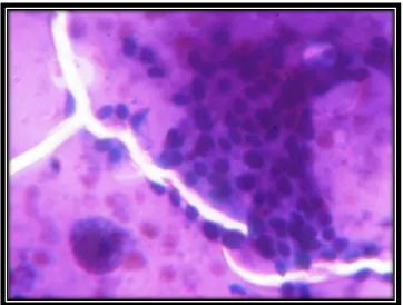
Cytological Criteria for diagnosis
Large multinucleate giant cell with numerous nuclei, phagocytosed colloid (Fig-1)
Epithelioid cells.
Degenerating follicular cells.
The gland may be enlarged either unilaterally or bilaterally. Firm in
consistency, usually with an intact capsule. Cut surface shows yellow-white firm
areas and these affected areas stand out from the normal thyroid parenchyma.
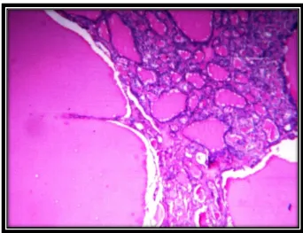
Microscopically the changes usually patchy and depends upon the stage of
the disease. In the early active inflammatory phase,the scattered follicles are
completely disrupted and may be replaced by polymorphs forming
microabscesses. In later stage, the more characteristic features may appear in the
form of aggregates of lymphocytes, plasma cells and activated macrophages
around damaged and collapsed thyroid follicles. Multinucleate giant cells may
enclose naked pools or fragments of colloid, hence the name granulomatous
thyroiditis (Fig-2). In later stages of the disease, fibrosis occurs.
Hashimoto thyroiditis. (Hashimoto’s disease, struma lymphomatosa)
Hashimoto thyroiditis is one of the most common immunologically
mediated disorder of the thyroid. First described by Hakaru Hashimoto in 1912 30. This disorder is most prevalent between the ages of 45 -65 years. Hashimoto
thyroiditis is common in women when compared to men. It has Female
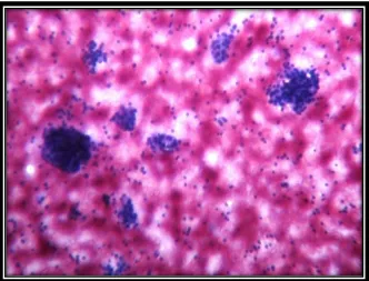
Cytological Criteria for diagnosis (Fig-3,4)
Oxyphilic transformation of epithelial cells (askanazy cells) Moderate number of lymphocytes and plasma cells
Scanty or no colloid
The thyroid is often diffusely enlarged with intact capsule. The gland is well
demarcated from the adjacent structures. Usually the cut surface appears pale,
yellow tan and firm (Fig- 5).
Histologic examination shows extensive infiltration of the thyroid
parenchyma with mononuclear inflammatory cell infiltrate containing
lymphocytes, plasma cells, and well-developed germinal center formation(Fig-6) .
The follicles are atrophic and are lined by Hurthle cells. These epithelial cells are
distinguished by the presence of abundant eosinophilic, granular cytoplasm. This
change is a metaplastic response of the normally low cuboidal thyroid follicular
epithelium to ongoing injury. Hashitoxicosis shows features of both Hashimoto
thyroiditis and Graves disease.
Graves’ disease - (Diffuse toxic goiter)
This is one of the common immunologically mediated disorder of the
thyroid. Graves disease has a peak incidence between 20 to 40 years of age.
Cytological Criteria for diagnosis
Blood stained smear with scanty colloid
Moderate amounts of thyroid follicular epithelial cells Cells have abundant vacuolated pale cytoplasm with
mild nuclear enlargement and showing moderate
anisokaryosis.
Fire flares/colloid suds/marginal vacuoles
Grossly,the thyroid gland is enlarged symmetrically because of diffuse
hyperplasia with hypertrophy of thyroid follicular epithelial cells . On cut
section the parenchyma appears as a soft and looks like normal muscle tissue.
Histologically, the thyroid follicular epithelial cells appear taller and
more crowded than usual in untreated cases. This crowding of thyroid
follicular epithelial cells results in the formation of small papillae (lack
fibrovascular cores) which may project into follicular lumen .The colloid
within the follicular lumen is pale with scalloped margins. Lymphoid infiltrates
consisting predominantly of T cells with fewer B cells and mature plasma cells
Goiters
Enlargement of the thyroid or Goiter is the most common manifestation
of thyroid disease.
DIFFUSE NONTOXIC GOITER (SIMPLE GOITER)
Diffuse nontoxic goiter (Simple Goiter)causes enlargement of the entire gland
without producing nodularity.
Cytological Criteria for diagnosis
Abundant colloid of varying thickness or excessive thick colloid with
normal Cytological appearance of follicular cells.
Two phases can be identified in the evolution of diffuse nontoxic goiter
hyperplastic phase and phase of colloid involution. In the hyperplastic phase,the
thyroid gland is diffusely and symmetrically enlarged although the increase is
usually modest and the gland rarely exceeds 100 to 150 grams. The follicles are
lined by crowded columnar cells which may pile up and form projections. In
theInvolutary phase cut surface appears as brown. Histologically the follicular
MULTINODULAR GOITER :
Repeated episodes of hyperplasia with involution combined to produce
more irregular enlargement of the thyroid gland producing Multinodular goiter.
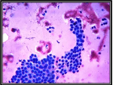
Cytological Criteria for diagnosis (Fig-7,8)
Abundant thin and thick colloid.
Small to moderate number of follicular epithelial cells in monolayered
sheets, poorly cohesive groups and single cells.
Both Involutional and hyperplastic follicular epithelial cells often some
Oxyphilic cells. Fragile cytoplasm.
Variable number of histiocytes.
Degenerative changes:old blood,debris.
Grossly Multinodular goiters are multilobulated, asymmetrically enlarged glands
that can reach weighs of more than 2000 grams. Cut section shows irregular
nodules containing variable amounts of brown gelatinous colloid. Older lesions
Microscopically colloid rich follicles lined by flattened inactive epithelium and
areas of follicular hyperplasia , areas of degenerative changes like hemorrhage,
calcification, fibrosis, and cystic change. (Fig-10)
TUMORS OF THE THYROID GLAND
World Health Organization (WHO) classification (2004) Of Tumors
of the thyroid gland is enclosed in Annexure-II
Benign tumors:
Follicular adenoma.
It is the most common tumor of the thyroid derived from follicular
epithelium hence they are known as Follicularadenoma .
Cytological Criteria for diagnosis(Fig-11,12) Cellular often bloody smear.
Many equal sized epithelial cell clusters scattered
throughout the smear.
Syncytial aggregates, nuclear crowding and overlapping.
Micro follicles.
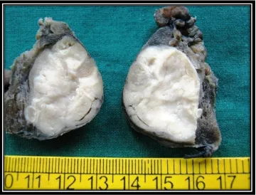
The typical thyroid adenoma is a solitary, spherical, encapsulated lesion that
is well demarcated from the surrounding thyroid parenchyma (Fig-13). On freshly
resected specimens the adenoma bulges from the cut surface and compresses the
adjacent thyroid. The colour ranges from graywhite to redbrown depending on the
cellularity of the adenoma and its colloid content.
Microscopically, the tumor cells are often arranged in uniform appearing
follicles that may contain colloid . The follicular growth pattern within the
adenoma is usually quite distinct from the adjacent non-neoplastic thyroid. The
epithelial cells composing the follicular adenoma reveal little variation in cell and
nuclear morphology. The hallmark of all follicular adenomas is the presence of an
intact well-formed capsule encircling the tumor.(Fig-14)
The most common patterns seen in follicular adenomas are as follows
Trabecular (embryonal),
Microfollicular (fetal),
Normofollicular (Simple),
The most important variants of follicular adenomas are
o Hurthle cell adenoma,
o Adenoma with clear cell change,
o Signet-ring cell adenoma,
o Hyalinizing trabecular adenoma,
o Adenoma with bizarre nuclei ,
o Adenoma with papillary hyperplasia ,
o Atypical adenoma,
o Adeno lipoma,
o Adeno chondroma.
PAPILLARY CARCINOMA
Papillary carcinomas are the most common form of thyroid cancer
accounting for nearly 85% of primary malignant thyroid neoplasm. They occur
throughout life but most often between the ages of 25 -50years.
Cytological Criteria for diagnosis (Fig-15,16) Cellular smears
Syncytial aggregates and sheets of cells with a distinct anatomical border. Papillary tissue fragments with or without a fibrovascular core
Multiple distinct nucleoli , intranuclear cytoplasmic inclusions and nuclear
grooves
Dense cytoplasm with distinct cell border.
Scanty viscous and stringy colloid(chewing gum colloid)
Squamoid or histiocyte-like metaplastic epithelial cells
Psammoma bodies
Macrophages and debris
Grossly presents as solitary or multifocal lesion ( 20% of cases),
encapsulated ( 10% of cases) or infiltrative lesion with variable degenerative
changes like fibrosis, calcification and cystic degeneration. The cut surface
sometime shows papillary foci that maybe useful to point the diagnosis.
(Fig-17)
Microscopically branching papillae have fibrovascular stalk covered
by single to multiple layers of epithelial cells. In many of them, the lining
epithelium of the papillae consists of well-differentiated, uniform and orderly
arranged cuboidal epithelial cells.(Fig-19)
The nuclei of tumor cells show finely dispersed chromatin, which gives
Annie eye or ground-glass nuclei .Invaginations of cytoplasm may in
cross-sections give the appearance of intranuclear inclusions (“pseudo-inclusions”) or
intranuclear grooves. (Fig-20) The diagnosis of papillary carcinoma made
based upon these nuclear features even in the absence of papillary
architecture.32
Psammoma bodies (concentrically calcified structures) are frequently
present within the papillary core.(Fig-18)
Variants of papillary thyroid carcinoma
Diffuse follicular variant
Cribriform-morular variant
Encapsulated variant
Diffuse sclerosing variant
Encapsulated follicular variant
Follicular variant
Macrofollicular variant
Microcarcinoma variant :
Oncocytic Variant
Tall Cell Variant
Clear Cell Variant
Solid variant of papillary carcinoma
Columnar cell variant
Nodularfasciitis like stroma variant
PROGNOSTIC FACTORS IN PAPILLARY CARCINOMA
Age: Mortality low in patients under the age of 40 years
Sex: Male sex associated with worse prognosis
Size: 1-1.5 cm excellent prognosis, >4cm poor prognosis.
Stage: Extra thyroidal extension-poor prognosis.
Tumor encapsulation confers a favourable prognosis.
Histological variants: Tall cell, Diffuse follicular, Diffuse
sclerosing, Solid variants, Cribriform-morular variant -more
Anaplastic (Undifferentiated) Carcinoma
Anaplastic carcinomas are undifferentiated neoplasm of the thyroid follicular
epithelium accounting for less than 5% of thyroid tumors. Manifests in older age
than those with other types of thyroid cancer. The mean age of presentation is 65
years.
Cytology : (Fig-21)
Highly cellular with bizarre large malignant cells showing
epithelial or spindle sarcomatoid type.
Prominent nuclear pleomorphism,multinucleation and mitotic
figures
Background shows necrotic cell fragments and debris
Gross: cut section shows large bulky ,soft, fleshy and lobulated mass with areas
of necrosis, hemorrhage and cystic degeneration.
Microscopically these tumors composed of highly anaplastic cells with variable
morphology including: (1) large pleomorphic cells including occasional
osteoclast-like multinucleate giant cells (2) spindle cells showing sarcomatous
appearance (Fig-22,23) (3) mixed spindle and giant cells. Foci of papillary or
follicular differentiation may be present in some tumors suggesting an origin from
Medullary carcinoma of thyroid gland
Thyroid Medullary carcinomas are neuroendocrine neoplasms and derived
from C cells of thyroid or parafollicular cells.It accounts for 5% of thyroid
neoplasms.
Cytological Criteria for diagnosis
Cellular smears with dispersed cells, some clustering may be seen .
Variable cell pattern showing plasmacytoid, spindle and small cells
Moderate anisokaryosis, occasional scattered very large nuclei with bi and
multinucleate forms
Uniform stippled nuclear chromatin
Amorphous pink/violet background (amyloid)
Grossly, sporadic medullary thyroid carcinomas present as a solitary nodule.
In contrast, bilaterality and multicentricity are common in familial cases. Larger
lesions often contain areas of necrosis and hemorrhage and may extend through
the capsule of the thyroid. The tumor tissue is firm, pale gray to tan and
Microscopically medullary carcinomas are composed of spindle shaped to
polygonal cells which may form trabeculae ,nests and even follicles. (Fig-24)
Small more anaplastic cells are present in some tumors and may be the
predominant cell type. Acellular amyloid deposits are present in the adjacent
stroma in many casesthat can be demonstrated by congo –red stain (Fig-25).
Variants of medullary thyroid carcinoma
Medullary microcarcinoma
Paraganglioma-like variant
Small cell variant
Tubular (follicular) variant
FOLLICULAR CARCINOMA
Follicular carcinomas account for 5% to 15% of primary thyroid cancers.
They are more common in women (3 : 1) and manifests at an older age than
papillary carcinomas .The Peak incidence is found between 40 - 60 years of age.
Follicular carcinomas presents as single nodule that may be well
circumscribed or widely infiltrative.They are gray tan to pink on cut section.
Degenerative changes such as central fibrosis and foci of calcification are
Microscopically most follicular carcinomas composed of fairly uniform
cells arranged in small follicles and containing colloid. In some cases follicular
differentiation may be minimal and there may be sheets and nests of cells without
colloid. Whatever the pattern ,the nuclei lack the features of typical of papillary
carcinoma.
Hurthle cell or oncocytic variant of follicular carcinoma Tumor cells with
abundant eosinophilic granular cytoplasm .
Minimally invasive follicular carcinoma. This variant requires extensive
histologic sampling from the tumor-capsule-thyroid interface to exclude capsular
or vascular invasion.
Widely invasive follicular carcinomas. Infiltrate the thyroid and extra-thyroidal
soft tissues.
Recommendation of Rosai in classifying definitive follicular
carcinomas as follows:
-Encapsulated
- With capsular invasion only
-With extensive (more than 4 vessels ) vascular
invasion
-Widely invasive
Touch Impression Cytology (TIC)
Intraoperative cytological diagnosis is required for the optimal extent of
surgery and to know the nature of lesion whether the lesion is malignant or not.
Both Frozen Section (FS) and Touch Impression Cytology (TIC) serve this purpose
well. Both provide accurate results in minutes while the patient is under anesthesia.
Surgeon then modifies his surgical plan based on the intraoperative consultation
with pathologist. While FrozenSection tissue architecture closely approximates
permanent histology sections, enabling a degree of comfort, Touch Impression
Cytology provides better, crisp cellular details and even some tissue architecture
with fewer artifacts.
Immunohistochemistry
Role of Ki-67 as a proliferative marker in lesions of thyroid
Ki-67 is an IgG1 type murine monoclonal antibody raised against a crude
nuclear fraction of Hodgkin's disease-derived cell line L-428. The ki 67was named
Ki67antibody was grown in the sixty seventh well of tissue culture plate. Ki-67 is
a novel proliferative marker that can be readily detected by immunohistochemistry.
Gerdes et al. have shown that all stages of the cell cycle will express Ki-67 except
G-0 because resting cells entering from G-0 lack Ki-67 in early part of G1.
Saad et al. determined the proliferative rate of normal human thyroid cells in
different age groups using Ki-67 and found Ki-67 Labeling Index to be 7.4 ±
6.10%in 25 fetal thyroids, 0.23 ± 0.15% in 55 childhood thyroids and 0.08 ±
0.04% in 37 adults at autopsy.
Ki- 67 marker study may be helpful in distinguish undifferentiated areas from
differentiated areas in a mixed type of thyroid cancer.
Ki-67 labeling index (LI) show progressive increase from multinodular goiter to
MATERIAL AND METHODS
In the two and half year study period from January 2010 to May 2012, 20908
specimens were received in the Department of pathology, Madurai Medical
College, Madurai for histopathological examination from Government Rajaji
Hospital, Madurai. Among these 1123 cases were from head and neck lesions and
626 cases from thyroid gland lesions.
During the study period 1026 Fine Needle Aspiration Cytology from
thyroid were received for cytological examination. Out of these 117 cases had post
surgical followup. A range of cytological diagnosis was offered on all satisfactory
smears. A correlative cytological and histopathological study was done. Imprint
cytology was done for 51 cases and a final correlative study was done between
Fine Needle Aspiration Cytology , Imprint cytology and Histopathology .
1123 specimens were from various sites in head and neck region such as
Scalp, periorbital region, ear, nose, cheek , lip, tonsil , tongue, thyroid, salivary
glands and lymph nodes. Out of these 117 specimens were from thyroid and these
cases were taken for this study .Out of these 117 cases imprint cytology was
done for 51 cases .
The detailed clinical history of these 117 patients including the duration of
expectoration etc. were obtained and tabulated in the proforma and which was
enclosed in annexure I.
Fine Needle Aspiration Cytology was done for 117 thyroid cases. The
aspiration syringes used were 10-20 ml and the needle size between 22-23
gauges .The cytological materials obtained were fixed in ninety five(95)% ethyl
alcohol then stained with haemotoxylin and eosin .The reports were recorded in
master chart-A.
Touch Imprint cytology was done for 51 cases on freshly cut surface of the
specimen by gently pressing the glass slide. Then the slides were immediately
wet fixed in ninety five percent ethyl alcohol for five to six seconds. Then the
smears were stained with haematoxylin and eosin. Results were recorded in
master chart-B.
The specimens of lobectomy, hemi thyroidectomy, near total
thyroidectomy and total thyroidectomy with modified neck dissection were
received for histopathological examination.
The specimens were fixed in 10% formalin for 24 – 48 hours .Then
detailed gross examination including weight, measurement, shape , colour and
consistency were noted. They were cut into parallel and longitudinal slices
cystic degeneration, calcification, necrosis and distance from the line of resection
were noted. The representative sections were taken from the lesions as shown in
the table number.1
Table 1
Thyroid lesion Number of sections41
For diffuse or inflammatory lesions Three sections from each lobe and one from isthmus
Solitary encapsulated nodule Sections from the entire circumference including tumor capsule and adjacent thyroid tissue
Multi nodular thyroid gland One section from each nodule including adjacent thyroid tissue Papillary carcinoma Entire thyroid gland was blocked Grossly invasive carcinoma (other than
papillary carcinoma)
Three sections from tumor and three sections from non neoplastic gland and one from line of resection
The tissue slices were processed in various grades of alcohol and xylol and
subsequently embedded in paraffin wax. Paraffin sections of 4 μm thickness were
subjected to haemotoxylin and eosin staining .(H and E staining technique is
A correlative study between Fine Needle Aspiration Cytology and
histopathology was done for these 117 cases and recorded in masterchart-A.
FNAC, Touch imprint cytology, Histopathological reports of 51 cases
were recorded seperately in master chart-B and final correlative study was done.
Immunohistochemistry ki67 marker study was done for some selective
cases and reports were recorded. The procedure of Immunohistochemistry ki67
marker study was enclosed in annexure-III.
Statistical Tools
Data analysis was done with the help of computer using Epidemiological
Information Package (EPI 2002).
The Statistical datas are calculated by using the following formulae
Sensitivity = True positive x 100
True positive + False negative
Specificity = True negative x 100
False positive + True negative
Positive predictive value = True positive x 100
True positive + False positive
Negative predictive value = True negative x 100
True negative + False negative
Accuracy = True positive + True negative x 100
OBSERVATION AND RESULTS
In the two and half year study period from January 2010 to May 2012, 626
thyroid specimens were received in the Department of pathology, Madurai
Medical College, Madurai for histopathological examination from Government
Rajaji Hospital, Madurai. The average incidence of thyroid lesions in this hospital
was 2.99%.
117 cases of Fine Needle Aspiration Cytology from thyroid had post
surgical followup. A range of cytological diagnosis was offered on all satisfactory
smears. A correlative cytological and histopathological study was done. Imprint
cytology was done for 51 cases and a final correlative study was done between
Fine Needle Aspiration Cytology , Imprint cytology and Histopathology.
AGE INCIDENCE:
The age incidence of various thyroid lesions were categorized and
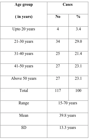
Table- 2 : Age distribution
Age group
( in years)
Cases
No %
Upto 20 years 4 3.4
21-30 years 34 29.0
31-40 years 25 21.4
41-50 years 27 23.1
Above 50 years 27 23.1
Total 117 100
Range 15-70 years
Mean 39.8 years
SD 13.3 years
In the present study, the youngest patient was 15 years old
Chart 1 -Age distribution
4% 29%
21% 23%
23%
Age group wise distribution of thyroid lesions
The age group wise distribution of thyroid lesions were tabulated in table number- 3.
Table- 3 Age group wise distribution of thyroid lesions
AGE
GROUP
MNG HASH
THY
GRA.
THY
FOLL
ADE
PAP.CA MEDU.
CA
ANA.CA
11-20 2 2
21-30 12 7 9 6
31-40 12 3 8 1 1
41-50 15 3 4 4 1
51-60 11 3 2 5
61-70 2 2 1 1
Non neoplastic and neoplastic (benign and malignant) lesions were found to be
SEX INCIDENCE:
The sex incidence of thyroid lesions were tabulated in table number -4 and chart
number -2.
Table -4 : Sex incidence
Sex
Cases
No %
Male 8 6.8
Female 109 93.2
Total 117 100
Chart- 2
Among total 117 cases, 109 patients were female (93.16%) and 8 patients were male
(6.84%). There is a female preponderance with a Female to Male ratio of 13.6:1.
The age of female patients were ranging from 15-70 years and male patients
ranging from 30-69 years
93% 7%
Sex distribution of thyroid lesions
Sex distribution of thyroid lesions tabulated in table number- 5
Table - 5 . -Sex distribution of thyroid lesions
HPE diagnosis Female Male
Non neoplastic lesions 69 4
Benign neoplastic lesions 24 1
Malignant neoplastic lesions 16 3
Total 109 8
Non neoplastic lesions
Among 73 non neoplastic lesions , 69 cases were female and the remaining 4
cases were male
Table 6- Sex Distribution Of Non Neoplastic Thyroid Lesions
HPE diagnosis Female Male
Nodular goiter 50 4
Hashimatothyroiditis 18
Granulomatous thyroidits 1
Benign Neoplastic Lesions
Among 25 benign neoplastic lesions 24 cases were female and one case
was male
Malignant Neoplastic Lesions
Among 19 Malignant neoplastic lesions 16 cases were female and 3 cases were
male .
Table- 7. Sex distribution Malignant neoplastic thyroid lesions
HPE diagnosis Female Male
Papillary carcinoma 14 2
Medullary carcinoma 1 1
Anaplastic carcinoma 1
CYTOLOGICAL EVALUATION OF THYROID LESIONS:
The cytological diagnosis was offered for 117 cases which had post
Table -8 : FNAC Diagnosis
Out of these 117 FNAC studies, 81cases were reported as nodular goiter , 12
cases as papillary carcinoma, 9 cases as hashimato thyroiditis, 5 cases as
FNAC Diagnosis Cases
No %
Nodular goiter 81 69.23
Papillary Carcinoma 12 10.3
Lymphocytic Thyroditis 5 4.3
Hashimato Thyroditis 9 7.7
Follicular neoplasm 8 6.8
Anaplastic carcinoma 1 0.85
Granulomatous thyroiditis 1 0.85
lymphocytic thyroiditis, 8 cases as follicular neoplasm, one case as
Granulomatous thyroiditis and another one case as anaplastic carcinoma .
Chart-3. Cytological distribution Of Thyroid Lesions
69% 10%
4%
8% 7%
2 %
Nodular Goitre Papillary Ca.
Lymphocytic Thyroiditis Hashimato Thyroiditis
Imprintcytology diagnosis
Imprintcytological diagnosis was offered for 51 cases and shown in table
number -9 and chart number -4.
Table 9: Imprint cytology Diagnosis
Imprint cytology diagnosis Number of cases
Nodular goiter 27
Papillary carcinoma 7
Hashimato thyroiditis 6
Follicular neoplasm 7
Lymphocytic thyroiditis 2
Anaplastic carcinoma 1
Granulomatous thyroiditis 1
TOTAL 51
Out of these 51 thyroid imprint cytology studies 27 cases were reported as
nodular goiter , 7 cases as papillary carcinoma, 6 cases as Hashimato thyroiditis,
2cases as lymphocytic thyroiditis, 7 cases as follicular neoplasm, one case as
Among them nodular goiter was the commonest lesion found in this study.
Chart-4 Imprint Cytological distribution Of Thyroid Lesions
HISTOPATHOLOGICAL DIAGNOSIS;
The histopathological diagnosis was offered for 117 cases of thyroid
lesions which had preoperative cytological diagnosis. The distribution of various
non neoplastic and neoplastic thyroid lesions tabulated in table number -10 and chart number -5. Out of these 117 lesions 73 cases were non neoplastic lesions
and 44 were neoplastic lesions.
53% 12%
14%
14% 7%
Nodular Goitre Papillary Ca.
Hashimato Thyroiditis Follicular neoplasm
Table -10. histopathological diagnosis;
LESION Number of cases Percentage
NONNEOPLASTIC
LESIONS
73 62
NEOPLASTIC LESIONS 44 38
Chart-5 Histopathological Distribution Of Thyroid Lesions
NONNEOPLASTIC LESIONS
Among the 73 non neoplastic lesions 54cases were reported as nodular goiter ,18 cases as hashimatothyroiditis and 1 case as Granulomatous thyroidits and shown
in table number -11 and chart number -6.
62% 38%
Table 11- Nonneoplastic Lesions
Diagnosis Number of cases
Nodular goiter 54
Hashimotothyroiditis 18
Granulomatous thyroidits 1
Total 73
Chart 6- Distribution Of Non Neoplastic Thyroid Lesions
NEOPLASTIC LESIONS
Out of the 44 neoplastic lesions 25 cases were reported as benign neoplastic
lesions and 19 as malignant neoplastic lesions and shown in table number -12 and
chart number - 7.
74%
25% 1%
Table-12. Neoplastic Thyroid Lesions
Diagnosis Number of cases
BENIGN NEOPLASTIC LESIONS
Follicularadenoma 25
MALIGNANT NEOPLASTIC LESIONS
Papillary carcinoma 16
Medullary carcinoma 2
Anaplastic carcinoma 1
Total 44
Among the 19 malignant neoplastic lesions 16 cases were reported as
papillary carcinoma , 2 cases as medullary carcinoma thyroid and 1 case as
anaplastic carcinoma thyroid .
Chart 7- Distribution Of Neoplastic Thyroid Lesions
57% 36%
5%
CORRELATIVE STUDY BETWEEN FINE NEEDLE ASPIRATION
CYTOLOGY(FNAC) AND HISTOPATHOLOGY(MASTER CHART –A)
A Correlative Study Between Fine Needle Aspiration Cytology And Histopathology
was done for 117 cases and tabulated in table number -13.
Table-13 Correlation between FNAC AND HPE
FNAC AND HPE
CORRELATION
NUMBER OF
CASES
PERCENTAGE
CORRELATED 65 55.5%
NOT CORRELATED 52 44.5%
Among 117cases , 65 Fine Needle Aspiration cytology reports (55.5%) were well
correlated with histopathological diagnosis. The remaining 52 Fine Needle
Aspiration cytology reports(44.5%) were not correlated with histopathological
diagnosis and tabulated in table -14. Among them 36 cases were reported as
Nodular goiter, 5 cases as Lymphocytic thyroiditis, 4 cases as
Hashimotothyroiditis , 5 cases as Follicular neoplasm and 2 cases as papillary
Table -14.Correlation Between FNAC And Histopathology FNAC DIAGNOSIS NO.OF CASES HPE DIAGNOSIS
MNG HAS
THY FOLL. ADE PAP CA MEDU CA
ANA CA Gran.thy
Nodular goiter 81 45 10 20 5 1
Papillary carcinoma 12 1 1 10
Hashimotothyroiditis 9 2 5 1 1
Lymphocytic
thyroiditis
5 1 2 2
Follicular neoplasm 8 5 3
Anaplasticcarcinoma 1 1
Gran. thyroiditis 1
In the present study 81 cases of Nodular goiter on Fine Needle Aspiration
cytology were found to be Nodular goiter in 45 cases, Follicular adenoma in 20 cases, Hashimato thyroiditis in 10 cases ,Papillary carcinoma in 5 cases and
Medullary carcinoma in one case on subsequent histopathological examination .
5 cases of Lymphocytic thyroiditis on Fine Needle Aspiration cytology were
found to be Nodular goiter in 1 case, Follicular adenoma in 2 cases and Hashimato
8 cases of Follicular neoplasm on Fine Needle Aspiration cytology were found to be Nodular goiter in 5 cases and Follicular adenoma in 3 cases on subsequent
histopathological examination.
9 cases of Hashimato thyroiditis on Fine Needle Aspiration cytology were found
to be Hashimato thyroiditis in 5 cases, Nodular goiter in 2 cases, Medullary
carcinoma in one case and Papillary carcinoma in one case on subsequent
histopathological examination.
12cases of Papillary carcinoma on Fine Needle Aspiration cytology were found to
be Papillary carcinoma in 10 cases, Hashimato thyroiditis in one case and Nodular
goiter in one case on subsequent histopathological examination.
One case of Anaplastic carcinoma and another one case of Granulomatous
thyroiditis on Fine Needle Aspiration cytology were confirmed by subsequent
histopathological examination .
FINAL CORRELATION BETWEEN FINE NEEDLE ASPIRATION
CYTOLOGY, IMPRINT CYTOLOGY AND HISTOPATHOLOGICAL
DIAGNOSIS (MASTER CHART –B)
Final Correlative study was done by comparing the results of Fine Needle
Correlation Between Fine Needle Aspiration Cytology (FNAC)And
Histopathology.
Out of 51 thyroid Fine Needle Aspiration cytology studies, 32cases were
reported as nodular goiter, 6 cases as papillary carcinoma, 2 cases as
Hashimotothyroiditis, 4cases as lymphocytic thyroiditis, 5 cases as follicular
neoplasm, one case as Granulomatous thyroiditis and another one case as
anaplastic carcinoma.
A correlation done between Fine Needle Aspiration cytology and
histopathology showed the following results. 33 Fine Needle Aspiration cytology
reports (64%) were correlated with histopathological diagnosis and tabulated in
table -15, chart number- 8.
In the remaining 18 (36%) Fine Needle Aspiration cytology reports were not correlated with histopathological diagnosis. Among them 11 cases were
reported as Nodular goiter, 4 cases as Lymphocytic thyroiditis and 3 cases as
Table-15. Correlation Between FNAC And Histopathology.
FNAC
DIAGNOSIS
HISTOPATHOLOGICAL DIAGNOSIS
NO.OF
CASES
MNG HASH
THY
GRA
THY
FOLL
ADE
PAP CA ANA CA
Nodular goiter 32 21 3 5 3
Lymphocytic
thyroiditis
4 1 2 1
Hashimatothyroiditis 2 2
Granulomatous
Thyroiditis
1 1
Follicular neoplasm 5 3 2
Papillary carcinoma 6 6
Anaplastic carcinoma 1 1
In this study 32 cases of Nodular goiter on Fine Needle Aspiration cytology were
found to be Nodular goiter in 21 cases, Follicular adenoma in 5 cases, Hashimato
thyroiditis in 3 cases and Papillary carcinoma in 3 cases on subsequent
4 cases of Lymphocytic thyroiditis on Fine Needle Aspiration cytology were found to be Nodular goiter in 1 case, Follicular adenoma in 1 case and Hashimato
thyroiditis in 2 cases on subsequent histopathological examination.
5 cases of Follicular neoplasm on Fine Needle Aspiration cytology were found to
be Nodular goiter in 3 cases and Follicular adenoma in 2 cases on subsequent
histopathological examination.
2 cases of Hashimato thyroiditis, 6 cases of Papillary carcinoma , one case of
Granulomatous thyroiditis and another one case of Anaplastic carcinoma were
subsequently confirmed on histopathology.
CHART-8 -Correlation between FNAC And HPE
0 5 10 15 20 25 30 35
Foll.ade Papillary ca Mng Anaplastic ca Hashi
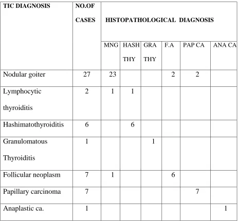
Correlation Between Imprintcytology And Histopathology
A correlative study was done between Touch imprint cytology
and histopathology showed the following results and tabulated in table number-
16, chart number -9. Among them 44(86%) Touch imprint cytology reports were
[image:62.612.66.529.282.712.2]correlated with histopathological diagnosis.
Table-16 Correlation Between Touch imprint cytology And Histopathology
TIC DIAGNOSIS NO.OF CASES
HISTOPATHOLOGICAL DIAGNOSIS
MNG HASH THY
GRA THY
F.A PAP CA ANA CA
Nodular goiter 27 23 2 2
Lymphocytic
thyroiditis
2 1 1
Hashimatothyroiditis 6 6
Granulomatous
Thyroiditis
1 1
Follicular neoplasm 7 1 6
Papillary carcinoma 7 7
In the present study 27 cases of Nodular goiter on Touch imprint Cytology were
found to be Nodular goiter in 23 cases, Follicular adenoma in 2 cases and Papillary
carcinoma in 2 cases.
2 cases of Lymphocytic thyroiditis on Touch imprint Cytology were found to be
Nodular goiter in 1 case and Hashimotothyroiditis in one case on subsequent
histopathological examination.
7 cases of Follicular neoplasm on Touch imprint Cytology were found to be
Follicular adenoma in 6 cases and Nodular goiter in 1 case on subsequent
histopathological examination.
6 cases of Hashimotothyroiditis, 6 cases of Papillary carcinoma,one case of
Granulomatous Thyroiditis and another one case of Anaplastic carcinoma on
Touch imprint Cytology were subsequently confirmed by histopathology.
CHART-9 Correlation Between TIC And HPE
0 5 10 15 20 25 30
Folli. Ade Pap. Ca Mng Ana.ca Hashi
Immunohistochemistry
Ki-67 Immunohistochemical staining was done for six different types of
thyroid lesions such as Granulomatous thyroiditis , Hashimotothyroiditis, Nodular
goiter, Follicular adenoma ,Papillary carcinoma and Anaplastic carcinoma .
An area with the maximum proliferation was chosen to evaluate the labeling index. Labeling index was expressed as percentage of positively stained cells
(Brown granular nuclear reactivity) per 100 follicular epithelial cells after counting
at least 1000 cells in each case. The staining pattern in various thyroid lesions
tabulated in table number -17
Table-17.Ki-67 staining pattern in various thyroid lesions
SNO HPE DIAGNOSIS Ki-67 staining
1 Granulomatous thyroiditis Negative
2 Hashimato thyroiditis Positive in germinal centre of follicles.
3 Multi Nodular goiter Very few cells positive
4 Follicular adenoma Positive
5 Papillary carcinoma Positive(1 to 2%)
6 Anaplastic carcinoma Strong positivity
In the present study, the mean values of Ki-67 Labeling index was increasing
DISCUSSION
Fine Needle Aspiration Cytology of thyroid has become the most common
and well established preoperative diagnostic procedure used in the management
of patients with thyroid lesions. It is relatively cost effective procedure that provide
diagnosis rapidly.
Incidence of thyroid lesions :
We received 117 gross specimens for histopathological examination
following initial cytological evaluation by fine needle aspiration cytology. In
present study non neoplastic lesions accounts for 73 cases and neoplastic lesions
accounts for 44 cases. The ratio between non neoplastic and neoplastic thyroid
lesions in this study is 1.66:1.
Incidence of nonneoplastic and neoplastic thyroid lesions in this study is
Table – 18 Incidence of thyroid lesions
S.NO Series
Non
Neoplastic
Neoplastic Ratio
1. Pepper G.M 35 84 18 4.66:1
2. Dorairajan N 7 78 20 3.90:1
3. Sarda AK 45 87 59 8.25:1
4. Naggada HA 33 51 18 2.83:1
5. Gupta C 17 470 30 15.66:1
6. Kaur K 23 32 15 2.13:1
7. Due k SD 145 61 2.37:1
8. Hurtado – LopezLM 19 80 50 1.60:1
9. Talepoor M 53 325 75 4.33:1
10. Prakash H.M 37 138 24 5.75:1
Incidence of Malignancy :
In the present study, the incidence of malignant neoplastic thyroid lesions
accounts for 16.24% which well correlates with studies conducted by various
research workers as well as in literature and tabulated in the Table number -19.
Table19 – Incidence of Malignancy
Sl.
No.
Study Percentage
1. Mary Jo Welker et al 28 5-10%
2. Kaur et al 23 18%
3. YS Chenug et al 56 5-10%
4. Munsad B et al 31 4.16%
5. Alexander Kessler 2 10%
6. Suresh et al 51 10%
7. GG Swamy et al 13 18.33%
8. Prakash H.M 37 14.81%
9. present study 16.24%
AGE INCIDENCE:
In the present study the mean age of presentation is 39.8 years which
correlates with the literature of various authors and tabulated in table number -20.
Table 20– Comparative Incidence of Mean age in Different Studies
Sl.No Studies Mean Age
1. Quari F et al 39 36.17 years
2. Wasser MH et al 54 44 years
3. Suresh Kumar et al 51 38.5 years
4. Talepoor M et al 53 38.6 years
5. Das DK et al 6 35 years
6. Prakash HM et al 37 35.67 years
7. Manoj Gupta et al 26 38.7 years
8. Martin et al 27 39.5years
SEX INCIDENCE :In this study , majority of them were females. Female to male
ratio of 13.6 : 1 and correlates with observation of other various authors as
indicated in table number-21.
Table – 21
Comparative sex incidence of thyroid lesions
in different studies
Sl.
No.
Studies
Sex Incidence
(female : male) ratio
1. Das DK 6 5.39 : 1
2. Manoj Gupta 26 11;1
3. Martin etal 27 6.4:1
4. Prakash H.M 37 7.1 : 1
5. Dorairajan N7 9:1
FINAL CORRELATIVE STUDY BETWEEN FINE NEEDLE ASPIRATION
CYTOLOGY , TOUCH IMPRINT CYTOLOGY AND HISTOPATHOLOGY
In 2008 Handa u et al reported that Fine Needle Aspiration Cytology(FNAC)
is routinely used preoperativily for the assessment of thyroid lesions and it
cutsdown the number of patients subjected to thyroidectomy for benign diseases of
the thyroid. Intraoperative cytological diagnosis is required for the optimal extent of
surgery and to know either the lesion is malignant or not. Both Touch Imprint
Cytology (TIC) and Frozen Section (FS) serve this purpose well. Both provide
accurate results within minutes.
In the present study , specimens of lobectomy,hemi thyroidectomy, near
total thyroidectomy and total thyroidectomy were received for histopathological
examination which offers final and confirmatory postoperative diagnosis of the
specimens.
Fine Needle Aspiration Cytology was done pre operatively for 1026 cases in
our institution during the study period and imprint cytology was undertaken
intraoperatively for 51 cases followed by histopathological examination
postoperatively and final diagnosis was made . Since imprint cytology was available
for only 51 cases a correlative study between FNAC, imprint cytology and
Correlation Between Fine Needle Aspiration Cytology and Histopathology
In the present study, non neoplastic lesions accounts for 39cases out of 51
cases and neoplastic lesions accounts for 12 cases out of 51 cases. Among them
Fine Needle Aspiration Cytology and Histopathology reports correlated well in
33cases 64%. (24 cases in non neoplastic lesions and 9 cases in neoplastic lesions) .
Non neoplastic lesions:
In the present study among the non neoplastic lesions Nodular goiter was the most
common lesion.
In this study 32 cases 0f Nodular goiter on Fine Needle Aspiration Cytology
were well correlated with 21 cases in histopathology. Among the other non
neoplastic lesions 2cases of Hashimato thyroiditis , 1case of Granulomatous
Thyroiditis were well correlated with histopathology.
4cases of Lymphocytic thyroiditis on Fine Needle Aspiration Cytology showed
varied diagnosis in histopathology.
Neoplastic lesions: In our study among the neoplastic lesions Papillary carcinoma
was the most common lesion followed by follicular neoplasm.
6 cases of Papillary carcinoma , 1case of Anaplastic carcinoma on Fine
Needle Aspiration Cytology were well correlated with subsequent histopathology.
Among the 5cases of follicular neoplasm 2 cases were well correlated with
subsequent histopathology, the remaining 3cases showed different
histopathological diagnosis.
Diagnostic problems were experienced in 11 cases of Nodulargoiter ,4 cases
of Lymphocytic thyroiditis and 3cases of follicular neoplasm.
Among the preoperatively diagnosed 11 cases of Nodulargoiter 5 cases
turned out to be Follicular adenoma, 3 cases as Hashimotothyroiditis and 3 cases
as Papillary carcinoma in subsequent histopathology .The reasons are discussed
below.
Problems are experienced in diagnosing the following thyroid lesions
1Nodulargoiter
2.Follicular neoplasm
3.Hyper plastic nodules
4.Cystic nodule/Cystic papillary carcinoma
5.Thyroiditis
The cytological picture of Nodulargoiter can overlap with follicular
neoplasm at times. Smears from microfollicular area in nodular goiter may show
nodule will show marked cellularity of the smear which may mimic follicular
neoplasm. Since this is a focal phenomenon, samples from other different areas
should be taken to avoid misdiagnosis.
Cystic lesions of thyroid constitute a particular problem in Fine Needle
Aspiration cytology .Cystic change and hemorrhage can occur not only in non
neoplastic lesions but also in neoplastic lesions like follicular neoplasm and
papillary carcinoma. If only cystic fluid containing macrophages but no epithelial
cells are obtained neoplasm with cystic change cannot be ruled out 49. In such cases fine needle aspiration biopsy should be done. Recurrent cysts greater than
3-4 cm is identified for surgery with ultra sound guidance 14. To identify the neoplastic lesion with cystic change fine needle aspiration biopsy is advised along
with ultra sound guidence to minimise the false negative diagnosis.
Cystic Papillay Carcinomas often contain abundant colloid. This can cause
diagnostic problem especially if smears are poor in cells. In Nodulargoiter groups
of large cells with irregulur nuclei of uncertain origin are frequently seen. They
may be regenerating epithelial cells consistent with repair or may be histiocytes .
These aggregates of histiocytes can mimic cells of papillay carcinomas in some