Copyright © 1999, American Society for Microbiology. All Rights Reserved.
Genetic Interaction of Flavivirus Nonstructural Proteins NS1
and NS4A as a Determinant of Replicase Function
BRETT D. LINDENBACHANDCHARLES M. RICE*
Department of Molecular Microbiology, Washington University School of Medicine, St. Louis, Missouri 63110-1093 Received 11 January 1999/Accepted 24 February 1999
Nonstructural protein 1 (NS1) of yellow fever virus (YF) is a glycoprotein localized to extracytoplasmic
compartments within infected cells. We have previously shown that NS1 can be supplied in trans and is
required for viral RNA replication, a process thought to occur in membrane-bound cytoplasmic complexes. Here we report that the NS1 gene from a related virus, dengue virus (DEN), is unable to function in the process of YF RNA replication. This virus-specific incompatibility leads to a lack of initial minus-strand accumulation, suggesting that DEN NS1 is unable to productively interact with the YF replicase. Based on a YF deletion
mutant that requires NS1 intrans, a genetic screen for suppressor mutants was used to select virus variants
able to utilize DEN NS1. In three independent selections, a single mutation was mapped to the NS4A gene, which encodes a putative transmembrane replicase component. This mutation, as well as several additional mutations, was engineered into the NS1-deficient genome and confirmed a genetic interaction between NS1 and NS4A. These findings suggest a potential mechanism for integrating NS1 into the cytoplasmic process of RNA replication.
Replicases of eukaryotic positive-strand RNA viruses have intricate relationships with cellular membranes, a fact which often complicates their detailed analysis. Nevertheless, impres-sive advances have been made in understanding the structure and function of several viral replicases (18, 19, 53). We have been investigating the replication of yellow fever virus (YF), which also utilizes a membrane-bound replicase (10, 17). YF is the prototype for the flaviviruses, an important group of hu-man pathogens that includes dengue virus (DEN), Japanese encephalitis virus, and tick-borne encephalitis virus. In this paper, we reveal a genetic interaction that exists between a flavivirus protein residing in the secretory pathway and a mem-brane protein postulated to be part of the viral replicase and show that this interaction is crucial for the process of flavivirus RNA replication.
The flavivirus genome consists of an;11-kb, single-stranded RNA molecule of positive polarity (36). This RNA is capped at its 59end and serves as the mRNA for the translation of a large polyprotein. Mature viral proteins are processed from this pre-cursor by a viral serine protease and multiple host proteinases (34). The virion structural components are encoded in the 59 one fourth of the genome, while viral nonstructural (NS) pro-teins are encoded in the remainder of the genome. The overall gene order is C-prM-E-NS1-NS2A-NS2B-NS3-NS4A-NS4B-NS5. The major function of NS proteins is the replication of viral RNA, and several relevant activities have been mapped to NS proteins. NS5 is the RNA-dependent RNA polymerase as well as a putative methyltransferase thought to be involved in the formation of the RNA cap (6, 46). The N-terminal region of NS3 contains the catalytic residues of the viral serine pro-tease, which requires the protein cofactor NS2B for activity (9, 15). The remainder of NS3 comprises an RNA triphosphatase, which may contribute to RNA capping (49), as well as an RNA helicase with nucleoside triphosphatase activity (16, 48).
Sev-eral other NS proteins (NS2A, NS4A, and NS4B) are small hydrophobic proteins with unknown functions, although it has been suggested that they may serve to anchor the viral repli-case to cellular membranes (8).
NS protein 1 (NS1) is inserted into the endoplasmic reticu-lum (ER) by a signal sequence located in the C terminus of the E protein and is processed by host signal peptidase (13). Shortly after synthesis, NS1 is cleaved from NS2A by an un-known ER-resident host proteinase (14). NS1 is a glycoprotein that forms homodimers and interacts with membranes via an unknown mechanism (42, 51, 52). It is largely retained within the cell, where it has been localized to presumed sites of RNA replication (26, 50). A significant proportion of NS1 is also secreted, and a minor amount associates with the cell surface. The flavivirus genome is replicated via a minus-strand inter-mediate that continues to accumulate over the course of in-fection, although the ratio of plus-strand RNA to minus-strand RNA may change over time (12). Despite its extracytoplasmic localization, NS1 is an essential component of the viral repli-case. Defined mutations in NS1 can result in dramatic de-creases in the level of viral RNA accumulation, and such mu-tations provided the first functional data indicating that NS1 is involved in this process (29, 30). We have recently demon-strated that a YF genome bearing a large in-frame deletion in the NS1 gene, YFDSK, is severely defective in viral replication (25). In order to study this defect, we demonstrated that YFDSK could be complemented intransby the expression of a functional NS1 gene via a noncytopathic Sindbis virus vector. This strategy permitted the analysis of YFDSK replication with or withouttrans complementation. YFDSK required NS1 for growth, plaque formation, and RNA replication. Examination of early events in RNA replication revealed a requirement for NS1 prior to or at initial minus-strand synthesis.
The objective of this investigation was to analyze the ability of a heterologous NS1 gene to function in thetrans comple-mentation of YFDSK. We initially found that YFDSK did not complete early minus-strand synthesis in cells expressing DEN NS1 and thus was not complemented by DEN NS1 supplied in trans. These results led us to hypothesize that NS1 requires * Corresponding author. Mailing address: Department of Molecular
Microbiology, Washington University School of Medicine, 660 S. Eu-clid Ave., St. Louis, MO 63110-1093. Phone: (314) 362-2842. Fax: (314) 362-1232. E-mail: [email protected].
4611
on November 9, 2019 by guest
http://jvi.asm.org/
interactions with other replicase components for function and that these interactions might be virus specific. Based on this hypothesis, we undertook a genetic screen for YFDSK suppres-sor mutants able to overcome this block in replication. The results presented here indicate that a single position in NS4A can influence the ability of YFDSK to utilize DEN NS1.
MATERIALS AND METHODS
Cell cultures, virus stocks, and plaque assays.BHK-21 cells were transfected with pSINrep21 DNAs by use of Lipofectamine (Life Technologies, Inc., Gaith-ersburg, Md.) and selected with media containing puromycin (Sigma, St. Louis, Mo.) essentially as described previously (1). BHK-SINrep21 cells were cultured in minimal essential medium (Life Technologies) containing 7.5% fetal calf
serum, nonessential amino acids, and 5mg of puromycin per ml at 37°C in 5%
CO2.
YFs were propagated by low-multiplicity passage (multiplicity of infection
[MOI],,0.1) for 36 to 48 h. Virus culture media were clarified by centrifugation
(3,0003gfor 10 min), divided into aliquots, and stored at280°C. DEN type 2
(DEN-2) strain PR-159(S1) (kindly provided by Ellen Strauss, California Insti-tute of Technology) was amplified by low-multiplicity passage on C6/36 cells in minimal essential medium containing 10% heat-inactivated fetal calf serum for 5
days at 30°C in 5% CO2. Plaque assays were done as previously described (25),
except that DEN plaques were allowed to develop on BHK-21 cells for 4 days. For plaque purification of viruses, cells were overlaid with minimal essential medium containing 2% fetal calf serum and 1% SeaPlaque agarose (FMC Bio-Products, Rockland, Maine) and allowed to incubate for 3 days. Plaques were visualized by 3 h of incubation with phosphate-buffered saline (PBS) containing 0.012% neutral red (Sigma). Plaques were picked with a sterile 1-ml pipette tip and resuspended in fresh media, and viruses were eluted from the agarose at 4°C for 4 h. The eluate was diluted and used for another round of plaque formation and picking. The final eluate was amplified in the appropriate cell type and stored. We found that for efficient amplification of the final eluate, it was necessary to remove the inoculum following adsorption by washing the cells a few times with PBS, perhaps due to the toxicity of neutral red.
Plasmid constructions.Standard molecular biology techniques were used (2, 40). The structures of all plasmids were checked by sequencing and restriction analysis. pSINrep21 is a DNA plasmid containing the genome of a noncytopathic
SINrep vector under the transcriptional control of the Rous sarcoma virus 59
long terminal repeat promoter (1, 25). pSINrep21/DEN NS1 was constructed as follows. The DEN NS1 signal sequence and the NS1-2A genes [DEN PR-159(S1) codons 754 to 1345] were amplified from pDEN 32 (a kind gift from Young Hahn, University of Virginia Health Science Center) by PCR (forward primer,
59-GCGTCTAGAACATGTCACTGTCTGTGTCACTGG-39; reverse primer,
59-GCGAGCTCCTAGGTTAGCTCCTTTTCTTGCTGG-39) and cloned into
theXbaI andSacI sites of pBSIISK2to yield pBSII/DEN NS1-2A. The DEN
NS1 gene was subcloned as a 1,131-bpXbaI-bluntedMspI fragment, prepared by
use of mung bean nuclease, into theXbaI/PmlI sites of pSINrep21. This
proce-dure introduced an in-frame termination codon precisely after the DEN NS1
gene as well as a novelDraIII site.
pSINrep21/DEN NS1-2A was constructed via a three-piece ligation with a
6,652-bp BsrGI/Bsp120I fragment of pSINrep21/DEN NS1, a 5,441-bpPmlI/
BsrGI fragment of pSINrep21, and an 839-bpBsp120I/Ecl136II fragment of
pBSII/DEN NS1-2A.
We have recently participated in the construction of a stable full-length infec-tious YF clone by subcloning the YF genomic regions into a low-copy-number plasmid (4). This construct, pACNR/FLYF, gave rise to RNA that had a greater specific infectivity than that from the previously described two-plasmid system (35), most likely due to the greater efficiency of preparing high-quality templates for transcription. Virus stocks derived from this construct were indistinguishable
from several YF 17D strains by multiple criteria. Therefore, pACNR/YFDSK
was constructed by use of the commonNsiI andAatII sites of pYFMDSK (25)
and pACNR/FLYF. Derivatives of this plasmid containing mutations in NS4A were constructed as follows. To reconstruct the NS4A-N42Y (A5783T)
muta-tion, pYFMDSK was amplified withPfuDNA polymerase and mutagenic
prim-ers (59-GCTTACCGCTATGCACTATCA-39and 59-CGAATGGCGATACGT
GATAGT-39[mutated sites are underlined in these and subsequent primers]),
followed byDpnI digestion of the wild-type plasmid. The mutated region was
then subcloned into pACNR/YFDSK by use of the commonStuI andNgoMIV
sites. To facilitate further mutagenesis of NS4A, an intermediate construct was
made by subcloning theNheI/SphI region of pACNR/FLYF into theXbaI/SphI
sites of pNEB193 (New England Biolabs, Beverley, Mass.). This construct was
mutated as described above with primers having the sequences 59-AAGGCTC
TAGAGCTTACCGCCATGCACTATCGATGATGCCTGA-39 and 59-CAGG
CATCATCGATAGTGCATGGCGGTAAGCTCTAGAGCCTT-39. The
result-ing construct, pNEB/YF5459-6897H, contained the NS4A-N42H mutation
flanked by silent mutations introducing novelXbaI andBspDI sites. TheStuI/
NgoMIV fragment of this construct was subcloned into pACNR/YFDSK as
described above. Additional NS4A mutations were constructed by cloning
an-nealed oligonucleotides into theXbaI/BspDI sites of pNEB/YF5459-6897H,
fol-lowed by subcloning theStuI/NgoMIV fragment as described above.
The oligonucleotides used for mutagenesis were as follows: NS4A-N42F, 59
-CTAGAGCTTACCGCTTTGCACTAT-39and 59-CGATAGTGCAAAGCGG
TAAGCT-39; NS4A-N42W, 59-CTAGAGCTTACCGCTGGGCACTAT-39and
59-CGATAGTGCCCAGCGGTAAGCT-39; NS4A-N42Y2, 59-CTAGGGCTTA
TAGGTACGCTCTGT-39 and 59-CGACAGAGCGTACCTATAAGCC-39;
NS4A-N42Q, 59-CTAGAGCTTACCGGCAGGCACTAT-39and 59-CGATAG
TGCCTGCGCGGTAAGCT-39; NS4A-N42D, 59-CTAGAGCTTACCGCGAC
GCCCTAT-39 and 59-CGATAGGGCGTCGCGGTAAGCT-39; NS4A-N42M,
59-CTAGAGCCTACAGGATGGCATTAT-39and 59-CGATAATGCCATCCT
GTAGGCT-39; and NS4A-N42S, 59-CTAGAGCTTACAGATCTGCACTAT-39
and 59-CGATAGTGCAGATCTGTAAGCT-39.
RNA transcriptions and transfections.RNA transcripts were synthesized in
reaction mixtures containing 40 mM Tris (pH 7.9); 6 mM MgCl2; 2 mM
sper-midine-HCl3; 1 mM each UTP, GTP, CTP, and ATP; 0.6 mM cap analog
[m7G(59)ppp(59)G; New England Biolabs];;0.5mM [5,6-3H]UTP (1.0 mCi/ml,
50 Ci/mmol; New England Nuclear Corp., Boston, Mass.); 10 mM dithiothreitol; 16 U of RNasin (Promega, Madison, Wis.); 20 U of SP6 RNA polymerase
(Epicentre, Madison, Wis.); and 100 ng ofXhoI-linearized template DNA.
Nu-cleotide utilization was monitored by measuring [5,6-3H]UTP incorporation via
RNA adsorption to DE-81 (Whatman, Maidstone, United Kingdom) filter paper (40). Cells were electroporated with RNAs essentially as described previously (25) on a model T820 square wave generator (BTX Inc., San Diego, Calif.). To measure RNA infectivity by infectious-center assays, cells were electroporated
with 1mg of RNA, serially diluted, and mixed with 53105untransfected cells to
serve as substrates for plaque formation. Cells were allowed to adhere to 35-mm dishes for 4 to 6 h before cell monolayers were overlaid with agarose-containing medium as described above.
RNA analysis.Analysis of viral RNA accumulation was performed with an RNase protection assay (RPA) as previously described (25). For analysis of early minus-strand accumulation, cells were synchronously infected at 4°C, the inocula were removed by extensive washing in the cold with chilled PBS, and infections were initiated by the addition of warm media. At various times postinfection, total cellular RNAs were extracted with Trizol (Life Technologies) and analyzed with a two-cycle RPA (31). Synthetic minus-strand standards were added directly to separate mock-infected cell lysates during the extraction procedure in order to determine the sensitivity of the extraction and detection methods as well as to serve as quantitation markers.
Protein analysis. Cells were labeled in methionine- and cysteine-deficient Dulbecco’s modified Eagle medium (Life Technologies) containing 2% fetal calf
serum and Expre35S35S protein labeling mix (100mCi/ml; New England Nuclear
Corp.) for 3 h. Cells were lysed in 50 mM Tris (pH 7.5)–1 mM EDTA–0.5% sodium dodecyl sulfate (SDS), and DEN NS1 was immunoprecipitated essen-tially as described previously (7) with monoclonal antibody 3E9 (a kind gift from Robert Putnak, Walter Reed Army Institute of Research) and Pansorbin (Cal-biochem, San Diego, Calif.). The amount of antibody used in immunoprecipita-tions was shown to be saturating in pilot experiments testing different antibody dilutions. Proteins were resolved on an SDS–10% polyacrylamide gel prior to fluorographic treatment and exposure to film.
Viral sequence and computational analyses.Viruses were pelleted with poly-ethylene glycol 8000 as previously described (25), and RNAs were extracted with Trizol. cDNAs were synthesized with Superscript II reverse transcriptase (Life Technologies) and amplified by PCR with internal primer pairs. Following silica gel purification (Qiagen Inc., Chatsworth, Calif.), PCR products were used directly in BigDye cycle sequencing reactions (Perkin-Elmer, Foster City, Calif.) run on a model ABI 377 electrophoresis unit (Perkin-Elmer). Sequences were interpreted and assembled with Lasergene software (DNAStar, Madison, Wis.) by use of at least two overlapping reactions in each direction. Sequence changes were confirmed by ThermoSequenase cycle sequencing reactions (Amersham Inc., Arlington Heights, Ill.) and manual electrophoresis.
Computer predictions of NS4A topology and secondary structure were based on an alignment of 63 flavivirus NS4A coding regions obtained from the PFAM database (43). Structural predictions were made via the EMBL-Heidelberg PHD server (37–39). The threading of NS4A (see Fig. 8A) was based on the refined
output of PHDRhtm, which has an expected average accuracy of.82% at each
position. Secondary-structure predictions were the refined output of PHDsec
based on an expected average accuracy of .82%. However, as this neural
network was trained on water-soluble globular proteins, the accuracy of the NS4A secondary-structure prediction is indeterminable.
RESULTS
Expression of DEN NS1.Populations of BHK-21 cells
ex-pressing the NS1 or NS1-2A gene from DEN-2 strain PR-159(S1) were derived by use of the pSINrep21 vector, which has previously been used to express the homologous regions of YF. In order to discriminate between the two flavivirus NS1 genes, we refer to them as DEN NS1 and YF NS1.
The patterns of DEN NS1 expression by metabolically la-beled BHK-SINrep21 cells or DEN-2-infected cells were
on November 9, 2019 by guest
http://jvi.asm.org/
amined by immunoprecipitation, separation by SDS-polyacryl-amide gel electrophoresis, and fluorography (Fig. 1). The major cell-associated form of DEN NS1 migrated as a single band of 48 kDa after boiling (Fig. 1, lanes 1, 3, and 4) or as a dimeric form of about 84 kDa in unboiled samples (lanes 9 to 11). Conditioned media from these cells contained secreted forms of DEN NS1 which migrated as a ladder of bands in the range of 50 to 53 kDa (boiled, Fig. 1, lanes 5, 7, and 8) or 90 to 96 kDa (not boiled, lanes 12 to 14) and representing prod-ucts of additional glycan processing. Small amounts of the slower-migrating forms were also found to be cell associated and were most likely DEN NS1 destined for secretion. In addition, a minor band of 63 kDa, which may be uncleaved NS1-2A, was associated with DEN-2-infected cells. The secre-tion of DEN NS1 from BHK-SINrep21 cells appeared to be more efficient than that from DEN-2-infected cells. This find-ing may represent a specific retention of DEN NS1 by the DEN-2 replication machinery or may result from the disrup-tion of secretory pathway funcdisrup-tion during viral infecdisrup-tion. These data suggest that pSINrep21-expressed DEN NS1 and DEN NS1-2A were processed, glycosylated, and targeted for secre-tion in a manner similar to that of authentic DEN NS1.
Protein band intensities should reflect the relative abun-dance of labeled proteins, since immunoprecipitations were performed with an excess of antibodies. The amount of pSIN-rep21-expressed DEN NS1 was about twofold smaller than the amount expressed in cells infected with DEN-2 from 22 to 25 h postinfection, when viral replication is expected to be expo-nential. Parallel analysis of [35S]Cys- and [35S]Met-labeled
BHK-SINrep21 cells expressing DEN NS1 or YF NS1 revealed similar NS1 band intensities (data not shown). Given that both flavivirus NS1 genes contain 12 Cys and 9 Met residues, the amounts of DEN NS1 and YF NS1 expressed by pSINrep21 were comparable. These data suggest that the quantities of DEN NS1 expressed by BHK-SINrep21 cells were likely to be sufficient for it to function intranscomplementation.
DEN NS1 does not support YFDSK.YFDSK is a YF genome
containing a large in-frame deletion in the NS1 gene. We have previously demonstrated that this virus is severely defective in RNA replication and growth but can be complemented intrans by the expression of YF NS1 or YF NS1-2A (25). In the current report, we used two methods of generating YFDSK. As previously described, YFDSK was originally constructed by use of a YF infectious clone residing in two separate plasmids that can be ligated together to produce a full-length template for RNA transcription (35). This method yielded YFDSK RNAs with a moderate specific infectivity (;103PFU/mg of RNA). In
addition, YFDSK was reconstructed in the context of a recently developed stable full-length YF infectious clone to produce pACNR/YFDSK (see Materials and Methods). Transcription of the resultant plasmid yielded highly infectious YFDSK RNAs that were dependent on the expression of YF NS1 in transfor RNA replication and virus growth (see below; data not shown).
To assess the ability of DEN NS1 to trans complement YFDSK, we examined YFDSK plaque formation on cells ex-pressing DEN NS1, YF NS1, or green fluorescent protein (GFP). Our original stocks of YFDSK were derived from the two-plasmid clone of the deletion mutant and had plaque titers of 33106to 43106PFU/ml on YF NS1-expressing cells and
,5 PFU/ml on DEN NS1- or GFP-expressing cells, indicating that DEN NS1 was not capable of supporting YFDSK plaque formation.
The growth of YFDSK in YF NS1- and DEN NS1-expressing cells in several independent single-cycle growth experiments was monitored. In all cases, the growth of YFDSK was not detected in cells expressing DEN NS1, while cells expressing YF NS1 were able to support the growth of this virus. A representative experiment is shown in Fig. 2A. At 0 h postin-fection, similar levels of input virus were present in both cul-tures. The titers of YFDSK increased over time in BHK-SIN-rep21/YF NS1 cells, demonstrating viral growth via trans complementation. By 23 h postinfection, the cytopathic effects of viral replication had eliminated a majority of these cells, and the decrease in viral titers after 23 h probably represents in-activation of the virus under the conditions of incubation. In contrast, no detectable increase in viral titers was seen for DEN NS1; only the decay of the initial inoculum was seen. Note that the decay of virus occurred with similar kinetics in both cultures. In this particular experiment, a large amount of residual infectivity was present at early times, most likely due to insufficient washing of the cells, which could have obscured low levels of YFDSK growth. However, in other experiments, the input inoculum was removed by more extensive washing to below the limit of detection used in the plaque assays (typically 50 PFU/ml). In these experiments, the level of YFDSK re-mained below detectable levels in BHK-SINrep21/DEN NS1 cells, confirming an inability to grow in these cells (data not shown). Similar experiments also demonstrated the inability of DEN NS1-2A to support the growth of YFDSK. Because YF NS1-2A was less efficient at complementing YFDSK (25), fur-ther studies with DEN NS1-2A were not pursued. These data indicated that NS1 was functioning in a flavivirus-specific man-ner.
YF NS1 was previously shown to be required for very early events of RNA replication. To determine iftrans complemen-tation with DEN NS1 was blocked in these early events, the accumulation of YFDSK minus-strand RNA was monitored with a sensitive strand-specific RPA. BHK-SINrep21 cells (23 106/well) expressing DEN NS1, YF NS1, or GFP were
synchro-nously infected at an MOI of 10, and total cellular RNAs were extracted at specific times postinfection for analysis. In
addi-DEN
-2
DEN
-2 NS1
MOCK DEN
-2 NS1-2A
DEN
-2
DEN
-2 NS1
MOCK DEN
-2 NS1-2A
DEN
-2
DEN
-2 NS1
DEN
-2 NS1-2A
DEN
-2
DEN
-2 NS1
DEN
-2 NS1-2A
cellular NS1 secreted NS1 cellular NS1 secreted NS1 monomers (boiled) dimers (not boiled)
96
69
46
30
[image:3.612.58.289.73.282.2]-1 2 3 4 5 6 7 8 9 10 11 12 13 14
FIG. 1. Expression of DEN NS1 and NS1-2A. BHK-21 cells (mock infected or infected with DEN-2 [MOI, 10] for 22 h) or BHK-SINrep21 derivatives were metabolically labeled for 3 h. NS1 was immunoprecipitated from equivalent portions of cellular SDS extracts or conditioned labeling media and eluted into loading buffer. Half of the eluate was boiled prior to electrophoresis. Numbers to the left indicate relative molecular masses in kilodaltons.
on November 9, 2019 by guest
http://jvi.asm.org/
tion, in vitro-synthesized minus strands were added to unin-fected cell lysates during RNA extraction to serve as quanti-tation standards, and the levels of a cellular mRNA encoding glyceraldehyde-3-phosphate dehydrogenase (GAPDH), were examined as a loading control. As shown in Fig. 2B, a back-ground level of YF minus strands was detected immediately following infection; this level most likely represents a small amount of YF minus strands present in the virus preparation, consistent with our previous results (25). Following infection, YF minus strands accumulated over time in BHK-SIN-rep21/YF NS1 cells (Fig. 2B, lanes 11 to 14). Based on the quantitation standards (Fig. 2B, lanes 2 to 6) and the amount of infectious input virus, the first YFDSK minus strands ap-peared to accumulate between 4 and 8 h postinfection in cells expressing YF NS1, consistent with earlier findings (25). Fur-thermore, minus-strand accumulation required the expression of NS1, as no increase in signal was seen in cells expressing GFP (Fig. 2B, lanes 15 to 19). Cells expressing DEN NS1 also failed to show any accumulation of YFDSK minus strands, even by 20 h postinfection. Taken together, these data indicate that DEN NS1 is unable to support the growth of YFdDSK due to a block prior to or at initial minus-strand synthesis.
Selection of YFDSKden variants.We wondered whether we
could use these observations to drive the selection of YFDSK variants able to utilize DEN NS1. YFDSK RNAs transcribed from pACNR/YFDSK were transfected into BHK-SINrep21/ DEN NS1 cells. We reasoned that the majority of YFDSK RNAs would not be complemented by DEN NS1, while those rare RNAs containing changes allowing them to utilize DEN NS1 would be complemented intrans and should therefore replicate. In one representative experiment, BHK-SINrep21 cells expressing YF NS1, DEN NS1, or GFP (;23106each)
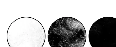
were electroporated with 4mg of YFDSK RNA, and a portion of the cells was plated on 100-mm dishes. At 50 h posttrans-fection, the media were recovered for analysis and the cell monolayers were stained with crystal violet. As shown in Fig. 3, nearly all cells expressing YF NS1 were destroyed by the cy-topathic effects of YFDSK replication, while cells expressing GFP were unaffected due to the lack of a functional NS1 gene. Surprisingly, comet-form plaques appeared on cells expressing DEN NS1, suggesting the replication and spread of YFDSK
within these cells. The variable shapes and sizes of these plaques were likely due to the use of a liquid overlay on the plates. When dilutions of cells from these same transfections were overlaid with a semisolid medium, DEN NS1-expressing cells yielded a small number of plaques that were heteroge-neous in size. The specific infectivity of YFDSK RNA was 33 103PFU/mg in this cell type. Transfection of this same RNA
into YF NS1-expressing cells yielded large round plaques at a specific infectivity of 9 3 105 PFU/mg. Thus, only a small
fraction of the YFDSK RNA was capable of replicating in DEN NS1-expressing cells.
The conditioned media from these transfected cells were tested for plaque formation on cells expressing DEN NS1 or YF NS1 (Table 1). YFDSK that was initiated in cells expressing YF NS1 had a high plaque titer on cells expressing YF NS1 but did not produce plaques on cells expressing DEN NS1. This result confirmed that YFDSK was not complemented by DEN NS1. However, transfection of cells expressing DEN NS1 yielded YFDSK capable of forming plaques on both YF NS1-and DEN NS1-expressing cells. The amount of virus produced by the DEN NS1-expressing cells was less than that produced by the YF NS1-expressing cells, a result which most likely
[image:4.612.88.519.73.230.2]YF NS1 DEN NS1 GFP
FIG. 3. Selection of YFDSKden by RNA transfection. BHK-SINrep21 cells
expressing YF NS1, DEN NS1, or GFP were transfected with YFDSK RNA and
[image:4.612.315.549.598.693.2]plated on 100-mm dishes. Following incubation, the cells were fixed and stained for plaque formation.
FIG. 2. DEN NS1 does not complement YFDSK. (A) Growth of YFDSK on cells expressing DEN NS1 (■) or YF NS1 (}). Cells were infected with YFDSK (MOI,
10) for 1 h. The inoculum was removed, the cells were briefly rinsed with PBS, and fresh growth medium was added. Virus growth media were sampled at 0, 12.5, 23,
37, and 46 h postinfection, clarified, and stored at280°C. Virus titers were determined on BHK-SINrep21/YF NS1 cells and plotted here on a semilogarithmic scale.
(B) RPA of viral minus strands. RNAs were harvested at 0, 4, 8, or 20 h postinfection. Equivalent portions of protected minus-strand reactions were subjected to denaturing electrophoresis, dried, and exposed to film. Numbers above lanes 2 to 5 refer to the numbers of synthetic minus-strand molecules analyzed in parallel;
numbers above lanes 6 to 19 refer to hours postinfection. The 109standard, which appeared as a smear in this exposure, did yield a distinct band in a lighter exposure
and is included here to further demonstrate the sensitivity of this assay to the quantity of input RNA.
on November 9, 2019 by guest
http://jvi.asm.org/
reflected the difference in specific infectivities. Note that the titers of the DEN NS1-initiated virus were similar on both types of cells, suggesting that a given virus particle might be capable of infecting either cell type. Passage of the DEN NS1-initiated virus on YF NS1-expressing cells yielded similar virus titers on both cell types (data not shown), further supporting this hypothesis and also demonstrating that the ability to form plaques on DEN NS1-expressing cells was retained after pas-sage. Thus, not only was there a difference in YFDSK RNA specific infectivity between these two cell types, but also the resultant viruses exhibited a large quantitative difference in their ability to form plaques on these cell types. These results indicated that there were two YFDSK populations: YFDSK, which can replicate only in YF NS1-expressing cells, and a minor population able to replicate in either YF NS1- or DEN NS1-expressing cells. We termed this latter virus YFDSKden. From a genetic standpoint, it appeared that viruses with the YFDSKden phenotype were variants of YFDSK that had gained the ability to utilize DEN NS1 for replication. If indeed YFDSKden was a subpopulation of YFDSK, it should also have been possible to select YFDSKden from the larger population by passage on DEN NS1-expressing cells. This idea was exam-ined in several experiments with a YFDSK stock that had been initiated from pACNR/YFDSK in YF NS1-expressing cells. This virus stock had a titer of 23107PFU/ml on YF
NS1-expressing cells and undetectable infectivities (,5 PFU/ml) on DEN NS1- and GFP-expressing cells. These three cell types were infected for 1 h with the same amounts of virus to yield an MOI of 10 on YF NS1-expressing cells, the cells were washed, and virus accumulation was monitored over time. The accu-mulation of YFDSK was detected by plaque formation on YF NS1-expressing cells (Fig. 4A). YFDSK produced robust growth on YF NS1-expressing cells and undetectable growth on GFP-expressing cells. After a short lag, modest growth was seen on DEN NS1-expressing cells. The accumulation of YFDSKden in these same samples was monitored by plaque formation on DEN NS1-expressing cells (Fig. 4B). In contrast to the high titer of YFDSK produced by YF NS1-expressing cells, the titer of YFDSKden remained below the detection limit of this assay on DEN NS1-expressing cells. Note that in the sample taken at 36 h postinfection, there was a difference in infectivity of at least 7 orders of magnitude between BHK-SINrep21/YF NS1 and BHK-SINrep21/DEN NS1 cells (Fig. 4). As predicted, YFDSKden emerged from the infection of DEN NS1-expressing cells with YFDSK. The appearance of this virus paralleled the growth of YFDSK on these cells, but at slightly lower titers. These data further support the hypothesis that YFDSK is incapable of utilizing DEN NS1 for replication but that a subset of YFDSK able to utilize DEN NS1 can be selected by growth on DEN NS1-expressing cells.
Isolation of YFDSKden variants. In order to examine the
genetic basis of the YFDSKden phenotype, three YFDSKden
variants were independently derived from separate transfec-tions of YFDSK RNA and isolated by two rounds of plaque purification on DEN NS1-expressing cells. Two of these vi-ruses, YFDSKden 1.7 and YFDSKden 2.5, were picked from medium-sized plaques, while a third virus, YFDSKden 3.1, was a large-plaque variant.
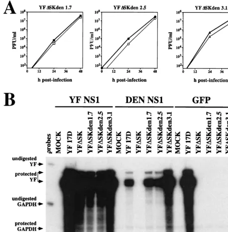
The growth characteristics of the isolated YFDSKden vari-ants were examined on both YF NS1- and DEN NS1-express-ing cells. All three viruses replicated to similar levels on both cell types (Fig. 5A). This result confirmed the hypothesis that YFDSKden can infect either cell type and, therefore, that YFDSKden is indeed a subset of YFDSK. YFDSKden 3.1 dem-onstrated a slightly higher level of growth at 24 h which may be related to its larger plaque size. All viruses required the ex-pression of NS1 intrans for growth (data not shown). Thus, these data demonstrate that these three virus isolates exhibit the YFDSKden phenotype.
[image:5.612.313.551.74.184.2]The RNA replication of YFDSKden isolates was compared to that of YF 17D and YFDSK on YF NS1-, DEN NS1-, and GFP-expressing cells by examination of the accumulation of viral plus strands at 16 h postinfection by an RPA. Shown in Fig. 5B is a relatively long exposure used in order to reveal any weak signals that might have been present. Plus strands were detected as a major 260-nucleotide band and a minor ;270-nucleotide band corresponding to a product of incomplete RNase digestion, as previously described (25). Abundant viral plus strands were specifically detected in all BHK-SIN-rep21/YF NS1 cells that were infected with a YF isolate (Fig. 5B, lanes 2 to 7). The requirement for NS1 expression by all YFDSK derivatives was shown by the absence of any plus-strand signal in cells expressing GFP (Fig. 5B, lanes 14 to 19). In DEN NS1-expressing cells, only YF 17D and the three YFDSKden isolates demonstrated plus-strand accumulation (Fig. 5B, lanes 9 and 11 to 13). The parental virus, YFDSK, failed to show evidence of viral RNA replication on this cell type (Fig. 5B, lane 10). While quantitation standards were not analyzed in this experiment, the relative intensities of these bands reflect differences in RNA replication rates over the first 16 h of infection. Notably, DEN NS1 appeared to have a somewhat inhibitory effect on the replication of YF 17D (see Discussion). Furthermore, YFDSKden 3.1 replicated to a higher level than the other YFDSKden isolates on this cell type, consistent with the larger plaque size of this isolate. TABLE 1. YFDSK produced by transfected cells
Transfected cell
typea
Plaque titer on the following cell type
(PFU/ml)b:
YF NS1 DEN NS1
YF NS1 2.83107 ,5
DEN NS1 3.83105 2.83105
GFP ,5 ND
aCells expressing the indicated protein.
bDetermined on the indicated plaque substrate cells. Shown are the averages
[image:5.612.52.292.84.155.2]from two independent experiments. ND, not determined.
FIG. 4. Growth of YFDSK on various cell populations. (A) BHK-SINrep21 cell monolayers (expressing YF NS1 [F], DEN NS1 [h], or GFP [}]) were
infected with YFDSK (MOI, 10) for 1 h, followed by three washes with PBS. At 12-h intervals, the growth media were removed and replaced with fresh media, clarified, and stored at280°C. Virus titers were determined on YF NS1-express-ing cells and plotted on a semilogarithmic scale. (B) The titers of the same samples from panel A determined on DEN NS1-expressing cells. The broken line indicates the detection limit of these plaque assays. The data represent the cumulative virus yields and are expressed as the means of the sum6standard deviations for three independent experiments.
on November 9, 2019 by guest
http://jvi.asm.org/
These data strongly indicate that the selected YFDSKden iso-lates are YFDSK variants that have gained the capacity to utilize DEN NS1 for the process of RNA replication.
Genetic analysis of the YFDSKden phenotype. Consensus
sequences were determined for regions of the viral genomes by isolating virion RNAs and amplifying the regions by reverse transcriptase PCR. We hypothesized that NS1 would likely interact with a membrane-spanning viral NS protein, so we initially focused our attention on the NS2A, NS4A, and NS4B regions of the genomes as well as the residual NS1 gene. In sequencing approximately 2.5 kb from each of the genomes, we
found only a single point mutation. Surprisingly, the same change was found in all three independently derived virus genomes, a point substitution at position 5783, coding for a change of Asn to Tyr at codon 42 of the NS4A gene (Fig. 6). The sequence at this position of YFDSK and the YFDSKden derivatives was confirmed in multiple sequencing reactions.
In order to determine if the NS4A-N42Y change could con-fer the YFDSKden phenotype, this single substitution was in-troduced into pACNR/YFDSK by site-directed mutagenesis. The infectivities of wild-type and mutant YFDSK RNAs were measured by infectious-center assays following RNA
electro-FIG. 5. YFDSKden can utilize DEN NS1 for RNA replication. (A) Growth analysis of independently derived YFDSKden 1.7, 2.5, and 3.1. BHK-SINrep21 cells
expressing YF NS1 (solid symbols) or DEN NS1 (open symbols) were infected with the indicated YFDSKden isolates (MOI, 10) for 1 h, followed by three washes with
PBS. Virus growth media were sampled at 0, 24, and 48 h postinfection, clarified, and stored at280°C. Virus titers were determined on BHK-SINrep21/YF NS1 cells.
The lower limit of the graph indicates the limit of detection in the plaque assays. (B) RNA replication of YFDSKden. BHK-SINrep21 cells expressing YF NS1 (lanes
2 to 7), DEN NS1 (lanes 8 to 13), or GFP (lanes 14 to 19) were infected with the indicated viruses (MOI, 10). Total cellular RNAs were harvested at 16 h postinfection and subjected to RPA analysis of viral plus strands. MOCK, mock infection.
on November 9, 2019 by guest
http://jvi.asm.org/
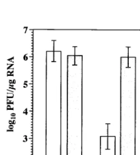
[image:6.612.74.528.76.538.2]poration of cells expressing YF NS1 or DEN NS1. The results of several experiments are summarized in Fig. 7. Both types of RNAs were equally infectious in YF NS1-expressing cells, pro-ducing roughly 106PFU/mg of RNA transfected. In DEN
NS1-expressing cells, wild-type YFDSK RNA had a low level of infectivity (;103 PFU/mg). This result is consistent with the
lack of replication and de novo selection of additional variants, as described above. The NS4A-N42Y mutation conferred a wild-type level of infectivity to YFDSK RNA in DEN NS1-expressing cells. Neither type of RNA was infectious (,5 PFU/ mg) in GFP-expressing cells (data not shown). These data indicated that a single point mutation within NS4A was suffi-cient to confer the YFDSKden phenotype.
To better define the requirements at this position, several additional substitutions were introduced into the NS4A gene and tested as described above. The results from a representa-tive experiment are shown in Table 2. All RNAs were tested at least twice, with similar results. In order to determine if the coding change of NS4A-N42Y rather than the local RNA sec-ondary structure was responsible, a second NS4A-N42Y mu-tant was made. This mumu-tant, NS4A-N42Y2, utilized a different Tyr codon and included nine additional silent mutations in the surrounding codons. These mutations gave rise to RNA that was as infectious as that resulting from the NS4A-N42Y change, formally demonstrating that the coding change of Asn to Tyr was indeed responsible for the YFDSKden phenotype. A variety of other substitutions were tolerated at this position by YF NS1, while DEN NS1 seemed to exclude the uncharged
polar residues Asn and Gln but not Ser. NS4A-N42D, which substituted a carboxyl group for the terminal amide on Asn, produced a partial restoration of DEN NS1 function. Another residue that led to an intermediate phenotype was the moder-ately hydrophobic Met. Aromatic residues at this position, as well as the cyclic basic residue His, were all tolerated.
DISCUSSION
The results presented here demonstrate that DEN NS1 is unable to trans complement YFDSK due to its inability to participate in early RNA replication. Based on this system, YFDSK variants able to utilize DEN NS1 were selected, and genetic analysis of the adapted variants indicated that a single point substitution in NS4A was sufficient to mediate this phe-notype. These findings have implications for understanding the role of NS1 in the replication of viral RNA, a cytoplasmic process occurring at membrane interfaces.
DEN NS1 does not function in YF RNA replication. Our
initial stocks of YFDSK did not grow or form plaques on DEN NS1-expressing cells, although efficienttranscomplementation was demonstrated by YF NS1. Most significantly, there were undetectable levels of RNA replication by this virus in DEN NS1-expressing cells. An inoculum of 107PFU must contain at
least this number of genomes. Thus, we infer that when;107
minus strands are detected in the samples during synchronized infection, the first minus strands are being synthesized. This time appeared to be shortly after 4 h postinfection of YF NS1-expressing cells with YFDSK, consistent with our previous report (25). Note that this level of minus-strand accumulation was never detected in YFDSK-infected DEN NS1-expressing cells, indicating that RNA replication was blocked prior to or at initial minus-strand synthesis.
The patterns of DEN NS1 expression via the pSINrep21 vector closely resembled those of authentic DEN NS1 in cells infected with DEN-2. Furthermore, the levels of protein ex-pression were comparable to that produced by DEN-2-infected cells as well as to the levels of YF NS1 expression sufficient for complementation of YFDSK. Thus, these considerations do not sufficiently explain the inability of DEN NS1 totrans com-plement YFDSK.
NS1 is among the more conserved of the flavivirus NS pro-teins. The NS1 proteins used in this study have 44% identity and 65% homology (PAM 250 index [12a]) at the amino acid level. Both Asn-linked glycosylation sites and all 12 cysteine residues are conserved, suggesting conservation of structural features as well. While it has never been formally
[image:7.612.79.270.72.163.2]demon-FIG. 6. Summary of YFDSKden sequence analysis. Shown at the top is the YFDSK genome. Thick bars below the genome indicate regions of the viral cDNAs that were sequenced. A single point substitution at position 5783, coding for a change of Asn (N) to Tyr (Y), is indicated.
[image:7.612.312.552.83.217.2]FIG. 7. NS4A-N42Y can confer the YFDSKden phenotype. The specific in-fectivities of wild-type YFDSK RNA (white bars) or mutant YFDSK RNA (gray bars) were assayed by transfection of cells expressing YF NS1 (left bars) or DEN NS1 (right bars). Values represent the log geometric means6standard devia-tions for four independent experiments.
TABLE 2. Effects of mutations at NS4A position 42
Amino acid substitution
RNA specific infectivity (log10PFU/mg)a
on cells expressing:
YF NS1 DEN NS1
N 6.1 2.9
Y 5.8 6.2
Y2 6.0 6.3
H 5.9 6.1
W 5.8 6.2
F 5.9 6.0
Q 6.3 2.0
D 6.0 4.8
M 6.1 4.3
S 6.1 5.7
aSpecific infectivities of mutant YFDSK RNAs determined as described in the legend to Fig. 7.
on November 9, 2019 by guest
http://jvi.asm.org/
[image:7.612.102.244.526.684.2]strated that DEN and YF utilize their NS1 proteins for the same function(s), it seems unlikely that a protein required for RNA replication would demonstrate high functional diver-gence among closely related viruses. To address this point, as well as to examine the reciprocal trans complementation of DEN-2 with YF NS1, we constructed a deletion variant ho-mologous to YFDSK by using an infectious DEN-2 cDNA clone (a kind gift from Richard Kinney, Centers for Disease Control and Prevention). We were unable to demonstrate complementation of this genome (DENDSK) with either DEN NS1 or YF NS1 (data not shown), preventing direct analysis of these points. The inability of DEN NS1 totranscomplement DENDSK could be due to differences in DEN-2 strains, since the NS1 gene was derived from DEN-2 strain PR-159(S1), while the infectious cDNA was from DEN-2 strain 16681. Alternatively, the tolerance of DEN-2 for deletions in the NS1 gene may be different from that of YF. Nevertheless, it appears that NS1 is required for DEN-2 replication (23), and we look forward to understanding its role in this viral system. Taken together, the above data indicated that NS1 functions in a flavivirus-specific manner and suggested that DEN NS1 does not productively interact with the YF replication machinery.
Genetic screen for YFDSKden variants.In order to define
such an interaction, we selected for YFDSKden variants able to utilize DEN NS1. Because the NS1 gene has been deleted from YFDSK and was supplied intrans, such variants were likely to contain suppressor mutations with the potential to reveal other viral genes involved in the function of NS1. As in all genetic selections, the ability to select a given phenotype is dependent on the genetic heterogeneity present in the initial population. To facilitate this selection, we capitalized on the improved specific infectivity of transcripts derived from pACNR/YFDSK in order to explore variant YFDSK genomes. In this regard, it may be instructive to consider the potential sources of adaptive mutations.
Given that YFDSK demonstrated undetectable RNA repli-cation on DEN NS1-expressing cells (Fig. 2B), the possibility that adaptive mutations existed in the population of input RNA molecules that were used for transfection must be con-sidered. The fidelity of SP6 RNA polymerase has not been reported; however, the related enzyme from bacteriophage T7 has an estimated error frequency of 1024(3, 44). Thus, for a
genome the size of YFDSK (;10 kb), RNA transcripts con-taining single point mutations are expected to be as abundant as the wild type, each about 37% of the RNA population. One microgram of this transcript should contain approximately 23 106copies of YFDSK with the single point mutation A5783T.
However, the actual misincorporation rate at any given posi-tion is likely to be influenced by other factors specific for the polymerase and the local template structure. This might ex-plain why Tyr seemed to be preferred although several other substitutions were found to work in this position. Furthermore, our ability to sample an RNA population is limited by the efficiency with which transfected RNAs initiate a productive infection. For reasons that remain obscure, only a small frac-tion of RNA transcripts actually yielded plaque-forming units, ;106 PFU/mg, which represents an RNA transfection
effi-ciency of ;1025 PFU/RNA molecule. The latter point may
help to explain why we initially saw no sign oftrans comple-mentation of our original YFDSK stock on DEN NS1-express-ing cells. Recall that this virus was generated via the two-plasmid YF infectious clone and was initiated at a specific infectivity of 73 102 PFU/mg (25). Thus, this virus stock is
likely to contain less genetic diversity initially due to this lower sampling rate. Based on these considerations, it is clear that the process of RNA transcription may produce sufficient
ge-netic diversity to examine the constellation of YFDSK single-point mutations but that this examination is subject to the RNA sampling rate.
Another potential source of adaptive mutations was misin-corporation by the YF replicase itself. Although YF replicase has an unknown fidelity, studies of other RNA viruses indicate error rates on the order of 1023to 1024 (45, 47). Thus, one
would expect YFDSKden variants to arise naturally in a pop-ulation of YFDSK isolates replicating in YF NS1-expressing cells. Indeed, passage of YFDSK on DEN NS1-expressing cells selected for rare variants in a population of YFDSK isolates (Fig. 4). However, it is unknown whether these variants existed in the RNA population used to derive the YFDSK stock or whether they arose during virus passage. To address this ques-tion more directly, two YFDSK isolates were independently derived by RNA transfection and isolated by plaque purifica-tion on YF NS1-expressing cells (data not shown). YFDSKden variants arose spontaneously after passage of these virus stocks several times on YF NS1-expressing cells (data not shown), suggesting that adaptive mutations could have arisen during the process of YF RNA replication. These results may also explain the absence of the YFDSKden phenotype in our orig-inal YFDSK stock (Fig. 1 and 2), which was passaged once following transfection and was therefore the product of a small (albeit unknown) number of RNA replication cycles.
YFDSKden isolates are variants of YFDSK able to utilize
DEN NS1.Given the above arguments for the generation of
genetic diversity in RNA virus populations, several lines of evidence support the hypothesis that YFDSKden isolates are variants of YFDSK able to utilize DEN NS1. Only a small fraction of YFDSK RNA transfected into DEN NS1-express-ing cells was infectious, yieldNS1-express-ing plaques with a variety of sizes and morphologies. Unlike YFDSK initiated on YF NS1-ex-pressing cells, DEN NS1-initiated virus demonstrated the abil-ity to form plaques on both cell types. Similarly, YFDSKden isolates could also be derived by passage of YFDSK on cells expressing DEN NS1 but not on cells expressing YF NS1. The lag that was observed in the growth of YFDSK was consistent with the emergence of a subpopulation with the ability to replicate in DEN NS1-expressing cells. Furthermore, YFDSKden isolates obtained by plaque purification were able to grow on both cell types and demonstrated the ability to replicate their viral RNA on DEN NS1-expressing cells. The lack of detectable viruses with the YFDSKden phenotype in YF NS1-initiated virus populations (,5 PFU/ml) may suggest that YFDSKden is at a selective disadvantage for growth in YF NS1-expressing cells when coinfected with YFDSK. Taken to-gether, these data argue that YFDSKden isolates, unlike the larger YFDSK population, were able to utilize DEN NS1 for RNA replication and growth.
It was remarkable that only a single substitution, NS4A-N42Y, was identified multiple times by sequencing of the iso-lated YFDSKden genomes. While it is entirely possible that other adaptive mutations exist in YFDSKden populations, we have not yet observed other sequence changes. Nevertheless, this single mutation was sufficient to confer high infectivity to YFDSK RNA in DEN NS1-expressing cells. We may conclude that NS4A-N42Y is sufficient to mediate this phenotype, al-though this particular change may not be necessary. In this regard, several additional substitutions were tested and also found to permit mutant YFDSK genomes to utilize DEN NS1. Of the amino acids tested, the only ones that were not toler-ated by DEN NS1 were Asn and Gln, while Asp and Met gave intermediate phenotypes. Thus, the rules of amino acid sub-stitution at this position for DEN NS1 utilization are not yet clear. However, these results support the hypothesis that a
on November 9, 2019 by guest
http://jvi.asm.org/
genetic interaction exists between NS1 and NS4A. Note that the type of genetic interaction that we are discussing here does not necessarily imply a direct protein-protein interaction of NS1 and NS4A gene products. Because the general features of flavivirus RNA replication appear to be conserved across the genus (11, 12, 21), these findings may extend to other flavivi-ruses, although this awaits experimental proof.
In addition to the complementation of YFDSKden, DEN NS1 had an inhibitory effect on wild-type YF 17D, which was seen as reduced levels of viral plus-strand accumulation (Fig. 5B, lanes 9 and 15). Inhibition of YF 17D replication was also evident as a reproducible 1- to 2-log10decrease in viral titers
during single-cycle growth experiments (data not shown). This result is in contrast to a slight increase in YF 17D replication with the expression of YF NS1 (Fig. 5B, lanes 3 and 15; see also Fig. 5, 6, and 7 in reference 25). To determine if this inhibition could be mediated by a dominant-negative effect of DEN NS1, perhaps through the usurping of NS4A, we introduced the NS4A-N42Y mutation into the wild-type YF 17D genome. The resultant viruses did not, however, overcome the inhibitory effect of DEN NS1 (data not shown), suggesting that other mechanisms may also be involved. For instance, YF NS1 and DEN NS1 may form inactive heterodimers. As mentioned above, YFDSKden 3.1 seemed to have an enhanced capacity for replication in DEN NS1-expressing cells (see, for instance, Fig. 5B, lanes 11, 12, and 13). This virus is likely to contain, in addition to NS4A-N42Y, adaptive mutations which may over-come this inhibition. Therefore, further characterization of this virus is under way.
Prospects for understanding replicase ultrastructure. It is
interesting that an adaptive mutation was mapped to NS4A. The product of this gene, a small hydrophobic protein, has an unknown function in the life cycle of flaviviruses. Although recent results suggest that NS4A is associated with flavivirus
replicase components (27), our results provide the first genetic data indicating that NS4A is involved in the process of RNA replication. NS4A is produced from the viral polyprotein via cleavage at its N and C termini by the viral serine protease (Fig. 8A). Both cleavage events appear to be delayed or reg-ulated, since the putative NS4A precursors NS3-4A and NS4A-4B have been identified in flavivirus-infected cells (7, 33). Protease cleavage of NS4A-B appears to coordinate a downstream cleavage event for signal peptidase, which gener-ates a small “2K” signal peptide and the N terminus of NS4B (24, 32). This coordinated cleavage may represent a mecha-nism for the regulation of replicase function by the viral pro-tease, perhaps analogous to the switch from minus-strand to plus-strand synthesis mediated by the alphavirus cysteine pro-tease (22, 41). Because little is known about the importance of NS4A-processing events, further investigation will be required to understand the relevant context in which the NS4A-N42Y mutation functions.
[image:9.612.143.454.75.297.2]Genetic interaction between NS1 and NS4A helps to resolve an important topological issue, namely, how a protein in the secretory pathway can be required for RNA replication, a process that occurs in the cytoplasm of infected cells in close association with virus-induced membrane structures. The two major enzymatic components of the replicase, NS3 and NS5, are predicted to reside in the cytoplasm (34). In support of this model, both were shown to be sensitive to trypsin digestion in extracts made from infected cells in the absence of detergents (5). The interaction of NS1 and cellular membranes remains more complex, although several lines of evidence indicate that NS1 resides in the luminal side of an extracytoplasmic com-partment. As mentioned earlier, processing at the N and C termini of NS1 is mediated by enzymes in the ER; NS1 is glycosylated and disulfide bonded, and a portion of it is se-creted. All of these processes indicate that NS1 forms in the
FIG. 8. Model of NS4A topology and secondary structure. (A) The topology of NS4A/2K was modeled as described in Materials and Methods. The arrows, open box, and black dot indicate viral serine protease cleavage sites, a signal peptidase cleavage site, and the approximate location of NS4A residue 42, respectively. (B) Several flavivirus NS4A coding regions were aligned with YF NS4A residues 11 to 60. The virus sequences shown (and their GenBank accession numbers) are as follows: YF 17D (P03314), DEN-1 (P33478), DEN-2 (P123823), DEN-3 (P27915), DEN-4 (P09866), Japanese encephalitis (JE) (P32886), Kunjin (KUN) (P14335), tick-borne encephalitis (TBE) (P14336), and West Nile (WN) (P06935). Shown below the alignment is a prediction of the secondary structure: H, helix; L, loop; E, strand; dot, not predictable. Also shown is the beginning of the first putative transmembrane domain, depicted as a box. The location of YF NS4A residue 42 is indicated above the alignment by a black dot.
on November 9, 2019 by guest
http://jvi.asm.org/
secretory pathway. Shortly after synthesis, NS1 acquires hydro-phobic properties and associates with membranes (51); these steps may correlate with the formation of NS1 homodimers (52). NS1 does not contain any putative transmembrane do-mains (36) or known posttranslational lipid modifications. Mu-tagenesis of the first glycosylation site of YF NS1 demon-strated that this modification is important for efficient RNA replication (29), indicating that NS1 requires translocation into the ER for function. We have recently expressed a version of NS1 lacking its signal sequence and found that it does not function in trans complementation (28). Furthermore, resis-tance to trypsin in the absence of detergents largely supports the extracytoplasmic sequestration of NS1, although a propor-tion of NS1 was sensitive to this treatment (5). However, these authors did not exclude the possible disruption of an NS1-containing compartment during the extraction procedure, which may explain this partial sensitivity. Taken together, these data indicate that NS1 is in a compartment topologically dis-tinct from the cytoplasm, where the process of RNA replica-tion occurs.
NS4A is predicted to be membrane spanning (34), although biochemical data on the topology of this protein are lacking. We have modeled the secondary structure of this protein and its interaction with cellular membranes by using current pre-dictive algorithms as described in Materials and Methods. The N and C termini of NS4A are expected to be cytoplasmic, consistent with their generation by the viral serine protease (Fig. 8A). The N-terminal region of NS4A (YF 17D codons 2108 to 2160) is predicted to be cytoplasmic and possibly rich in alpha helices and to be followed by two transmembrane domains separated by a short peptide (YF 17D codons 2161 to 2178, 2179 to 2194, and 2195 to 2219) and a short C-terminal domain (YF 17D codons 2220 to 2233). The NS4A-N42Y mutation localizes to a putative alpha-helical region in the cytoplasmic N-terminal domain, an observation that is incon-sistent with this residue being involved in a direct protein-protein interaction with NS1. Perhaps the N-terminal region of NS4A affects the structure of the luminal peptide, which might interact with NS1. Alternatively, an interaction between NS1 and the luminal peptide might induce a conformational effect on this region of NS4A, allowing it to recruit cytoplasmic replicase components. As noted above, the genetic interaction between NS1 and NS4A does not exclude the involvement of other replicase components, and we do not mean to necessarily imply a direct protein-protein interaction. From an alignment of several flavivirus NS4A genes, it is apparent that a His residue is conserved at this position between DEN isotypes, while Met is preferred at NS4A position 42 in tick-borne and other mosquito-borne flaviviruses (Fig. 8B). Clearly, more work needs to be done to examine the structure and function of this region of NS4A in order to determine the basis for our genetic observations. Nevertheless, the involvement of NS4A, a putative transmembrane protein, in NS1 utilization may pro-vide a solution to the puzzle of NS1 versus replicase topology. Cytological studies of the flavivirus replicase have indicated that NS1 and NS4A can both be localized to perinuclear clus-ters of vesicles bound by a larger membrane, termed vesicle packets (26, 27, 50). It is unclear whether this larger mem-brane, which appears to be contiguous with the ER, represents an enclosed vesicle or a transverse slice of a membranous pocket. An indication that vesicle packets are associated with replication has been provided by labeling studies with an an-tiserum that recognizes double-stranded RNA. NS2A and NS3 are also associated with these structures, while other NS pro-teins have been excluded (27, 50). In addition to vesicle pack-ets, NS4A localizes to other virus-induced membrane
struc-tures, termed convoluted membranes or paracrystalline arrays. The role of these structures in infected cells is unclear, al-though it has been suggested that they represent sites of polyprotein processing (50). The colocalization of NS1, NS4A, and a marker of RNA replication in vesicle packets supports our genetic data indicating that an NS1-NS4A interaction is important in the process of RNA replication.
Biochemical studies have also indicated that NS4A interacts with other replicase components. Both NS1 and NS4A appear to cosediment with dense membrane fractions enriched in RNA-dependent RNA polymerase activity (10). Furthermore, it was recently reported that a fusion protein of NS4A and glutathione-S-transferase could retain several virus-specific proteins on glutathione beads, including NS1, NS2A, NS3, NS3-4A, NS4A, and NS5 (27). The binding of NS2A and NS3-4A appears to be sensitive to RNase treatment, suggest-ing that an indirect interaction is involved. The involvement of both NS1 and NS4A in these complexes further supports a model of their interaction.
Our approach to understanding the flavivirus replicase has been to use genetic means. A particularly useful technique has been thetrans complementation of NS proteins, which previ-ously revealed a requirement for NS1 in early RNA replication (25). We have now extended our studies to demonstrate a genetic interaction between NS1 and NS4A. It was recently reported that a Kunjin virus genome bearing lethal mutations in the RNA polymerase or putative methyltransferase domains of NS5 could also be complemented intransby use of a Kunjin virus replicon lacking structural genes (20). This approach shows promise for demonstrating the transcomplementation of multiple NS genes. However, this system differs from ours in at least two respects. It appears that a significant amount of Kunjin virus replicon is packaged in trans, eliminating the ability to examine helper-free mutant virus stocks. Further-more, the expression of the complementing NS protein is cou-pled to its function, preventing the future application of this system to the probing of NS protein structure and function. We have used our system, which circumvents both of these disad-vantages, to reveal the functional roles of flavivirus NS1.
Future directions.This study reveals a genetic interaction
between NS1 and NS4A and demonstrates the significance of this interaction for RNA replication. Further studies are needed to directly examine the nature of this interaction and of other proteins in the replicase. The integration of biochemical, structural, and genetic approaches shows great promise in un-raveling the intricacies of flavivirus replicase structure and function. Here we have demonstrated a tool that we expect will be useful in this endeavor.
ACKNOWLEDGMENTS
We thank Rebecca Moran for expert technical assistance. We are also grateful to many colleagues for helpful discussions during the course of this work and to Sean Amberg, Richard Hardy, and Mara Lippa for critical reading of the manuscript.
This work was supported in part by a grant from the Public Health Service (AI31501).
REFERENCES
1.Agapov, E. V., I. Frolov, B. D. Lindenbach, B. M. Pra´gai, S. Schlesinger, and C. M. Rice.1998. Noncytopathic Sindbis virus RNA vectors for heterologous
gene expression. Proc. Natl. Acad. Sci. USA95:12989–12994.
2.Ausubel, F. M., R. Brent, R. E. Kingston, D. D. Moore, J. G. Seidman, J. A. Smith, and K. Struhl (ed.).1993. Current protocols in molecular biology. Greene Publishing Associates, New York, N.Y.
3.Boyer, J. C., K. Bebenek, and T. A. Kunkel.1992. Unequal human immu-nodeficiency virus type 1 reverse transcriptase error rates with RNA and
DNA templates. Proc. Natl. Acad. Sci. USA89:6919–6923.
4.Bredenbeek, P. J., E. Kooi, B. D. Lindenbach, M. Lucassen, N. Huijkman,
![FIG. 1. Expression of DEN NS1 and NS1-2A. BHK-21 cells (mock infectedor infected with DEN-2 [MOI, 10] for 22 h) or BHK-SINrep21 derivatives were](https://thumb-us.123doks.com/thumbv2/123dok_us/549876.73508/3.612.58.289.73.282/expression-cells-infectedor-infected-moi-bhk-sinrep-derivatives.webp)