Copyright © 1999, American Society for Microbiology. All Rights Reserved.
La Autoantigen Specifically Recognizes a Predicted Stem-Loop
in Hepatitis B Virus RNA†
TILMAN HEISE,‡ LUCA G. GUIDOTTI,
ANDFRANCIS V. CHISARI*
Department of Molecular and Experimental Medicine, The Scripps
Research Institute, La Jolla, California 92037
Received 1 February 1999/Accepted 14 April 1999
We recently identified three nuclear proteins (p45, p39, and p26) that bind to a 91-nucleotide (nt) RNA
element between nt 1243 and 1333 in hepatitis B virus (HBV) RNA, and we showed that these proteins and HBV
RNA are regulated coordinately by gamma interferon and tumor necrosis factor alpha. Purification and
sequence analysis of tryptic peptides obtained from p39 revealed sequence homology to the mouse La protein.
Immunoprecipitation experiments showed that p45, p39, and p26 were recognized by La-specific
anti-serum, indicating that p45 is the full-length La protein and that p39 and p26 are likely to be proteolytic La
cleavage products. Furthermore, in competition experiments we found that all three La proteins bind, in a
phosphorylation-dependent manner, to the same predicted stem-loop structure located between nt 1275 and
1291 of HBV, with
K
ds of approximately 1.0 nM. Collectively, these results support the notion that the La
protein may contribute to HBV RNA stability, constitutively and in response to inflammatory cytokines.
RNA-protein interactions regulate gene expression by
con-trolling the processing (62), export (43, 51, 62), translation
(65), intracellular localization (67), and degradation (39, 58) of
mRNA. mRNA stability is regulated by hormones and
cyto-kines (72) that induce the phosphorylation or
dephosphoryla-tion of RNA-binding proteins (17, 26, 46) and modulate
RNase activities (39a, 41, 57). Furthermore, prokaryotic
mul-tiprotein complexes consisting of RNase E, polynucleotide
phosphorylase (11, 55), ATP-dependent RNA helicases (56),
heat shock-chaperone proteins GroEL and DnaK, and the
glycolytic enzyme endolase (49) are thought to coordinate the
stabilization or destabilization of certain mRNAs. In addition,
it was shown that these complexes also contain cellular RNAs
(49).
The hepatitis B virus (HBV) is a DNA virus that replicates
through an RNA intermediate and encodes four unspliced,
overlapping messages that terminate at a common
polyadenyl-ation signal (61). The vigor and kinetics of the cytotoxic
T-lymphocyte (CTL) response to HBV determine the outcome of
infection (19). By the use of transgenic mice that express some
(20, 21, 30) or all of the viral proteins and replicate the virus
(35), many of the host-virus interactions responsible for viral
clearance and disease pathogenesis during HBV infection have
been defined (3, 4, 18, 50). Recently, we have shown that
inflammatory cytokines, especially gamma interferon (IFN-
g
)
and tumor necrosis factor alpha (TNF-
a
), induced by
adop-tively transferred HBV-specific CTLs or during lymphocytic
choriomeningitis virus and murine cytomegalovirus infections,
can abolish hepatic HBV gene expression and replication in
the livers of these animals (12, 13, 29–34). Importantly, the
cytokines suppress HBV gene expression by a
posttranscrip-tional mechanism (36, 69), presumably reflecting
destabiliza-tion of HBV mRNA.
Recently, we demonstrated that liver nuclear extracts from
untreated, CTL-injected, lymphocytic choriomeningitis
virus-infected, and murine cytomegalovirus-infected HBV
trans-genic mice contain three proteins (p45, p39, and p26) that bind
a 91-nucleotide (nt) in vitro transcript of HBV (40). This
tran-script is located in the 5
9
region of the HBV posttranscriptional
regulatory element (between nt 1200 and 1650), which is
thought to mediate the nuclear export of HBV RNA (23, 42).
A tight correlation was observed among the downregulation of
HBV RNA, the disappearance of p45, and the appearance of
p26, suggesting that these proteins might contribute to the
regulation of HBV mRNA stability by the cytokines (40).
Fur-thermore, we showed that the elimination of p45 and the
induction of p26 are coupled events that require IFN-
g
and
TNF-
a
, suggesting that cytokine-induced signal transduction
pathways might regulate the RNA-binding activity of these
proteins (40).
The goal of the current study was the purification and
mo-lecular identification of p45, p39, and p26 and the mapping of
the RNA target element(s) to which they bind. In this report,
we demonstrate that all three proteins are recognized by
an-ti-La antibodies and that they bind HBV RNA in a
phospho-rylation-dependent manner. Furthermore, we showed that
they bind to a predicted stem-loop in the 91-nt transcript with
high affinity and that the stem structure and specific loop
nu-cleotides are required for binding. Collectively, these results
are consistent with the notion that the La protein, especially
p45, may be part of a complex mechanism that controls HBV
RNA stability, constitutively and in response to inflammatory
cytokines.
MATERIALS AND METHODS
HBV transgenic mice.The HBV transgenic mouse lineages 1.3.32 (official designation, Tg{HBV 1.3 genome}Chi32) and 1.3.46 (official designation, Tg{HBV 1.3 genome}Chi46) used in this study have been described previously (35). Lineages 1.3.32 and 1.3.46 express all of the HBV transcripts under the control of their respective promoters and replicate HBV at high levels in the liver and kidney without any evidence of cytopathology (35). Mice were matched for age (8 to 10 weeks), sex (male), and serum hepatitis B e antigen concentration
* Corresponding author. Mailing address: Department of Molecular
and Experimental Medicine, The Scripps Research Institute, 10550 N.
Torrey Pines Rd., La Jolla, CA 92037. Phone: (619) 784-8228. Fax:
(619) 784-2160. E-mail: [email protected].
† Paper no. 11631-MEM from The Scripps Research Institute.
‡ Present address: Heinrich-Pette-Institut fu¨r Experimentelle
Vi-rologie und Immunologie, Universita¨t Hamburg, D-20251 Hamburg,
Germany.
5767
on November 9, 2019 by guest
http://jvi.asm.org/
by a commercially available solid-phase radioimmunoassay (Sorin Biomedica, Saluggia, Italy).
HBsAg-specific CTLs.Ld-restricted, CD31CD42CD81hepatitis B surface antigen (HBsAg)-specific CTL clones that recognize an epitope located between residues 28 and 39 of HBsAg (HBsAg28–39) and secrete IFN-gand TNF-aupon antigen recognition (3) were used for the studies. In all experiments, 107CTLs were injected intravenously into transgenic mice 5 days after in vitro stimulation with irradiated P815 cells that stably express the HBV large envelope protein (50). CTL-induced liver disease was monitored by measuring serum alanine aminotransaminase levels at various time points after CTL injection. Liver tissue obtained at autopsy was either processed for histological analysis or snap frozen for subsequent molecular analyses.
RNA analyses.Snap-frozen (liquid nitrogen) liver tissues were mechanically pulverized, and total genomic RNA was isolated for Northern blot analyses exactly as previously described (35).
Preparation of liver nuclear and cytosolic extracts from HBV transgenic mice.
Frozen liver tissue (0.2 to 0.5 g) was thawed and homogenized in a fivefold volume of ice-cold homogenization buffer containing 10 mM Tris-HCl (pH 7.4), 10 mM NaCl, 2.5 mM MgCl2, 1 mM EDTA (buffer A) containing 0.5 mM dithiothreitol (DTT), and a 1/25 volume of proteinase inhibitor mix (Boehringer Mannheim, Indianapolis, Ind.) by five strokes in a glass homogenizer with a loose-fitting motor-driven (50 rpm) Teflon pestle. The homogenate was centri-fuged at 2,0003gfor 20 min, and the resulting supernatant was stored at280°C. The pellet was resuspended in 6 ml of buffer A containing 0.88 M sucrose (buffer B), loaded on a 7-ml cushion of buffer B, and centrifuged at 10,0003gfor 30 min. The supernatant was discarded, and the pellet was dissolved in 5 ml of buffer A containing 2.0 M sucrose (buffer C). The slurry was loaded on a 7-ml cushion of buffer C and centrifuged at 180,0003gfor 70 min. The supernatant was discarded, and the nuclei was resuspended in 100ml of storage buffer containing 20 mM Tris-HCl (pH 8.0), 75 mM NaCl, 2.5 mM MgCl2, 0.5 mM EDTA, 50% glycerol, 0.5 mM DTT, and a 1/10 volume of proteinase inhibitor mix (Boehr-inger Mannheim). Nuclei were counted by light microscopy and lysed by adding 53lysis buffer containing 100 mM Tris-HCl (pH 8.0), 2.1 M NaCl, 7.5 mM MgCl2, 1.0 mM EDTA, and 25% glycerol to final concentrations of 33 mM Tris-HCl (pH 8.0), 420 mM NaCl, 1.5 mM MgCl2, 0.2 mM EDTA, 5% glycerol, and 0.5 mM DTT, and a 1/10 volume of proteinase inhibitor mix (Boehringer Mannheim). The viscous lysate was transferred into dialysis tubes (molecular weight cutoff, 6,000 to 8,000; Spectro/Por; Spectrum Companies, Gardena, Cal-if.) and dialyzed three times against 500 ml of dialysis buffer F containing 10 mM Tris-HCl (pH 7.4), 100 mM NaCl, 3 mM MgCl2, 0.5 mM EDTA, 10% glycerol, 0.5 mM DTT, and proteinase inhibitor mix (Boehringer Mannheim). The dia-lyzed nuclear extract was cleared by centrifugation for 10 min at 24,0003g, and the protein content was determined by the Bradford dye-binding procedure, with a commercial kit (Bio-Rad Laboratories, Hercules, Calif.).
Protein purification.All the following steps were performed on ice or at 4°C. Nuclear extract was brought to 35% saturation with ammonium sulfate and stirred for 30 min. After centrifugation of the slurry for 30 min at 20,0003g, the supernatant was brought to 65% saturation with ammonium sulfate and stirred for 30 min. The slurry was centrifuged at 20,0003gfor 30 min, the pellet was suspended in dialysis buffer F (see above), and the extract was dialyzed overnight. The next purification steps were performed with fast protein liquid chromatog-raphy (Pharmacia, Piscataway, N.J.). The extract was loaded (1 ml/min) on a heparin column (Pharmacia) which was preequilibrated with wash buffer con-taining 10 mM Tris-HCl (pH 7.4), 3 mM MgCl2, 0.5 mM EDTA, and 200 mM NaCl. The column was washed until protein was no longer detectable in the flowthrough. Bound protein was eluted in a linear gradient ranging from 200 mM to 2 M NaCl at a flow rate of 1 ml/min. Fractions (1 ml) were assayed for p45, p39, and p26 by UV cross-linking as described below. RNA-binding-protein-containing fractions were pooled, concentrated by ultrafiltration on Centricon 10 (Millipore, Bedford, Mass.), and loaded (1 ml/min) onto a source 30Q column (Pharmacia), and the flowthrough was further run through a source 30S column (Pharmacia). The final flowthrough was tested for p45, p39, and p26 by UV cross-linking; concentrated by ultrafiltrations as described above; and subse-quently loaded (0.7 ml/min) onto a Superdex 75-pg gel filtration column (Phar-macia) equilibrated with 10 mM Tris-HCl (pH 7.4)–3 mM MgCl2–0.5 mM EDTA–100 mM NaCl. Elution was performed in equilibration buffer at a flow rate of 0.17 ml/min, and 0.51-ml fractions were collected and assayed for p45, p39, and p26 by UV cross-linking (ranging of fractions 13 to 26). For molecular mass determinations, the Superdex column was calibrated with standard proteins in a gel filtration calibration kit (Sigma). The gel filtration fractions containing p45, p39, and p26 (ranging of fractions 15 to 20) were separately concentrated and subjected to sodium dodecyl phosphate–14% polyacrylamide gel electro-phoresis (SDS-PAGE). To determine the exact positions of p45, p39, and p26, UV cross-linking samples were loaded onto the same gel, and the gel was stained with Coomassie blue (Bio-Rad), destained, and exposed to Kodak Biomax (Kodak, Rochester, N.Y.) overnight at 4°C. The autoradiogram was matched with the gel, and a dominant protein band corresponding to the position of the p39 UV cross-linking signal was cut out. The protein band was subsequently submitted for tryptic in-gel digestion, and high-pressure liquid chromatography (HPLC) fractionation of tryptic peptides obtained from p39 and N-terminal sequencing of several peptides were performed by the Scripps Protein and Nucleic Acid Core Facility.
Immunoprecipitation and Western blotting.One hundred milligrams of pro-tein A-coupled Sepharose CL-4B beads (Pharmacia) was swollen in 1.5 ml of TS buffer (15 mM Tris-HCl [pH 7.4], 150 mM NaCl) for 4 h at room temperature. The swollen gel was collected by centrifugation and washed three times with TS buffer. The final pellet was resuspended in a total volume of 600ml of TS buffer. Three hundred microliters was combined with 100ml of control human serum or with anti-La-positive human serum (Centers for Disease Control and Prevention prototype, kindly provided by E. Chan, The Scripps Research Institute [14]). The mixture was incubated overnight at 4°C and subsequently for 4 h at room temperature. The slurry was washed three times with TS buffer and resuspended in 300ml of TS buffer. One hundred microliters from this mixture was incubated with 20mg of nuclear extracts prepared from untreated or CTL-injected mice and rotated for 6 h at 4°C. Precipitates were collected by centrifugation at 14,0003g. The supernatant was taken off, and 5mg of protein was analyzed by UV cross-linking and Western blotting. For Western blotting (semidry proce-dure), the SDS gel was incubated in 25 mM Tris-HCl (pH 9.0)–20% methanol at room temperature for 20 min. The nitrocellulose membrane was equilibrated in 25 mM Tris-HCl (pH 10.6)–20% methanol. The transfer was performed at 1.2 mA/cm2for 90 min. After the transfer, the membrane was incubated in blocking buffer (10 mM Tris-HCl [pH 7.5], 100 mM NaCl, 0.001% Tween 20, and 10% milk powder) at room temperature for 45 min. The blocking solution was changed, and anti-La-positive human serum (Centers for Disease Control and Prevention prototype, dilution 1:600) was added and further incubated overnight at 4°C. Subsequently, the membrane was washed six times for 5 to 10 min at room temperature with washing buffer (10 mM Tris-HCl [pH 7.5], 100 mM NaCl, 0.001% Tween 20) and replaced by blocking buffer. Peroxidase-conjugated rabbit human immunoglobulin A (IgA), IgG, IgM, and kappa and lambda anti-bodies (DAKOPATTS, Glostrup, Denmark) were added in a 1:1,000 dilution and incubated for 1 h at 37°C. The membrane was washed six times for 5 to 10 min at room temperature with washing buffer followed by detection with the ECL detection system (Amersham, Arlington Heights, Ill.) according to the manufacturer’s instructions.
In vitro transcription.A plasmid containing the entire HBV genome (ayw
subtype) was used for the production of DNA templates for the generation of HBV transcripts. Two primers were used. Primer 1 (59-CCATCGAT-TAATACG ACTCACTATAG-39) contained a restriction site forClaI (shown in italics), the T7 RNA polymerase promoter sequence (shown in boldface), and the sense HBVaywDNA sequences (28) spanning nt 1243 to 1261 (59-GAACCTTTTCG GCTCCTCT-39). Primer 2 contained antisense HBV sequences from nt 1312 to 1333 (59-GTCCCGATAATGTTTGCTCCAG-39, RNA.B), 1317 to 1294 (59-CT CCAGACCTGCTGCGAGCAAAAC-39, RNA.C), and 1293 to 1276 (59-AAG CGGCTAGGAGTTCCG-39, RNA.D). For the generation of templates with mutations in the binding region of the RNA-binding proteins (stem-loop 2; see Fig. 1B and 6), the following primers containing antisense HBV sequences (nt 1293 to 1276) and nucleotide changes (shown in boldface and underlined) were used: 59-AAGCGGATCTTAGTTCCG-39(RNA.D-M1), 59-AAGCTTCTAGG AGTTCTT-39(RNA.D-M2), 59-AAGCGGATAGGAGTTCCG-39 (RNA.D-M3), 59-AAGCGACTAGGAGTTCCG-39 (RNA.D-M4), 59-AAGCGGCTA
GGAGTTCCG-39 (RNA.D-M5), and 59-AAGCGGCTAGGCGTTCCG-39
(RNA.D-M6). RNA.E is a synthetic oligoribonucleotide spanning HBV nt 1243 to 1281 (59-GAACCUUUUCGGCUCCUCUGCCGAUCCAUACUGCGGAA C-39, produced by Oligos Etc., Wilsonville, Oreg.). The mouseb-actin template was generated by using the following primers. Primer 1 included aClaI site and a T7 RNA polymerase promoter followed byb-actin-specific DNA sequences spanning nt 27 to 45 (59-GGGCCGCTCTAGGCACCAA-39) (2). Primer 2 con-tained antisenseb-actin-specific sequence between nt 121 and 140 (59-TGTTC AATGGGGTACTTCAG-39) (2). The mouse glyceraldehyde-3-phosphate dehy-drogenase (GAPDH) template was generated by using the following primers. Primer 1 included aClaI site and a T7 RNA polymerase promoter followed by GAPDH-specific DNA sequences spanning nt 383 to 401 (59-GGAGCCAAAC GGGTCATCA-39) (60). Primer 2 contained antisense GAPDH-specific se-quence between nt 478 and 497 (59-TGCAGGATGCATTGCTGACA-39) (60). The human immunodeficiency virus (HIV) Rev response element (RRE) tem-plate was generated by using the following primers. Primer 1 included aClaI site and a T7 RNA polymerase promoter followed by RRE-specific DNA sequences spanning nt 1565 to 1583 (59-GAGCAGTGGGAATAGGAGC-39) (25); primer 2 contained antisense RRE-specific sequence spanning nt 1826 to 1819 (59-TC CCTAGGAGCTGTTGAT-39) (25). The Mason-Pfizer virus constitutive trans-port element (CTE) (52) template was generated by using the following primers. Primer 1 included aClaI site and a T7 RNA polymerase promoter followed by Mason-Pfizer virus-specific DNA sequences spanning nt 8007 to 8025 (59-CCT CCCCTCTGAGCTAGAC-39) (64). Primer 2 contained antisense Mason-Pfizer virus-specific sequence between nt 8238 and 8221 (59-AAGACATCATCCGGG CAG-39) (64). PCRs for HBV, RRE, and CTE templates were produced with 1 ng of plasmid (plasmids containing specific RRE and CTE sequences were a generous gift from T. Hope). For the production of the mouseb-actin and GAPDH templates reverse-transcribed mouse liver RNA, the mixture contained 80 pmol of each primer in 13PCR buffer; 0.2 mM GTP, ATP, TTP, and CTP; and 2.5 U ofTaqDNA polymerase (Boehringer Mannheim). PCR was per-formed as follows: 5 min at 95°C, followed by 35 cycles of 1 min at 95°C, 1 min at 56°C, 1 min at 72°C, and 5 min at 72°C. The PCR products were purified by size exclusion with a commercial kit (PCR purification kit; Boehringer
Mann-5768
HEISE ET AL.
J. V
IROL.
on November 9, 2019 by guest
http://jvi.asm.org/
heim), ethanol precipitated, and used as templates to generate transcripts. Tran-scription reactions were carried out with 0.5 to 1.0mg of PCR product in a final volume of 20ml in transcription buffer (Promega, Madison, Wis.) containing 0.31 mM ATP, CTP, and GTP; 7.5 mM [a-32P]UTP (800 Ci/mmol) (NEN, Boston, Mass.); 5 mM DTT; and 20 U of RNasin (Promega). The reaction was started by addition of 20 U of T7 RNA polymerase (Promega). After incubation for 45 min at 37°C, another 20 U of T7 RNA polymerase was added and the reaction was continued for 45 min at 37°C, another 20 U of T7 RNA polymerase was added and the reaction was continued for 45 min at 37°C. The reaction was terminated by adding 10mg of yeast tRNA and 1 U of DNase I (Promega), and the reaction mixture incubated for 15 min at 37°C. After phenol-chloroform extraction and ethanol precipitation, transcripts were dissolved in 10 mM Tris-HCl (pH 7.4)– diethyl pyrocarbonate-treated water.
UV cross-linking experiments.Standard binding reactions were carried out in a final volume of 40ml with 5mg of total nuclear protein and 40 fmol of the 32P-labeled transcripts in binding buffer containing 10 mM Tris-HCl (pH 7.4), 3 mM MgCl2, 1.5 mM EDTA, 450 mM NaCl, 0.01% Triton X-100, 20mg of yeast tRNA, and 6 mg of heparin for 20 min at room temperature. The reaction mixtures were incubated on ice, irradiated for 10 min with UV light (254 nm) in a Stratalinker (Stratagene, La Jolla, Calif.) approximately 3 cm under the bulbs, and then digested with 40mg of RNase A and 100 U of RNase T1for 45 min at 37°C. Forty microliters of SDS sample buffer (2% SDS, 5% mercaptoethanol, 63 mM Tris-HCl [pH 6.8], 10% glycerol, and 0.01% bromophenol blue) was added, and samples were boiled for 5 min, placed on ice, and resolved on an SDS–12.5% PAGE gel. After electrophoresis, the gels were stained with Coomassie blue, destained, dried, and exposed to Kodak Biomax overnight at280°C. Competi-tion experiments were carried out by addiCompeti-tion of excess cold competitor to the binding reaction 3 min before or after the addition of the labeled transcript.
Dephosphorylation of nuclear proteins.In the dephosphorylation reaction, 5 to 10mg of nuclear extract was treated with 0.5 to 3 U of calf intestine alkaline phosphatase (CIAP) (Ambion, Austin, Tex.) for 30 min at 37°C in 20ml of 13
dephosphorylation buffer (Ambion). Control assays were performed in the pres-ence of reaction buffer but without CIAP; in addition, we showed that the strength of the RNA-protein complex signal after UV cross-linking was un-changed by CIAP. To rule out the possibility that CIAP dephosphorylates the bound32P-labeled in vitro transcript RNA.B, we performed the following control experiment. After UV irradiation of the binding reaction mixture, the samples were digested with RNase and then treated with CIAP for 30 min at 37°C, and we showed that the strength of the RNA-protein complex signal after UV cross-linking was unchanged by CIAP treatment (data not shown).
RESULTS
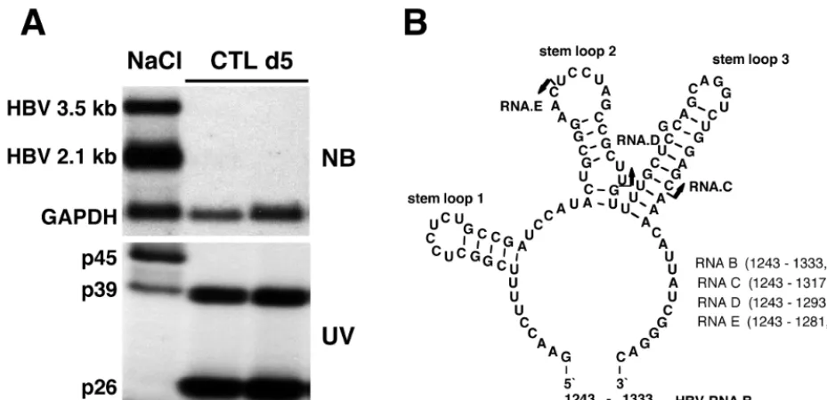
Characteristics of nuclear HBV RNA-binding activities.
As
shown in Fig. 1A, nuclear extracts prepared from HBV
trans-genic mouse liver contain three proteins that form
RNA-pro-tein complexes with a 91-nt
32P-labeled HBV transcript
[image:3.612.67.532.73.298.2](des-ignated RNA.B, shown in Fig. 1B) with apparent molecular
masses of 45 kDa (p45), 39 kDa (p39), and 26 kDa (p26) in UV
cross-linking experiments. In addition, an RNA-protein
com-plex with an apparent molecular mass of 42 kDa was
occasion-ally detected in variable amounts in all extracts containing p45.
Note that p45 activity is constitutively present in the liver, as
are the overlapping 3.5- and 2.1-kb HBV transcripts, both of
which contain RNA.B, and it disappears following CTL
injec-tion, concomitant with the appearance of p26 and the
disap-pearance of HBV RNA. In contrast, p39 is present under both
conditions, appearing to be induced following CTL injection in
FIG. 1. HBV RNA-binding proteins p45 and p39 are detectable in liver nuclear extracts from NaCl-injected mice, while p39 and p26 are detectable in CTL-injected mice. (A) Northern blotting and UV cross-linking analysis of 20mg of total liver RNA or 5mg of liver nuclear extract prepared from the same liver were performed as described in Materials and Methods. Sex and serum HBsAg-matched mice (lineage 1.3.32) were intravenously injected with 107CTLs or with saline and sacrificed on day 5 after CTL administration. The upper panel shows the Northern blot analysis, and the lower panel shows the UV cross-linking analysis of nuclear extracts. (B) Predicted secondary structure of HBV in vitro transcript RNA.B used in this study. The secondary structure was calculated with the program MFOLD version 3 by Zuker and Turner available on the MFOLD server (71, 74). Arrows indicate the 39ends of in vitro transcripts RNA.C and RNA.D and of an oligoribonucleotide, RNA.E. The positions for all RNAs are shown according to the HBVaywsubtype sequence.FIG. 2. Sequence analysis of tryptic peptides obtained from p39 revealed 100% homology to the mouse La protein. p39 was purified and processed as described in Materials and Methods. The sequence tags observed by N-terminal sequencing of three tryptic peptides obtained from p39 are shown in boldface.
on November 9, 2019 by guest
http://jvi.asm.org/
this experiment. The predicted secondary structure of RNA.B
is shown in Fig. 1B.
Molecular characterization of p45, p39, and p26.
In order to
characterize the HBV RNA-binding proteins at the molecular
level, we subjected them to ammonium sulfate precipitation
and heparin-affinity, ion-exchange, and molecular-exclusion
chromatography (see Materials and Methods). Duplicate
sam-ples of gel filtration fractions containing p39 were separated by
SDS-PAGE and subjected to UV cross-linking and Coomassie
blue staining to locate the precise position of p39. The
Coo-massie blue-stained protein band corresponding to the
mobil-ity of p39 detected by UV cross-linking was cut out of the gel
and digested with trypsin. After HPLC separation of the tryptic
peptides, several peptides were subjected to N-terminal
se-quencing in the Scripps Research Institute Molecular Biology
Core Facility. Three peptide sequences showed striking
homol-ogy to the mouse La protein sequence (Fig. 2), identifying p39
as La protein (68). The mouse La protein has an apparent
molecular mass of 47.7 kDa (68), and it is known that the La
protein prepared from rabbit thymus and calf thymus was
protease sensitive and displayed distinct 39- and 26-kDa
cleav-age products after repeated freezing and thawing or incubation
of the samples at 37°C (14, 16, 37). Hence, we assayed whether
p45 might be the full-length La protein while p39 and p26
might be proteolytic cleavage products of La, by attempting to
deplete them from liver nuclear extracts with an La
anti-serum. Figure 3 shows the Western blot (top) and UV
cross-linking (bottom) results obtained with immunodepleted
nu-clear extracts analyzed on the same membrane. Lanes 1 and 2
demonstrate that similar patterns of proteins are detected by
Western blotting and by UV cross-linking, with the exception
of two high-molecular-weight protein bands that were detected
only by Western blotting. Importantly, all three HBV
RNA-binding proteins were depleted from the nuclear extracts by
the anti-La antiserum (Fig. 3, lanes 5 and 6) but not by the
control serum (Fig. 3, lanes 3 and 4). These results suggest that
p45 is the full-length La protein while p39 is a constitutively
detectable proteolytic product and p26 is a La proteolytic
product that was induced following CTL injection.
FIG. 3. HBV RNA-binding proteins are recognized by anti-La-positive human serum. Five micrograms of liver nuclear extract prepared from untreated or CTL-injected mice (lanes 1 and 2) and 5mg of liver nuclear extract from untreated or CTL-injected mice after immunoprecipitation with control human serum (lanes 3 and 4) or with anti-La-positive human serum (lanes 5 and 6) were incubated with 40 fmol of in vitro-labeled RNA.B. The UV cross-linking reaction was performed as described in Materials and Methods. The gel was transferred to a nitrocellulose membrane and analyzed for La protein by Western blotting (WB) and by autoradiography to detect p45, p39, and p26 as described in Materials and Methods.
5770
HEISE ET AL.
J. V
IROL.
on November 9, 2019 by guest
http://jvi.asm.org/
Binding of p45, p39, and p26 to HBV RNA is
phosphoryla-tion dependent.
Since IFN-
g
and TNF-
a
, induced in the liver
following CTL injection and other intrahepatic inflammatory
processes (12, 13, 34), mediate the switch from p45 to p26 (40),
and since these cytokines are known to activate cellular kinases
at a proximal step in their signal transduction cascade, we
assayed whether the phosphorylation status of these proteins
might influence their RNA-binding activity by treating the
nuclear extracts with CIAP before the binding reaction was
performed. As shown in Fig. 4, the RNA-binding activity of
p45, p39, and p26 was strongly reduced after
dephosphoryla-tion. Since CIAP can dephosphorylate nucleic acids, control
experiments were performed to see if the UV cross-linked
labeled RNA was dephosphorylated by the phosphatase,
thereby artifactually reducing the signal from the RNA-protein
complex. No change in signal intensity was observed in this
control experiment (data not shown). While these results
sug-gest that the RNA-binding activity of these proteins is
regu-lated by phosphorylation and dephosphorylation, it remains to
be determined whether specific protein phosphatases and/or
kinases actually regulate the RNA-binding activity of p45, p39,
and p26 in vivo.
Affinity and specificity of protein binding to RNA.
Experi-ments were performed to determine the binding affinity and
specificity of the RNA-binding proteins for RNA.B. First, we
added increasing amounts of nuclear extract to two different
concentrations of RNA.B to determine the optimal protein
concentration required for the titration of RNA.B. UV
cross-linking results were analyzed by phosphorimaging with
arbi-trary units to quantitate the ribonucleoprotein complexes
formed at varying nuclear extract concentrations. We observed
a linear increase in RNA binding at nuclear extract
concentra-tions between 0.5 and 4
m
g per reaction mixture (data not
shown). Based on these results, 0.5 and 2.0
m
g of nuclear
extract were used to measure the binding affinity of p45, p39,
and p26 at increasing RNA.B concentrations. UV cross-linking
results were analyzed by phosphorimaging to quantitate
RNA-protein complex formation at increasing RNA.B
concentra-tions. The apparent
K
dwas calculated according to the mass
action equation (44):
K
d5
[
r
] [
p
]/[
c
], where [r], [p], and [c] are
the molar concentrations of free RNA, protein, and complex,
respectively, assuming that the total RNA concentration (
R
t) is
much higher than the total protein concentration (
P
t) (
R
t2
[
c
])
'
R
t. The final equation was 1/[c]
5
(
K
d/
P
t) (1/
R
t)
1
(1/
P
t).
Plotting 1/[c] (1/arbitrary unit) versus 1/
R
t(1/RNA.B
concen-tration) generated a straight line with the apparent
K
ddefined
as the point of intersection with the
x
axis. By this method, we
determined the apparent
K
ds as 1.4 to 1.5 nM for p45, 0.4 to 1.1
nM for p39, and 0.9 to 1.0 nM for p26.
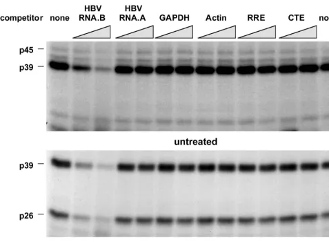
In order to establish the binding specificity of the
RNA-binding proteins, competition experiments were performed
with unlabeled transcripts, including RNA.B and several other
transcripts derived from an AU-rich region of HBV (nt 767 to
870) designated RNA.A or from mouse GAPDH (nt 383 to
497 [60]), mouse
b
-actin (nt 27 to 140 [2]), the HIV RRE (nt
1565 to 1826 [25]; a generous gift from T. Hope), and the
Mason-Pfizer monkey virus CTE (nt 8007 to 8238 [64], also a
generous gift from T. Hope). All experiments were done with
a 10- and a 30-fold molar excess of unlabeled competitors. As
shown in Fig. 5, unlabeled RNA.B inhibited the binding of p45,
p39, and p26 to labeled RNA.B in a concentration-dependent
manner while the other transcripts did not, indicating that the
binding interaction is specific.
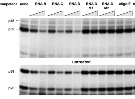
To map the La-binding domain within the 91-nt HBV
RNA.B element more precisely, additional competition
exper-iments were performed with in vitro transcripts RNA.C (nt
FIG. 4. RNA-binding activity of p45, p39, and p26 depends on their phosphorylation status. Two micrograms of liver nuclear extract from untreated or CTL-injected mice was treated with 0.5 and 1.0 U of CIAP (alk. phos.) prior to addition of 40 fmol of32P-labeled in vitro transcript RNA.B. The dephosphorylation reaction was performed in 20ml of 13reaction buffer for 30 min at 37°C. The binding reaction was performed and analyzed as described in Materials and Methods.on November 9, 2019 by guest
http://jvi.asm.org/
1243 to 1317) and RNA.D (nt 1243 to 1293) and an RNA
oligonucleotide (RNA.E, nt 1243 to 1281) representing 3
9
deletions of RNA.B (Fig. 1B). As shown in Fig. 6, RNA.B,
RNA.C, and RNA.D inhibited the binding of p45, p39, and p26
to labeled RNA.B in a concentration-dependent manner, while
RNA.E did not compete. These results suggest that a sequence
or structural element between nt 1275 and 1291 (i.e., stem-loop
2 in Fig. 1B) was recognized by these RNA-binding proteins.
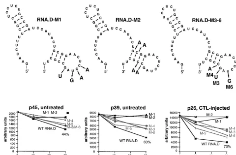
The secondary structure of RNA.D (nt 1243 to 1293)
pre-dicted by using MFOLD version 3 by Zuker and Turner (71,
74) is shown in Fig. 7. The 3
9
stem-loop was of interest since it
is included in RNA.C and RNA.D, both of which compete for
the binding of RNA.B, but not in RNA.E, which does not.
Therefore, we introduced two sets of mutations into the
tem-plate for the in vitro transcription of RNA.D, designated
RNA.D-M1 (containing mutations in the predicted loop) and
RNA.D-M2 (containing mutations in the predicted stem) in
Fig. 6 and 7. As shown in Fig. 6, neither of these mutant
homologues of RNA.D was able to inhibit the binding of p45,
p39, or p26 to RNA.B. Finally, we introduced single mutations
into the RNA.D template, creating RNA.D-M3, -M4, -M5,
and -M6, shown in Fig. 7. The ability of a 10- and a 30-fold
molar excess of unlabeled RNA.D and mutant RNA.D
homo-logues to inhibit the binding of p45, p39, and p26 to labeled
RNA.B was assessed in competitive UV cross-linking
experi-ments and analyzed by phosphorimaging. As shown in Fig. 7,
the relative signal intensities of p45, p39, and p26 (expressed in
arbitrary units) plotted against the relative concentrations of
competitor indicate that single nucleotide substitutions in
RNA.D-M3, -M4, -M5, and -M6 reduced their ability to inhibit
the binding of p26, p39, and p45. These results suggest that
both the structure and the sequence of stem-loop 2 are
prob-ably important for its recognition by the RNA-binding proteins.
DISCUSSION
Intrahepatic inflammatory processes characterized by the
production of IFN-
g
and TNF-
a
downregulate HBV gene
ex-pression in the livers of transgenic mice (12, 13, 29–34, 36, 69)
by a posttranscriptional mechanism (69) that could contribute
to viral clearance during HBV infection. To begin to define this
mechanism, we have recently shown that liver cell nuclei
con-tain a family of three proteins that bind to a 91-nt element
(RNA.B) located at the 5
9
end of the HBV posttranscriptional
regulatory element (40). Furthermore, we suggested that these
proteins might contribute to HBV RNA stability, since they
are regulated by the same cytokines that destabilize the viral
transcripts and because their relative abundance is tightly
linked to the presence or absence of HBV RNA (40).
[image:6.612.68.546.71.421.2]In the studies reported herein, we demonstrate that the
FIG. 5. Competition of various in vitro transcripts with RNA.B for binding of p45, p39, and p26. UV cross-linking experiments were performed with liver nuclear extracts from untreated or CTL-injected mice under standard conditions described in Materials and Methods. A 30- and a 60-fold molar excess of unlabeled in vitro transcripts RNA.B, GAPDH, actin, RRE, and CTE were added into the binding reaction mixture prior to the addition of 40 fmol of32P-labeled RNA.B.5772
HEISE ET AL.
J. V
IROL.
on November 9, 2019 by guest
http://jvi.asm.org/
HBV RNA-binding proteins p45, p39, and p26 are recognized
and depleted from nuclear extracts by anti-La antibodies.
These results strongly suggest that p45 is the full-length mouse
La protein, that p39 is a constitutive proteolytic cleavage
prod-uct of p45, and that p26 is generated from p45 in an IFN-
g
-and/or a TNF-
a
-dependent manner. The La protein is a
well-described RNA-binding protein (15, 16, 45, 70) that binds to
poly(U)-rich elements in RNA polymerase III transcripts
(tRNA precursors and 5S RNA) as well as several other
cel-lular and viral RNAs (1, 66, 70). La appears to be necessary for
the processing of tRNA (73); stimulates translation of
polio-virus (10, 48) and hepatitis C polio-virus RNA (1); has helicase
activity (8); and seems to be translocated from the nucleus to
the cytoplasm during cell stress secondary to virus infection (5,
6), transformation (59), and UV irradiation (7). Recently, the
La protein was reported to stabilize histone mRNA (47). In
addition, IFN-
g
and TNF-
a
have been shown to induce
mem-brane expression of La in cells (22, 24). The La protein carries
a nuclear localization sequence and a nuclear retention signal
(63), and it coprecipitates with certain viral RNAs (38). La is a
phosphoprotein (9, 27, 53, 54) that binds RNA in a
phospho-rylation-dependent manner in some (9, 53) but not all (27) of
the systems in which it has been studied. In the present study,
we demonstrated that the ability of all three La proteins to
bind HBV RNA is phosphorylation dependent. Since these
experiments were performed with crude nuclear extracts, we
do not know whether phosphorylation of La itself or that of
other accessory molecules is required for the binding
interac-tion to occur. This should be clarified in future experiments
with purified recombinant La proteins instead of nuclear
ex-tracts. Such studies will also allow more precise measurement
of the binding affinity of La for HBV RNA in the absence of
possible cellular cofactors.
It is important to note that the RNA-binding activity of p45,
p39, and p26 La depends on the phosphorylation status of the
nuclear extracts used (Fig. 4) and that the disappearance of
p45 and the appearance of p26 are regulated by IFN-
g
and
TNF-
a
(40). Therefore, the activation of signal transduction
pathways by IFN-
g
and TNF-
a
following CTL injection could
reflect a phosphorylation-dependent proteolytic cleavage of
p45 into a 26-kDa RNA-binding fragment and one or more
fragments that are unable to bind HBV RNA. Others have
shown that La is sensitive to proteolytic cleavage, yielding
several cleavage products similar in size to p39 and p26 (14, 16,
37).
In this study, the specificity of the interaction of p45, p39,
and p26 with the 91-nt HBV RNA.B target element was
con-firmed in the competition experiments shown in Fig. 5 to 7.
Importantly, mutational analysis of RNA.B revealed that all
three RNA-binding proteins recognize a single 17-bp target
element, located between nt 1275 and 1291 (Fig. 7).
Interest-ingly, this element displays a predicted stem-loop structure
that appears to be recognized by all three proteins, since
mu-tational disruption of the predicted stem (RNA.D-M2)
abol-FIG. 6. Mapping of RNA.B for a sequential-structural element recognized by p45, p39, and p26. UV cross-linking experiments were performed with liver nuclear extracts from untreated or CTL-injected mice under standard conditions described in Materials and Methods. A 30- and a 60-fold molar excess of unlabeled in vitro transcripts RNA.B, RNA.C, RNA.D, and RNA.E (Fig. 1B) were added into the binding reaction mixture prior to the addition of 40 fmol of32P-labeled RNA.B.on November 9, 2019 by guest
http://jvi.asm.org/
[image:7.612.74.538.71.409.2]ished their ability to bind the RNA. Similarly, binding was
reduced by point mutations in the loop (RNA.D-M1),
suggest-ing that the proteins display sequence specificity as well as
structural specificity for their substrate. Additional
experi-ments with recombinant La protein and mutant RNA
sub-strates containing single nucleotide mutations in the loop and
other substrates containing compensatory mutations that
maintain the structure of the stem will be necessary to further
define the nature of the binding site(s) within the substrate.
In summary, we have previously shown that a close
relation-ship exists between the presence of three HBV RNA-binding
proteins (p45, p39, and p26) and the abundance of HBV RNA
in the livers of HBV transgenic mice (40). The current results
demonstrate that all three proteins are related to the cellular
nucleoprotein, La, and that p45 is probably the full-length
protein while p39 and p26 are constitutive and
cytokine-induc-ible proteolytic cleavage products, respectively. We also
dem-onstrate that all three La isoforms bind the same predicted
stem-loop in HBV RNA between nt 1275 and 1291 with high
affinity in a phosphorylation-dependent manner. These results
suggest that conditions, such as inflammation, that alter the
content, metabolism, and distribution of La in the hepatocyte
may contribute to the posttranscriptional control of the
steady-state content of HBV RNA and, thereby, influence the
out-come of HBV infection.
ACKNOWLEDGMENTS
We thank Edward K. Chan (The Scripps Research Institute, La
Jolla, Calif.) for providing anti-La human antiserum; Joel Gottesfeld
(The Scripps Research Institute) for consultations and advice; Thomas
J. Hope (Salk Institute, La Jolla, Calif.) for providing plasmids carrying
the HIV RRE and Mason-Pfizer monkey virus CTEs; the Scripps
Molecular Biology Core Facility for the production of
oligonucleo-tides; the Scripps Protein and Nucleic Acid Core Facility for tryptic
digestion of the proteins, HPLC purification, and N-terminal
sequenc-ing of peptides; and Jennifer Newmann for help with manuscript
prep-aration.
This work was supported by grants R37-CA40489 and R01-AI40696
from the National Institutes of Health.
REFERENCES
1.Ali, N., and A. Siddiqui.1997. The La antigen binds 59noncoding region of the hepatitis C virus RNA in the context of the initiator AUG codon and stimulates internal ribosome entry site-mediated translation. Proc. Natl. Acad. Sci. USA94:2249–2254.
2.Alonso, S., A. Minty, Y. Bourlet, and M. Buckingham.1986. Comparison of three actin-coding sequences in the mouse; evolutionary relationships be-tween the actin genes of warm-blooded vertebrates. J. Mol. Evol.23:11–22. 3.Ando, K., L. G. Guidotti, A. Cerny, T. Ishikawa, and F. V. Chisari.1994. CTL
access to tissue antigen is restricted in vivo. J. Immunol.153:482–488. 4.Ando, K., T. Moriyama, L. G. Guidotti, S. Wirth, R. D. Schreiber, H. J.
Schlicht, S. N. Huang, and F. V. Chisari. 1993. Mechanisms of class I restricted immunopathology. A transgenic mouse model of fulminant hep-atitis. J. Exp. Med.178:1541–1554.
[image:8.612.60.540.70.385.2]5.Baboonian, C., P. J. Venables, J. Booth, D. G. Williams, L. M. Roffe, and
FIG. 7. Sequential and/or structural features of stem-loop 2 are substantive for the binding of p45, p39, and p26. UV cross-linking experiments were performed with liver nuclear extracts from untreated or CTL-injected mice under standard conditions described in Materials and Methods. A 10- and a 30-fold molar excess of unlabeled in vitro transcripts RNA.D-M1 to RNA.D-M6 were added into the binding reaction mixture prior to the addition of 40 fmol of32P-labeled RNA.B. The
decreases in signal intensity for p45, p39, and p26 were separately analyzed by phosphorimaging. The upper panel shows the predicted structure of RNA.D with nucleotide changes indicated by the arrows. The lower panel shows the plot of complex formation (arbitrary units) versus competitor concentrations (10- and 30-fold). WT, wild type.
5774
HEISE ET AL.
J. V
IROL.
on November 9, 2019 by guest
http://jvi.asm.org/
R. N. Maini.1989. Virus infection induces redistribution and membrane localization of the nuclear antigen La (SS-B): a possible mechanism for autoimmunity. Clin. Exp. Immunol.78:454–459.
6.Bachmann, M., H. Althoff, H. Troster, C. Selenka, D. Falke, and W. E. Muller.1992. Translocation of the nuclear autoantigen La to the cell surface of herpes simplex virus type 1 infected cells. Autoimmunity12:37–45. 7.Bachmann, M., S. Chang, H. Slor, J. Kukulies, and W. E. Muller.1990.
Shuttling of the autoantigen La between nucleus and cell surface after uv irradiation of human keratinocytes. Exp. Cell Res.191:171–180.
8.Bachmann, M., K. Pfeifer, H. C. Schroder, and W. E. Muller.1990. Char-acterization of the autoantigen La as a nucleic acid-dependent ATPase/ dATPase with melting properties. Cell60:85–93.
9.Bachmann, M., H. C. Schroder, K. G. Wagner, W. J. Mayet, K. Pfeifer, and W. E. Muller.1986. Purification and characterization of the Ro and La antigens. Modulation of their binding affinities to poly(U) by phosphoryla-tion and the presence of ATP. Biol. Chem. Hoppe-Seyler367:671–680. 10.Belsham, G. J., N. Sonenberg, and Y. V. Svitkin.1995. The role of the La
autoantigen in internal initiation. Curr. Top. Microbiol. Immunol.203:85– 98.
11. Carpousis, A. J., G. Van Houwe, C. Ehretsmann, and H. M. Krisch.1994. Copurification of E. coli RNAase E and PNPase: evidence for a specific association between two enzymes important in RNA processing and degra-dation. Cell76:889–900.
12. Cavanaugh, V. J., L. G. Guidotti, and F. V. Chisari.1998. Inhibition of hepatitis B virus replication during adenovirus and cytomegalovirus infec-tions in transgenic mice. J. Virol.72:2630–2637.
13. Cavanaugh, V. J., L. G. Guidotti, and F. V. Chisari.1997. Interleukin-12 inhibits hepatitis B virus replication in transgenic mice. J. Virol.71:3236– 3243.
14. Chan, E. K., A. M. Francoeur, and E. M. Tan.1986. Epitopes, structural domains, and asymmetry of amino acid residues in SS-B/La nuclear protein. J. Immunol.136:3744–3749.
15. Chan, E. K., K. F. Sullivan, and E. M. Tan.1989. Ribonucleoprotein SS-B/La belongs to a protein family with consensus sequences for RNA-binding. Nucleic Acids Res.17:2233–2244.
16. Chan, E. K., and E. M. Tan.1987. The small nuclear ribonucleoprotein SS-B/La binds RNA with a conserved protease-resistant domain of 28 kilo-daltons. Mol. Cell. Biol.7:2588–2591.
17. Chen, F. Y., F. M. Amara, and J. A. Wright.1993. Mammalian ribonucle-otide reductase R1 mRNA stability under normal and phorbol ester stimu-lating conditions: involvement of a cis-trans interaction at the 39untranslated region. EMBO J.12:3977–3986.
18. Chisari, F. V.1997. Cytotoxic T cells and viral hepatitis. J. Clin. Investig.
99:1472–1477.
19. Chisari, F. V., and C. Ferrari.1995. Hepatitis B virus immunopathology. Springer Semin. Immunopathol.17:261–281.
20. Chisari, F. V., P. Filippi, A. McLachlan, D. R. Milich, M. Riggs, S. Lee, R. D. Palmiter, C. A. Pinkert, and R. L. Brinster.1986. Expression of hepatitis B virus large envelope polypeptide inhibits hepatitis B surface antigen secre-tion in transgenic mice. J. Virol.60:880–887.
21. Chisari, F. V., C. A. Pinkert, D. R. Milich, P. Filippi, A. McLachlan, R. D. Palmiter, and R. L. Brinster.1985. A transgenic mouse model of the chronic hepatitis B surface antigen carrier state. Science230:1157–1160. 22. Clark, D. A., P. J. Lamey, R. F. Jarrett, and D. E. Onions.1994. A model to
study viral and cytokine involvement in Sjogren’s syndrome. Autoimmunity
18:7–14.
23. Donello, J. E., A. A. Beeche, G. J. Smith, G. R. Lucero, and T. J. Hope.1996. The hepatitis B virus posttranscriptional regulatory element is composed of two subelements. J. Virol.70:4345–4351.
24. Dorner, T., M. Hucko, W. J. Mayet, U. Trefzer, G. R. Burmester, and F. Hiepe.1995. Enhanced membrane expression of the 52 kDa Ro(SS-A) and La(SS-B) antigens by human keratinocytes induced by TNF alpha. Ann. Rheum. Dis.54:904–909.
25. Duchet, S., F. Letourneur, I. Loussert-Ajaka, C. Chaplain, E. Gomas, F. Brun-Vezinet, F. Simon, and S. Saragosti.1997. gag and env sequences of an A/G/H recombinant from a Zairian HIV type 1 isolate. AIDS Res. Hum. Retroviruses13:1351–1354.
26. Eisenstein, R. S., P. T. Tuazon, K. L. Schalinske, S. A. Anderson, and J. A. Traugh.1993. Iron-responsive element-binding protein. Phosphorylation by protein kinase C. J. Biol. Chem.268:27363–27370.
27. Fan, H., A. L. Sakulich, J. L. Goodier, X. Zhang, J. Qin, and R. J. Maraia.
1997. Phosphorylation of the human La antigen on serine 366 can regulate recycling of RNA polymerase III transcription complexes. Cell88:707–15. 28. Galibert, F., E. Mandart, F. Fitoussi, P. Tiollais, and P. Charnay.1979.
Nucleotide sequence of the hepatitis B virus genome (subtype ayw) cloned in E. coli. Nature281:646–650.
29. Gilles, P. N., G. Fey, and F. V. Chisari.1992. Tumor necrosis factor alpha negatively regulates hepatitis B virus gene expression in transgenic mice. J. Virol.66:3955–3960.
30. Guidotti, L. G., K. Ando, M. V. Hobbs, T. Ishikawa, L. Runkel, R. D. Schreiber, and F. V. Chisari.1994. Cytotoxic T lymphocytes inhibit hepatitis B virus gene expression by a noncytolytic mechanism in transgenic mice.
Proc. Natl. Acad. Sci. USA91:3764–3768.
31. Guidotti, L. G., P. Borrow, M. V. Hobbs, B. Matzke, I. Gresser, M. B. Oldstone, and F. V. Chisari.1996. Viral cross talk: intracellular inactivation of the hepatitis B virus during an unrelated viral infection of the liver. Proc. Natl. Acad. Sci. USA93:4589–4594.
32. Guidotti, L. G., and F. V. Chisari.1996. To kill or to cure: options in host defense against viral infection. Curr. Opin. Immunol.8:478–483. 33. Guidotti, L. G., S. Guilhot, and F. V. Chisari.1994. Interleukin-2 and
alpha/beta interferon down-regulate hepatitis B virus gene expression in vivo by tumor necrosis factor-dependent and -independent pathways. J. Virol.
68:1265–1270.
34. Guidotti, L. G., T. Ishikawa, M. V. Hobbs, B. Matzke, R. Schreiber, and F. V. Chisari.1996. Intracellular inactivation of the hepatitis B virus by cytotoxic T lymphocytes. Immunity4:25–36.
35. Guidotti, L. G., B. Matzke, H. Schaller, and F. V. Chisari.1995. High-level hepatitis B virus replication in transgenic mice. J. Virol.69:6158–6169. 36. Guilhot, S., L. G. Guidotti, and F. V. Chisari.1993. Interleukin-2
downregu-lates hepatitis B virus gene expression in transgenic mice by a posttranscrip-tional mechanism. J. Virol.67:7444–7449.
37. Habets, W. J., J. H. den Brok, A. M. Boerbooms, L. B. van de Putte, and W. J. van Venrooij.1983. Characterization of the SS-B (La) antigen in adenovirus-infected and unadenovirus-infected HeLa cells. EMBO J.2:1625–1631.
38. Hamelin, R., E. K. Chan, E. M. Tan, and R. B. Arlinghaus.1986. Antibodies against small nuclear ribonucleoproteins immunoprecipitate complexes con-taining ts110 Moloney murine sarcoma virus genomic and messenger RNAs. Virology152:87–99.
39. Harford, J. B., and D. R. Morris.1997. mRNA metabolism & post-tran-scriptional gene regulation. Wiley-Liss, New York, N.Y.
39a.Heise, T.Unpublished data.
40. Heise, T., L. G. Guidotti, V. J. Cavanaugh, and F. V. Chisari.1999. Hepatitis B virus RNA-binding proteins associated with cytokine-induced clearance of viral RNA from the liver of transgenic mice. J. Virol.73:474–481. 41. Heise, T., A. Krones, A. Nath, K. Jungermann, and B. Christ.1998. Parallel
acelleration of phosphoenolpyruvate carboxykinase mRNA degradation and increase in ribonuclease activity induced by insulin in cultured rat hepato-cytes. Biol. Chem. Hoppe-Seyler379:875–883.
42. Huang, J., and T. J. Liang.1993. A novel hepatitis B virus (HBV) genetic element with Rev response element-like properties that is essential for ex-pression of HBV gene products. Mol. Cell. Biol.13:7476–7486.
43. Izaurralde, E., and I. W. Mattaj.1995. RNA export. Cell81:153–159. 44. Lehninger, A. L.1975. Biochemistry, 2nd ed., p. 1152–1154. Worth
Publish-ers, New York, N.Y.
45. Lerner, M. R., J. A. Boyle, J. A. Hardin, and J. A. Steitz.1981. Two novel classes of small ribonucleoproteins detected by antibodies associated with lupus erythematosus. Science211:400–402.
46. Malter, J. S., and Y. Hong.1991. A redox switch and phosphorylation are involved in the post-translational up-regulation of the adenosine-uridine binding factor by phorbol ester and ionophore. J. Biol. Chem.266:3167– 3171.
47. McLaren, R. S., N. Caruccio, and J. Ross.1997. Human La protein: a stabilizer of histone mRNA. Mol. Cell. Biol.17:3028–3036.
48. Meerovitch, K., Y. V. Svitkin, H. S. Lee, F. Lejbkowicz, D. J. Kenan, E. K. Chan, V. I. Agol, J. D. Keene, and N. Sonenberg.1993. La autoantigen enhances and corrects aberrant translation of poliovirus RNA in reticulocyte lysate. J. Virol.67:3798–3807.
49. Miczak, A., V. R. Kaberdin, C. L. Wei, and S. Lin-Chao.1996. Proteins associated with RNase E in a multicomponent ribonucleolytic complex. Proc. Natl. Acad. Sci. USA93:3865–3869.
50. Moriyama, T., S. Guilhot, K. Klopchin, B. Moss, C. A. Pinkert, R. D. Palmiter, R. L. Brinster, O. Kanagawa, and F. V. Chisari.1990. Immuno-biology and pathogenesis of hepatocellular injury in hepatitis B virus trans-genic mice. Science248:361–364.
51. Nigg, E. A. 1997. Nucleocytoplasmic transport: signals, mechanisms and regulation. Nature386:779–787.
52. Pasquinelli, A. E., R. K. Ernst, E. Lund, C. Grimm, M. L. Zapp, D. Rekosh, M. L. Hammarskjold, and J. E. Dahlberg.1997. The constitutive transport element (CTE) of Mason-Pfizer monkey virus (MPMV) accesses a cellular mRNA export pathway. EMBO J.16:7500–7510.
53. Pfeifle, J., F. A. Anderer, and M. Franke.1987. Multiple phosphorylation of human SS-B/LA autoantigen and its effect on poly(U) and autoantibody binding. Biochim. Biophys. Acta928:217–226.
54. Pizer, L. I., J. S. Deng, R. M. Stenberg, and E. M. Tan.1983. Characteriza-tion of a phosphoprotein associated with the SS-B/La nuclear antigen in adenovirus-infected and uninfected KB cells. Mol. Cell. Biol.3:1235– 1245.
55. Py, B., H. Causton, E. A. Mudd, and C. F. Higgins.1994. A protein complex mediating mRNA degradation in Escherichia coli. Mol. Microbiol.14:717– 729.
56. Py, B., C. F. Higgins, H. M. Krisch, and A. J. Carpousis.1996. A DEAD-box RNA helicase in the Escherichia coli RNA degradosome. Nature381:169– 172.
57. Rao, K. S., R. Sirdeshmukh, and P. D. Gupta.1994. Modulation of cytosolic
on November 9, 2019 by guest
http://jvi.asm.org/
RNase activity by endogenous RNase inhibitor in rat vaginal epithelial cells on estradiol administration. FEBS Lett.343:11–14.
58. Ross, J.1995. mRNA stability in mammalian cells. Microbiol. Rev.59:423– 450.
59. Rother, R. P., and P. S. Thomas.1991. La/SSB ribonucleoprotein levels increased in transformed cells. Clin. Exp. Immunol.83:369–374.
60. Sabath, D. E., H. E. Broome, and M. B. Prystowsky.1990. Glyceraldehyde-3-phosphate dehydrogenase mRNA is a major interleukin 2-induced tran-script in a cloned T-helper lymphocyte. Gene91:185–191.
61. Schaller, H., and M. Fischer.1991. Transcriptional control of hepadnavirus gene expression. Curr. Top. Microbiol. Immunol.168:21–39.
62. Sharp, P. A.1994. Split genes and RNA splicing. Cell77:805–815. 63. Simons, F. H., F. J. Broers, W. J. Van Venrooij, and G. J. Pruijn.1996.
Characterization of cis-acting signals for nuclear import and retention of the La (SS-B) autoantigen. Exp. Cell Res.224:224–236.
64. Sonigo, P., C. Barker, E. Hunter, and S. Wain-Hobson.1986. Nucleotide sequence of Mason-Pfizer monkey virus: an immunosuppressive D-type ret-rovirus. Cell45:375–385.
65. Standart, N., and R. J. Jackson.1994. Regulation of translation by specific protein/mRNA interactions. Biochimie76:867–879.
66. Stefano, J. E.1984. Purified lupus antigen La recognizes an oligouridylate stretch common to the 39termini of RNA polymerase III transcripts. Cell
36:145–154.
67. St. Johnston, D.1995. The intracellular localization of messenger RNAs. Cell81:161–170.
68. Topfer, F., T. Gordon, and J. McCluskey.1993. Characterization of the mouse autoantigen La (SS-B). Identification of conserved RNA-binding motifs, a putative ATP binding site and reactivity of recombinant protein with poly(U) and human autoantibodies. J. Immunol.150:3091–3100. 69. Tsui, L. V., L. G. Guidotti, T. Ishikawa, and F. V. Chisari.1995.
Posttran-scriptional clearance of hepatitis B virus RNA by cytotoxic T lymphocyte-activated hepatocytes. Proc. Natl. Acad. Sci. USA92:12398–12402. 70. van Venrooij, W. J., R. L. Slobbe, and G. J. Pruijn.1993. Structure and
function of La and Ro RNPs. Mol. Biol. Rep.18:113–119.
71. Walter, A. E., D. H. Turner, J. Kim, M. H. Lyttle, P. Muller, D. H. Mathews, and M. Zuker.1994. Coaxial stacking of helixes enhances binding of oligo-ribonucleotides and improves predictions of RNA folding. Proc. Natl. Acad. Sci. USA91:9218–9222.
72. Williams, D. L., M. Sensel, M. McTigue, and R. Binder.1993. Hormonal and developmental regulation of mRNA turnover, p. 161–197.InJ. Belasco and G. Brawermann (ed.), Control of messenger RNA stability. Academic Press, Inc., New York, N.Y.
73. Yoo, C. J., and S. L. Wolin.1997. The yeast La protein is required for the 39
endonucleolytic cleavage that matures tRNA precursors. Cell89:393–402. 74. Zuker, M., and D. H. Turner.1995–1999, copyright date. [Online.] MFOLD,
version 3. Michael Zuker, Washington University School of Medicine. http: //mfold2.wustl.edu/;mfold/rna/form1.cgi. [6 May 1999, last date accessed.]
5776
HEISE ET AL.
J. V
IROL.
on November 9, 2019 by guest
http://jvi.asm.org/