R E S E A R C H
Open Access
The solution structure of the prototype foamy
virus RNase H domain indicates an important role
of the basic loop in substrate binding
Berit Leo, Kristian Schweimer, Paul Rösch, Maximilian J Hartl and Birgitta M Wöhrl
*Abstract
Background:The ribonuclease H (RNase H) domains of retroviral reverse transcriptases play an essential role in the replication cycle of retroviruses. During reverse transcription of the viral genomic RNA, an RNA/DNA hybrid is created whose RNA strand needs to be hydrolyzed by the RNase H to enable synthesis of the second DNA strand by the DNA polymerase function of the reverse transcriptase. Here, we report the solution structure of the separately purified RNase H domain from prototype foamy virus (PFV) revealing the so-called C-helix and the adjacent basic loop, which both were suggested to be important in substrate binding and activity.
Results:The solution structure of PFV RNase H shows that it contains a mixed five-strandedβ-sheet, which is sandwiched by fourα-helices (A-D), including the C-helix, on one side and oneα-helix (helix E) on the opposite side. NMR titration experiments demonstrate that upon substrate addition signal changes can be detected predominantly in the basic loop as well as in the C-helix. All these regions are oriented towards the bound substrate. In addition, signal intensities corresponding to residues in the B-helix and the active site decrease, while only minor or no changes of the overall structure of the RNase H are detectable upon substrate binding. Dynamic studies confirm the monomeric state of the RNase H domain. Structure comparisons with HIV-1 RNase H, which lacks the basic protrusion, indicate that the basic loop is relevant for substrate interaction, while the C-helix appears to fulfill mainly structural functions, i.e. positioning the basic loop in the correct orientation for substrate binding. Conclusions:The structural data of PFV RNase H demonstrate the importance of the basic loop, which contains four positively charged lysines, in substrate binding and the function of the C-helix in positioning of the loop. In the dimeric full length HIV-1 RT, the function of the basic loop is carried out by a different loop, which also harbors basic residues, derived from the connection domain of the p66 subunit. Our results suggest that RNases H which are also active as separate domains might need a functional basic loop for proper substrate binding.
Keywords:Foamy virus, Retroviral RNase H, C-helix, Basic loop, NMR, Basic protrusion, Solution structure, Substrate binding
Background
RTs of retroviruses are multifunctional enzymes, which in addition to a polymerase domain harbor a C-terminal RNase H domain whose activity is essential during re-verse transcription. The RNase H function is necessary for degrading the RNA in the emerging RNA/DNA hy-brid [1] to allow synthesis of the second DNA strand. In contrast to orthoretroviruses, spumaretroviruses, also
called foamy viruses (FVs), possess a mature protease-reverse transcriptase (PR-RT) in the virion, whereas all other retroviruses cleave off the PR domain from the Pol precursor [2,3]. Similarly to the RT from Moloney mur-ine leukemia virus (MoMLV) [4], the PR-RT of the prototype foamy virus (PFV) is also monomeric [3].
We have shown previously, that the isolated RNase H domain of PFV exhibits activity, albeit to a much lower extent than the mature full length PR-RT [5]. This be-havior is comparable to that of the separate RNase H domain of MoMLV [6-8]. Furthermore, NMR spectros-copy of PFV RNase H revealed the existence of the so-* Correspondence:[email protected]
Universität Bayreuth, Lehrstuhl Biopolymere, Universitätsstr. 30, D-95447 Bayreuth, Germany
called“basic protrusion”which consists of anα-helix, re-ferred to as the “C-helix” followed by a basic loop [5]. The basic protrusion of RNases H has been implicated in substrate binding and activity [9-11] and has also been found in the retroviral RNase H domain of MoMLV [12] and in the recently solved x-ray structure of xenotropic murine leukemia virus-related virus (XMRV) [13]. XMRV is highly related to MoMLV and appears to have arisen through recombination events during passaging of human tumors in mice [14].
A basic protrusion can also be found in the human and Escherichia coli (E. coli) RNases H, whereas the RNase H fromBacillus halodurans (B. halodurans) con-tains only the basic loop but no C-helix. However, an N-terminal RNA/DNA-hybrid binding domain (RHBD) compensates for the lack of the helix [15]. In contrast, HIV-1 RNase H, which is not active when expressed in isolation [16-20], does not harbor the basic protrusion, rather, positively charged residues in the connection do-main assume this function.
Here, we show the solution structure of PFV RNase H and titration experiments with an RNA/DNA substrate indicating an important role of the basic loop in sub-strate binding.
Results and discussion
To determine the solution structure of the wild type (wt) PFV RNase H we purified the recombinant15N or
15
N,13C labeled protein PFV RNase H-(Q591-N751) from E. coli. We have shown previously, that the purified do-main exhibits RNase H activity and possesses a defined tertiary structure including the C-helix [5].
Structure of the isolated PFV RNase H
To verify the oligomerization state of the PFV RNase H domain 15N spin relaxation experiments were performed. Comparison of 15N transverse (R2) and
longitudinal (R1) relaxation rates at two different
con-centrations (1 mM and 0.4 mM) reveals deviations of less than 6 % for R2and 3 % for R1, indicating that no
aggregation is observed under the conditions applied for NMR spectroscopy. From the relaxation rates, a rota-tional correlation time (tc) of 12.4 ns was determined for an isotropic model. In addition, the data were fitted to an axial symmetric as well as a fully asymmetric rota-tional diffusion tensor using the coordinates from the lowest energy structure, but no further statistical im-provement was obtained. The higher tc of PFV RNase H as compared to the isolated RNase H domain of HIV-RT (9.7 ns, [21]) reflects the larger size of the PFV RNase H (165 aa vs. 138 aa, 18 kDa vs. 15.3 kDa), and confirms the monomeric character of the domain already demon-strated by size exclusion chromatography [5].
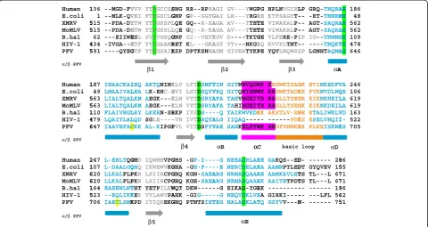
Sequence comparisons (Figure 1) indicated that similar to MoMLV and XMRV RNase H, PFV RNase H contains a basic protrusion, including the so-called C-helix fol-lowed by a basic loop structure. We have shown previ-ously that the purified, separate PFV RNase H domain is weakly active [5]. Structural analyses by NMR spectros-copy revealed the presence of a structured C-helix [5]. Thus, we set out to determine the solution structure of PFV RNase H (Figure 2).
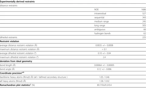
Using multidimensional heteronuclear edited NOESY experiments, 1686 distance restraints together with 66 restraints for hydrogen bonds and 225 dihedral angle restraints based on chemical shift analyses could be derived (Table 1). The final structure calculation resulted in an ensemble of structures with no distance restraint violation larger than 0.1 Å and no dihedral restraint vio-lation larger than 2.4° together with good stereochemical properties (Table 1). The structures superimpose well in the secondary structure regions (Figure 2A). For several loop regions no distance restraints could be derived be-cause of unassigned residues [5]. Their NMR signals are most likely broadened beyond detection due to exchange processes. Therefore the large coordinate variability reflects the dynamic character of these regions.
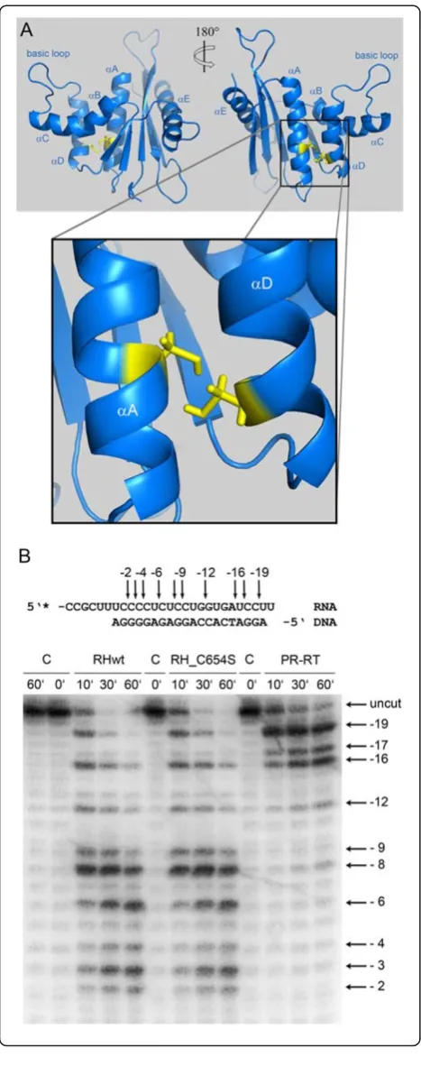
The structure of PFV RNase H shows the typical ter-tiary fold of an RNase H domain, which is formed by a five-stranded mixed β-sheet flanked by five α-helices. The long carboxy-terminal helix E packs on one side of the β-sheet, and the helices A, B, C and D are located on the other side of the β-sheet (Figure 2B). Due to a lack of structural restraints accessible by NMR, no attempts were made to characterize the active site. Des-pite the absence of certain restraints in this region, the active site residues (D599, E646, D669, D740) are closely enough together to allow coordination of mag-nesium ions.
A characteristic feature of PFV RNase H is the pres-ence of helix C which directly follows helix B after a kink. Helix C precedes a basic loop containing three neighboring lysines. Helix C as well as the adjacent loop is also found in XMRV RNase H. However, in XMRV RNase H the consecutive basic residues (3 x Arg) are part of helix C [13], whereas in PFV RNase H four lysines (KKKPLK) are located in the adjacent basic loop (Figure 1). The slightly different position of the basic residues might cause different distances to the active site and might thus contribute to possible differences in cleavage activities of the two enzymes [5,13,22]. The orientation of helix C is determined by numerous hydro-phobic contacts to helix D. This suggests that helix C acts as a molecular ruler to orient the basic loop in order to optimize substrate binding and specificity.
in helix A and D, respectively, are facing each other, thus allowing the potential formation of a disulfide bridge (Figure 3A). To find out whether this disulfide bridge is important for RNase H function, we mutated Cys654 to serine to abolish disulfide formation. However, analyses of the RNase H activities of the mutant RNase H-(C654S)
[image:3.595.58.539.90.345.2]and the wt enzyme revealed no differences in activity (Figure 3B), indicating that creation of the disulfide bridge is not relevant for RNase H activity. Sequence compari-sons of PFV RNase H with other foamy virus RNases H (feline, bovine) showed, that the cysteines are not con-served. Only the closely related RNase H from simian FV
Figure 2Solution structure of PFV RNase H.(A) Superposition of the 19 lowest energy structures. (B) Ribbon diagram of the lowest energy structure. The C-helix is highlighted in magenta, and the basic loop in orange. The active site residues D599, E646, D669 and D740 are highlighted in bright green.
Human 1 E.coli XMRV 5 MoMLV 5 B.hal HIV-1 4 PFV 5
α/β PFV
Human 1 E.coli XMRV 5 MoMLV 5 B.hal 1 HIV-1 4 PFV 6
α/β PFV
Human 2 E.coli 1 XMRV 6 MoMLV 6 B.hal 1 HIV-1 5 PFV 7
α/β PFV
136 --MGD-FV 1 --MLK-QV 515 --PDA-DY 515 --PDA-DH 62 ---EIIWE 434 -IVGA--E 591 ----QYEG
187 IHAACKAI
49 LMAAIVAL
563 LIALTQAL
563 LIALTQAL
110 FLAIVHGL
479 LQAIYLAL
647 IAAVEFAC
247 L-ERLTQG 107 L-DAALGQ
620 LLKALFLP 620 LLKALFLP 164 EAEEWLNT
523 --EQLIKK
706 IAECLSMK
VVV YTDGCCSS VEI FTDGSCLG YTW YTDGSSFL HTW YTDGSSLL ESL SVDVGSQG ETF YVDGAANR GVF YTDGSAIK
IEQ AKTQNINK
LEA LK-EHC-E
LKM AEGK---K
LKM AEGK---K
LRY LKERN-SR
LQD SG
LE----CKK AL-KIPGP
GMD IQWMHVPG
QHQ IKWEWVKG PKR LSIIHCPG PKR LSIIHCPG
THT YETPILKW
KEK VYLAWVPA KPD ITIQHEKG
β1
β5
SNG RR--RPRA GNP GP--GGYG LQE GQ--R-RA LQE GQ--R-KA GNP GI--VEYK RET KL---GKA KSP DPTKSNNA
KLV LYTDSMFT
EVI LSTDSQYV
KLN VYTDSRYA
KLN VYTDSRYA
RKP IYSD
S----VN IVTDSQYA
PVL VITDSFYV
GHS -G
F-I---GHA -G
H-P---GHQ KGN-SAEA
GHQ KGH-SAEA
WQT DKW---AHK -GI
G----GHQ PTNTSIHT
β4
AGI GV---YWGP GAI LR---YRGR AGA AV---TTET AGA AV---TTET KGV D----TKTG AGY VT---NKGR AGM GIVHATYKP
TIN GITNWVQGW
VRQ GITQWIHNW
AFA TAHVHGEIY
AFA TAHIHGEIY
-Q TAIKWVKDK ALG IIQAQ
----VAE SANKELPYW
-G NEEADRLAR -E NERCDELAR ARG NRMADQAAR ARG NRMADQAAR
-G EIKAD-YGR
-G NEQVDKLVS TEG NALADKLAT
β2
αB α
αE
PG HPLNVGIRL RE KTFSAGY T-TE VIWARAL P-TE VIWAKAL P-GE VLFERE-PI RQ KVVPL TNT-PE YQVLNQWSI
WK KNGWKTSAG
WK KRGWKTADK
YR RRGLLTSEG
YR RRGLLTSEG KK AKSTLV-RN
-- ---PDK
WK SNGFVNNKK
RE GAKQS--ED
RA AAMNPTLED
RE AAMKAVLET
RK AAITETPDT RK
---SA
GIRKI----TQ GSYVV---N β3
αC basic
LP GRQ-TNQRA
-- -RT-TNNRM
-- AGT-SAQRA
-- AGT-SAQRA
IP IG--TNNMG
-- ----TNQKT
IP LGNHTAQMA
GK EVINKEDFV KK PVKNVDLWQ GR EIKNKNEIL GK EIKNKDEIL NE ETALIWKLV KS ESELVNQII KK PLKHISKWK
D- --- 28 DT GYQVEV 15 TS TL---L 67 TS TL---L 67 -- --- 19 -- ---LFL 56 N- --- 75 αA
αD loop
AE 186
ME 48
AE 562
AE 562
GE 109
TE 478
AE 646
VA 246
QR 106
LA 619
LA 619
VD 163
I- 522
KS 705
[image:3.595.58.538.504.694.2]86 55 71 71 96 62 51 A
from macaques (SFVmac) retains the cysteines (data not shown). Other RNases H also do not contain two cysteines facing each other (Figure 1).
Substrate binding
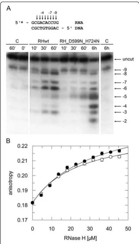
To analyze the interaction of PFV RNase H with an RNA/DNA substrate, we had to abolish RNase H activity to avoid cleavage of the substrate during long-term NMR analyses. Thus, we mutated the active site residue Asp599 and, in addition, residue His724, located in the loop region between β-strand 5 and helix E to aspara-gine. The corresponding histidine residue in HIV-1 RNase H had been shown to severely reduce RNase H activity [23,24]. The RNase H activity of the double mu-tant D599N-H742N was then analyzed qualitatively with the same 10/10-mer RNA/DNA substrate, which we wanted to use further in NMR analyses. Our results demonstrate that the RNase H activity of the mutant is seriously impaired (Figure 4A). However, only minor changes were observed in the recorded NMR spectra, in-dicating that the overall tertiary structure of the RNase H domain is unaffected by the amino acid exchanges (data not shown).
Furthermore, we introduced the two amino acid exchanges into the full length PR-RT to determine the impact of the mutations in DNA polymerization. Ana-lysis of the polymerization activities on a homopolymeric substrate resulted in similar specific activities of 8.2 U and 8.8 U (perμg of enzyme) for the wt and mutant PR-RT-(D599N-H742N), respectively, revealing that the mutations do not impair polymerase activity and overall structure of PFV PR-RT.
To determine the binding affinity of the mutant RNase H-(D599N-H742N) we used the same 10/10-mer sub-strate, now labeled with the fluorescence marker DY647 to allow fluorescence anisotropy measurements [5]. Des-pite of its low RNase H cleavage activity, the mutant revealed comparable binding affinities for the RNA/ DNA substrate (KD31 μM ± 5) as the wt enzyme
(KD23 μM ± 3) (Figure 4B). This characteristic made
the double mutant D599N-H742N suitable for NMR ti-tration experiments.
NMR titration experiments
1
[image:4.595.57.539.102.393.2]H-15N-HSQC spectra of 50μM mutant PFV RNase H-(D599N-H742N) were recorded using increasing molar
Table 1 Solution structure statistics
Experimentally derived restraints
distance restraints
NOE 1686
intraresidual 527
sequential 343
medium range 245
long range 505
ambiguous 66
hydrogen bonds 62
dihedral restraints 225
Restraint violation
average distance restraint violation (Å) 0.0035 +/−0.0008
maximum distance restraint violation (Å) < 0.1
average dihedral restraint violation (°) 0.10 +/−0.04
maximum dihedral restraint violation (°) 2.4
deviation from ideal geometry
bond length (Å) 0.00064 +/−0.00005
bond angle (Å) 0.12 +/−0.006
Coordinate precisiona,b
backbone heavy atoms (Rmsd) (Å) (all / defined secondary structure ) 1.05 / 0.46
all heavy atoms (Rmsd) (Å) 1.58 / 0.92
Ramachandran plot statisticsc(%) 89.7/9.6/0.3/0.4
a
The precision of the coordinates is defined as the average atomic root mean square difference between the accepted simulated annealing structures and the corresponding mean structure calculated for the given sequence regions.
b
calculated for residues 593–747 (all) or 594–604, 612–621, 629–637, 642–659, 664–668, 671–686, 700–712, 717–720, 731–747 (defined secondary structure).
c
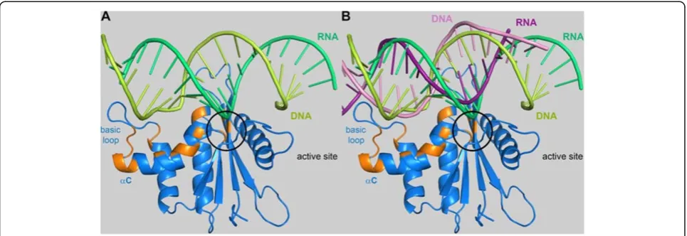
ratios of RNA/DNA:protein of 0, 1, 2, 3, 4 and 5. Upon substrate addition a decrease of the signal intensities of several residues (G600, A614, T641, Q643, D669, F671, A674, E675, S676, E680, K685, S686, N687, F689, V690, N691, H699, I700) could be observed. A signal change was considered relevant, if the signal decreased to 50 % or less of the original value. The residues potentially involved in substrate binding are highlighted in orange in Figure 5. Apart from the active site residues, helix B and C as well as the basic loop appear to be involved in substrate binding. The similarity of PFV RNase H and human RNase H allowed the generation of a simple model of PFV RNase H in complex with substrate. Thus, we superimposed the PFV RNase H structure with that of the human RNase H/substrate complex (PDB: 2QK9) to determine whether the residues, which show signifi-cant effects in the NMR titration experiments, are located spatially close to the substrate (Figure 5A). The alignment indicates a potential role of helix C in posi-tioning the basic loop close to the DNA strand, thus supporting substrate binding, whereas the active site residues are close to the RNA strand, which has to be cleaved during catalysis.
[image:5.595.57.291.87.679.2]Further information on substrate binding is provided by the alignment of the complex shown in Figure 5A with HIV-1 RT (PDB: 1HYS) in complex with a polypur-ine tract (PPT) containing RNA/DNA hybrid [25]. Only the PPT hybrid substrate modeled onto the PFV RNase H is shown (Figure 5B). The modeling reveals that the PPT RNA of the hybrid is not as close to the active site as the RNA strand from the other substrate depicted in Figure 5A. This is not surprising since the PPT-RNA/ DNA hybrid, which is not cleaved by the RNase H dur-ing reverse transcription, adopts a peculiar structure in the complex with HIV-1 RT [25]. In the PPT region, which is rich in adenines, an unpaired base on the DNA strand takes the base pairing out of register leading to two offset base pairs. Then, an unpaired base on the RNA strand re-establishes the normal register. This structural deviation of PPT containing substrates was suggested to play a role in the resistance of the PPT to RNase H cleavage [25].
Figure 3Orientation of Cys654 and Cys709 in PFV RNase H.(A) Enlargement ofα-helices A and D of PFV RNase H.The blow-up shows the structural elementsαA andαD, harboring residues C654 and C709, respectively.(B) Qualitative RNase H activity assay.
A model of XMRV RNase H complexed with sub-strate, published recently, suggests that several aromatic residues (Y586, H594 and Y598) located in helices B and C are capable of hydrogen-bond or stacking interactions with the substrate [13]. The corresponding residues in PFV RNase H are Y672 (located in helix B), K679 (located between helix B and C) and Y683 (located in helix C). Since these residues are solvent exposed in PFV RNase H, substrate interaction might also be pos-sible. These results suggest that monomeric RNase H domains that are active in isolation might need the basic protrusion to confer sufficient affinity for the substrate to permit cleavage.
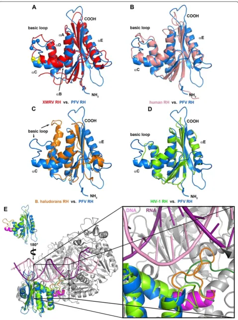
To further analyze the structural features of PFV RNase H we performed structural comparisons with the RNases H of B. halodurans [15], human origin [26], XMRV [13] as well as HIV-1 [20] (Figure 6). The struc-ture of PFV RNase H most closely resembles those of XMRV and HIV-1, followed by human RNase H, while the orientation of the basic loop inB. haloduransRNase H is strikingly different (Figure 6C). However, similar to HIV-1 RNase H (Figure 6D), B. halodurans RNase H also lacks the C-helix, but harbors an additional N-terminal extension instead, which was shown to be involved in substrate binding [15]. Superposition of PFV RNase H with HIV-1 RNase H (Figure 6D) demonstrates that due to the lack of the C-helix in HIV-1 the adjacent loop is too short and is not positioned correctly for sub-strate binding.
To obtain a clearer picture of the orientation of these loops, we superimposed the structure of the full length HIV-1 RT in complex with the PPT containing hybrid (PDB: 1HYS) with PFV RNase H (Figure 6E). The en-largement of the region where the basic protrusion of PFV RNase H is located clearly shows that the basic loop is close to the substrate, whereas the corresponding loop of HIV-1 RNase H is much too short to contact the nucleic acid. Rather, a different loop derived from the connection domain (R356–N363) of the p66 subunit of the heterodimeric HIV-1 RT appears to fulfill this func-tion [27]. Similarly to the basic loop of PFV RNase H, which contains several lysine residues, this loop of HIV-1 RT also contains basic residues (R356, R358).
Conclusions
[image:6.595.56.290.87.496.2]In summary, we were able to identify regions in the RNase H domain - and by structural alignments also in the connection domain of HIV-1 RT - that interact with the substrate. Our data suggest that the basic loop plays an important role in substrate binding and that the major role of the C-helix is to orientate the basic loop properly towards the substrate. Furthermore, regions in the B-helix, which are also present in HIV-1 RNase H, have been identified to be involved in substrate binding.
Figure 4RNase H activity and binding affinities of mutant RNase H (D599N-H724N).(A) Qualitative RNase H assay. The 10/10-mer RNA/DNA substrate, 5’labeled at the RNA strand with32P, is depicted on top of the figure. Cleavage products obtained with 670 nM substrate and 20μM wt RNase H (RHwt) or mutant RNase H-(D599N-H724N) (RH_D599N_H724N) were separated on a 15 % sequencing gel and visualized by phosphoimaging. Arrows and numbers indicate the cleavage sites identified on the gel. The first nucleotide of the RNA hybridized to the 3’OH nucleotide of the DNA strand is designated−1. The incubation times are shown on top of the gel. C, control assays were performed in the absence of enzyme for 0 minute, 60 minutes and 6 hours. (B) Determination of KD-values by fluorescence anisotropy measurements. 25 nM of the
10/10-mer RNA/5’-DY647-DNA substrate were titrated with increasing amounts of wt RNase H (open circles) or mutant RNase H-(D599N-H724N) (closed circles). The fit of the curve to the data [5] resulted in KD-values of 23μM (± 3) for the wt, and 31μM (± 5) for
Together, this information might lead to a better under-standing of the mechanism of action of HIV RNase H inhibitors and can contribute to the design and develop-ment of more specific and more potent inhibitors.
Methods
Mutant plasmid construction
The plasmid pET-GB1a-RNase H-(Q591-N751) coding for the gene of the wt PFV RNase H-(Q591-N751) as described previously [5] was used as a template for site-directed mutagenesis to obtain the mutant proteins RNase H-(C654S) and RNase H-(D599N-H724N) using the following primers:
(i) mutation C654S:
5’
-GCTGCAGTTGAATTTGCCAGTAAAAAAG-CTTTAAAAATACCTGG;
5’
-CCAGGTATTTTTAAAGCTTTTTTACTGGC-AAATTCAACTGCAGC; (ii) mutation D599N:
5’
- GAAGGAGTGTTTTATACTAATGGCTCGGCC-ATCAAAAGTCC
5’
- GGACTTTTGATGGCCGAGCCATTAGTATAA-AACACTCCTTC
(iii) mutation H724N:
5’
-CTATTCAACATGAAAAAGGGAATCAGCCT-ACAAATACCAGTAT 5’
- ATACTGGTATTTGTAGGCTGATTCCCTTTTT-CATGTTGAATAG.
Point mutations were introduced according to the QuikChange kit (Stratagene, Waldbronn, Germany) and confirmed by sequencing (LGC Genomics, Berlin, Ger-many). Mutations coding for the amino acid exchanges D599N and H724N were introduced into the wt full length PR-RT using the same primers as for the muta-tion of the isolated RNase H domain.
Gene expression and protein purification
Protein purifications of PFV RNase H wt, RNase H-(C654S) and RNase H-(D599N-H724N) were performed as described previously [5]. PFV PR-RT(D599N-H724N) as well as 15N-labeled mutant RNase H-(D599N-H724N) and 15N,13C labeled wt RNase H used in NMR studies were also purified as described previously [3,5].
Qualitative RNase H assay
[image:7.595.57.540.89.255.2]Reactions were performed as described previously [5] with 20 μM wt RNase H, RNase H-(C654S) and RNase H-(D599N-H724N), respectively, using a 10/10-mer or a 27/20-mer RNA/DNA substrate, whose RNA was 5’end labeled with 32P [5]. The sequence of the 10-mer RNA was 5’-GCGACACCUG, the 10-mer DNA sequence was complementary to it. Sequences of the 27/20-mer RNA/ DNA hybrid have been published previously [5]. After preincubation of the sample for 5 minutes at 25°C, reac-tions were started by the addition of ca. 670 nM 10/10-mer or 240 nM 27/20-10/10-mer RNA/DNA hybrid. Aliquots were taken as indicated in the figures and processed fur-ther as described previously [5].
Fluorescence anisotropy measurements
Fluorescence equilibrium titrations were performed as described [5] to determine the dissociation constant (KD) of wt RNase H and mutant RNase
H-(D599N-H724N) employing 25 nM of the 10/10-mer RNA/DNA substrate used above for the qualitative RNase H activity assay, however with the fluorescence label DY647 attached at the 5’ end of the DNA (DNA-5’ -DY647-CAGGTGTCGC) (biomers.net GmbH, Ulm, Germany). The excitation wavelength was 652 nm and emission was detected at 673 nm. The slit widths were set at 8 nm and 7 nm for excitation and emission, respectively. The standard deviation for each data point is repre-sented by error bars in Figure 4B. Calculation of the KD
-values was performed as described previously [5].
NMR analyses
NMR samples containing 0.4 – 1.0 mM uniformly 15N or 13C, 15N labeled RNase H were analyzed in 5 mM Na-phosphate, pH 7.0, 100 mM NaCl, 0.5 mM DTT, 10 % D2O (v/v), 6 mM MgCl2. All NMR experiments
were conducted at 298 K on a Bruker Avance 600 MHz or a Bruker Avance 700 MHz (equipped with a cryo-genic probe) spectrometer. In addition to the previously described experiments for resonance assignments [5]
15
N- and 13C-edited NOESY experiments (mixing time 120 ms) were acquired for obtaining distance restraints. Standard 15N spin relaxation experiments [28,29] were conducted at 600 MHz 1H NMR frequency for obtain-ing transverse and longitudinal15N relaxation rates.
For NMR titration experiments a 1.2 fold molar excess of the non-labeled 10-mer DNA was hybridized to the comple-mentary non-labeled 10-mer RNA in 50 mM Na-phosphate, pH 7.0, 100 mM NaCl by heating the sample to 95°C for 2 minutes, followed by transfer to a heating block at 70°C and slow cooling to room temperature. 50μM15N-labeled mutant RNase H-(D599N-H724N) was dissolved in 50 mM Na-phosphate, pH 7.0, 100 mM NaCl, 6 mM MgCl2, 2 mM DTT, 10 % D2O (v/v). Different molar ratios
of RNA/DNA:protein of 0, 1, 2, 3, 4 and 5 were employed.
Structure calculation
Distance restraints for structure calculation were derived from15N-edited NOESY and13C-edited NOESY spectra.
NOESY cross peaks were classified according to their relative intensities and converted to distance restraints with upper limits of 3.0 Å (strong), 4.0 Å (medium), 5.0 Å (weak), and 6.0 Å (very weak). For ambiguous dis-tance restraints the r-6summation over all assigned pos-sibilities defined the upper limit. Experimental NOESY spectra were validated semi-quantitatively against back-calculated spectra to confirm the assignment and to avoid bias of upper distance restraints by spin diffusion. Dihedral restraints were taken from analysis of chemical shifts by the TALOS software package [30]. Hydrogen bonds were included for backbone amide protons in regular secondary structure, when the amide proton did not show a water exchange cross peak in the15N-edited NOESY spectrum.
The structure calculations were performed with the program XPLOR-NIH 1.2.1 ([31]) using a three-step simulated annealing protocol with floating assignment of prochiral groups including a conformational database potential. The 19 structures (out of total 120 structures) showing the lowest values of the target function exclud-ing the database potential were further analyzed with X-PLOR, PROCHECK [32] and pyMOL [33].
Polymerization assay
RNA-dependent DNA polymerase activities of PFV PR-RT and PFV PR-PR-RT(D599N-H724N) were quantitated at 37°C on a poly(rA)/oligo(dT)15 substrate (0.2 U/ml)
(Roche Diagnostics GmbH, Mannheim, Germany) in a reaction volume of 24 μl and 50 mM Tris/HCl, pH 8.0, 80 mM KCl, 6 mM MgCl2, 0.5 mM DTT with 150μM
unlabeled TTP and 1.95μCi of [3H]TTP (120 Ci/mmol; MP Biomedicals Inc., Irvine, CA. USA) [34]. Samples were preincubated for 5 min at 37°C. The reaction was started by the addition of 12 nM enzyme. After 5 min, 7.5μl aliquots were taken out and spotted on DEAE fil-ter paper. Filfil-ters were washed twice with 2 x SSC (300 mM NaCl, 30 mM Na-citrate, pH 7.0) for 10 min-utes, twice with 96 % ethanol for 10 min and dried. Incorporation of radiolabeled precursor into polydeoxy-nucleotide was determined by liquid scintillation count-ing. Under the conditions used 1 unit (U) of enzyme catalyzes the incorporation of 1 nmol TTP into poly (rA)/oligo(dT)15in 10 min at 37°C.
(See figure on previous page.)
Structure alignments
Structure alignments shown in Figure 5 were performed using the program WinCoot [35].
Accession codes
The structure coordinates of the wt PFV RNase H-(Q591-N751) domain were deposited in the Protein Data Bank under the accession code 2LSN. Chemical shift assignments were deposited in the BioMagResBank, ac-cession number: 17745 [5].
Abbreviations
B. halodurans:Bacillus halodurans; DTT: Dithiothreitol;E. coli:Escherichia coli; FV: Foamy virus; GB1: Immunoglobulin binding domain B1 of streptococcal protein G; HIV-1: Human immunodeficiency virus type 1; PFV: Prototype foamy virus; MoMLV: Moloney murine leukemia virus; PPT: Polypurine tract; PR: Protease; RHBD: RNA-hybrid binding domain; RNase H: Ribonuclease H; RT: Reverse transcriptase; SFVmac: Simian foamy virus from macaques; TEV: Tobacco etch virus; XMRV: Xenotropic murine leukemia virus-related virus.
Competing interests
The authors declare that they have no competing interests.
Authors' contributions
BMW conceived and coordinated the study. BL performed the experiments. BL, BMW, MJH and KS analyzed the data. BMW, BL and KS wrote the paper. PR provided conceptual input. All authors read and approved the manuscript.
Acknowledgement
We thank the DFG (Wo630/7-3), the Bavarian International Graduate School of Science (BIGSS) and the University of Bayreuth for financial support. We thank Prof. Dr. Paul Rösch for continuous support and Ramona Heissmann for excellent technical assistance.
Received: 14 June 2012 Accepted: 10 August 2012 Published: 10 September 2012
References
1. Moelling K, Bolognesi DP, Bauer H, Büsen W, Plassmann HW, Hausen P:
Association of retroviral reverse transcriptase with an enzyme degrading the RNA moiety of RNA-DNA hybrids.Nat New Biol1971,234:240–243. 2. Hartl MJ, Bodem J, Jochheim F, Rethwilm A, Rösch P, Wöhrl BM:Regulation
of foamy virus protease activity by viral RNA - a novel and unique mechanism among retroviruses.J Virol2011,85:4462–4469. 3. Hartl MJ, Mayr F, Rethwilm A, Wöhrl BM:Biophysical and enzymatic
properties of the simian and prototype foamy virus reverse transcriptases.Retrovirology2010,7:5.
4. Das D, Georgiadis MM:The crystal structure of the monomeric reverse transcriptase from moloney murine leukemia virus.Structure2004,
12:819–829.
5. Leo B, Hartl MJ, Schweimer K, Mayr F, Wöhrl BM:Insights into the structure and activity of the prototype foamy virus RNase H.Retrovirology2012,
9:14.
6. Tanese N, Goff SP:Domain structure of the moloney murine leukemia virus reverse transcriptase: Mutational analysis and separate expression of the DNA polymerase and RNase H activities.Proc Natl Acad Sci U S A
1988,85:1777–1781.
7. Schultz SJ, Champoux JJ:RNase H domain of moloney murine leukemia virus reverse transcriptase retains activity but requires the polymerase domain for specificity.J Virol1996,70:8630–8638.
8. Zhan X, Crouch RJ:The isolated RNase H domain of murine leukemia virus reverse transcriptase. Retention of activity with concomitant loss of specificity.J Biol Chem1997,272:22023–22029.
9. Kanaya S, Katsuda-Nakai C, Ikehara M:Importance of the positive charge cluster inEscherichia coliribonuclease HI for the effective binding of the substrate.J Biol Chem1991,266:11621–11627.
10. Telesnitsky A, Blain SW, Goff SP:Defects in moloney murine leukemia virus replication caused by a reverse transcriptase mutation modeled on the structure ofEscherichia coliRNase H.J Virol1992,66:615–622. 11. Lim D, Orlova M, Goff SP:Mutations of the RNase H C helix of the
moloney murine leukemia virus reverse transcriptase reveal defects in polypurine tract recognition.J Virol2002,76:8360–8373.
12. Lim D, Gregorio GG, Bingman C, Martinez-Hackert E, Hendrickson WA, Goff SP:Crystal structure of the moloney murine leukemia virus RNase H domain.J Virol2006,80:8379–8389.
13. Zhou D, Chung S, Miller M, Grice SF, Wlodawer A:Crystal structures of the reverse transcriptase-associated ribonuclease H domain of xenotropic murine leukemia-virus related virus.J Struct Biol2012,177:638–645. 14. Paprotka T, Delviks-Frankenberry KA, Cingoz O, Martinez A, Kung HJ, Tepper
CG, Hu WS, Fivash MJ Jr, Coffin JM, Pathak VK:Recombinant origin of the retrovirus XMRV.Science2011,333:97–101.
15. Nowotny M, Gaidamakov SA, Crouch RJ, Yang W:Crystal structures of RNase H bound to an RNA/DNA hybrid: Substrate specificity and metal-dependent catalysis.Cell2005,121:1005–1016.
16. Evans DB, Brawn K, Deibel MR Jr, Tarpley WG, Sharma SK:A recombinant ribonuclease H domain of HIV-1 reverse transcriptase that is enzymatically active.J Biol Chem1991,266:20583–20585. 17. Hostomsky Z, Hostomska Z, Hudson GO, Moomaw EW, Nodes BR:
Reconstitution in vitro of RNase H activity by using purified N- terminal and C-terminal domains of human immunodeficiency virus type 1 reverse transcriptase.Proc Natl Acad Sci U S A1991,88:1148–1152. 18. Smith JS, Roth MJ:Purification and characterization of an active human
immunodeficiency virus type 1 RNase H domain.J Virol1993,
67:4037–4049.
19. Smith JS, Gritsman K, Roth MJ:Contributions of DNA polymerase subdomains to the RNase H activity of human immunodeficiency virus type 1 reverse transcriptase.J Virol1994,68:5721–5729.
20. Davies JF 2nd, Hostomska Z, Hostomsky Z, Jordan SR, Matthews DA:Crystal structure of the ribonuclease H domain of HIV-1 reverse transcriptase.
Science1991,252:88–95.
21. Mueller GA, Pari K, DeRose EF, Kirby TW, London RE:Backbone dynamics of the RNase H domain of HIV-1 reverse transcriptase.Biochemistry2004,
43:9332–9342.
22. Kirby KA, Marchand B, Ong YT, Ndongwe TP, Hachiya A, Michailidis E, Leslie MD, Sietsema DV, Fetterly TL, Dorst CA, Singh K, Wang Z, Parniak MA, Sarafianos SG:Structural and inhibition studies of the RNase H function of xenotropic murine leukemia virus-related virus reverse transcriptase.
Antimicrob Agents Chemother2012,56:2048–2061.
23. Wöhrl BM, Volkmann S, Moelling K:Mutations of a conserved residue within HIV-1 ribonuclease H affect its exo-and endonuclease activities.
J Mol Biol1991,220:801–818.
24. Tisdale M, Schulze T, Larder BA, Moelling K:Mutations within the RNase H domain of human immunodeficiency virus type 1 reverse transcriptase abolish virus infectivity.J Gen Virol1991,72:59–66.
25. Sarafianos SG, Das K, Tantillo C, Clark AD 2nd, Ding J, Whitcomb JM, Boyer PL, Hughes SH, Arnold E:Crystal structure of HIV-1 reverse transcriptase in complex with a polypurine tract RNA:DNA.EMBO J2001,20:1449–1461. 26. Nowotny M, Gaidamakov SA, Ghirlando R, Cerritelli SM, Crouch RJ, Yang W:
Structure of human RNase H1 complexed with an RNA/DNA hybrid: Insight into HIV reverse transcription.Mol Cell2007,28:264–276. 27. Julias JG, McWilliams MJ, Sarafianos SG, Alvord WG, Arnold E, Hughes SH:
Mutation of amino acids in the connection domain of human immunodeficiency virus type 1 reverse transcriptase that contact the template-primer affects RNase H activity.J Virol2003,77:8548–8554. 28. Kay LE, Torchia DA, Bax A:Backbone dynamics of proteins as studied by
15N inverse detected heteronuclear NMR spectroscopy: Application to
staphylococcal nuclease.Biochemistry1989,28:8972–8979.
29. Farrow NA, Muhandiram R, Singer AU, Pascal SM, Kay CM, Gish G, Shoelson SE, Pawson T, Forman-Kay JD, Kay LE:Backbone dynamics of a free and phosphopeptide-complexed src homology 2 domain studied by15N NMR relaxation.Biochemistry1994,33:5984–6003.
30. Cornilescu G, Delaglio F, Bax A:Protein backbone angle restraints from searching a database for chemical shift and sequence homology.
J Biomol NMR1999,13:289–302.
31. Schwieters CD, Kuszewski JJ, Tjandra N, Clore GM:The Xplor-NIH NMR molecular structure determination package.J Magn Reson2003,
32. Laskowski RA, Rullmann JAC, MacArthur MW, Kaptein R, Thornton JM:AQUA and PROCHECK-NMR: Programs for checking the quality of protein structures solved by NMR.J Biomol NMR1996,8:477–486. 33. Schrödinger L:The PyMOL molecular graphics system, version 1.3.
Schrödinger, LLC: Mannheim, Germany; 2010.
34. Hartl MJ, Kretzschmar B, Frohn A, Nowrouzi A, Rethwilm A, Wöhrl BM:AZT resistance of simian foamy virus reverse transcriptase is based on the excision of AZTMP in the presence of ATP.Nucleic Acids Res2008,
36:1009–1016.
35. Emsley P, Cowtan K:Coot: Model-building tools for molecular graphics.
Acta Chrystallogr D2004,60:2126–2132.
doi:10.1186/1742-4690-9-73
Cite this article as:Leoet al.:The solution structure of the prototype foamy virus RNase H domain indicates an important role of the basic loop in substrate binding.Retrovirology20129:73.
Submit your next manuscript to BioMed Central and take full advantage of:
• Convenient online submission • Thorough peer review
• No space constraints or color figure charges
• Immediate publication on acceptance
• Inclusion in PubMed, CAS, Scopus and Google Scholar
• Research which is freely available for redistribution