RESEARCH
ARTICLE
MALDI-MSI for the analysis of a 3D tissue-engineered
psoriatic skin model
Amanda Harvey
1, Laura M. Cole
1, Rebecca Day
1, Maggie Bartlett
2, John Warwick
2,
Richard Bojar
2, David Smith
1, Neil Cross
1and Malcolm R. Clench
11Centre for Mass Spectrometry Imaging, Biomolecular Sciences Research Centre, Sheffield Hallam University, Sheffield, UK
2Innovenn, Sand Hutton Innovation Campus, York, UK
Received: January 22, 2016 Revised: May 5, 2016 Accepted: May 23, 2016
MALDI-MS Imaging is a novel label-free technique that can be used to visualize the changes in multiple mass responses following treatment. Following treatment with proinflammatory cy-tokine interleukin-22 (IL-22), the epidermal differentiation of Labskin, a living skin equivalent (LSE), successfully modeled psoriasis in vitro. Masson’s trichrome staining enabled visualiza-tion and quantificavisualiza-tion of epidermal differentiavisualiza-tion between the untreated and IL-22 treated psoriatic LSEs. Matrix-assisted laser desorption ionization mass spectrometry imaging was used to observe the spatial location of the psoriatic therapy drug acetretin following 48 h treat-ments within both psoriatic and normal LSEs. After 24 h, the drug was primarily located in the epidermal regions of both the psoriatic and nonpsoriatic LSE models whereas after 48 h it was detectible in the dermis.
Keywords:
Acetretin / Interleukin-22 / Living skin equivalent / MALDI-MSI / Psoriasis / Skin / Tissue engineering / Technology
1
Introduction
Tissue engineering has enabled the development of skin models. These living skin equivalents (LSEs), derived from primary human skin cells, self-assemble to form stratified lay-ers comparable to human skin [1–8]. LSEs offer treatment for burns patients following serious injury, avoiding the need for meshed skin grafts and donor skin [6–9]. Additionally, LSEs are used for toxicity screening with the ability to replace ani-mal models for cosmetic and drug development. The use of skin models in drug development has been reviewed recently [10]. Whilst a 3D in vitro model would not be expected to show a full range of histopathological responses due to the absence of an immune system, blood supply, or innervation, LSE of-fers an alternative to animal models which can give useful data for studies, for example of drug absorption/penetration.
Correspondence: Professor Malcolm R. Clench, Centre for Mass Spectrometry Imaging, Biomolecular Sciences Research Centre, Sheffield Hallam University, Howard Street Sheffield, UK S1 1WB
E-mail: [email protected]
Abbreviations: ALI, air–liquid interface;IL-22, interleukin-22;LSE, living skin equivalent;MSI, mass spectrometry imaging
Psoriasis is a skin condition that causes red, flaky, crusty patches of skin covered with silvery scales or plaques that affects 2% of people in the United Kingdom (NHS 2015). One of the markers of psoriasis as an inflammatory disease is the interruption of normal cytokine pathways affecting cellular communication with keratinocytes. This interruption to the interleukin-22 (IL-22) pathway is one of the primary causes of adverse effects to these normal inflammatory pathways, which can cause keratin atherosclerosis, or thickening and unevening of the epidermis within patients with psoriasis, as shown in Fig. 1 [11–15]. Acanthosis in psoriasis is visible with the induction of psoriatic plaques on the skin surface. It is also responsible for the hypoproliferation of cells in the epidermis [16]. In this study, we have added IL-22 in order to induce psoriasis in vitro by mimicking the inflammatory response environment present [15–17].
One of the strengths of mass spectrometry imaging (MSI) is the ability to visualize the distribution of multiple com-pounds within tissues simultaneously and in a label-free man-ner [18]. We have previously applied MALDI-MSI to the study of both ex vivo human skin and 3D skin models [19–23]. MSI
Color Online: See the article online to view Figs. 1–5 and 7 in color.
C
Significance of the study
The work reported here represents the first reported mass spectrometric study of a 3D skin model modified to repre-sent a disease state.
has recently been used to examine drug absorption in LSE, with the aim to identify the metabolism of drugs within LSE [24]. MALDI-MSI is continuing to grow as a robust technique for the analysis of tissues and offers great prospects for tis-sue engineering to compare the profile of native tistis-sues and diseases with those of their tissue-engineered counterparts, overall enabling greater development of the latter.
In the study reported here, we have added IL-22 to a develop-ing LSE in order to induce psoriasis in vitro by mimickdevelop-ing the inflammatory response environment present. We have exam-ined the effects of the IL-22 treatment on both the structure of the LSE and the barrier function by the use of MALDI-MSI.
2
Materials and methods
2.1 Psoriatic development and LSE culture
LSE skin models at day 7 air–liquid interface (ALI; Labskin, Innovenn, York, UK) were treated with IL-22 (R&D Sys-tems) for 14 days to induce psoriasis. Labskin maintenance medium (Innovenn, York, UK) was supplemented with IL-22 (10 ng/mL) and medium was refreshed daily keeping the LSEs at ALI. The epidermal differentiation and metabolic ac-tivity were monitored by AlamarBlueۚstaining every 2 days. Psoriasis drug treatments of topical acetretin (30L of 300
g/mL in acetone/olive oil 4:1) were applied on day 21 and
the efficacy and drug penetration within the skin models was compared over 48 h.
2.2 Fresh frozen tissue and cryosectioning
Directly following drug treatment, LSEs were removed from the well insert and snap frozen in a bath of precooled isopen-tane (2–5 min) in a Styrofoam box containing liquid nitrogen and a suitable float. LSEs were stored at−80⬚C for at least 24 h before sectioning. LSEs were mounted onto cork rings with dH2O at−25⬚C and allowed to equilibrate to temperature for
30 min before being cryosectioned at 12m and freeze-thaw mounted onto “polycat” microscope slides (SLS, Hessle, UK).
2.3 Hematoxylin and eosin staining
LSE sections were stained using Mayers hematoxylin (VWR) and aqueous eosin Y (VWR) solutions. Each slide was sub-merged in hematoxylin for 8 min, washed in running tap water for 4 min, submerged in eosin y solution for 30 s, dH20
for 1 min, and mounted with DPX (distyrene, a plasticizer, and xylene used as a synthetic resin mounting media) follow-ing dehydration through a series of alcohol and xylene dehy-dration baths. Slides were imaged using an Olympus BX60 microscope fitted with UPlanFl 10x/0.30, 20x/0.50/0.17, 40x/0/75/0.17 objectives and analyzed with QCapture-Pro 8.0 software (QImaging, Surry, BC, Canada).
2.4 Masson’s trichrome staining
LSE sections were stained using Masson’s trichrome stain-ing kit (VWR). Slides were submerged in hematoxylin for 10 min, washed in running tap water for 4 min, stained in
ponceau fuchsin for 5 min, rinsed in dH2O, submerged in
phosphomolybdic acid for 3 min, and colored without rins-ing in vert lumiere, before berins-ing rinsed in two baths of acidi-fied water to remove excess dye. Following staining samples were mounted with DPX following dehydration through a serious of alcohol and xylene dehydration baths. Slides were imaged using an Olympus BX60 microscope fitted with UP-lanFl 10x/0.30, 20x/0.50/0.17, 40x/0.75/0.17 objectives and analyzed with QCapture-Pro 8.0 software (QImaging). Epidermal thickness was measured from recorded images using ImageJ (https://imagej.nih.gov/ij/) and statistical anal-yses were carried out using StatsDirect (StatsDirect, Cheshire, UK). A Tukey analysis of variance was performed on multi-ple measurements taken from random sections, taken from treated and untreated LSEs.
2.5 Immunohistochemistry
Fresh frozen tissue sections were fixed in ice-cold acetone and endogenous peroxidases quenched. Antigen retrieval was performed in a microwave oven in 0.05 M Tris, pH 9.5. Samples were blocked in rabbit serum, then incubated overnight at 4⬚C with mouse anti-human Psoriasin (1:200; Abcam, ab13680) and with preimmune mouse IgG (Abcam) as a negative control (1:50). After washing, samples were incubated with biotinylated rabbit anti-mouse secondary an-tibody (1:500; Abcam) and binding detected by formation of streptavidin–biotin complexes (Vector Laboratories, Pe-terborough, UK) with 3,32-diaminobenzidine tetrahydrochlo-ride solution (Sigma–Aldrich). Sections were counterstained with Mayers Hematoxylin (Leica Microsystems), dehydrated, cleared, and mounted in Pertex (Leica). Sections were visual-ized and images captured on an Olympus BX60 Microscope using QCapture Pro version 8.0 software (Media Cybernetics, Marlow, UK).
2.6 Small-molecule MALDI-MS imaging
For small-molecule MALDI-MS imaging, sections were coated with MALDI matrix (␣-cyano-4-hydroxycinnamic acid, 5 mg/mL in 70:30 methanol: 0.5% TFA) using a SunCollectۛ
[image:3.595.313.519.80.284.2](SunChrom Friedrichsdor, Germany) sprayer. Slides were coated with four layers of CHCA matrix, spraying each layer approximately 50 L per layer at 2 L/min. Mass spectra and images were acquired in positive ion mode on an Ap-plied Biosystems/MDS Sciex hybrid quadrupole TOF mass spectrometer (Q-Star Pulsar-i) with an orthogonal MALDI ion source (Applied Biosystems, Foster City, CA, USA) and a high-repetition neoddymium-doped yttrium aluminium gar-net (Nd:YAG) laser (355 nm, 1 KHz) (Applied Biosystems). Image acquisition was performed at a spatial resolution of 100m×100m in “Raster Image” mode; images were generated using the freely available Novartis Biomap 3.7.5.5
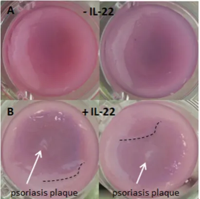
Figure 2. Labskin stained with AlamarBlueR at day 21 ALI. (A)
Without treatment with IL-22 there are no visible changes to the epidermis of the LSEs. (B) LSEs treated with IL-22 show disregula-tion to the epidermis (dotted line) and psoriatic plaques (arrows) after treatment.
software (www.maldi-msi.org). Post-MS imaging sections were also examined by histology as described in this section.
3
Results and discussion
3.1 Psoriatic development within LSEs
Following treatment with IL-22 from day 7 ALI for 2 weeks to day 21 ALI, psoriatic plaques were visible on the LSE con-structs as shown in Fig. 2B (arrows). Costaining with Alamar-Blueۚhighlighted abnormalities in epidermal differentiation within the LSE constructs visible by deregulation of the epi-dermal layer (acanthosis) visible in Fig. 2B (dashed lines) with IL-22 as contrasted with the smooth epidermal layer observed in Fig. 2A. The formation of this acanthosis within the epi-dermis of the treated LSEs creates a similar structure to that observed with in vivo psoriasis.
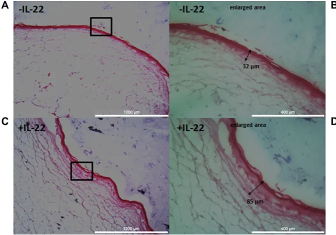
Standard H&E staining of the LSEs yielded results indicat-ing thickenindicat-ing of the epidermis. In Fig. 3D followindicat-ing treat-ment with IL-22, it is possible to see an upper epidermal thickness of85m as opposed to32m in Fig. 3B with-out treatment of IL-22. Additionally, the character of the LSEs becomes quite characteristically wavy (i.e. exhibits disregula-tion) in Fig. 3C following treatment with IL-22 as opposed to Fig. 3A without.
Figure 3. Histology H&E staining of Labskin at day 21 ALI. (A) Non-IL-22 treated LSE, “normal skin.” (B) En-larged area showing the measurable upper epidermal thickness of non-IL-22 treated LSE to be 32 m. (C) IL-22 treated LSE, “psoriatic skin,” char-acterised by wavy acanthosis. (D) En-larged area showing the measurable upper epidermal thickness of IL-22 treated LSE to be 85m.
clear that there is very little stratification of the epidermis and its structure is fairly flat, however in Fig. 4C and D, the “wavy” psoriatic characteristic is clearly observable. The measured total epidermal thickness (comparing Fig. 4B and D) has in-creased from approximately 132±14.6m to 179±13.6m (p=0.01), i.e. the thickening is statistically significant. The measured epidermal stratification of non-IL-22 treated Lab-skin is 41±5.9m and for the treated samples 78±21.6m (p=0.0568), i.e. the difference is not statistically different.
In order to validate the psoriatic-like nature of the IL-22 treated LSE, immunohistochemical staining for the presence of psoriasin was carried out. As can be seen in Fig. 5, pso-riasin is detectable in low abundance in untreated Labskin (Fig. 5A) but is much more highly expressed in the IL-22 treated samples (Fig. 5C). There is little indication of non-specific binding as shown by the non-specific isotype controls (Fig. 5B and D). Figure 5C also shows the epidermal thicken-ing observed in both the H&E and Masson’s stained samples.
3.2 Drug treatment of LSEs and analysis with MALDI-MSI
Acetretin, a psoriatic drug was chosen for its known effect to-ward psoriatic skin [25] and also for the ability of the drug to be visualized by MSI. The phosphatidylcholine head group (m/z
184) was chosen to enable visualization of the LSE structure as phospholipids are abundant in tissues such as skin. The positive phospholipid head group is key to their ionization efficiency. Figure 6A–F show representative LSE samples, Fig. 5A control without drug, Fig. 6B drug treated without
IL-22 (24 h posttreatment), Fig. 6C IL-22 treated, drug-treated psoriatic LSE (24 h posttreatment) with Fig. 6E–F showing enlarged areas of these samples. In the enlarged images fo-cusing on the epidermis of the LSEs, drug was not observed within the untreated control, Fig. 6D, as expected. The “red” drug signal is, however, clearly observable in the representa-tive images of treated samples (Fig. 6E and F). The red ace-tretin signal (m/z326.2) appears to be solely located within the epidermis, by comparison with the H&E image. From these images the depth of penetration of the drug into the LSE can be evaluated. For the non-IL-22 treated Labskin (Fig. 6E) the apparent depth of penetration is 114 m and for the IL-22 treated Labskin (Fig. 6F) the apparent penetration is 177m, an increase of approximately 63m. This is in agreement with the previously measured (Fig. 4) epidermal stratification. Therefore at the 24 h time point, the distance penetrated appears to correspond to the thickness of the epi-dermis.
A 48 h posttreatment sample was imaged to examine drug distribution following longer treatment (Fig. 7). In this case a sodiated sphingomyelin (SM 34:1 [M+Na]+) atm/z725.4 was chosen to visualize the epidermis. In Fig. 7A, the epider-mal regions of the various LSEs can be clearly seen. In this case, images were taken of LSEs with and without acetretin drug treatment and both with and without IL-22 treatment. In these preliminary results it is not possible to see a signif-icant difference in the thickness of the epidermis using MSI of sphinogomyelins at the 100m spatial resolution used. Figure 7B shows a closer look at the LSE treated with both IL-22 and acetretin. Figure 7B shows the distribution of the
Figure 4. Histology Masson’s Trich-rome staining of Labskin at day 21 ALI. (A) Non-IL-22 treated LSE, “nor-mal skin.” (B) Enlarged area show-ing the measurablestratum corneum thickness of non-IL-22 treated LSE as 20 m and stratification of the epi-dermal region of 19 m. (C) IL-22 treated LSE, “psoriatic skin,” charac-terised by wavy acanthosis. (D) En-larged area showing the measurable stratum corneum thickness of IL-22 treated LSE as 26m and stratification of the epidermal region of 55m. (E) Statisical analysis of epidermal thick-ening observed in IL-22 treated and un-treated Labskin (p=0.01).
through the epidermis of the LSE into the dermal region with now an apparent depth of penetration of approximately 557
m, the full thickness of the LSE.
Tissue engineering, and specifically skin tissue engineer-ing has far from reached its limits. While much work was undertaken in the 1980–2000s to develop 3D culture of cells,
Figure 5. (A–D) Immunohistochem-istry showing Psoriasin expression and localization in LabSkin. DAB staining (brown) shows Psoriasin is localized to the epidermal lay-ers of the LSE model (A and C). LabSkin treated with IL-22 has a thick-ened epidermis and increased expres-sion of Psoriasin (C).
immunohistochemistry and may be visible by MALDI-MSI. As with many diseases much of the specific mechanisms of psoriasis are still greatly unknown, and observing the pres-ence of lipids, cytokines, chemokines, proteins, and peptide markers associated with the disease would greatly impact our understanding of the disease. This is therefore an ideal area for further study using MSI.
4
Concluding remarks
[image:6.595.58.392.80.336.2]In this study we have been able to produce in vitro LSE models of psoriasis using Labskin commercial LSE, as confirmed by histology and have used MSI to determine the special location of the psoriatic drug acetretin. It was possible to demonstrate that 24 h posttreatment, the acetretin was located
Figure 6. MALDI-MSI data overlay ofm/z326.2 acetretin (red) onm/z184 phosphatidylcholine lipid (green) of LSEs 24 h posttreatment. (A) Non-IL-22 and nondrug-treated control. (B) Non-IL-22 and acetretin drug treated. (C) IL-22 and acetretin treated. Location of the drug is visible in the epidermis of treated samples. (D–F) Enlarged areas enable measurement of apparent depth of penetration of acetretin in the epidermal regions. (D) No drug signal present in the control. (E) The apparent depth of penetration within the non-IL-22 treated LSE is 114
Figure 7. (A) MALDI-MSI distribution of sodiated sphingomyelin (SM 34:1 [M+Na]+) atm/z725.4 of LSEs 48 h post-treatment across all post-treatment samples. (B) Enlarged area MALDI-MSI data over-lay ofm/z326.2 acetretin (green) on so-diated sphingomyelin (SM 34:1 [M+Na]+) atm/z725.4 of LSEs 48 h posttreatment. Apparent acetretin depth of penetration is 557m, i.e. it has penetrated to within the dermal region.
in the epidermal region in both psoriatic and nonpsoriatic skin models whereas 48 h posttreatment it had penetrated into the dermis.
We intend to utilize sphingomyelins (or different lipid, peptide, or protein species) in combination with matrix sub-limation to obtain high-resolution images of the epidermal region of the LSE model in future studies.
This work was funded by NC3Rs Grant No. NC/L001896/1. The authors would also like to express their gratitude to the tech-nical team at Innovenn for helping to provide the Labskin LSEs.
The authors have declared no conflict of interest.
5
References
[1] Groeber, F., Holeiter, M., Hampel, M., Hinderer, S. et al., Skin tissue engineering–in vivo and in vitro applications.Adv. Drug Deliv. Rev.2011,63, 352–366.
[2] MacNeil, S., Progress and opportunities for tissue-engineered skin.Nature2007,445, 874–880.
[3] Priya, S. G., Jungvid, H., Kumar, A., Skin tissue engineering for tissue repair and regeneration.Tissue Eng. Part B Rev. 2008,14, 105–118.
[4] Lu, G., Huang, S., Bioengineered skin substitutes: key el-ements and novel design for biomedical applications.Int. Wound J.2013,10, 365–371.
[5] Yildirimer, L., Thanh, N. T. K., Seifalian, A. M. et al., Skin regeneration scaffolds: a multimodal bottom-up approach. Trends Biotechnol.2012,30, 638–648.
[6] Auger, F. A., Berthod, F., Moulin, V., Pouliot, R. et al., Tissue-engineered skin substitutes: from in vitro constructs to in vivo applications Skin TE approaches. Biotechnol. Appl. Biochem.2004,39, 263–275.
[7] MacNeil, S., Shepherd, J., Smith, L., in: ,3D Cell Culture: Methods and Protocols, Methods in Molecular Biology, 2011, pp. 129–153.
[8] Bell, E., Parenteau, N., Gay, R., Nolte, C. et al., The living skin equivalent: its manufacture, its organotypic properties and its responses to irritants.Toxicol. Vitr.1991,5, 591–596.
[9] Demling, R. H., DeSanti, L., Management of partial thick-ness facial burns (comparison of topical antibiotics and bio-engineered skin substitutes). Burns 1999, 25, 256– 261.
[10] Mathes, S. H., Ruffner, H., Graf-Hausner, U., The use of skin models in drug development.Adv. Drug Deliv. Rev.2014, 69–70, 81–102.
[11] Wolk, K., Haugen, H. S., Xu, W., Witte, E. et al., IL-22 and IL-20 are key mediators of the epidermal alterations in psoriasis while IL-17 and IFN-gamma are not.J. Mol. Med.2009,87, 523–536.
[12] Hao, J. Q., Targeting interleukin-22 in psoriasis. Inflamma-tion2014,37, 94–99.
[13] Ma, H., Liang, S., Li, J., Napierata, L. et al., IL-22 is re-quired for Th17 cell-mediated pathology in a mouse model of psoriasis-like skin inflammation. 2008,118, 597–607. [14] Niv-Spector, L., Shpilman, M., Levi-Bober, M., Katz, M.
et al., Preparation and characterization of mouse IL-22 and its four single-amino-acid muteins that act as IL-22 receptor-1 antagonists.Protein Eng. Des. Sel.2012,25, 397– 404.
[15] Danilenko, D. M., Review paper: preclinical models of psori-asis.Vet. Pathol.2008,45, 563–575.
[16] Portugal-Cohen, M., Horev, L., Ruffer, C., Schlippe, G. et al., Non-invasive skin biomarkers quantification of psoriasis and atopic dermatitis: cytokines, antioxidants and psoriatic skin auto-fluorescence. Biomed. Pharmacother.2012, 66, 293– 299.
[17] R ¨uffer, C., Psoriatic in vitro epidermis A human tissue culture model for testing cosmetical and medical skin care products. Househ. Pers. Care Today2011, 30–33.
[18] Cornett, D. S., Reyzer, M. L., Chaurand, P., Caprioli, R. M. et al., MALDI imaging mass spectrometry: molecular snap-shots of biochemical systems.Nat. Methods2007,4, 828– 833.
[19] Mitchell, C. A., Long, H., Donaldson, M., Francese, S. et al., Lipid changes within the epidermis of living skin equivalents observed across a time-course by MALDI-MS imaging and profiling.Lipids Health Dis.2015,14, 84.
[21] Hart, P. J., Francese, S., Woodroofe, M. N., Clench, M. R., Matrix assisted laser desorption ionisation ion mobility sep-aration mass spectrometry imaging of ex-vivo human skin. Int. J. Ion Mobil. Spectrom.2013,16, 71–83.
[22] Cole, L. M., Mahmoud, K., Haywood-Small, S., Tozer, G. M. et al., Recombinant “ IMS TAG” proteins—a new method for validating bottom-up matrix-assisted laser des-orption/ionisation ion mobility separation mass spectrom-etry imaging. Rapid Commun. Mass Spectrom.2013, 27, 2355–2362.
[23] Cole, L. M., Clench, M. R., Mass spectrometry imaging for the proteomic study of clinical tissue.Proteomics Clin. Appl. 2015,9, 335–341.
[24] Avery, J. L., McEwen, A., Flinders, B., Francese, S. et al., Matrix-assisted laser desorption mass spectrom-etry imaging for the examination of imipramine ab-sorption by Straticell-RHE-EPI/001 an artificial model of the human epidermis. Xenobiotica 2011, 41, 735– 742.
[25] Hsia, E., Johnston, M. J., Houlden, R. J., Chern, W. H. et al., Effects of topically applied acitretin in reconstructed human epidermis and the rhino mouse.J. Invest. Dermatol.2008, 128, 125–130.