0022-538X/92/041899-08$02.00/0
CopyrightC)1992, American SocietyforMicrobiology
BHRF1
of
Epstein-Barr Virus,
Which Is
Homologous
to
Human
Proto-Oncogene
bc12,
Is Not Essential for Transformation
of B Cells
or
for Virus
Replication
In
Vitro
MAY-ANN LEEANDJOHNL. YATES*
Department of Human Genetics, Roswell ParkCancerInstitute, Buffalo, New York 14263 Received 14 May 1991/Accepted 26 December 1991
The Epstein-Barrvirus(EBV)genomecontainsanopenreadingframe, BHRF1,that encodesapresumptive
membrane protein with sequence similarity to the proto-oncogene bcI2, which is linked to human B-cell follicular lymphoma. Potential roles for BHRF1 inEBV's abilitytogrowth transform human B cells and to replicate inB cells in culturewereinvestigated by generatingEBV mutantsthat lack most of theopenreading
frame. This was accomplished by recombination of plasmids carrying mutations in BHRF1 with the
transformation-defective EBV strain P3HR1. Because BHRF1 resides close to the deletion in P3HR1 that renders this straintransformation defective, B-cell transformation could be used toselectfor recombination events intheregion. B-cell clones were established byrecombinants which lacked mostof the BHRF1 open
reading frame, although most ofthese initial B-cell transformants also carried nonrecombinant (BHRF1+) P3HR1 genomes, at levels ranging from a fraction of a copy to four copies per cell. Secondary B-cell transformants that lacked BHRF1+ EBVat detectable levelswere found to release transforming, BHRF1-deficientEBVatlevels thatwerewithin the normalrangeforEBV-immortalizedB-cell clones. These studies
demonstrate that BHRF1 is nonessential forgrowthtransformation of B cells and for virus replication and release from thesecells in culture.
Epstein-Barrvirus(EBV) is associated with nasopharyn-geal carcinoma, B lymphomas, and other lymphoprolifera-tive disorders in humans. Invitro, EBV establishesalatent infection inrestingBlymphocytesandtransforms these cells intoindefinitely proliferatingblast cells. EBV's involvement inlymphoproliferativedisorders ismostlikelyaresult of this
capacity to stimulate the proliferation of B cells when infectingthem latently (reviewedin references 15 and 19).
Several EBVgene productsarebelieved tocontribute to B-cell growth transformation, either directly or indirectly.
All of the gene products that are expressed in latently
infected, growth-transformed B-cell lines have been
gener-allyconsideredascandidates forinvolvement in theprocess
and, in sum, sufficient for it. These gene products include two membrane proteins, six nuclearproteins (or antigens; EBNAs), and two abundant small RNAs, called EBERs (15). Geneticprooffor theircontributions to growth
trans-formation exists for only two of these gene products:
EBNA2,which isessential forgrowthtransformation (6, 9), and EBNA-LP(orEBNA4),whichmakesanimportant,but not essential, contribution to the outgrowth of B cells in culture (9). EBNA2, and to a lesser extent EBNA3C, can
cause the induction ofantigens associated withB-cell
acti-vation whenexpressedinparticular EBV-negative Burkitt's lymphoma cell lines(37, 38). EBNA2 mayalsobe required for theexpressionofseveral ofthe otherEBVgenesthatare
expressed latently (25, 40). The productof anotherlatently expressed gene, the latent membrane protein, is strongly suspectedofplayingadirect role insupportingB-cellgrowth
becauseitcanalso contribute totheinductionofactivation antigens and,moreover,cantransformthegrowth properties
of rodentfibroblastcell lines (3, 35-37). EBNA1 islikelyto
playatleastasupportingrole ingrowthtransformation ofB
* Correspondingauthor.
cells by sustaining the autonomous maintenance of the circularized EBVgenome (43). Activities for the remaining
latently expressed proteinshavenotbeenreported. The assumption that the genes mentioned above (i.e.,
those that are invariably expressed in EBV-established
B-cell lines) are togethersufficient for transformation ofB
cellsrequiresthefollowingcaveat:additionalgenesmightbe necessaryduring the initial stagesofinfectionbut unneces-sarylater. Thissituation could exist forbiological reasons;
certain EBV genes might be expressed only early after
infection, serving to establish the patterns of gene
expres-sion thatdefinelatencybutnot tomaintainthem,oractingto initiate B-cell proliferation but not to sustain it. Or the situationmight arise forthe artifactual reasonthat as
EBV-transformed B cells adapt to growing in culture, variants could emerge that have obviated the need forone or more
viral gene product, whose expression could then be lost. These considerations seemed apt for BHRF1 because of severalpublished findings.
BHRF1 has significant colinear sequence similarity with
the proto-oncogene bc12,which hasbeen implicated in the development of B-cell follicular lymphoma in humans (5). Expression ofbc12 from retrovirus vectors or from trans-genesin miceledtoexpanded restingB-cellpopulationsand contributedtoB-cellneoplasia(16-18, 29, 34). Invitro, Bor
Tcells expressingbc12were foundtosurvive longer in low
serum orwhen stressed(20, 23,24, 33),inonesuchexample
by avoiding the rapid, apoptotic cell death that would otherwiseensuefollowing deprivationofanessentialgrowth
factor (12). In group I EBV-positive Burkitt's lymphoma
cells,arestrictedstateof EBVlatentgeneexpressionexists
and thecellsarepronetoapoptosis. Forced expression ofan
introduced copyofbc12oran introduced EBV latent
mem-braneproteingene,whichinduced bc12 expression, rendered
these cells resistantto apoptosis (11).
It is conceivable that BHRF1 possesses bcl2-like
activi-1899
on November 9, 2019 by guest
http://jvi.asm.org/
ties, but the appropriate functional tests of BHRF1 have not been reported. The 24-kDa Bc12 protein was found to associate with the mitochondrial inner membrane (12). The 17-kDa BHRFl-encoded protein is associated with mem-branes in the cytoplasm (21) but has not been localized further. The regulation ofBHRF1 expression appears to be complex and is somewhat controversial. Abundant expres-sion of BHRF1 mRNA and protein appears early after lytic induction (la, 10, 21), suggesting a function for BHRF1 during virus replication. In addition, at lower levels, at least twoforms ofBHRF1-containing mRNAs whose expression levels appeared unrelated to the productive cycle were detectedby Northern (RNA) analysis of RNA from several EBV-transformed B-cell lines,including a tightly latent one (la). One such form ofBHRF1-containing mRNA carries the spliced leader sequences characteristic of the EBNA family oflatently expressed mRNAs (la, 4, 21). However, neither the BHRF1 protein nor an mRNAwith known potential to direct its synthesis has been detected in latently infected cells(21).
Reports from two laboratories suggested a method of isolating EBV carrying mutations in BHRF1 (6, 9). These laboratories showed that EBVstrain P3HR1, which cannot transform B cells becauseof a deletion that removes EBNA2 coding sequences, can generate transforming virus by ge-netic recombination with plasmids carryingEBVsequences spanning the deleted region. Because the region deleted in P3HR1 is only 2 kb away from BHRF1, this presented a method of introducing deletions affecting the open reading frame into a viralstrain that would otherwisehave anormal phenotype. It was found that viruses mutant for BHRF1 couldreadily be obtained by this method and propagated in the absence of helper virus by the sequential isolation of immortalized B-cell clones.
MATERIALSANDMETHODS
Cell lines and culture. The subclone HH514, clone 16, of P3HR1,which lacks the rearranged defective viral genomes carriedbyP3HR1(22),wasusedthroughoutthis work andis referredto asHH514-16. All cell linesweregrown inRPMI 1640medium(GIBCO) supplementedwith10%fetalbovine serum and 5% iron-supplemented calf serum (HyClone, Inc.).
Transformation of B cells.Leukocytes from theperipheral
blood of normal adult donors wereseparated from
erythro-cytesbycentrifugationoverHistopaque(Sigma), depletedof monocytesby adherence toplasticculturedishes, and then
depletedofTcellsbythe method ofrosette formation with
2-aminoethylisothiouroniumbromide-treatedsheep
erythro-cytes
(14).
In someexperiments,
the step ofdepleting
monocytes was omitted without a noticeable effect on the results. Although someof the blood donors were
seroposi-tive for EBV viral capsid antigens, B cells from such individuals were used in these studies only iftheydid not give risetotransformantsat adetectablefrequencywithout the addition ofEBV. Cells from twodifferent donorswere usedto testeach supernatant in eachexperiment,with little variation in the numbers of transformants observed.
Typically, 5 x
106
HH514-16 cells suspendedin0.6mlof complete mediumwere electroporated with3 ,ugof thetest plasmid plus 3pg
of pCMV-BZLF1 (8) to induce the lytic cycle. Electroporation was done by using a Bethesda Re-search Laboratories Cell-Porator set at 330 ,uF and 300V. Cellswerecultured in 10 ml ofmedium,whichwascollectedafter4
days
andpassed
through
a0.22-pm-pore-size
filter.Bcells(6 x 10⁴ to 8 x 10⁴) were added to each well of 24-well culture plates alongwith 0.5 ml of growth medium and 0.1 ml of cell supernatant to be tested for transforming virus. Human fibroblasts, lethally gamma irradiated with 3,000 rads, were also included in the culture dishes at a 1:3 or 1:4 split toserve asfeeder cells. Some of thedata in experiments 1 and 2 ofTable 1 were obtained by testing the electropo-ratedcells for release of transforming virus by cocultivation with B cells. Four days after electroporation, the cells were lethallyirradiated, and 5 x 10⁴cells were added along with B cells to each well of a 24-well plate. Results were very similar to those obtained with the cell supernatants. Cultures were observed for up to 2 months for the growth of trans-formants.
Plasmids. The plasmids indicated in Fig. 1 were con-structed fromplasmidspW2YHSalG(43) and pHEBo (31) by placing the EBV DNA indicated between the BamHI and
Sall sites ofpHEBo. In p530, sequences between the BclI siteat53767 and the BamHI site at 54853 were deleted. For p531, between the sameBclI and BamHI sites was placeda 2.9-kb BamHIfragment carrying the neo gene between the human cytomegalovirus (CMV) immediate-early (IE) pro-moterand the simian virus 40small-t-antigen splice sites and
early polyadenylation site. This CMVIE-neo selective
marker was constructed by excising the cat gene from pCATwt760 (28) by usingXbaI and HpaI and replacing it with aBglII-to-HpaI fragment from pSV2neo (27) carrying theneo gene.
DNA analysis. Cells were lysed in sodium sarcosine in buffercontaining EDTA and proteinase K; DNA was then isolated by phenol-chloroform extraction followed by pre-cipitation with ethanol (26). Afterdigestion with restriction enzymes, the DNAs were recovered by phenol extraction and ethanol precipitation. The final concentrations of the digested DNAs were determined with a DNA fluorometer (HoeferScientificInstruments)to ensurethatequal amounts ofdigested DNA from each sample would be analyzed.DNA samples(5 ,ugeach)wereelectrophoresed on0.7% agarose
gels; Southern analysis was done as described previously
(42).
RESULTS
Transformationproficiency can berestored to the trans-formation-defective strain P3HR1 by recombination with cloned EBV DNA spanning a deletion in the P3HR1 viral genome (6, 9).The strategyfortestingthepotential require-ment for BHRF1 in B-cell transformation by EBV was to generate transformation-proficient recombinants of P3HR1 byusingDNAcarryingmutations inBHRF1, whichhappens to reside close to the P3HR1 deletion. BHRF1 is
approxi-mately 2 kb away from the deleted region, so a deletion introduced intoarecombinant virus could becomplemented
by a coinfecting nonrecombinant P3HR1 genome. Were
BHRF1 to be required forB-cell transformation, recombi-nantslackingBHRF1would be observedin B-cell transfor-mants only in the company of a complementing viral ge-nome.
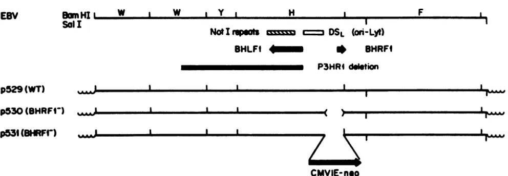
Aplasmid,p529,wasconstructed for these studiessothat it contained EBV DNA spanning the P3HR1 deletion and extending8.4 kbbeyondBHRF1, asillustrated inFig. 1. A 1.1-kb deletion removing all but the carboxy-terminal 31 codons ofBHRF1 and 0.6 kb of5'-flanking sequenceswas introducedbetweenBcll and BamHIsites.BecauseBHRF1 isover2kbawayfromthe P3HR1deletion, itwas
expected
that recombination mightoften occurbetween BHRF1 and
on November 9, 2019 by guest
http://jvi.asm.org/
BwnHII. W Sal I
I W I Y I H
NOtI
So'-=
DSL(Ori-LLYt)
BHLF * BHRFI
P3HRI dleton
p529(WT) p530(BHRFI-)
p531 (BRF-)
, I
I I
CMVIE-neo
FIG. 1. Schematicdiagramof part of the EBVgenome(top), extending rightwardfrom within the 3.1-kbinternalrepeat(BamHI fragment W), and of the EBV DNA present withinplasmidsconstructed for thisstudy (shown below).Positionsof theNotI repeats, DSL (ori-Lyt), leftward and rightwardopenreading framesBHLF1andBHRF1(2),and theregiondeleted in strain P3HR1(13)areindicated. Theplasmids consistof the EBVsequencesshownlinkedtopHEBo (31),indicatedbythewavylines,atits BamHI andSallsites. The deletion introduced
intoBHRF1, indicated by< >,and the substitutionofCMVIE-neoDNAformostof BHRF1arealso shown.BamHI andSallcleavagesites
areindicatedby upwardand downward marks from the horizontallines, respectively.
theregion deleted in P3HR1, restoring transformation
pro-ficiencywithout introducing the intended BHRF1 mutation into the recombinant viral genome. For this reason, a
selectivemarker, CMVIE-neo,wasintroduced into thesame
restriction sites used to generate the deletion in BHRF1. BecauseCMVIE-neo conferstomammalian cells resistance toG418,the selective markerpermitted quickdetermination of whether the recombinant virus carriedby aB-cell clone had acquiredthe desired substitution.
Generation of recombinant viralgenomescarryingdeletions
in BHRF1. Each plasmidofFig. 1 was mixed with
pCMV-BZLF1(8),whichexpressesBZLF1toinduce the EBVlytic cycle (7), and introducedby electroporationinto the P3HR1 subclone HH514-16. After 4days inculture, approximately 2.5% of the cells from each electroporation were observed
by immunofluorescence to express viral capsid antigens, comparedwith0% of cells withoutelectroporation, indicat-ing that electroporation was successful and that lytic viral
replication had been induced. Filtered growth media from the cell cultures, or the cells themselves following lethal gammairradiation,werecombined with Blymphocytesfrom adult peripheral blood and plated in 24-well culture dishes
overgamma-irradiated human fibroblasts. The numbers of
wells which produced proliferating B-cell clones for four experiments arepresented inTable 1.
The BHRF1-deleted plasmid, p530, consistently yielded fewer transformants (13% of wells positive) than did its parentplasmid, p529 (60% positive).This differencedidnot likelyresult fromthe absence of BHRF1function sincep531, which carries the CMVIE-neo construct within the same
BHRF1 deletion, gave rise to transformants as efficiently
(72% positive) asdid thewild-type plasmid. These perhaps minordifferences in transformation efficiencies could have arisen because of effects that the deletion or substitution
could have had on the rate of recombination between the plasmids and the EBVgenome (see Discussion).
Because of the 2-kbdistanceseparatingthe P3HR1 dele-tion and BHRF1, transformation-proficient recombinants could have been generatedwithp530 and p531without the transfer of the BHRF1 deletion to the viral genome. The
CMVIE-neo selective marker which was substituted for
mostof BHRF1 in p531 allowed foraquickexamination of several B-cell transformantsobtainedbyrecombination with
this plasmid.All of29 transformants tested for resistanceto G418 at 1,500 ,ug/ml (-750 ,g of active drug per ml), a concentration sufficienttokillcells of other EBV-established B-cell lines,werefullyresistant. Since p531also carries the hygromycin B resistance genewithin the vectorportion of theplasmid (not shown inFig.1), thesesametransformants were also tested for resistance to hygromycin B; none expressed noticeable resistance. These results suggest that the transforming virus present in these cell lines resulted from double homologous recombinationeventsbetween the P3HR1 genome and p531, flanking the P3HR1 deletion on the left andflankingthe BamHIsite within the BHRF1 gene on theright.
Analysis
ofrecombinant viral genomes. For each plasmid usedtogeneraterecombinants, several B-cell transformants from individual wellswereexpandedinasfewgenerationsas possible to approximately 20 million cells, and theirDNAs were isolated for Southern analysis. Analysis of BamHI-digested DNAs of three clones obtained by using p530showed that in all three the BHRF1 deletion had been
[image:3.612.65.561.75.247.2]incorporated into the viral genome. This deletion removed the BamHI site between the H and Ffragments, resulting in
TABLE 1. Generation oftransforming P3HR1 virus recombinants byusing plasmids carryingmutations in BHRF1
No.of wellspositive/no.
Plasmid tested in expt: Total %ofwells
positive
la 2 3 4
p529(wild type) 55/96 56/96 28/48 35/48 174/288 60 p530 (BHRFl-) 8/72 11/72 9/72 5/48 33/264 13
p531(BHRF1-) 52/72 52/72 72
Noneb 0/72 0/48 0/48 0/168 0
aForexperiment 1,electroporatedHH514 cells werecocultivated with B
cells. For the otherexperiments,filtered cell supernatants from
electropo-rated cells were used for infection.
bAsfor theelectroporationswith the testplasmids, pCMV-BZLF1DNA waspresent forthese controlelectroporations.
EBV F
a a I
l1
,I ww
L- I
on November 9, 2019 by guest
http://jvi.asm.org/
[image:3.612.320.561.617.694.2]A.
BarmHi w H r j
OSL ~g BHRF
deleted irnp530andp531 -- deweted ir: P3H R'
C.
probeprobe2 _
Clones Immortalized withHH514+Plasmid:
I I
0~ C
O s N r'e 'J -0 - In In to _ L cL
r--Ia I J1 2 3 4 5
N
C\j
cLLe LL Ikc
I DI
Ir
G4IBRClones, Recombinant virus from:
HH514 B95-8
-1r
kw
0-_
i.
-=DSR
-H-neo
A
-
10~~~~~~~~~~~~~~~~~~~._
H-F-DSR
H-~4
A-
to
D. H-1 s*
**H *; W
pI-FIG. 2. Southernanalysis of BamHI-digestedDNAofB-cell lines established by using recombinant viruses harboring deletions inBHRF1.
(A) BamHI cleavage sites, relevant genetic features, and probes 1 and 2. Probe 1 is a 6.6-kb HpaI fragment; probe 2 is the 1.1-kb
BclI-to-BamHlDNA deleted inp530 and p531. (B) Southern analysis of 5 ,ug of cell DNA fromB95-8-immortalized clone DF1, from HH514,
and fromB-cell clones obtainedby recombination of P3HR1 with the indicatedplasmid, performed with probe 1. Asstandards, in the first twolanes,52and 156pgofp529 DNA,correspondingto twoand sixmoleculespercell-equivalent of DNA,weremixedwith salmonsperm
DNAanalyzedinparalleltoprovidestandards forquantification. Theexpected DNA fragmentsareindicatedatthe left (see text). Marksat
therightindicatethepositionsof DNA sizestandards,withsizes(from the top) of 27.5, 9.4, 6.6, 4.4, 2.3, and 2.0 kb. (C)Similar analysis of
DNA fromB-cell linesestablishedby usingvirusesconferringG418resistance, obtained byrecombinationofp531 withP3HR1orwithB95-8, asindicated.H529-2,alsoanalyzedinpanel B,isaclone establishedbyrecombinationof P3HR1 withp529 (containing wild-type DNA).The
p529standardscorrespondto(fromlefttoright) 1, 3, 9,and 18moleculespercell. (D) Regionofthe blotfrompanel C after being stripped andreprobedwithprobe 2,the1.1-kbBamHI-to-BclI DNA thatwasreplacedwith the CMVIE-neo DNA inp531.
the appearanceof alarge, H-FBamHI fragment (Fig. 2B).
Theprobe used, a6.6-kbHpaI fragment indicated asprobe
1 inFig. 2A, also detected theadjacentYBamHIfragment, which was restored to these viral genomes by recombina-tion. Theprobe spanned ori-Lyt,which isduplicatedinEBV strains other than B95-8 and called DSL and DSR (for duplicated segments left and right). Thus, a 10-kb BamHI
fragmentspanning DSRwasalso detectedinstrainscarrying
the P3HR1 viral genome or recombinants derived from it.
Similar analysis ofBamHI-digested DNAs from 10 of the B-cell linesobtained withp531confirmed thepresencein all casesof the7.8-kb, alteredBamHIfragmentHlinkedtothe CMV-neoDNA(labeled H-neo inFig. 2C).
Most of the clones established by the BHRF1-deleted recombinants also appeared to carry nonrecombinant
P3HR1genomes, asindicatedbythepresenceof the 4.1-kb BamHIfragmentthatspansthe P3HR1 deletion. Asseenin Fig. 2B, the P3HR1 deletion fragment was detected at severalcopiespercell inoneofthe clones obtainedby using p530 (lane 4). Inthe cell lines establishedby using p531, the deletionfragment appeared tobepresentinall 10 examined (Fig. 2C andD).This isseenmostclearlyinFig. 2D,which shows the relevantportionofthe blot ofFig.2C after it had been reprobedwith probe 2 (theBclI-to-BamHI DNA seg-ment that equals the BHRF1 deletion of p531; Fig. 2A) because this probedetects theBamHI H fragment and the
B.
11,0W
_-_w_~
--"
4.
I
w
on November 9, 2019 by guest
http://jvi.asm.org/
[image:4.612.61.553.77.480.2]deletion fragment of P3HR1 (A in Fig. 2) equally. In one
clone, the number of P3HR1 nonrecombinant genomes,
aboutthreetofourpercell, appearedtoexceed the number of recombinantgenomes,abouttwopercell(Fig. 2C and D, leftmost G418'clone). With threeof theclones, the P3HR1 deletion fragment was present at much less than one copy per cell.
Examination of DNA from two cell lines established by recombination with the wild-type plasmid, p529, showed thatonedidnotcarry detectable copiesof nonrecombinant P3HR1genomes(H529-2; Fig.2C andD);for theother,lane background due to the very high number ofrecombinant EBVgenomespresent(100to200 percell; datanotshown) made it impossible to determine whether a low number of
nonrecombinant genomes was also present. However, in similar studies bythis and other laboratories, nonrecombi-nantP3HR1genomesarecommonly found coinfectingB-cell lines immortalized by P3HR1 recombinants (see Discus-sion). This finding indicates that as these experiments are
commonly performed, nonrecombinant virus is present in sufficient quantitiesto infect a significant fraction of the B
cells, whilerecombinants arepresent atmuch lowerlevels, usually10orfewer transforming unitsperml inourstudies (not corrected for the cloning efficiency oftransformed B cells).
The fact that P3HR1was found to be present at signifi-cantlyless than one copyper cell in someof the cell lines establishedby infection with aBHRFl-deficientvirus dem-onstrates that BHRF1 isnot essentialto sustain the prolif-eration of latently infected B cells after 2 to 3 months in culture,the times atwhichDNAwas isolated fromthe cell
lines foranalysis. However,theseobservationsalone donot excludetheformalpossibilitythat BHRF1mightberequired during the initial infection of B cells. The low numbers of recombinant virus present in the supernatants made it im-practical to reduce the multiplicityof infection as atest of this hypothetical contribution by the nonrecombinant P3HR1. However, by obtaining secondary and tertiary B-cell transformants with virus released from an initial
transformant, it was possible to show that the BHRF1-deficient EBVcouldpropagateandimmortalize Bcells in the absence of the parentalvirus.
BHRFl-deleted EBVcanimmortalize B cellsandpropagate inthe absenceof detectable helpergenomes.
EBV-immortal-ized B-cell lines typically release little or novirus into the
culture medium, because very few cells enter into the productive phaseof infectionandbecausemostvirus that is released remains associated with the cells(41). One of the B-cell lines carrying avirus in whichCMVIE-neo replaced BHRF1, H531-7,wasfoundtorelease transforming virusat the low levelstypicalofEBV-transformed B-celllines. Two secondary B-cell transformants were establishedby
infect-ing106 B cellswith0.5ml ofculture medium ofH531-7 after passing the medium through a 0.22-,um-pore-size filter. As
expectedfrom theverylowmultiplicity of infection, thetwo secondary transformants did notcarry thenonrecombinant P3HR1genome at detectable levels (limit ofdetection, less than 0.03copy percell; Fig. 3, 2°H531 clones 1 and 2).
By using the more sensitive method ofcocultivation to
detect transforming virus (30, 41), many more secondary
transformantswereobtained, and severalwereexaminedfor
viral DNAcontent and forrelease of transformingvirus.Of
sevenclonesexamined,fourcontainednodetectable P3HR1
genomes,twoclones contained 2and4P3HR1genomes per
cell,andonecarriedapproximately 0.03P3HR1genome per
[image:5.612.319.562.109.225.2]cell, justabovethedetection limit forthe Southern analysis.
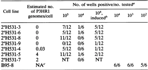
TABLE 2. Cocultivation assayfor releaseoftransformingEBV
by B-cell lines carrying BHRFl- EBV with and
withoutP3HR1as apotential helper virus Estimated no. No. of wells positive/no.testeda
Cell line of P3HR1 104
genomes/cell 105 1i0 induced" 10 0 102
2°H531-3 0 7/12 1/6 5/12
2°H531-6 0 5/12 1/6 5/12
2°H531-8 0 11/12 0/6 5/12
2°H531-9 0 0/12 0/6 1/12
2°H531-4 0.03 5/12 0/6 1/12
2°H531-5 4 11/12 1/6 2/12
2°H531-7 2 NT 0/6 NT
B95-8 NAC 6/6 6/6 5/6
aCocultivation ofBcellswiththeindicated numberofy-irradiated cells of each cell lineperwellwasdoneasdescribedin Materials andMethods.NT,
nottested.
bCellsweretreated withinducingagents3 mMsodiumbutyrateand 30 nM TPAfor3daysandthen washedwithmedium before cocultivation.
cNA, notapplicable.
Theresults of the Southernanalysisaresummarized in Table 2; the autoradiographs are shown in Fig. 3 for secondary
clone 3, carrying no detectable P3HR1 genomes, and for
secondaryclone4,carrying0.03 P3HR1 genome.(The signal
for the latterwasclearly visible, though faint,in theoriginal autoradiogram but lost during photographic reproduction.)
All secondary transformants carried the BHRF1-deficient EBV genome at7 to 20 copies per cell. It is notsurprising that during cocultivation, acell in the process ofreleasing
EBVmightoften pass on more than onevirusparticleto a recipient cell, although this was not known previously for EBV. Six of thesevensecondarytransformantsweretested with or without treatment with sodium butyrate and TPA
(tetradecanoyl phorbol acetate), which can induce
produc-tive EBVreplicationinsomecell lines. Allfourcelllinesthat lacked detectable BHRF1+ virus released transforming
EBV, and all but one did so within the range of one transformingeventper104to 105donor cells without induc-ers.ButyrateplusTPAappearedtoincreasemoderatelythe amounts of virus released from some of the lines. Four
tertiaryH531cloneswereanalyzed for the presence of EBV genomes, three obtained from two secondary clones that lacked P3HR1 genomes and one obtained from secondary
clone4 which contained 0.03 P3HR1 genome per cell. The P3HR1 genome couldnotbedetected in any of thesetertiary
transformants, while all carried the altered H-neofragment; the Southern analysis forthree of these clones is shown in
Fig. 3. One of thetertiary cloneswastestedsubsequently in the cocultivation assay and found to release transforming
virus. Theseexperimentsclearly demonstrate that BHRF1-deficientEBVcanreplicate and immortalize B cells without
requiring ahelper virus.
Forthetwosecondarytransformants that carried P3HR1 genomes at more than one copy per cell, one released
transforming EBV at levels similarto those of three of the foursecondarytransformants that lacked detectable P3HR1 genomes(Table2). The otherwastested only at104cells per well, but since all wellswerenegative, it is unlikely that this cell line releasedmuchmoretransforming virus than did any of the others.
The secondary and tertiary transformants all had the appearance andclumping tendencytypical of EBV-immor-talized B-cell lines and grewattypicalrates, withpopulation
doublingtimes of2 to3days.Allcell lineswereresistantto
on November 9, 2019 by guest
http://jvi.asm.org/
A B
20 30
I 7P
-to \ > H531 H531
,,;
p529 Stds. E .Im U) 1 2 3 4 1 2 3 in
H
--_ Long
-
Exposure
7 10
Sts - int
I
~~~~~~~~~~~~~~~~~~r~~~~~~~~~~~~~~~~.
Short
Exposure
C
--H-
NM1PO
FIG. 3. Southern analysis of EBV genomes present in secondary and tertiary B-cell transformants obtained withBHRF1-deficientEBV. FivemicrogramsofDNAfromeachcellline,exceptH529-1 (0.5 ,ug), was digested withBamHIandanalyzed. Symbols are as described for Fig.2.(A) Blot probedwith the 1.1-kb DNAof the BHRF1 deletion (probe 2; Fig. 2A); (B) much shorter exposure of the central portion of thesameblot; (C) similar blotprobedwithaplasmidcarryingtheBamHIHsegment of EBV. Increasing amounts ofBamHI-digestedp529 wereaddedasstandardscorresponding to (from the left) 0.03, 0.05, 0.1, 0.5, 1, 5, and 10 copies per cell. The 7.0-kb DNA seen in the plasmid standards inpanelCis the vectorportion ofp529.Thelowestthree standards were omitted from this blot. A signal from the lowest standard wasclearly visible ontheoriginal X-rayfilmof the blot in panel A.
G418, and the resistant phenotype was maintained if cells werepropagated for months in the absence of the drug.
Replacement of BHRF1 with the CMVIE-neo selective marker instrainB95-8.Since the CMV-neo selective marker could be substituted for BHRF1 in the EBV genome and express G418 resistance without interfering with B-cell
transformation,wetested whetherthis substitution could be
introduced into the B95-8 strain of EBVby recombination
andisolatedbyG418 selection in immortalizedBcells.B95-8 cells werecoelectroporated with p531 and pCMV-BZLF1. After4days inculture, thefraction of cellsexpressingviral
capsid antigens increased from approximately 4% to 8%.
Filtered cell supernatantswere usedto infectBcells in five 24-well culturedishes, using0.1 mlof supernatant and 8 x 10⁴ Bcells perwell. After 3weeks, atwhichtime
transfor-mantsweregrowingvigorously in allwells, G418wasadded at1,500 ,ug(-750pgof activedrug)perml.One G418' clone was identified and expanded for analysis of its DNA. In a BamHIdigest of this DNA,therecombinant H-neofragment waspresent, asexpected, inmultiple copies per cell-equiv-alent ofDNA(Fig. 2C andD, rightmostlanes).The normal BamHIHfragment of B95-8 could also be detected clearly, but at alevel ofonlyonecopy per 20cells, indicating thatthe clone had been infected initially by nonrecombinant virus also (Fig. 3, lane B531; the band may not be visible after
reproduction). Release oftransforming virus from this cell linewas notdetected in the cocultivation assay.
DISCUSSION
Themainfindingof this work is that EBV mutantslacking almost all of the BHRF1 openreadingframecanimmortalize human B cells andreplicate in these cells in culture. B cells immortalized by BHRFl-deleted EBV alone, in the absence of detectableBHRFl-containingEBV genomes,werefound to release transforming virus at levels that are within the range typical of EBV-immortalized B cells. The normal
frequency with which EBV-infected B-cell clones escape
latency to release transforming virus varies widely among
clones,from1/103cells inapopulationtoless than1/106 (30, 41). Because of this natural variation and because only a limited number of clones wereexamined, these studies do not rule out small quantitative contributions of BHRF1 to eitherphase ofEBVinfection, growth transformation of B cells orvirusreplication.
InisolatingtheBHRF1-deficientEBVrecombinants,most initial B-cell transformants also carried the parentalP3HR1 viral genome because of the large excessofparental virus present during the initial infection. Under such conditions that a large fraction of cells are initially infected with parental virus which can complement the mutant being generated, the number of transformants obtained with a givenmutantmaynotbemeaningful.Therearegoodreasons tobelieve that the deletion and substitutionmutations intro-duced into BHRF1 could have affected the rates of
recom-
on November 9, 2019 by guest
http://jvi.asm.org/
bination of the plasmids with the viral genome. Since the
studies of this report were performed, we have found that
the activity of ori-Lyt, which is adjacentto BHRF1, greatly stimulates the rate ofrecombination between the plasmids
and EBV DNA (ourunpublished studies). Previously itwas
found thatadeletion removing DNA justtotheright of DSL (right of 53820, similar to the BHRF1 deletion in p530) significantly reduced replication activity; in addition, the CMVIEenhancerwasfoundtorestore activitytoadeletion
fromthe leftextending into DSL (8). Thus, areducedrateof recombination should be expected with p530, as perhaps
reflected in the observed reduction in the number of
trans-formants, and it would not be unexpected for the CMV enhancer tohaverestored the recombination rate in p531.
During these studies, it was observed that a cell line
carrying bothBHRF1-deficient EBVgenomesand BHRF1+
P3HR1genomesoftenwould, after cocultivationwith B cells,
yieldB-cell clones carrying both EBVgenomes. This
obser-vationpromptedustocompare theefficiencywithwhichthe
secondary clones carryingorlacking P3HR1 genomeswould
yieldimmortalized B-cell clones following cocultivation with Bcells.Asignificantcontribution ofBHRF1 either to repli-cation and release of virus from cells or to efficiency of immortalization and outgrowth of B cells would result in significantly fewer B-cell transformants being obtainedfrom cocultivation with cell lines lacking P3HR1 genomes
com-pared with those carrying this potential helper virus. Al-though only a small number of cell lines with and without
P3HR1genomes wereavailableforthisanalysis (Table 2), the
seven linesundercomparison all had been established from
thesameindividual atthesametime andhad been in culture forthesamelength of time. While studieswithamuch larger
number of cell lines would be required to support a null
hypothesis, itmaybestatedthat the results failedto suggest aconspicuous contribution of the BHRF1+ P3HR1genomes
tothenumbers ofB-cell transformants obtained.
It is a matter ofcuriosity that P3HR1 genomes were so
readily detected in the initial B-cell lines transformed by viruslackingBHRF1. In similarstudies, the P3HR1genome
wasdetected atmuch lessthanone copy per cell in atleast
one of five clones in a study of BHLF1 (our unpublished
data)and inonecloneobtainedwitharecombinant deficient
in EBNA-LP (1). In a recent study in which B-cell clones
were established with P3HR1 recombinants lacking the
EBER genes, 5 of 14 clones transformed by an
EBER-deletedgenome also carried the EBER-containing (P3HR1) genomeaswell (32). In thecaseof thecell lines established
with viruses with deletions in BHLF1 or EBNA-LP, no
apparentbenefitwould be provided by the nonrecombinant P3HR1, since P3HR1 does not carry these genes intact.
Apparently, in these experiments performed in different lab-oratories, a significant fraction of all of the B cells were
infected with the parent P3HR1 virus, and a coinfecting
P3HR1genome wasoftenmaintainedgratuitouslyin the cells thatacquired atransforming, recombinant viral genome.
Whatmaybeunusualaboutthe cell lines initiallyobtained by using BHRF1-deficient recombinants is that the P3HR1 genomewasoftenpresent atone or morethanonecopy per
cell(7of13clones), whereasthiswas neverobserved in the
isolation of other recombinantviruses that were generated
from P3HR1 in ourlaboratory (0 of 7). Too few studies of this kind have been performed for the significance of this differencetobeevaluated. (For the generationof the EBER-deficient recombinants, it was not reported whether the coinfecting P3HR1 genomes were present at more or less
than one copy per cell [32].) Little is known about the
dynamics of amplification and loss of EBV genomes after infection of B cells. It is conceivable that BHRF1 could provide a slight growth advantage to EBV-infected B cells during the early stages of clonal outgrowth, although no differences were apparent in the growth properties ofB-cell lines carrying or lacking viral BHRF1 once the cell lines were established.
Through similarlydesigned experiments, Swaminathan et al. (32) recently constructed EBV mutants that lacked the EBER genes but appeared normal in the transformation of B cells and in virus replication. Presumably, conditions exist during infection of humans when BHRF1 or the EBERs contribute either to EBV replication or to survival of in-fected cells. It is possible that at some stage of infection of humans, EBV-infected B cells are prone to apoptosis, for example, when entering the productive phase of infection, and that BHRF1 functions as doesbc12 in promoting survival of the infected cells (see the introduction).
The demonstration that a selective marker replacing BHRF1could be recombined into the B95-8 genome, with the mutant virussubsequently identified as a drug-resistant B-cell clone, suggests a general method for the disruption of any EBV gene by homologous recombination. If the selective marker disrupted a gene required for B-cell transformation, drug-resistant B-cell clones would be obtained only if the clones were coinfected with a nonrecombinant, helper virus. For the EBV recombinants generated in these studies, as well as theEBER-deleted mutants constructed by Swaminathan et al. (32), the plasmids used for homologous recombination carried the EBV lytic replication origin, on-Lyt. Recent studies have shown that homologous recombination between introduced plasmids and the EBV genome is greatly stimu-lated by thepresence ofoni-Lyt on the plasmid; homologous recombination targeted to distant regions of the EBV genome also can be obtained efficiently if on-Lyt is placed on the plasmid carrying EBV DNA from the region of interest (15a). Asimilar method was reported recently by Wang et al., who found that certain Burkitt'slymphoma cell lines can be used for the isolation and study of EBV mutants carrying selective markers (39). The development of methods such as these should permit full geneticanalyses of the potential of EBV to replicate and to transform B cells.
ACKNOWLEDGMENTS
We thank Ted Torrey and GarthAnderson for comments on the manuscript.
This work was supported by grants CA4312203 and RR0564823 from theNational Cancer Institute.
REFERENCES
1. Allen,G., and P.Farrel.Personal communication.
la.Austin, P. J., E. Flemington, C. N. Yandava, J. L. Strominger, and S. H. Speck. 1988. Complex transcription of the Epstein-Barr virus BamHIfragment H rightward open readingframe 1 (BHRF1)inlatently andlyticallyinfected Blymphocytes. Proc. Natl. Acad. Sci. USA85:3678-3682.
2. Baer, R., A. T. Bankier, M. D. Biggin, P. L. Deininger, P. J. Farrell, T. J. Gibson, G. Hatfull, G. S. Hudson, S. C. Satchwell, C. Sequin, P. S. Tuffnell, and B. G. Barrell. 1984. DNA sequence and expression of the B95-8 Epstein-Barr virus ge-nome. Nature (London) 310:207-211.
3. Baichwal, V. R., and B. Sugden. 1989. Themultiple membrane-spanning segments of the BNLF-1 oncogene from Epstein-Barr virus are required fortransformation. Oncogene 4:67-74. 4. Bodescot, M., and M. Permcaudet. 1986. Epstein-Barr virus
mRNAs produced by alternative splicing. Nucleic Acids Res. 14:7103-7114.
on November 9, 2019 by guest
http://jvi.asm.org/
5. Cleary, M. L., S. D. Smith, and J. Sklar. 1986. Cloning and structural analysis of cDNAs for bcl-2 and a hybrid bcl-2/ immunoglobulin transcript resulting from the t(14;18) transloca-tion. Cell 47:19-28.
6. Cohen, J. I., F. Wang, J. Mannick, and E. Kieff. 1989. Epstein-Barr virus nuclear protein 2 is a key determinantof lymphocyte transformation. Proc. Natl. Acad. Sci. USA 86:9558-9568. 7. Countryman,J., and G. Miller. 1985. Activation ofexpression
of latent Epstein-Barr herpesvirus after gene transfer with a small cloned subfragment of heterogenous viral DNA. Proc. Natl. Acad. Sci. USA82:4085-4089.
8. Hammerschmidt, W., and B. Sugden. 1988. Identification and characterization oforiLyt, a lytic originof DNA replication of Epstein-Barr virus. Cell55:427-433.
9. Hammerschmidt, W., and B. Sugden. 1989.Genetic analysis of immortalizing functions of Epstein-Barr virus in human B lymphocytes. Nature (London)340:393-397.
10. Hardwick, J. M., P. M. Lieberman,and S. D. Hayward. 1988. A new Epstein-Barr virus transactivator, R, induces expression of a cytoplasmic early antigen. J. Virol. 62:2274-2284.
11. Henderson, S., M. Rowe, C. Gregory, D. Croom-Carter, F. Wang,R.Longnecker,E.Kieff, and A. Rickinson. 1991. Induc-tion ofbcl-2 expressionby Epstein-Barr virus latent membrane protein 1 protects infected Bcells from programmed cell death. Cell 65:1107-1115.
12. Hockenbery, D., G. Nunez, C.Milliman, R. D. Schreiber, and S. J. Korsmeyer. 1990. Bcl-2 is an inner mitochondrial mem-brane proteinthat blocks programmed cell death. Nature (Lon-don) 348:334-336.
13. Jeang, K.-T., and S. D. Hayward. 1983. Organization of the Epstein-Barr virusDNA molecule III. J. Virol. 48:135-148. 14. Kaplan, M., andC. Clark. 1974. An improved rosetting assay
fordetection of human T lymphocytes. J. Immunol. Methods 5:131-135.
15. Kieff, E., and D. Liebowitz. 1990. Epstein-Barr virus and its replication, p. 1889-1990. InB. N. Fields, D. M. Knipe, et al. (ed.), Virology. Raven Press, Ltd., NewYork.
15a.Lee, M. A.,0.J. Kim, andJ. L. Yates.Unpublished data. 16. McDonnell, T.J., N. Deane, F. M. Platt,G. Nunez, L. Jaeger,
J. P. McKearn, and S. J.Korsmeyer. 1989.bcl-2 immunoglob-ulin transgenic mice demonstrate extended B cellsurvival and follicular lymphoproliferation. Cell57:79-88.
17. McDonnell, T. J., G. Nunez, F. M. Platt, D. Hockenberry, L. London, J. P.McKearn, and S. J. Korsmeyer. 1990.Deregulated Bcl-2-immunoglobulin transgene expands aresting but respon-sive immunoglobulin M and D-expressing B-cell population. Mol. Cell. Biol. 10:1901-1907.
18. McDonnell, T. J., G. Nunez, F. M. Platt, D. Hockenberry, L. London, J. P. McKearn, and S. J. Korsmeyer. 1991.Deregulated Bcl-2-immunoglobulin transgene expands a resting but respon-sive immunoglobulin M- and D-expressing B-cell population. Mol. Cell. Biol.349:254-256.
19. Miller, G. 1990. Epstein-Barr virus, biology,pathogenesis, and medical aspects, p.1921-1957. In B. N.Fields, D. M.Knipe,et
al. (ed.), Virology. Raven Press, Ltd., NewYork.
20. Nunez,G., M. Seto,S. Seremetis, D. Ferrero, F.Grignani, S. J. Korsmeyer, and R. Dalla-Favera. 1989. Growth- and
tumor-promoting effects of deregulated BCL2 in human B-lymphoblas-toid cells. Proc.Natl. Acad. Sci.USA 86:4589-4593.
21. Pearson, G.R., J. Luka, L. Petti, J. Sample, M.Birkenbach, D. Braun, and E. Kieff. 1987. Identification of an Epstein-Barr virusearlygene encoding a secondcomponent of the restricted earlyantigen complex. Virology 160:151-161.
22. Rabson, M., L. Heston, and G. Miller. 1983. Identificationofa
rareEpstein-Barr virus variant that enhances early antigen
ex-pression in Raji cells. Proc. Natl. Acad. Sci. USA80:2762-2766. 23. Reed, J. C., M. Cuddy, S. Haldar, C. Croce, P. Nowell, D. Makover, and K. Bradley. 1990. BCL2-mediatedtumorigenicity of a human T-lymphoid cell line: synergy with MYC and inhibition by BCL2 antisense. Proc. Natl. Acad. Sci. USA 87:3660-3664.
24. Reed, J. C., S.Haldar,M. P. Cuddy, C.Croce, and D. Makover. 1989. Deregulated BCL2 expression enhances growth of a
human B cellline. Oncogene 4:1123-1127.
25. Rooney, C., J. G. Howe, S. H. Speck, and G. Miller. 1989. Influences ofBurkitt'slymphoma and primaryBcellsonlatent gene expression by the nonimmortalizing P3J-HR-1 strain of Epstein-Barrvirus. J. Virol.63:1531-1539.
26. Sambrook, J., E. F. Fritsch, and T. Maniatis. 1989.Molecular cloning: a laboratory manual, 2nd ed. Cold Spring Harbor Laboratory,Cold Spring Harbor,N.Y.
27. Southern, P. J., and P.Berg. 1982. Transformation of
mamma-lian cells to antibiotic resistance with a bacterial gene under control of theSV40earlyregionpromoter.J.Mol.Appl. Genet. 1:327-341.
28. Stinski, M. F., and T. J. Roehr.1985. Activation of themajor immediate early gene of humancytomegalovirusbycis-acting elements in the promoter-regulatory sequence and by virus-specific trans-acting components.J.Virol. 55:431-441. 29. Strasser, A., A. W.Harris,M. L.Bath,and S.Cory. 1990. Novel
primitivelymphoidtumoursinducedintransgenic miceby coop-erationbetween myc andbcl-2. Nature(London)348:331-333. 30. Sugden, B. 1984. Expression of virus-associated functions in
cells transformed in vitroby Epstein-Barr virus: Epstein-Barr virus cell surface antigen and virus-release from transformed cells,p.165-177.In D. T.Purtilo(ed.),Immunedeficiencyand cancer.PlenumMedical BookCo.,NewYork.
31. Sugden, B., K. Marsh, and J. L. Yates. 1985. A vector that replicates as aplasmid andcanbeefficiently selectedin B-lym-phoblasts transformed byEpstein-Barr virus. Mol. Cell. Biol. 5:410-413.
32. Swaminathan, S., B. Tomkinson, and E.Kieff.1991. Recombi-nantEpstein-Barrvirus with small RNA(EBER)genesdeleted transforms lymphocytes and replicates in vitro. Proc. Natl. Acad.Sci. USA88:1546-1550.
33. Tsujimoto, Y.1989.Stress-resistance conferredbyhighlevel of bcl-2-alpha protein in human B lymphoblastoidcell. Oncogene 4:1331-1336.
34. Vaux, D. L., S. Cory, and J. M. Adams. 1988. Bcl-2 gene promoteshaemopoieticcellsurvival andcooperateswithc-myc toimmortalize pre-B cells. Nature(London)355:440-442. 35. Wang,D.,D.Liebowitz,and E. Kieff.1985. An EBVmembrane
proteinexpressedin immortalized lymphocytes transforms
es-tablished rodent cells. Cell 43:831-840.
36. Wang, D., D.Liebowitz,and E.Kieff.1988. The truncated form of the Epstein-Barr virus latent-infection membrane protein expressedin virusreplication does nottransform rodent fibro-blasts.J. Virol. 62:2337-2346.
37. Wang,F., C.Gregory, C. Sample, M. Rowe, D. Liebowitz, R. Murray, A. Rickinson, and E. Kieff. 1990. Epstein-Barr virus latent membraneprotein(LMP1)andnuclearproteins2 and 3C areeffectors ofphenotypicchangesin Blymphocytes:EBNA-2 and LMP1cooperatively induce CD23. J.Virol. 64:2309-2318. 38. Wang, F.,C. D.Gregory,M.Rowe,A. B.Rickinson, D.Wang, M.Rirkenbach, H.Kikutani, T.Kishimoto, and E.Kieff. 1987. Epstein-Barrvirusnuclearantigen2specificallyinduces expres-sion of the B-cell activation antigen CD23. Proc. Natl. Acad. Sci. USA84:3452-3456.
39. Wang, F., A.Marchini, and E. Kieff. 1991. Epstein-Barrvirus (EBV) recombinants: use ofpositive selection markers to
res-cue mutants in EBV-negative B-lymphoma lines. J. Virol. 65:1701-1709.
40. Wang, F., S. Tsang, M.G. Kurilla,J. I.Cohen, and E. Kieff. 1990.Epstein-Barrvirus nuclearantigen2transactivates latent membraneproteinLMP1. J.Virol.64:3407-3416.
41. Wilson,G., andG. Miller.1979.RecoveryofEpstein-Barrvirus fromnonproducer neonatalhumanlymphoidcelltransformants. Virology95:351-358.
42. Wysokenski, D. A., and J. L. Yates. 1989. Multiple EBNA1-binding sites are required to form an EBNAl-dependent en-hancer andtoactivateaminimalreplicativeoriginwithinoriPof Epstein-Barrvirus. J. Virol. 63:2657-2666.
43. Yates,J. L.,N.Warren,and B.Sugden.1985. Stable
replication
ofplasmidsderived fromEpstein-Barrvirus invarious
mamma-liancells. Nature (London)313:812-815.