Vol. 49. No. 2 JOURNALOF VIROLOGY. Feb. 1984. P. 325-332
0022-538X/84/02()0325-08$02.00!0
Copyright t 1984, American Society forMicrobiology
Role of the Mitogenic Property
and Kinase
Activity
of
p6Osr'
in
Tumor Formation by
Rous Sarcoma Virus
F. POIRIER,I* P. JULLIEN,' P. DEZELEE,' G. DAMBRINE, E. ESNAULT, A. BENATRE,3 AND G. CALOTHY' Institit Curlie-Biologie, Centre Unjiiersitaire, 91405 Orsav,1 Station dePathologieAiviaire de l'institlut National de Ia
Rec-her(
he Agr-onolniiqlue,
37830Monna(ie
2and
Centre
Hospitalier
Univ er-sitaire,
37000Tours,3 France
Received 5July 1983/Accepted 12 October 1983
Expression of the sr( gene of Rous sarcoma virus in chicken embryo neuroretinal cells results in
morphological transformation and sustained proliferation of this normally resting cell population. PA101 and PA104 are two mutants of Rous sarcoma virus which induce neuroretinal cell proliferation in the absence ofmorphologicaltransformation. Theirmitogenic propertyistemperature sensitive, andtheyboth encode p6O''' proteins with low kinase activity. To study the role of the mitogenic function and protein kinase activity of p6O0''(' in tumorigenesis, we investigated the oncogenicity of PA101 and PA104. Both
mutantswere lesstumorigenic thanwild-type virus when injected intochicks. Tumorigenicity wasfurther
assayed by inoculating infected chicken embryofibroblasts and neuroretinal cellsonto the chorioallantoid membrane of embryonated duck eggs. This system provides a nonpermissive and immunodeficient
environment for xenogenic cell grafting and allows the study of cell tumorigenicity within a temperature range of 37 to 39.5°C. Chicken embryo fibroblasts and neuroretinal cells infected with PAIOI were as
tumorigenicaswildtype-infected cellsat37°C,buttumordevelopmentwassignificantlyreducedat39.5°C. In contrast. both cell types infected with PA104 displayed sharply reduced tumorigenicity. Cell cultures derived from PA101 tumors induced on the chorioallantoid membrane were similar to the corresponding cells maintained in vitrointermsofmorphology, productionofplasminogenactivator, relativeamountsof phosphotyrosine in total cellular proteins, and phosphorylation of34,000-molecular-weight protein. These results indicate that the expression of the mitogenic function of src does not account per se for cell
tumorigenicity and that tumorformation is compatible withlow levels ofp6O"'' protein kinase activity. Transformation by Rous sarcoma virus (RSV) causes
significant changes in the regulation of cell growth, which may play an important role in tumorformation (10, 26). To study these changes, we have developed an in vitro model system in which RSVinfection interacts with growth regula-tory mechanisms taking place during differentiation of cells
from the chicken embryo neuroretina. Asaconsequence of such interactions, chicken embryo neuroretinal (NR) cells which normally do not grow in vitro are induced to prolifer-ateforseveralgenerationsupontransformationbyRSV (17). Thischaracteristic effect ofRSVonthegrowth propertiesof NRcellsis referred to as the mitogenic property of the virus. We have previously shown that both transformation and proliferation of NR cells require the expression ofa function-al src gene (2).
PA101 and PA104 are two mutants of RSV capable of
inducing NR cell division in the absence of morphological transformation (3, 4). Characterization of these viruses has shown that their mitogenic property istemperaturesensitive and that their mutatedsrc genes encodep60r'' proteins with low kinaseactivity(3,20). Becauseof thedissociation ofsrc genefunctions in infected NR cells. thesemutantsconstitute auseful tool to study the role of the mitogenic function and
protein kinase activity of p6O0''' in tumor formation. Oncogenicity ofPA101 and PA104 was studied by inject-ing these viruses into chicks and by inoculating mutant-infected cells onto the chorioallantoid membrane (CAM) of embryonated duck eggs. The latter assay of cell tumorigenic-ity was selected to fulfill five specific requirements. First, the CAMofembryonated eggsprovidesanimmunodeficient environment for xenogenic cell grafting (23). Second, duck
* Correspondingauthor.
cells are genetically resistant to infection by subgroup A viruses used in these experiments (6). Third, this system allows the study of tumorigenicity of the cell expressingthe mitogenic function of the src gene product. Preliminary experiments have shown thatNRcellstransformed by RSV couldreadilyformtumorsontheCAM of embryonated eggs but not in irradiated (700 rads) athymic mice (nu/nu, Swiss background). Fourth, inoculated eggs can be maintained at controlled temperatures within arangeof 37to39.5°C, thus
permitting the analysis of temperature-sensitive src gene
functions (19, 21). Fifth, cell cultures can be derived from explanted tumors, and their properties can be compared with those of the corresponding cells before inoculation.
Our results show marked differences in the tumorigenic
propertiesof the two mutants, suggestingthat expression of themitogenicfunctionof p60'' maynotbesufficient for cell
tumorigenicity. The dataalso indicate that tumorformation on CAM is compatible with low levels of p60"'' protein kinaseactivity and does not result from the selection of cell variants with increased p60''''-associated phosphorylating properties.
MATERIALS AND METHODS
Cell cultures and viruses. Chicken embryofibroblast cul-tures were prepared from 10-day-old lymphomatosis-free
Brown Leghorn embryos
(Gs'
Chf+) of the Edinburgh strain. Chicken embryo fibroblasts (CEF) were grown in Eagle minimal medium containing 3% newborn calf serum and 10%tryptose phosphate broth.Preparation of NR cell cultures from 7-day-old embryos has been described previously (18). NR cells were main-tained in Eagle basal medium supplemented with 5% fetal calfserum.
325
on November 10, 2019 by guest
http://jvi.asm.org/
326 POIRIER ET AL.
RAV1 is a subgroup A lymphomatosis virus. The Schmidt-Ruppin strain of RSV subgroup A (SR RSV-A) was used as the wild-type (wt) virus. PA101 and PA104 are two mutants derived from SR RSV-D and SR RSV-A, respectively. Their isolation and properties have been previously reported (3, 4, 20). SincePA101 bears a deletion in the envelope gene (R. Swanstrom and J. M. Bishop, personal communication), nonproducer clones ofPA101-infected CEF were isolatedin agar. One clone was superinfected with RAV1, and the pseudotypePA101 (RAV1) was used in these experiments.
Assay of viral infectivity in NR cells. NR cells were infected as previously described (17). The ability of wt or mutant viruses to induce NR cell multiplication was quantitated by infecting NR cells with serial 10-fold dilutions of the viruses. The mitogenic titer was defined as the reciprocal of the highest dilution inducing cell proliferation after one subculti-vation and is expressed as mitogenicunits per milliliter. End-point-transforming and mitogenic titers of SR RSV in NR cells were identical.
Tumorigenicity in chickens and isolation of virus from tumors. One-day-old White Leghorn chicks (PA-12 strain) were subcutaneously inoculated in each wing web with 0.1 ml of virus. The stocks of virus were adjusted to 106 mitogenic units per ml. Control chicks received 0.1 ml of medium. The birds were examined for tumor development every third day over a period of 10 weeks. Some tumors were excised and dissociated with trypsin. Fragments of tumors were mixed with fresh CEF, and culture fluids were used as a source of virus.
Preparation ofextracts from tumorsin vivo. Some tumors were excised and lysed on ice in Nonidet P-40 buffer (1% Nonidet P-40, 0.15 M NaCl, 0.01 M sodium phosphate (pH 7.2), 1% trasylol, 25 mM EDTA). After partial homogeniza-tion with a Vortex mixer, the lysates were clarified by
spinning at 10,000 x g for 15 min. The supernatants were stored at -70°C before being assayed for protein kinase activity.
Inoculation of cells on CAM of embryonated duck eggs. Inoculation of infected cells on the CAM of 12-day-old
embryonated duck eggs was performed as described else-where (19), and eggs were examined 10 days later. Some tumors were removed andfixed in 10% formaldehyde in 0.15 Mphosphate-buffered saline (pH 7.4). Sections, 5 p.mthick, were stained with hematoxylin and eosin for histological examination.
Culture of tumor cells. Tumors were sterilely excised from the CAM, chopped into small pieces, and dissociated first with collagenase and then with trypsin. The dispersed cells were seeded in 35-mm dishes and grown in Eagle minimal medium supplemented with 3% newborn calf serum-10%
tryptose phosphate broth.
Detection ofspecies-specific surface antigen. The cytotoxic-ity assay of Gorer and O'Gorman (9) was used to detect chicken or duck antigens on the surface of cells derived from the tumors developed on the CAM of duck eggs.
Plasminogen activator production. Plasminogen activator production (25) in serum-free culture media was determined by the caseinolysis of defatted dry milk in a gelled medium containing chicken serum as a source of plasmin (8). The cellsin 6-cm dishes were washed three times with serum-free medium and incubated for 6 h at 37°C in 2 ml of serum-free medium. The media were harvested, and the number of cells perplate were determined. Detection of plasminogen activa-tor was performed in wells of Costar multiplates (1.6 cm diameter) filled with 0.4 ml of 0.1 M Tris-hydrochloride buffer (pH 8.1) containing 1% agarose, 2% nonfat milk, and
2% of a batch of selected chicken serum. Samples (20 pLl) of dilutionsof culturemedium were layered on the gels,and the plates were incubated at 37°C in a humidified atmosphere. The highest dilution causing a zone of caseinolysis was detected 48 to 72 h later. The absence of plasminogen-independentcaseinolysis wasdetermined on separate plates without chicken serum.
Isotopic labeling of cells and preparation of cell extracts. CEF orNR cellswere seeded in 35-mm dishes. Cells were incubated for 1 h in 1 ml ofmethionine-free Eagle minimal medium (GIBCO Laboratories) or phosphate-free Eagle minimal medium, each supplementedwith 1% dialyzed calf serum. The medium was replaced with 0.4 ml ofthe same medium containing
L-3Sl
methionine (150 ,uCi/ml; specific activity, 1,000-1,200Ci/mmol; NewEngland Nuclear Corp.) or carrier-free [32p]p; (1 mCi/mI; Commissariat 'a l'EnergieAtomique, Saclay, France). Cell extracts were prepared 5 h later, usingmodified RIPA buffer to lyse the cells (20).
Immunoprecipitation. Immunoprecipitations were per-formedfollowing the procedure described before (20).
Antiserum directed against p60'r' was prepared as de-scribed by Brugge and Erikson (1). A single lot of serum obtained from atumor-bearing rabbit 8 weeks after inocula-tion was used throughout these experiments.
Anti-34,000-molecular-weight (34K) protein serum was kindly provided
by R. Erikson.
Protein kinase assay.
[y-32P]ATP
wasadded to the immu-noprecipitates containing p6Orc complexed to specific tu-mor-bearing rabbit antibodies, and the reaction products were analyzed by polyacrylamide gel electrophoresis as described by Collett and Erikson (5).Measurement of the amount of acid-stable phosphoamino acidsin total cellular proteins. Cells were labeledwith
[32p]p;
as described above. The medium was removed, and the cultures were washed twice with phosphate-buffered saline and then hydrolyzed in 1 ml of10% trichloroacetic acid at 70°C for 2 h. The cells were scraped off the plates and transferred to an Eppendorf tube. After three washes with 1 ml of 10% trichloroacetic acid at room temperature, the pellet was rinsed three times with chloroform-methanol (2:1 [vol/vol]). The dried pellet was hydrolyzed in 200
[LI
of 6 N HCI for 2 h at 110°C. The hydrolysates were lyophilized,washed once with water, lyophilizedagain, and dissolvedin 10 ,u1 of a marker mixture containing phosphoserine,
phos-phothreonine, and phosphotyrosine, each at 1 mg/ml. The hydrolysates were resolved in twodimensions on thin-layer cellulose-coated glass plates(Merck &Co.) by electrophore-sis toward the anode at pH 1.9 (750Vfor 150 min), followed
by electrophoresis toward the anode at pH 3.5 (1,000 Vfor 60
min).
Phosphoamino acid markers were revealed with ninhydrin, and the cellulose plates were exposed for 24 h. Colored spots were scraped off and transferred into scintilla-tion vials. Blanks were preparedby using twoareas ofequal surfaces in regions of the plates on which no radioactivity was detected by autoradiography. After the addition of 0.5 ml of distilled water, 10 ml of Aquasol (New EnglandNuclear Corp.) was added for liquid scintillation counting.
RESULTS
Biological properties of PA101 and PAl04. The in vitro biological properties of PA101 and PA104 have been
previ-ously described (3, 4, 20). A summary of these results is presented in Table 1. NR and CEFcultures infected withwt SR RSV are converted into typical round and refringent
J. VIROL.
on November 10, 2019 by guest
http://jvi.asm.org/
p60"' MITOGENIC FUNCTION AND TUMORIGENESIS 327 TABLE 1. Propertiesof PA101 and PA104 mutants"
Properties SRRSV-A PA101 PA104
Induction of NR cell multiplication wt" ts' ts
Morphology of infected CEF or NR cells Round Normal Normal
Growthin agarof infected CEF wt ts Negative
Invitrokinase activity of p60"" immuno- 100% 7% 10%
precipitated from CEF or NR cells maintained at 37°C
Tyrosinephosphorylation of p6O"r< 100% 86% 21%
Total amount of phosphotyrosine in pro- 100% 17% 12%
teins extracted from infected NR cells maintainedat37°C
Phosphorylation of 34K protein in infect- 100% 25% 30%
ed NR cells maintained at 37°C
"All data arefrom references
3.
4.
and20.
wtlevels werearbitrarilyset at100%.
hwt,Wild type.
' ts,Temperature sensitive.
cells. Such transformed cells are not observed when these culturesarechronically infectedwithPA101 orPA104. Both
mutants induce sustained proliferation ofNRcells, andthis
propertyistemperature sensitive. Cell division, whichtakes place at 35 to 37°C. is blocked at 41 to 42°C but resumes
rapidly when the cultures are returned to the permissive temperature. The p6O0"' proteins encoded by the two
mu-tantshavealow proteinkinaseactivityinvitroasmeasured
by their ability to phosphorylate heavy chains of specific immunoglobulin G molecules (5, 14). Similarly, theamounts
of phosphotyrosine in total cellular proteins (11. 24) are
reduced in mutant-infected NR cells, and a 34K protein. a
potentialinvivotargetofp60r"' kinase activity(7, 22). is four to five times less phosphorylated in NR cells infected with PA101orPA104 compared with thatinwt-transformedcells.
Despite the similar phenotypic changes they induce in infected cells, the two mutants differ essentially by two properties. First, theamountof phosphotyrosine in p6o0'' of PA104 is markedly reduced when compared with that of PA101 andwtSR RSV-A.Second, cells infected with PA101 acquire a thermolabile capacity to grow in semisolid medi-um, whereas CEF and NRcells infected with PA104 donot
formcolonies in soft agar.
Tumorigenicityof PAlO and PA104 in chickens. One-day-old chicks were subcutaneously injected with SR RSV-A, PA101, and PA104 adjusted to the same concentration of mitogenic units (Table 2). Both PA101 and PA104wereless
tumorigenic than wtvirus asindicated by the longer latency and lower tumor incidence. However, differences were observed in the patterns of tumor formation by the two mutants. PA104 was poorly oncogenic, yet the few tumors
inducedby thisvirusbecameaslargeasthose induced bywt virus. Oncogenicity of PA101wasintermediatebetween that
of SR RSV-A and PA104. PA101 tumors were slow to appear, some of them remained small and even regressed. Thesetumors were typical fibrosarcomas.
Replication of mutant viruses in the permissive host increases the chancesthattumorformation could be duetoa
wt virus generated by reversion or recombination with
homologous cellularsequences (13).Totestthispossibility, tumors were removed from the chickens and examined for
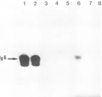
immunoprecipitable p60"' kinaseactivityand virus produc-tion. Elevated levels of kinase activity were detected in
extracts prepared from SR RSV-A induced tumors (Fig. 1. lane 1). High levels of p60"' kinase activity were also observed in the two tumors induced by PA104 which were
analyzed, and theresult obtainedwithoneof themis shown inFig. 1, lane 2. Characterization of the virusesreleased by thesetumors indicated thattheyhadacquired theproperties of a wt src gene. suggesting that these tumors arose as a
consequence of virus reversion. In contrast, phosphoryla-tionofimmunoglobulinG wasbarelydetectable in immuno-precipitates fromfour of fivetumorsinducedby PA101 (Fig. 1, lanes 3, 4, 5, 7). The kinase activity present in the fifth
tumor(Fig. 1, lane 6) was somewhat higher than thatin the others and in normal tissues (Fig. 1, lane 8) but was still lower in comparison with the wt-derived tumors. Unfortu-nately, noviruscould be isolated from thesetumors. Froma
total of 10 PA101 tumors examined, only one virus was
recovered. It differed from the original mutant: it
trans-formed CEF and NR cellsat37 and41°Cand proved highly oncogeniconsubsequent inoculation into chickens (datanot shown). The tumorreleasing thiswtviruswasnotexamined forproteinkinaseactivity.The failure torecovervirusfrom the majority of PA101-induced tumors could be explained eitherbyadefectinthe spreading ofthecomplexbetweena
TABLE 2. Incidence ofsarcomas inchicks"
Inoculated Tumorigenicity at week after infection
virus 1 2 3 4 5 6 7 8 9 t(
SR RSV-A 0/12 4/12 9/12 10/12 b
PA101 1/24 1/24 3/24 6/24 6/24 8/24 10/24 11/24 11/24 10/24'
PA104 0/26 0/26 2/26 2/26 2/26 3/26 4/26 4/26 4/26 5/26
'Groupsof1-day-old WhiteLeghorn chicks maintained in separate isolation rooms wereinoculated intoeach wing web with 0.1 ml
of
undiluted virus. The stocks of viruses had identical titers. Numbers representsiteswithapalpabletumor overthetotalamountof inoculation sites. These numbers are cumulative, the animals sacrificed during the experiment are included in the total until the last week.
b , All of the animals died fromtumors.
' Onetumorcompletely regressed. VOL. 49. 1984
on November 10, 2019 by guest
http://jvi.asm.org/
328 POIRIER ET AL.
G~~~~~~~
FIG. 1. Reaction products of proteinkinaseassayperformedon
immunoprecipitates of extracts prepared from tumors and from normal chicken tissue. Extracts were prepared (see text) from tumorsthat developed in chickens inoculated with wt SR RSV-A (lane 1), PA104 (lane 2),orPA101 (lanes 3through 7). Anextract
prepared from the muscle ofabirdwhichwasinoculated with 0.1 ml of mediumwasincludedas acontrol(lane 8). Immunoprecipitation
andprotein kinaseassay aredescribedin thetext.Thesampleswere
loadedon an8.5% acrylamide gel.
replication-defective virus and its helperor by more trivial
reasonssuchasthedenseconsistency of thesetumorswhich renderedthe dissociation of the tissue very difficult.
Since the original virus could not be recovered from the mutant-inducedtumors, definite conclusionsconcerningthe actual oncogenicity of PA101 and PA104 in chickens are
difficultto draw.
Tumorigenicity of virus-infected cells inoculated onto the CAM ofembryonated duckeggs.Thetumorigenic properties ofwtvirus, PA101,and PA104werefurtherinvestigated by
inoculating various numbers of infected CEF and NR cells ontotheCAM ofembryonated duckeggs.Duplicatecultures of thesamecellswereinoculated after lethal irradiation and served as controls in these experiments. The incidence of tumors at permissive temperature is shown inTable 3. NR cells infected with SR RSV-Aacquirethecapacityto prolif-erate and invade CAM, although with a slightly reduced
efficiencyascompared with transformedfibroblasts. Onlya
fewsmall tumors were observed whenirradiated cellswere
inoculated, indicating that minimal secondary infection of adjacent cells had occurred. CEFand NR cells infected with
PA101were astumorigenicaswt-infectedcells. Incontrast,
both cell types infected with PA104 displayed sharply
re-duced tumorigenicity.
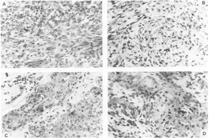
Histological examination of the tumors revealed that in-oculation of CEF infected with wt and PA101 gave rise to
typicalfibrosarcomasdevelopingin the mesodermal layerof CAM. In contrast, tumors derived from NR cells infected with the same viruses were epidermoid carcinomas with
areas offusiform fibroblast-like cells (Fig. 2).
We also examined the effects oftemperature on tumor formationby incubatingeggs inoculated withwt-or
PA101-infected CEFateither 37or39.5°C, the highesttemperatures
compatiblewith survival of duckeggs.CEF infected with SR
RSV-A are equally tumorigenic at both temperatures.
Al-though the absolute number oftumors induced by PA101-infected cellsis only slightly reducedat39.5°C,weobserved a marked decrease of their size in comparison to those developed atthe permissive temperature(data not shown).
[image:4.612.97.267.76.237.2]Characteristics of cell cultures and viruses derived from tumors. Itwas possible toderive cell cultures from tumors obtained by inoculating SR RSV-A- orPA101-infected cells
ontothe CAM. The fewtumorsinduced byPA104-infected cells weretoo smalltobeexplanted. SR RSV-Atumorcells
were round and refringent whether they originated from
infected CEForNRcells. Incontrast,alltumorcellsderived from CEF or NR cells infected with PA101 were flat and
nonrefringent, and they retained a morphology similar to that of the cells before inoculation.Hence, tumorformation induced by PA101 did not result from the selection of cells with analtered morphology.
Alltumor-derived cultures could be subcultured onlyonce
ortwicebefore the cellsenlarged, degenerated, and eventu-ally ceasedtodivide. Therefore, all investigations described belowwerecarriedoutonviable cells shortly after theywere
seeded in culture.
Acytotoxicityassay wasperformedtodetect thepresence
ofchickeh or duck antigens onthe surface of cells derived
from tumors. Of twelvetumor explantsexamined, allwere
highly sensitivetothecytotoxic effect of antichicken serum
prepared in mice and resistant to mouse antiduck serum,
indicating that the tumors originated from invasion and proliferation of chicken cells rather thanfrom the duck host cells(datanot shown).
Viruseswereharvestedfrom four independent explants of tumors obtained by inoculation of PA101-infected CEF. Their properties werenot distinguishable from those of the original mutant: they didnot alter themorphology of newly infected cells, they induced temperature-dependent prolif-eration of NRcells, and they displayed low protein kinase
TABLE 3. Tumorigenicity of infected CEForNRcellsonCAM ofembryonatedduckeggs"
No.of infected cells
CEF NR
Virus 4x 10' 4 x 10⁵
106 105 104 lo, Irradiated 4 x 10⁵ 4 x 10⁴ 104 Irradiated
cells" cells'
RAV1 0/9 0/6
SRRSV-A 12/13 7/14 1/14 1/11 21/31 16/24 4/13 0/13
PA101 14/14 11/12 4/14 2/14 22/32 4/14 3/14 0/17
PA104 2/13 0/10 0/13 4/16 0/9 0/18
a CEFwereinfectedwithundiluted virus 1 week beforeinoculation.NRcellswereinfectedandsubcultured twicebefore the experiment. The cells weremaintainedat 37°C, and theeggs were incubated at the same temperature. Results are expressed as a ratio of the number of eggs with atleast one tumor over the total number of surviving eggs.
bIrradiation(10,000 rads) was delivered to cell suspensions with a
'"Co
source at adose rate of5.000
rads/min.J. VlIROL.
on November 10, 2019 by guest
http://jvi.asm.org/
[image:4.612.58.557.600.694.2]p60-"' MITOGENIC FUNCTION AND TUMORIGENESIS 329
ti :r -
4k-,.Pt3 4'v
. s
-j.* ,
C~~~~~~~~~~~~~~~~~~C
.,s&&wsX ....ne * W~~~~~~~~~~~~~~Os
w_.> -, * t; 8 "s~~~~~~~3^ t ;, v _ w
go: ,*- ,* f *
_.,~
*11..
RL ~ ~ ~ ~
.. .' °..-.' 'f
4b 4.pA.
~ 0 Wi
0:M-ii
; a : t >_ ^, .
@ ; h v ^~
_S§}Rw ~~~~~i_ret,',
< 8 , ^{-g,, * _
C-''l,W#'..''.~~
S s .s\ef
FIG. 2. Histological sections of tumors developed on CAM. A. Tumor induced after inoculation of wt SR RSV-A-infected CEF (fibrosarcomawithfusiformcells). B, Tumorinduced after inoculation ofPA101-infectedCEF(fibrosarcomawithareasofmyxoidsarcoma).
C, Tumor inducedafter inoculation ofwtSRRSV-A-infected NR cells (epidermoid carcinoma). D. Tumorinducedafterinoculation of PA101-infectedNR cells (epidermoidcarcinoma). Magnification. x250.
activity associated with their p6O0"' proteins. Viruses with identical properties were also released in cultures derived from tumors induced by PAIOI-infected NR cells.
Plasminogeri activator production. Toinvestigate whether tumor formation correlates with the selection ofcells
pro-ducing high levels of plasminogen activator as reported by
Kahn et al. (12), this activity was measured in the culture
media collected from cells before inoculationontoCAM and from explanted cells. The results presented in Table 4 confirmed the previously reported data ofCalothy etal. (3) that PAIOI- or PA104-infected cells maintained in vitro
released less plasminogen activator than SR RSV-A-trans-formedcells. Moreover, this difference inplasminogen acti-vatorlevelswas maintained whenwe examined SR RSV-A-and PA101-infected cells derived from CAM tumors.
Levels of phosphotyrosine in total cellular phosphoproteins. Wehavepreviously reported that althoughtransformation of NR cells by RSV resulted in a 10-fold increase in total cellular phosphotyrosine, the levels of this phosphoamino acid in NRcells induced to proliferate by PA101 or PA104 wereonly slightly above those of uninfected cells (20).
To test whether the levels of total cellular phosphotyro-sine correlated with tumor formation, we measured the relative abundance of phosphotyrosine in virus-infected CEF maintained in vitro and in individual tumors resulting from inoculation of thesecells onto CAM (Table 5). Trans-formation of CEF with RSV resulted in an 8- to 10-fold increase oftotal cellular phosphotyrosine, and comparable levels were found in tumor explants derived from
wt-infectedcells. Incontrast, therelative amountsof phospho-tyrosine in CEF infected witheither PA101 or PA104 were
onlythree- tofourfold above those ofRAV1-infected cells. With theexceptionofone tumorin which elevated levels of phosphotyrosine were detected, therelativeamountsofthis phosphoaminoacid incultures derived from PA101-induced tumorswerecomparablewiththose measuredinCEF infect-ed with PA101 before inoculation.
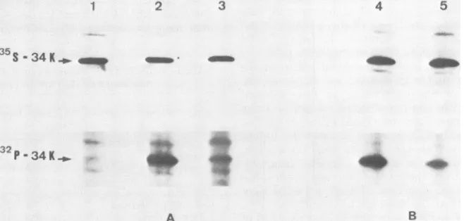
Phosphorylation of 34K protein. A 34K protein is phos-phorylated at both seririe and tyrosine residues in cells transformed by RSV and is considered to be a target for
p6Osr( kinase activity in vivo (7, 22). To study the relation-ship between the extent of phosphorylation of this protein and tumor formation,
[35S]methionine-
and[32P]P
-labeled cell extracts wereimmunoprecipitated with anti-34K protein serum(Fig. 3). In agreement with the data ofPoirieretal.on mutant-infected NR cells (20), we found that the extent of phosphorylation of 34K protein in PAIOI-infected CEFwas intermediate between that observed in RAV1-infected CEF and wt-transformed CEF. Similarly, the 34K protein re-mained under-phosphorylated in PAIOI-induced CAM tu-mors.DISCUSSION
We studied the oncogenic properties of two sr gene mutantsofRSV, PA101 and PA104, selected on NR cells for their ability to induce cell proliferation in the absence of
TABLE 4. Detection of plasminogen activator production in CEF maintained invitroand intumorexplants"
Plasminogen
Cells Virus activator
Infected CEF maintained RAV1 <1
invitro SR RSV-A 100
PA1(1 10
PA104 17.5
Cell culturesderived SR RSV-A 90
fromtumors PA101 12.5
"Plasminogenactivatorproductionwasdeterminedasdescribed inthetext.Results werenormalized accordingtothenumber of cells perdish andexpressedas apercentageof theactivitydetectedinthe mediumof SR RSV-A-transformed CEF maintained in vitro. Vol. 49. 1984
on November 10, 2019 by guest
http://jvi.asm.org/
[image:5.612.136.487.75.307.2] [image:5.612.319.560.597.693.2]330 POIRIER ET AL.
TABLE 5. Relative amounts of acid-stable phosphoamino acids in infected CEF maintained in vitro and in tumor explants"
Amtof phosphoaminoacid
Cells Virus andtumor
Phosphoserine Phosphothreonine Phosphotyrosine
InfectedCEFmaintained RAV1 93 7.3 0.jb
invitro SR RSV-A 89 8.6 1.03
PA101 91 9.0 0.39
PA104 91 8.6 0.38
Cell cultures derived SR RSV-A,tumor 1 87 12 0.87
fromtumors SRRSV-A, tumor 2 87 12 1.1
SRRSV-A,tumor 3 89 9.7 0.83
SRRSV-A,tumor 4 88 11 1.1
PA101, tumor 5 89 11 0.29
PA101, tumor6 93 6.1 0.49
PA101, tumor 7 91 8.7 0.41
PA101, tumor X 89 9.8 0.87
aCellswere labeled with
[32p]Pi
andthe relative amounts ofphosphoamino acids weredetermined. Cells explanted from four separate tumorsinducedafter inoculation of SRRSV-A-infectedCEF andfrom four separate tumors induced after inoculation ofPA101-infectedCEF wereanalyzed.bPhosphotyrosinespots in
RAV1-infected
cellscontainedca. 600 cpm whereas 60 cpm were detected in blanks (see text).morphological transformation. Concomitant with the disso-ciation of these src gene functions in mutant-infected NR
cells isamarked reductionin proteinkinase activity
associ-atedwith thep6Osrc proteins encoded by PA101 andPA104.
Onthe basisoftheresultsobtainedwith CAM of
embryonat-edduckeggs, weconcludethat, inspiteoftheirsimilarities,
the two mutants differ greatly in their tumorigenic
proper-ties: cells infected by PA101 are as tumorigenic as
wt-transformed cells at 37°C, whereas PA104-infected cells
exhibita sharply reducedtumorigenicity.
Analysis ofthe tumorigenic properties of PA104-infected cells on CAM at 37°C
paralleled
the results obtained withchicks, indicating thatPA104 ispoorly
oncogenic.
The few tumors obtained in chicks injected with this mutant dis-played elevated levels of p6Os''-associated protein kinaseactivity and released viruses which had acquired transform-ing properties similarto those ofwt SR RSV. Such rever-tantsprobablyaroseby back mutationsorby recombination
with homologous cellular sequences occurring during
sec-ondary infection in the
permissive
host (13).1
A comparison of the oncogenic properties of PA101 in
CAM and in chicks leads toapparently conflicting results. On the one hand, CEF and NR cells infected with PA101 were as tumorigenic as wt-infected cells when tested on
CAM at 37°C. Moreover, the viruses released by these tumors had retained the properties of the original mutant
indicatingthat, in this system, PA101 exhibitsanoncogenic potential identical to that of wt virus. On the other hand, incidence of sarcomas in chicks infected with PA101 was lower than thatofwtvirus. However, experimentsinwhich
the naturalpermissivehostisused to assay theoncogenicity ofPA101 sufferfromtwomain drawbacks. First,the subcu-taneoustemperatureofthechicken(38 to40°C)maylead to an underestimation of the tumorigenicity of this mutant, which bears a temperature-sensitive mutation in the src gene.Thismaybethe casesincetumorformationby
PA101-infectedcells on CAM wastemperature sensitive. Second,in contrast to theCAMtumors,itwas notpossibletoascertain
whether the tumors induced in chicks were due to the
original
mutant. The fact that immunoprecipitable kinase2 3 4
35S-34k
_5
_smil -4
32p-
34K_.
4qW
A B
FIG. 3. Immunoprecipitation ofradiolabeled 34K protein in cells maintained in vitro (A) and in tumor explants (B). Extracts from [35S]methionine- (top panel) or
[32P]Pi-
(bottompanel) labeled cells wereimmunoprecipitated with anti-34K proteinserum. Onlythe34K protein region of each gel is shown. Lanes: 1,RAV1-infected CEF; 2, SR RSV-A-infectedCEF; 3, PA101-infected CEF; 4, tumorcells derivedfromSR RSV-A-infectedCEF; 5, tumor cells derived fromPA101-infectedCEF.J. VIROL.
iftP'.
40
on November 10, 2019 by guest
http://jvi.asm.org/
[image:6.612.148.477.530.687.2]p60"' MITOGENIC FUNCTION AND TUMORIGENESIS 331 activity remained low in four of five tumorscausedbyPAIOI
suggested that, in these cases, tumor development was not related to changes in protein kinase activity of the mutant p60sc'. However, outof 10 tumors examined, the only virus
we could recover had acquired the transforming and
onco-genic properties ofawt virus.Therefore, webelieve that the
results obtainedbyinoculatingPA101-infected cellsontothe CAM ofembryonated duck eggs are best representative of
the tumorigenic properties of this mutant.
PA101 and PA104 have several properties in common. (i)
They both induce NR cell proliferation, (ii) cells infected
with these viruses are notmorphologically transformed and (iii) release lowamounts of plasminogen activator, (iv) their p60%SI proteins exhibit low kinase activity, and (v) 34K protein is under-phosphorylated in cells infected with either
mutant. Hence, the tumorigenic properties of PA101 cannot
be directly attributed toany of these phenotypic traits. Our
results differ from those of Kahn et al. (12), who found a
correlation between the phosphorylation of 34K protein and tumorigenicity in nude mice of CEF infected with RSV mutants. In contrast to PA104, PA101 has retained the abilityto induce cell growth in soft agar. Therefore, expres-sionof thisproperty appearstocorrelate with the
oncogenic-ity of this mutant.
Studies on tumorigenicity of mammalian cells have stressed the likelihood that tumor development maydepend
on continuous selection ofcell variants (15, 16). According-ly, Kahnet al. (12) have shown that tumorigenesis by RSV-infected cells in nude mice correlated with the selection of variants producingincreased levels of plasminogen activator relative to the corresponding cells before injection. In
con-trast,analysis ofseveral parametersin cultures derived from
tumorsinduced by PA101 onCAM did not pointto any such
selection of cell variants. First,tumorcells retainedanormal
morphology. Second, we did not find an overproduction of
plasminogen activator. Third, in three of four cases, the
relative amount ofphosphotyrosine in cells explanted from
PA101 tumors was comparable to that of infected cells before inoculation. Fourth, there was no increase in
phos-phorylation of34K protein in tumor-derived cultures. Ininitiating this work, we wishedtoassess the role of the
mitogenic function of p6O'"' in tumorigenicity and to estab-lish acorrelation between the levelsof p6O"' kinase activity
andtumorformation. Ourresults indicate that expressionof
the mitogenic property of p6O"' is not sufficient for tumor
induction, since PA104-infectedcellsarebarelytumorigenic.
They also suggest that tumor formation is compatible with
low levels ofprotein kinase activity, since PA101-infected
cellswerehighlyoncogenic,and thetumorstheyinduced did
not result from the selection of cell variants with increased p60src_associated kinase activity. Thus, the differences in tumorigenicity between PA101 and PA104 could be
ex-plained by assuming that p60"'( of PA101 has retained the ability to phosphorylate protein substrates specifically in-volved in tumorigenesis. Comparison ofphosphoproteins in
cellsinfected by the twomutants is now in progress.
ACKNOWLEDGMENTS
We are grateful to R. E. Karess and B. Mathey-Prevot for critically reading themanuscript. Wethank A. Leray for assistance in thepreparation of themanuscript.
This work was supported by grantsfrom Institut National de la Santeetde laRecherche Medicale(CRL 822003 and PRC 118001). from Ministere de la Recherche et de l'Industrie (ATP 3588). and fromtheFondationpourlaRechercheMedicaleFranqaise. F.P.was
alsosupported bythe Philippe Foundation.
LITERATURE CITED
1. Brugge, J. S., and R. L. Erikson. 1977. Identification of a
transformation-specific antigen induced by an avian sarcoma virus. Nature (London) 269:346-348.
2. Calothy, G., and B. Pessac. 1976. Growth stimulation of chick embryo neuroretinal cells infected with Rous sarcoma virus: relationship to viral replication and morphological transforma-tion. Virology 71:336-345.
3. Calothy, G., F. Poirier, G. Dambrine, P. Mignatti, P. Combes, and B. Pessac. 1980. Expression of viral oncogenes in differenti-ating chick embryo neuroretinal cells infected with avian tumor viruses. Cold Spring Harbor Symp. Quant. Biol. 44:983-990. 4. Calothy, G., F. Poirier, G. Dambrine, and B. Pessac. 1978. A
transformation defective mutantof Rous sarcoma virusinducing chick embryo neuroretinal cellproliferation. Virology 89:75-84. 5. Collett, M. S., and R. L. Erikson. 1978. Protein kinase activity associated with the avian sarcoma virus src gene product. Proc. Natl. Acad. Sci. U.S.A. 75:2021-2024.
6. Duff, R. G., and P. K. Vogt. 1969. Characteristics of two new avian tumor viruses. Virology 39:18-30.
7. Erikson, E., and R. L. Erikson. 1980. Identification of a cellular protein substrate phosphorylated by the avian sarcoma virus transforming gene product. Cell 21:829-836.
8. Goldberg, A. R. 1974. Increased protease level in transformed cells: a casein overlay assay for the detection ofplasminogen activator production. Cell 2:95-102.
9. Gorer, P. A., and P. O'Gorman. 1956. The cytotoxic activityof isoantibodies in mice. Transplant. Bull. 3:142-143.
10. Hanafusa, H. 1977. Cell transformation by RNA tumorviruses, p. 401-423. In H. Fraenkel-Conrat and R. R. Wagner (ed.), Comprehensive virology, vol. 10. Plenum Press, New York. 11. Hunter, T., and B. M. Sefton. 1980. Transforming geneproduct
of Rous sarcoma virus phosphorylates tyrosine. Proc. NatI. Acad. Sci. U.S.A. 77:1311-1315.
12. Kahn, P., K.Nakamura, S. Shin, R. E. Smith, and M. J. Weber. 1982. Tumorigenicity of partial transformation mutants of Rous sarcoma virus. J. Virol. 42:602-611.
13. Karess, R. E., W. S. Hayward, and H. Hanafusa. 1979. Cellular information in the genome of recovered avian sarcoma virus directs the synthesis of transforming protein. Proc. NatI. Acad. Sci. U.S.A. 76:3154-3158.
14. Levinson, A. D., H. Opperman, L. Levintow, H. Varmus, and J. M. Bishop. 1978. Evidence that the transforming gene of avian sarcoma virus encodes a protein kinaseassociated with a phosphoprotein. Cell 15:561-572.
15. Nowell, P. C. 1976. The clonal evolution of tumor cell popula-tions. Science 194:23-28.
16. Perez-Rodrigues,R., A. Franchi, B. F. Deys, and J. Pouyssegur. 1982. Evidence that hamster fibroblast tumors emerge in nude mice through the process of two in vivo selections leading to growth factor "relaxation' and to immune resistance. Int. J. Cancer 29:309-314.
17. Pessac, B., and G. Calothy. 1974. Transformation of chick embryo neuroretinal cells by Rous sarcoma virus in vitro: induction of cell proliferation. Science 185:709-710.
18. Pessac, B., and V. Defendi. 1972. Cell aggregation. Role of acid mucopolysaccharides. Science 175:898-900.
19. Poirier, F., D. Lawrence, P. Vigier,and P. Jullien. 1982. A ts-T mutant ofSchmidt-Ruppin strain of Rous sarcoma virus restrict-ed at 39.5°C for the morphological transformation and the tumorigenicity of chicken embryo fibroblasts. Int. J. Cancer 29:69-76.
20. Poirier, F., G. Calothy, R. Karess, E. Erikson, andH. Hanafusa. 1982. Role ofpp60"`kinaseactivity in the induction of neurore-tinal cellproliferation by Rous sarcoma virus. J. Virol. 42:780-789.
21. Poste, G., and M. K.Flood. 1979. Cellstransformed by tempera-ture-sensitive mutants of avian sarcoma virus cause tumors in vivo at permissive and nonpermissive temperatures. Cell 17:789-800.
22. Radke, K., and G. S. Martin. 1979. Transformation by Rous sarcoma virus: effects of src gene expression on the synthesis andphosphorylation of cellular polypeptides. Proc.Natl. Acad. VOL.49, 1984
on November 10, 2019 by guest
http://jvi.asm.org/
332 POIRIER ET AL.
Sci. U.S.A. 76:5212-5216.
23. Scher, C. D., C. Haudenschild, and M. Klagsbrun. 1976. The chick chorioallantoic membraneas amodelsystemfor the study of tissue invasion by viral transformed cells. Cell 8:373-382. 24. Sefton, B. M., T. Hunter, K. Beemon, and W. Eckhart. 1980.
Evidence that the phosphorylation of tyrosine is essential for cellular transformation by Roussarcomavirus.Cell 20:807-816.
25. Unkeless, J. C., A. Tobia, L. Ossowski, J. P. Quigley, D. B. Rifkin, and E. Reich. 1973. Anenzymatic function associated with transformation of fibroblasts by oncogenicviruses.J.Exp. Med. 137:85-111.
26. Vogt, P. K. 1977. The genetic of RNA tumor viruses, p.
341-455. In H. Fraenkel-Conrat and R. R. Wagner (ed.). Compre-hensivevirology, vol. 9. Plenum Press, New York.
J. VIROL.