JOURNALOFVIROLOGY, Mar. 1988,
p.
1071-1075 0022-538X/88/031071-05$02.00/0Copyright
© 1988, American
Society
for Microbiology
Identification
of the E5 Open Reading Frame of
Human Papillomavirus Type 16
CHRISTINE
L.HALBERT
ANDDENISE
A.GALLOWAY*
Fred Hutchinson
Cancer Research Center, 1124 Columbia Street, Seattle, Washington 98104, andDepartmentofPathology, University of Washington, Seattle, Washington
98105Received 28August 1987/Accepted 23 November 1987
Sequencing of the
E5
openreading frame (ORF) ofhuman papillomavirus type 16 revealedanadditionalnucleotide,athymidine residue,atposition 3903 compared with theoriginalsequence(Seedorfetal., Virology 145:181-185, 1985). The additional T hadtwoeffects;first, in reading frame 2, in which the original
E5
ORFwas predicted, the additional T changed the reading frame downstream of position 3903to create anORF,
which we designated E5*, that terminated at position 4018 and potentially encoded a 52-amino-acid
polypeptide. Secondly, in reading frame3,anewORFwascreated(positions 3807to4097), whichwepropose
is theauthenticpapillomavirustype 16
E5
ORF. Itcontainedamethionine residue and encodedanadditional82amino acids. BothORFs have been cloned into bacterial expressionvectors(pATH), and the fusion proteins havebeen used to generatepolyclonal antibodies in rabbits.
Human papillomavirus (HPV) DNA has been found in
manyhumanneoplasias of
squamousepithelial origin
(4, 8,11,
12),
andasubset of viral
genescontinues
tobe expressed
in thetumors(7,
15, 20, 22, 24, 29).
Inaddition,
both animal
(27)
and human(14, 16, 26, 28) papillomaviruses have been
shown totransform cells in culture.Together,
theseobser-vations
suggestthat HPVs have
arole in the
etiology of
somecancers. HPV type 16
(HPV-16),
a strain that has been shownto beassociated
with invasive cervicalneoplasia (8,
9),
has agenomic
sizeof
7,904 base pairs (23).
Recombinant
DNAtechnology
has been useful in thestudy
of HPV since these viruses so far have not beenpropagated
intissue culture.
Comparative
sequenceanalysis
of HPV and animalpapillomavirus
genomes have shownanoverall
genetic
relatedness(6,
21).
The genome is divided intoanearly region
that containsseven oreight
openreading
frames
(ORFs)
andalateregion
thatcontainstwoORFs,
Li andL2, coding
forcapsid proteins.
Various viralgeneshavebeen
identified
primarily
asORFsfrom nucleotide
sequencedatafor cloned viral DNA. These
ORFs
have been cloned into bacterial expression vectors, and the recombinant fu-sionproteins
have been used asimmunogens
to generate antibodies todetect
authentic viralproteins (2,
19, 25).
We have
cloned
andexpressed
allof the
ORFs
of HPV-16 to look forexpression
of variousputative
viralproteins
in clinicalspecimens
and in in vitro studies(10).
It isof interest tolook forE5 expression
because theE5 protein
of bovinepapillomavirus
type 1(BPV-1)
has been shown to havetransforming activity (18, 19). Although
evidence suggests that the E6 and E7ORFs of
HPV have a function intransformation
(1,
3, 25),
arole forE5
in HPV infections hasyettobedefined.
The DNA
fragment
of HPV-16from position 3877
to4284 obtainedfromanSfaNI
restrictiondigest
wascloned into the SmaI site ofpUC18
as shown inFig.
1. Thisfragment
containedthemajor portion
of theE5
ORF(3863
to4097)
asdefined
by
thepublished
sequence(23). Cloning
of thisE5
ORF to express afusionprotein
was doneby
isolating
the DNAfragment
from thepUC hybrid
clonefollowing
diges-tion with EcoRI andHindlIl,
enzymes which cleave within* Correspondingauthor.
the
polylinker
of thevector.Thefragment
wasligated
tothe
EcoRI-Hindlll-digested pATH1 expression
vector(gift of
T.J.
Koerner)
anddesignated
pl6E5sfl. The ligated
DNAwas
transfected into Escherichia coli HB101 cells. The
plasmid
wassequenced
toverify
thatthe
fragment
wasinserted
inthe
correctreading frame and
to confirm the nucleotide sequence of the geneencoding
theprotein.
Se-quencing of the plasmid
wasdoneby
thedideoxy
nucleotidetriphosphate
chain termination method(17)
withdenaturedplasmid
DNAas atemplate
(5),
withaprimer hybridizing
toa sequence 5' of the
multiple cloning
site in thepATH
vector.Analysis
of the sequence revealed that there werefour T's
starting
atposition 3903,
instead of the threereported
in thepublished
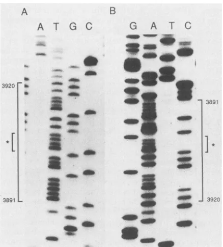
sequence(23). Figure
2Adisplays
thefour T's in thesequencing gel.
Aprimer
complementary
tosequencesof HPV-16 located 5' of theE5
ORF(primer
1,
nucleotides3821
to3839)
wasusedtodirectly
sequencethe HPV-16 DNAplasmid
obtained from H. zur Hausen. Thisconfirmed the presence of the four T's in the
original
pHPV16 clone (data
notshown).
To obtain sequence from the otherstrand,
aprimer complementary
to HPV-16se-quences located 3' of the
E5
ORF(primer 2,
nucleotides 4107 to4122)
wassynthesized. Figure
2Bdisplays
the four A's onthecomplementary
strand.The additional T at
position
3903 had two effects.First,
theoriginal
E5 ORF,
with the additionalT,
encoded aprotein
of 52 amino acids rather than theoriginally predicted
72 amino acids.Second,
the additional T created another ORFbeginning
atposition
3807andterminating
at4097,
and it encoded a methionine codon atposition
3849. This ORFoverlapped
theoriginal
E5
ORF and extended further up-stream. The newE5
ORFwas in a differentreading
frame andpotentially
encoded apolypeptide
of 83 amino acidsstarting
at the methionine residue.Figure
3 shows the size and location of the initialE5
ORF describedby
Seedorfetal.(23),
the effect of the additional Ton thatORF, designated
E5*,
and the newE5 ORF,
which contains a methionine residue. A further effect of the additional Twasthat it shiftedthe
reading
frame of the L2 and LI ORFs relative to the otherORFs. The fact that thenewORF contained a methi-onineresidue,
asdo theE5
ORFs of the otherpapillomavi-ruses, while the
previously
designated
onedidnot, suggests1071
Vol.
62,
No. 3on November 10, 2019 by guest
http://jvi.asm.org/
1072 NOTES
5,
3877
'-
3
DNA
Frogment
4284
Contoining
E5
polylinker
FIG. 1. Construction of hybrid clones pl6E5sfl andp16E5sfll. A pBR plasmid containing HPV-16 (pHPV16, originally from H. zur Hausen) wasdigested with SfaNI restriction enzyme. The location of the DNA fragment containing E5 is indicated by its nucleotideposition in the genome of HPV-16. This fragment was filled in by Klenow polymerase and inserted into the
SmaI
site of pUC18. E. coliJM103 was transfected withthe ligation product, and white colonies were screened by restriction digests followed by nucleotide sequence analysis across theligation junction. The pUC hybrid clone was subsequently digested withEcoRI andHindlIl,and the DNA fragment containingE5 was ligatedtosimilarlydigested pATH1 (designated pl6E5sfl) or pATH11 (pl6E5sfll) DNA.that the ORF located at positions 3807 to 4097 is
theauthentic
E5
ORF. However, biochemical or genetic
data areneeded to
confirm this prediction. It is also possible that
coding secquences of the E5* ORF are utilized as part of
aspliced
mR1NA.
Cloning
of
aportion of the new E5 ORF for the production
of
afusion
protein was
done
similarly to the cloning of the
old E5
ORF. The DNA fragment obtained from digestion of
the
pUC hybrid
clone was
ligated
to
EcoRI-HindIII-digested
pATH11 and designated pl6E5sfl1.
Induction of the trpE fusion proteins and the preparation
of
salt-insoluble
fractions
was
done as described previously
(10), and the products were analyzed on sodium dodecyl
sulfate
(SDS)-polyacrylamide
gels. Both E5 ORF fusion
proteins were expressed at very low abundance (data not
shown) compared with other HPV-trpE fusion proteins (10).
The
fusion
protein expressed from pl6ESsfl showed a
protein which
wasapproximately
4.0
kilodaltons (kDa)
larger
than the 37
kDa of
the trpE moiety, which correlated
closely with
the
predicted
size of 5
kDa.
The
fusion protein
expressed by
pl6ESsfl
corresponded to
asegment
of the
E5*
ORF
(Fig.
3). The fusion
protein expressed
from
pl6ESsfll should encode
an
HPV
moiety of
8
kDa;
how-ever,
additional proteins
2
kDa
larger
than trpE and
of higher
molecular weight
were seen.
The
fusion protein expressed
by pl6ESsfll
corresponded to
asegment
of the newly
designated
E5
ORF (Fig. 3). To visualize the E5 fusion
proteins
more adequately, a Western blot (immunoblot)
containing
both uninduced and
induced bacterial lysates as
well as the salt-insoluble fractions were reacted with an
antibody
which detected the trpE moiety (a gift of J. Firzlaff)
(Fig.
4).
This analysis indicated that
pl6ESsfl
expressed a
41-kDa
protein, as expected. The plasmid
pl6E5sfll
showed
atrpE
fusion protein migrating somlewhat slower than the
trpE
protein alone and another protein of 78 kDa. The most
likely
explanation for the aberrant mobility of the fusion
protein
is that the highly hydrophobic residues encoded by
E5
causethis
behavior in SDS gels. The
higher-molecular-A
A T
G C
3920
*[
3891
B
G
A
T
C
_M
-Ww4*
am_
_AN
_-a,4
_
40
.a.. _7
~~~~~
_* _~~~~~~*
4
-3891
]
3920
FIG. 2. Analysisof the nucleotide sequence of HPV-16 E5ORF. Dideoxy nucleotide sequencing of hybrid clones pl6E5sfl and pHPV16 was done with
32P-labeled
primers and double-stranded DNA templates. Denaturation of thetemplateandextension reac-tions were done by standard procedures (5, 17). Panels show reactionproductsrun on an8% polyacrylamide-SDS denaturinggel, with thenucleotide lanes indicated at the top of the gel. (A) Portion of the sequence for the sense strand from positions3891 to 3920 sequencedwithapATH primerandpl6E5sfl. (B) Complementary sequence on thereverse strand sequenced by using primer2 andpHPV16.
Asterisksdesignate positionof the four T's and four A'sonthe DNA strands.
J.VIROL.
on November 10, 2019 by guest
http://jvi.asm.org/
[image:2.612.141.471.65.260.2] [image:2.612.325.544.364.607.2]I
LI
JL
IUJI~
1
&727
i5
-I
r
.m
13 4 5
?ITNDTASTTLLMCFLLC= LCYCLLIRPLLLSVSTYTSLIILVLLLMITAASAFEWFIVYIIFVYIPILLIHTHARFLIT
E5 ORF
4097
'Nat 3949 ladditioal T at 3903
YCIHNIT¶ ALLLTCAFVCLPINTSARFCVYIHIIMIGITIVDNSSICV
2
2
E5*
ORF sa3863 4018
additional T at 393
YCIHNITGVLFAIVLICWVCLLIRPLLLSVSTYTSLIILVLLLWITAASAFWZFIVIIFVIPTFL-HTHARLT
E5
ORF
of Sedorf et al7891
3863 4096
3900 3900 4000 4100
FIG. 3. The E5ORF(3863to4096) described
by
Seedorfetal.(23) is shownbelow,
asis the aminoacidsequenceitpredicts.
Theinsertion ofanadditionalTinto thatreading framegeneratestheORF designated E5*, with the amino acidsequenceitpredicts shown above.Inreading frame3theadditionalT creates the newE5 ORF (3807 to 4097)which encodesamethionineatposition 3849. The additionalTchangesthe reading frame of theE5, L2,andLi
ORFs relativetothe otherpredictedORFs ofHPV-16, asshown inthetoppanel.aa,Amino acids.A pATH 1 p16E5sil pATH1 pl6E5sfl B pATH 1 1 pl6E5sfl'
D _
i_ _
D - D
-J iJ -J LL E. U.
¢: 3' 3' ¢: I: m 0a
_ 97,400
68.000
43 COO
_o
*-* 8.3vO
5. 00
- 25,700
18.000
FIG. 4. Western blotanalysis of the E5 fusion proteins with polyclonal rabbit antisera with reactivitytothetrpE moiety. Clonespl6E5sfl andpl6E5sfllwereinduced toproduce the E5* and E5 fusion proteins, respectively. Induction and purification of proteinsweredoneas
describedpreviously (10). Briefly, overnight cultures of bacteriawerediluted 1:10 in 50ml of M9 medium andgrownfor 1to2 h. For induction (I), idoleacrylic acidwasadded andculturesweregrownfor another 4 h. Uninduced (UI) culturesweregrownfor 6 h in mediumcontaining tryptophan. Cellswerepelleted, and whole bacteriallysates(WBL; 0.1 mlequivalent of culture) and high-salt-insoluble fractions (HSIF: 1 mlequivalent of culture) were'runon a 12.5% polyacylamide-SDS gel and transferredtonitrocellulose. The blot
was-
reactedtoantisera previously absorbed with E. coliHB101'bacteriallysates.(A) Induced trpEprotein from pATHl and the induced fusion protein from'pl6E5sfl (arrow). The induced proteinwasapproximately 41 kDa,or4kDalarger than trpE. (B) Induced trpE protein frompATH1l,whichseemsto be lesshigh-salt insolublethantrpE frompATH1, and the induced fusion protein for pl6E5sfll (indicatedwithanarrow). Molecuilar weights areshowntotheright.1073
RXg
Frame
1
2
3
0 3
27 h1al
r
_J
I 2
1 2
3
3807L
6 7 7905
pA-'H1.' vzEm
I i~~~
!C
;1.8*, A. A
Ago I
on November 10, 2019 by guest
http://jvi.asm.org/
[image:3.612.116.510.55.361.2] [image:3.612.127.486.401.638.2]1074 NOTES
BPV 1: MPNLWFLLFL GLVAAVMLLL LLFLLLFFLV YWDHFECSCT GLPF
HPV 6b: MEVVPVQIAA GTTSTFILPV IIAFVVCFVS IILIVWISEF IVYTSVLVLT LLLYLLLWLL LTTPLQFFLL TLLVCYCPAL
YIHYYIVTTQ
QHPV16: MTNLDTASTT LLACFLLCFC VLLCVCLLIR PLLLSVSTYT SLIILVLLLW ITAASAFRCF IVYIIFVYIP LFLIHTHARF LIT
HPV18: MLSLIFLFCF CVCMYVCCHV PLLPSVCMCA YAWVLVFVYI VVITSPATAF TVYVFCFLLP MLLLHIHAIL
SLQ
FIG. 5. Amino acid sequence of papillomavirus ES ORFs. The amino acid sequences are
displayed
with thehydrophobic
residues underlined.Acomparison of amino acid residues between theE5ORFsof BPV-1 and HPV-6B, -16, and-18shows that thesimilarity resides primarily in the large number of hydrophobic residues (approximately60%),
of whichasignificant portion areleucine residues(20%
for HPV-18, 30% for HPV-16 and -6B, and 50% for BPV-1).weight form
presumably
results from
incomplete
denatur-ation in SDS.
Rabbits were
immunized
with the
high-salt-insoluble
frac-tions of
induced bacterial
cultures to
produce
antibodies
to
both
E5
ORFs.
The
antibodies
produced
will be
used to
identify
the
authentic viral proteins in
clinical
specimens and
in in vivo
and
in
vitro viral expression
systems.
The
E5
protein
of
BPV-1 has
been shown
to
be
highly
hydrophobic (68%),
and
34% of the amino acids
are
leucine
(19).
Figure
5
displays
the
amino acid
sequence
for
the
newly
designated
E5 ORF of HPV-16
in
comparison with
other
papillomaviral
E5
ORFs.
The
E5*
ORF
was
not
closely
related
to
the other
E5
ORFs.
A
comparison of
the
amino
acid
sequences
of
the
E5
ORFs from
HPV-6b,
-16, and -18
and
BPV-1
revealed
that
the
homology resided
primarily
in
the
large number of hydrophobic residues
(approximately
60%), of which
many were
leucines (20
to
30%).
The new
HPV-16 E5
ORF, like
the
other E5
ORFs, contained
a
methionine
at
its amino terminus
and
Cys-X-Cys residues
interspersed
in a stretch
of
hydrophobic amino acids.
Al-though the
new
HPV-16
E5
polypeptide
had these
proper-ties,
the
distribution of
a
long
stretch
of
hydrophobic
resi-dues
at
the
5' end
with
some
charged residues
and a
Cys-X-Cys
at
the
3' end did
not
occur
in
it,
as
they do
in
the
BPV-1
E5
ORF.
Therefore, the function of
E5
in HPV-16
could be
different from its
transforming function in
BPV-1.
In
summary,
DNA sequence
analysis of
a
segment
of
HPV-16 DNA revealed an
additional nucleotide
at
position
3903.
This
insertion
created a new
ORF which contained
a
methionine residue followed by
82
amino
acids,
and
presum-ably
encoded the
authentic
E5
ORF.
The
identification of
the
E5
ORF is extremely important
in the
design
of
fusion
proteins
or
synthetic
peptides
which
will be used as
immu-nogens to create
HPV-16
E5
antibodies.
We thankMargaretSwain andJanette Valentine for
assistance,
Juliane Firzlaff and Steve Jenison forhelpful suggestions, and Marci Wright for typing this manuscript.This workwas supported by Public Health Service grants P01-CA42792 andR01-CA35568 from the National Cancer Institute.
LITERATURE CITED
1. Androphy, E. J., N. L. Hubbert, J. T.Schiller,and D.R.Lowy. 1987.Identification ofthe HPV-16 E6protein from transformed mousecells and humancervical carcinomacelllines.EMBO J. 6:989-992.
2. Androphy,E.J., J.T.Schiller,andD. R.Lowy. 1985. Identifi-cation of theprotein encodedby the E6 transforminggeneof bovine papillomavirus. Science230:442-445.
3. Baker, C. C., W. C. Phelps, V. Lindgren, M. J. Braun, M. A. Gonda,and P. M. Howley. 1987. Structuralandtranscriptional analysisof humanpapillomavirustype
16
sequencesincervical carcinoma cell lines.J. Virol. 61:962-971.4. Boshart, M., L. Gissmann, H. Ikenberg, A. Kleinheinz, W.
Scheurlen,and H.zurHausen. 1984. Anewtype of papilloma-virus DNA and its presence ingenitalcancerbiopsies and in cell lines derived from cervicalcancer. EMBO J.3:1151-1157. 5. Chen,E.Y., andP. H.Seeburg. 1985. Supercoilsequencing:a
fast and simple method of sequencing plasmid DNA. DNA 4: 165-170.
6. Danos, O., L. W. Engel, E. Y. Chen, M. Yaniv, and P. M. Howley. 1983. A comparative analysis of the human type la and bovine type 1 papillomavirus genomes. J. Virol. 46:557-566. 7. Doorbar, J., D. Campbell, R. J. A. Grand, and P. H. Gallimore.
1986. Identification of human papillomavirus-la E4 gene
prod-ucts. EMBO J. 5:355-362.
8. Durst, M., L.Gissmann, H.Ikenberg, and H.zurHausen.1983. A papillomavirus DNA from a cervical carcinoma and its prevalence in cancer biopsy samples from differentgeographic regions. Proc. Natl. Acad. Sci. USA 80:3812-3815.
9. Durst, M., A.Kleinheinz,M.Hotz,and L.Gissmann. 1985. The physical state of human papillomavirus type 16 DNA inbenign andmalignant genital tumors. J. Gen. Virol. 66:1515-1522. 10. Firzlaff, J. M., C. N. L.Hsia,C. L.Halbert,S. A.Jenison,and
D. A. Galloway. 1987. Polyclonalantibodies tohuman papillo-mavirustype 6b and type 16bacterially derived fusion proteins, p. 105-113. In B. M. Steinberg, J. L. Brandsma, and L. B. Taichman (ed.), Cancer cells 5: papillomaviruses. ColdSpring HarborLaboratory,Cold SpringHarbor, N.Y.
11. Gissmann, L., L. Wolnik, H. Ikenberg, U. Koldovsky, G. Sch-nurch, and H. zur Hausen. 1983. Humanpapillomavirus types 6 and 11 DNA sequences in genital and larynegeal papillomas and in some cervical cancers. Proc. Natl. Acad. Sci. USA 80:560-563.
12. Gissmann,L., and H. zur Hausen. 1980. Partial characterization of viral DNA from humangenitalwarts (condylomata acumi-nata). Int. J. Cancer25:605-609.
13. Kaur,P., andJ.K.McDougall. 1987. Transformation ofmouse cells by HPV-6b DNA and HPV-18 DNA, p. 249-252. In B. M. Steinberg, J. L.Brandsma, andL. B. Taichman(ed.), Cancer cells 5:papillomaviruses.ColdSpringHarborLaboratory,Cold SpringHarbor,N.Y.
14. Kreider, J. W., M. K.Howlett, N. L.Lill,G. L.Bartlett,R.J. Zaino, T. V. Sedlacek, and R. Mortel. 1986. In vivo transforma-tion of human skin with human papillomavirus type 11 from condylomataacuminata. J. Virol. 59:369-373.
15. Lehn, H., P.Krieg,andG. Sauer. 1985.Papillomavirusgenomes in human cervical tumors: analysis of their transcriptional activity. Proc. Natl. Acad. Sci. USA 82:5540-5544.
16. Pirisi,L., S.Yasumoto,M.Feller,J.Doniger,andJ.A.DiPaolo. 1987. Transformation of human fibroblasts and keratinocytes with human papillomavirus type 16 DNA. J. Virol. 61:1061-1066.
17. Sanger,F.,S.Nicklen,andA.R.Coulson. 1977. DNA sequenc-ing with
chain-terminating
inhibitors. Proc. Natl. Acad. Sci. USA74:5463-5467.18. Schiller, J. T., W. C. Vass, K. H. Vousdan, and D. R. Lowy. 1986. TheE5open
reading
frameofbovinepapillomavirus
type 1encodesatransforminggene. J. Virol. 57:1-6.19. Schlegel, R., M. Wade-Glass, M. S. Rabson,and Y. C.Yang. 1986. The E5
transforming
gene ofbovinepapillomavirus
en-codes asmallhydrophobicpolypeptide.Science 233:464 467. J. VIROL.
on November 10, 2019 by guest
http://jvi.asm.org/
NOTES 1075
20. Schneider-Gadicke, A., and E. Schwarz. 1986. Different human cervical carcinoma cell lines show similar transcriptionpatterns
of humanpapillomavirustype18 earlygenes.EMBO J. 5:2285-2292.
21. Schwarz, E., M. Durst, C. Demankowski, 0. Lattermann, R.
Zech, E. Wolfsperger, S. Suhai, and H.zurHausen. 1983. DNA
sequenceandgenomeorganization of genital human
papilloma-virustype6b. EMBOJ. 2:2341-2348.
22. Schwarz, E., U. K. Freese, L. Gissmann, W. Mayer,B.
Roggen-buck, A. Stremlau, and H. zur Hausen. 1985. Structure and
transcription of human papillomavirus sequences in cervical carcinoma cells. Nature(London) 314:111-114.
23. Seedorf, K., G. Krammer, M. Durst, S. Suhai, and W. Rowe-kamp. 1985. Human papillomavirus type 16 DNA sequence.
Virology 145:181-185.
24. Seedorf, K., T. Oltersdorf, G. Krammer, and W. Rowekamp. 1987. Identification ofearly proteins of the human papilloma viruses type 16 (HPV 16) and type 18 (HPV 18) in cervical
carcinoma cells. EMBO J. 6:139-144.
25. Smotkin, D., and F.0.Wettstein.1986.Transcription of human
papillomavirus type 16earlygenes inacervical canceranda
cancer-derived cell line and identification of the E7 protein. Proc.Natl. Acad. Sci. USA 83:4680-4684.
26. Watts, S. L., W. C. Phelps, R. S. Ostrow, K. R. Zachow, and A.J. Faras.1984. Cellular transformation by human papilloma-virus DNA invitro. Science 225:634-636.
27. Yang, Y.-C., H. Okayama, and P. M. Howley. 1985. Bovine papillomavirus contains multiple transforming genes. Proc. Natl. Acad. Sci. USA82:1030-1034.
28. Yasumoto,S., A. L. Burkhardt, J. Doniger, and J. A. DiPaolo. 1986. Human papillomavirustype 16 DNA-induced malignant transformation of NIH 3T3 cells. J. Virol. 57:572-577. 29. Yee, C., I. Krishnan-Hewlett, C. C. Baker, R. Schlegel, and
P. M. Howley. 1985. Presenceandexpression of human papil-lomavirus sequences in human cervical carcinoma cell lines. Am.J. Pathol. 119:361-366.
VOL.62, 1988