A PROSPECTIVE STUDY COMPARING ACROMIO
AXILLO SUPRASTERNAL NOTCH INDEX AND
MODIFIED MALLAMPATI TEST IN PREDICTING
THE DIFFICULTY IN VISUALIZATION OF LARYNX
Dissertation submitted in partial fulfillment of the regulations for the award of the degree of
M.D. DEGREE, BRANCH – X ANESTHESIOLOGY
Of
TAMIL NADU Dr. M.G.R. MEDICAL UNIVERSITY CHENNAI, TAMILNADU
ESIC- MEDICAL COLLEGE & POSTGRADUATE INSTITUTE OF MEDICAL SCIENCE AND RESEARCH,
KK NAGAR, CHENNAI- 78.
THE HEAD OF THE INSTITUTION
This is to certify that this dissertation titled “A PROSPECTIVE STUDY COMPARING ACROMIO AXILLO SUPRASTERNAL NOTCH INDEX AND MODIFIED MALLAMPATI TEST IN PREDICTING THE
DIFFICULTY IN VISUALIZATION OF LARYNX” submitted by
Dr.P.Ravindra Kumar, appearing for M.D Degree Branch – X ANESTHESIOLOGY examination in April 2016 is a bonafide record of
work done by him in partial fulfillment of the regulations of Tamilnadu
Dr. M.G.R Medical University, Chennai. I forward this to the
Tamilnadu Dr. M.G.R Medical University, Chennai Tamilnadu, India.
DEAN
Dr. SRIKUMARI DAMODARAM, M.S.,M.Ch(SGE), M.A.M.S.,
F.A.C.S., F.I.C.S., F.M.M.C
ESIC Medical College and PGIMSR
K.K. Nagar, Chennai – 78.
DATE:
This is to certify that dissertation entitled
“A PROSPECTIVE
STUDY COMPARING ACROMIO AXILLO SUPRASTERNAL NOTCH INDEX AND MODIFIED MALLAMPATI TEST IN PREDICTING THE DIFFICULTY IN VISUALIZATION OF LARYNX” is a bonafide research
work done by Dr.P.RAVINDRA KUMAR, in partial fulfillment of the requirement for the degree of M.D. in Anesthesiology
Signature
DATE: Prof.S.Gayathri, M.D.,D.A.,
PLACE: KK Nagar Professor and HOD,
Department of Anesthesiology
ESIC MC & PGIMSR
This is to certify that the dissertation named “A PROSPECTIVE STUDY COMPARING ACROMIO AXILLO SUPRASTERNAL NOTCH INDEX AND MODIFIED MALLAMPATI TEST IN PREDICTING THE DIFFICULTY IN VISUALIZATION OF LARYNX” is a bonafide work
performed by Dr.P.Ravindra Kumar, postgraduate student, Department
of Anesthesiology, ESIC Medical college & PGIMSR, Chennai – 78,
under my guidance and supervision in fulfillment of regulations of The
Tamilnadu Dr. M.G.R Medical University for the award of M.D. Degree
during the academic year 2014 – 2016.
GUIDE: CO-GUIDE:
Dr. Ilango Ganesan, M.D., Dr. K. Radhika, M.D.,
Associate Professor, Associate Professor,
Department of Anesthesiology, Department of Anesthesiology,
ESIC Medical College & PGIMSR, ESIC Medical College & PGIMSR,
I solemnly declare that this dissertation entitled “A PROSPECTIVE STUDY COMPARING ACROMIO AXILLO SUPRASTERNAL NOTCH INDEX AND MODIFIED MALLAMPATI TEST IN PREDICTING THE DIFFICULTY IN VISUALIZATION OF LARYNX” has been conducted by me at ESIC Medical College & PGIMSR,
Chennai, under the guidance and supervision of Dr. ILANGO GANESAN, M.D., and Dr.K.RADHIKA, M.D., Department of Anesthesiology, ESIC Medical College & PGIMSR, Chennai. This
dissertation is submitted to The Tamil Nadu Dr. M.G.R. Medical University, Chennai in partial fulfillment of the University regulations for the award of the degree of M.D. Branch X (Anesthesiology).
Date :
It is my immense pleasure to thank everybody who contributed in
compilation of this study.
I express my deep debt of gratitude to our respected Dean
Dr.Srikumari Damodaram, M.S., M.Ch (SGE), M.A.M.S., F.A.C.S., F.I.C.S., F.M.M.C., for permitting me to carry out this study.
I would like to thank Prof. Kamalini Sridharan, M.D., D.A, former HOD, Department of Anesthesiology, ESIC MC & PGIMSR for
her immense support and encouragement for the study.
I express my deep sense of gratitude and indebtedness to my
guide Dr.Ilango Ganesan, M.D., for suggesting the topic and for his scholarly guidance, dynamic interest and clinical acumen. His
considerable time and effort enabled me to give this study its ultimate
shape.
I acknowledge my heartfelt gratitude to my co-guide
Dr.K.Radhika, M.D., who provided valuable suggestions, guidance and encouragement to help me do better.
I am grateful in every possible way to the Assistant professors,
Specialists, Medical Officers, Senior Residents and the postgraduates of
Patil, Ph.D., for her guidance regarding the sample size and data analysis.
I am thankful to the Institutional Ethical Committee for their
guidance and approval for the study.
I also thank the theatre staff nurses and technicians for their help.
I would fail in my duty if I forget to thank all the patients for their
participation and co-operation.
I am thankful to my affectionate mother, beloved wife for their
constant support and encouragement, my daughters for their
unconditional love, all my family members and friends for their love
and support throughout my postgraduation.
S.NO. TITLE PAGE NO.
1 INTRODUCTION 1
2 AIM AND OBJECTIVES 6
3 REVIEW OF LITERATURE 8
4 HISTORY OF AIRWAY
MANAGEMENT
32
5 AIRWAY ANATOMY 36
6 AIRWAY ASSESSMENT 40
7 AIRWAY MANAGEMENT 44
8 GENERAL ANESTHESIA 45
9 MATERIALS AND METHODS 51
10 OBSERVATION AND RESULTS 59
11 DISCUSSION 75
12 SUMMARY 82
13 CONCLUSION 84
BIBLIOGRAPHY
TITLE :
A prospective study
comparing
Acromio
axillo
suprasternal
notch
index
and
Modified
mallampati
test
in
predicting
difficulty
in
visualization of larynx.
SUBMITTED BY:
DR. P. RAVINDRA KUMAR
GUIDE :
DR. ILANGO GANESAN, MD
CO- GUIDE :
DR.K.RADHIKA, MD
DEPT. OF
ANAESTHESIOLOGY, ESIC MC AND PGIMSR, CHENNAI 78.
EMAIL :
BACKGROUND:
The management of the airway is the primary responsibility of the anesthesiologist. Endotracheal intubation remains the gold standard in maintaining definitive airway and is conventionally facilitated by direct laryngoscopy. The Modified mallampati (MMP) test is a standard tool in the airway assessment and its low sensitivity is always a deterrent. Kamranmanesh MR et al tested a new simple bedside screening tool called Acromio Axillo Suprasternal notch index (AASNI) for airway assessment.
AIM:
To compare the AASNI and MMP in predicting the “Difficult visualization of the larynx”.
METHODS:
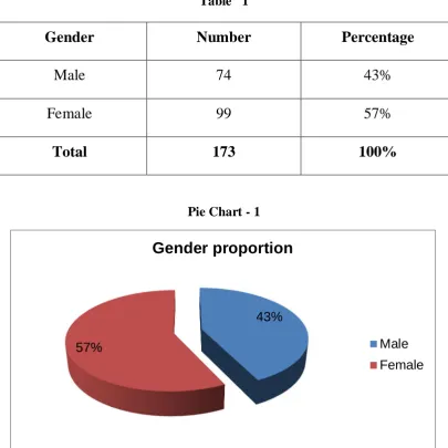
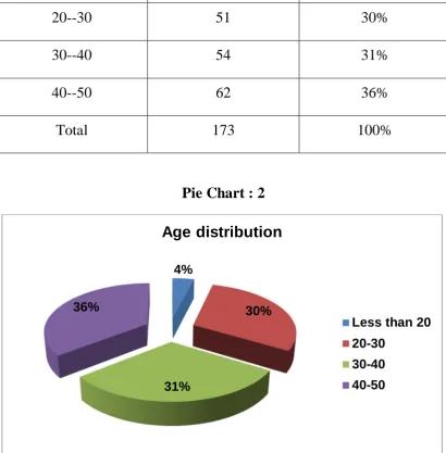
A total of 173 adult patients of ASA 1 and 2 (18 to 50 years), who were candidates for tracheal intubation in elective surgery were enrolled in this prospective observational study. Preoperative airway assessment was carried out with AASNI and MMP. After induction of anesthesia, direct laryngoscopy was done and the laryngeal view was recorded according to the Cormacke Lehane grading system.
+
RESULTS:
The validity of AASNI and MMP are analysed with Open epi (ver.2), SPSS software (ver.16) and Microsoft excel. The sensitivity, specificity, positive predictive value, negative predictive value of AASNI vs MMP are 70.5 vs 29.4, 84.6 vs 95.5, 33.3 vs 41.6, and 96.3 vs 92.5 % respectively. Thus, AASNI had better sensitivity and negative predictive value than MMP but lower specificity and positive predictive value than MMP.
CONCLUSION:
This study concludes that AASNI can be used as a predictive tool
for ‘Difficult visualization of larynx’ (DVL). The higher sensitivity of
AASNI makes it a better tool than MMP for screening DVL. As no single test predicts DVL precisely, AASNI can be used in conjunction with standard tool like MMP to increase the validity. AASNI may be investigated as a part of multivariate index to predict DVL.
KEYWORDS:
INTRODUCTION
The management of the airway is the primary responsibility of the
anesthesiologist. It includes maintaining airway patency, thereby
ensuring adequate ventilation and oxygenation. Airway management is
encountered by the anesthesiologist during the conduct of anesthesia or
resuscitation of the critically ill patients every day.
Traditionally, the airway is maintained by mask ventilation and
tracheal intubation with endotracheal tubes. In modern day practice the
Supraglottic airway devices like laryngeal mask airway (LMA) play a
crucial role in airway management.
Endotracheal intubation remains the gold standard in maintaining
definitive airway, inspite of many advances. The endotracheal
intubation is conventionally facilitated by direct laryngoscopy1. The
alternate methods include tracheal intubation using fibreoptic
bronchoscope, video laryngoscopy, video endoscopy, intubating LMA
and various other airway adjuncts. However the cost and availability of
these airway adjuncts force the anesthesiologist to use conventional
Hence, assessing the airway and predicting the difficulty in mask
ventilation or intubation is of utmost importance.
Difficult Airway (DA) is defined as “the clinical situation in
which a conventionally trained anesthesiologist experiences difficulty in
ventilation of upper airway via a mask , difficulty in tracheal intubation
or both”1.
Difficult Laryngoscopy (DL) is defined as a situation where “It is
not possible to visualize any portion of the vocal cords after multiple
attempts at conventional laryngoscopy”2. Difficult laryngoscopy implies
“Difficult visualization of larynx” (DVL). Failed intubation occurs in
75% of Difficult laryngoscopy (DL) cases and only in 3 % of Easy
Laryngoscopy (EL) cases.
Failed or Difficult intubation may lead to a “Cannot intubate
-Cannot ventilate” (CICV) situation3. CICV is a life threatening situation.
Failure to ensure adequate oxygenation either by mask ventilation or
intubation may lead to oxygen desaturation4.
ASA closed claims study in 1990 revealed that the “adverse
respiratory events” is the major contributor (34%) among the total
major causes were lack of adequate ventilation (38%), intubation into
esophagus (18%) and difficult tracheal intubation (17%)5.
Prior recognition of difficult airway may help to minimize the
above adverse effects.
The purpose of preoperative airway assessment is to diagnose the
potential for difficult airway which facilitates ‘preparedness’ such as:
1. Proper selection of airway equipments and techniques,
2. Procuring additional airway adjuncts and
3. Participation of experienced anesthesiologist in the
management when needed.
Anticipation and preparedness decreases the incidence of
catastrophic events due to difficult Airway. Surgical airway can be
avoided by predicting difficult airway and planning alternate method of
intubation.
The detailed history and physical examination will figure out the
risk factors that may predict a ‘Difficult airway’. Various scores and
tests have been used to assess the ‘Difficult airway’ but none can prove
The Mallampati test was proposed on the basis that the size of the
tongue in relation to the oropharynx may influence the laryngoscopy
and hence intubation6. Various other factors weren’t taken into account
by the Mallampati test.
The Modified mallampati (MMP) test is a standard tool in the
airway assessment and widely used for a long time. The low sensitivity
of MMP is always a deterrent for the anesthesiologist in predicting
‘Difficult airway’.
The ability to achieve Sniffing position (SP) or Magill’s position
could be assessed by measuring the degree of atlanto-occipital joint
extension. The mandibular space can be assessed by thyromental
distance (TMD), sternomental distance (SMD), mandibulo hyoid
distance (MHD) and inter incisor distance (IID)7.
As a diagnostic tool of DL, we need a predictor with higher
sensitivity, and specificity. Various predictors have been tested since
then either individually or together in the search for an ideal predictor.
Mohammad R.Kamranmanesh et al felt that DL is common in
people whose neck appear to be situated deep in the chest. Based on this
compared it along with MMP8. It was called Acromio Axillo
Suprasternal notch index (AASNI). AASNI is a simple bedside test and
claimed to have higher sensitivity, specificity and predictive values than
MMP.
A test / predictor with higher validity could help the fraternity of
anesthesiology in optimizing the resources and modify the plan of
airway management.
This study is intended to find out the validity of the new test
(AASNI) in prediction of DL or DVL and compare it with a standard
AIM OF THE STUDY
To compare Acromio axillo suprasternal notch index and
Modified Mallampati test in predicting the “Difficult visualization of the
PRIMARY OBJECTIVE
To test the validity (Sensitivity, Specificity, Positive predictive
value, Negative predictive value) of Acromio axillo suprasternal notch
Index and Modified mallampati test in predicting “Difficult
REVIEW OF LITERATURE
When the United Kingdom was recording an average of 14
maternal deaths per year in the era of 1980’s during obstetric anesthesia,
difficult intubation was the commonest cause for the deaths. The US
was also following a similar trend. There wasn’t a consensus regarding
the difficult intubation or its management although a drill for failed
intubation suggested by Tunstall was becoming popular. R.S.Cormack and J.Lehane9 in their article published in “Anesthesia” (1984, volume 39) discussed about the issues and the basics of anatomy of difficult
intubation. “Although people with thick or short neck, receding jaw,
prominent incisors can be identified, the size and mobility of the tongue
is difficult to assess” the authors quote.
As long as the cases of difficult intubation aren’t easily
predictable and definable, the recommendations even if made cannot be
implemented practically. Hence the author suggested a laryngoscopy
view grading system to classify or define easiness of intubation.
Grades 1 to 4 laryngoscopy views were defined and the probable
line of management for grade 3 or grade 4 views were also discussed in
grade 2 as “only posterior extremity of glottis visible”, grade 3 as “no
part of the epiglottis seen” and grade 4 as “not even epiglottis exposed”.
The management may be easy (grade 1), slightly difficult (grade 2),
fairly severe difficulty (grade 3), impossible except by special methods
(grade 4).
In 1985, S.Rao Mallampati et al6 tested his hypothesis of predicting intubation difficulty by assessing visibility of pharyngeal
structures in a total number of 210 patients (47 men and 163 women) at
Brigham and women hospital, Massachusetts. The mean age of the
study group was 39.32 years, mean height was 163.81 cms and mean
weight was 70.33 Kg.
All the above patients belonged to ASA physical status 1 or 2 and
were devoid of respiratory or cardiac problems. Four patients were
obese and underwent gastroplasty. Patients with Rheumatoid arthritis
were included in the group but however they didn’t have limitation of
joint mobility. Four of 210 patients had moderate limitation in neck
movement.
Both the assessment of pharyngeal aperture (Mallampati scoring)
and intubation were done by same anesthesiologist. The scoring was
pharyngeal structures were done with patient seated, mouth opened
widely and tongue protruded maximally.
Mallampati et al described Class 1 as visualization of all
structures, viz- “faucial pillars, soft palate and uvula”, Class 2 as
visualization of “faucial pillars and soft palate” but not uvula which was
masked by base of tongue and class 3 as visualization of “soft palate
only”.
Direct laryngoscopy was done using size 3 Macintosh blade with
patient in “air sniffing position”. The laryngoscopy views described
similar to Cormack Lehane (CL) grading. The ease / difficulty in
intubation were described in terms of adequate (grade 1 or 2) or less
than adequate (grade 3 or 4) exposure of pharyngeal structures.
S.Rao Mallampati et al observed that the visualization of
pharyngeal structures correlated significantly with the ease of
laryngoscopy. If the pharyngeal visualization was class 1 (155 patients),
the laryngoscopy was either grade 1 (80.6%) or 2 (19.3%). In other
words class 1 means easy laryngoscopy. Twenty six patients of class 2
had easy laryngoscopy and fourteen had difficult one. Only one of 15
Mallampati et al claimed that their classification of pharyngeal
aperture significantly predicts difficult laryngoscopy and hence aids in
anticipation of difficult intubation in those cases. The article was
published in Canadian Anesthesia society journal, volume 32.
It should be noted that there was no standardized definition of
‘Difficult airway’ until 1993.
G.L.T.Samsoon and J.R.B.Young10 did a retrospective analysis of airway structures in the patients who had experienced
difficult/impossible intubation earlier in the years between 1982 and
1985. Even though started with obstetric patients the study was extended
to non obstetric patients too. Seven among 1980 obstetric patients and
six among 13380 non obstetric patients have encountered failed
intubation. The incidence of failed intubation in non obstetric cases (1 in
2230) is less than obstetric cases (1 in 280). All were operated in
St.Mary hospital, Portsmouth. All the difficult intubations were
unexpected and those patients had class 4 MMP scoring.
The patients were recalled and oral / pharyngeal aperture
examined as suggested by Mallampati et al. But the original Mallampati
noted that the examination was repeated after asking the patient to relax
for 1 minute to confirm the classification.
S.Pilkington et al11 developed a photographic version of Mallampati test. It was tested in 242 pregnant patients at 12 weeks and
38 weeks gestational age. They found that the Mallampati class
increases with gestational age and correlates well with the weight gain.
The study supported the fluid retention concept (pharyngeal edema) for
difficult intubation in pregnant patients.
The impact of obesity on the ease of intubation is always
challenging. The obese patients especially with thick neck
circumference may be difficult to intubate. The relationship of anterior
neck thickness was studied by T.Ezri et al12 in 50 morbidly obese patients at Wolfson medical center, Israel (2003). The study also
included measurement of TMD, mouth opening, MMP, upper abnormal
tooth, limited neck mobility, neck circumference, and h/o sleep apnea.
Of the 50 cases, nine (18%) had difficult laryngoscopy. Seven of
the DL cases had h/o sleep apnea, whereas only 2 of other 41 had the
h/o sleep apnea. The neck circumference (50 vs. 43.5 cms) and pre
cases. However the other factors didn’t prove to be valuable predictor
for difficult laryngoscopy.
D.R.Hillman, P.R.Platt, and P.R.Eastwood13 in their study (2003) emphasized the importance of h/o snoring, obstructive sleep
apnea in addition to the previous anesthetic history and examination of
upper airway in the preoperative evaluation. Factors like obesity,
maxillary / mandibular abnormality, nasal obstruction – adenotonsillar
hypertrophy also significantly increases the risk.
The authors suggested that it may be difficult to maintain the
(upper) airway in these patients and hence airway may be secured prior
to anesthetizing the patient if needed.
In Nigeria, N.A.Merah, D.J.O.Foulkes-Crabbe, O.T.Kushimo and P.A.Ajayi14 studied (2004) a group of 80 consecutive obstetric patients over a period of one year to compare the Modified Mallampati
test, thyromental distance, sternomental distance, horizontal lenghth of
mandible and interincisor gap in prediction of difficult airway. Of 80,
eight patients had difficult airway (10%). They analysed and compared
the five bedside tests in terms of sensitivity, specificity, positive
predictive value and concluded that MMP can be used as a single
population. They also concluded that except MMP and thyromental
distance, no other tests predicts difficult airway significantly.
The search for newer bedside screening tests was evident when
Leopold.H.J.Eberhart et al15 published an article on Upper lip bite test (ULBT) in “Anaesth Analg” journal of 2005. In the test 1425 patients
were asked to cover upper lip with lower incisors and graded 1, 2, and 3
in addition to the Mallampati test. Then the ease of laryngoscopy was
assessed. They concluded that both ULBT and Mallampati were poor
predictors for difficult laryngoscopy as single screening tests.
Dr.Sunanda gupta, Dr.Rajesh Sharma, Dr.Dimpel Jain reviewed the conditions that predispose to difficult airway and the tests
or factors that are used to predict difficult airway7. The review article
was published in IJA 2005 Vol.49 (4). The importance of securing the
airway was emphasized by the point that 28 % of anesthesia related
deaths are attributed to inability to intubate and ventilate.
The authors even proposes the idea of “Difficult airway clinics”
to detect patients prone for difficult airway so that preparation of the
patient, equipments could be optimal and participation of experienced
The article lists many factors such as patency of nares, mouth
opening, abnormal dentition, high arched palate, large tongue,
prognathism, TM joint mobility, neck thickness, sub mental distance,
any infection of airway, obesity, pregnancy which should be noted in
PAC.
The difficulty in mask ventilation following difficult intubation
further endangers the patient’s life. Factors such as beard, BMI>28,
absence of teeth, age > 60 years, h/o snoring, jewellery in lips, cheek
make the ventilation difficult.
The anatomical tests include MMP, atlanto occipital joint
mobility, indicators of mandibular space (thyromental distance,
sternomental distance, mandibulo hyoid distance, and interincisor
distance), LEMON score, and radiological assessment (mandibulo hyoid
distance, atlanto occipital gap, C2-C3 gap, depth of mandible).
The article discussed about airway evaluation in diabetic and
pediatric cases.
intubation in the patients whose airway is normal16. The data were
obtained from electronic databases. The tests analyzed were MMP,
thyromental distance, sternomental distance, mouth opening, Wilson’s
scoring. The individual tests showed poor promise but the combination
of MMP and thyromental distance exhibited some usefulness in
predicting difficult intubation. Hence they concluded that the clinical
usefulness of bedside screening tools remains limited in prediction of
difficult intubation in normal patients.
One should not forget that a difficult intubation doesn’t lead to
catastrophe by itself but only when it is followed by difficult mask
ventilation. Recognizing the importance of Mask Ventilation (MV)
Sachin Kheterpal et al did a prospective observational study to identify cases of difficult mask ventilation (grade 3 or grade 4) as well as
difficult intubation17. The incidence and predictors of impossible mask
ventilation and intubation were analyzed in a group of 22660 patients,
which ended up in 313 cases of grade 3 mask ventilation, 37 cases of
grade 4 mask ventilation, and 84 cases of grade 3 or 4 mask ventilation
The risk factors for grade 3 MV were identified as BMI > 30,
beard, MMP 3 or 4, age >57 years, snoring, severely limited jaw
protrusion. Snoring along with thyromental distance > 6 cms were
identified as risk factors for grade 4 MV. BMI>30, snoring, sleep
apnea, abnormal neck, limited mandibular protrusion were identified as
risk factors for grade 3 or grade 4 MV with difficult intubation.
The beard is the only modifiable risk factor among the others and
the mandibular protrusion shouldn’t be missed in preop airway
examination - the authors concluded. The study was published in 2006
Anesthesiology journal.
In 2009, Zahid Hussain khan et al studied in a group of 380 patients comparing the composite score of ULBT, sternomental
distance, thyromental distance, inter incisor distance to individual scores
in predicting the difficult laryngoscopy18. They concluded that the
ULBT is superior to others in airway assessment. However the
composite score proved even better than the individual ones.
M.Boutonett, V.Faitot, A.Katz, L.Salomon, H.Keita (2010) evaluated the change in Mallampati class in pregnant women before,
during and after labour19. The Mallampati class was assessed at four
after delivery and 48 hours after delivery. 87 pregnant patients
underwent the study. They observed that Mallampati class increased
during labour (63% of patients) and the reversal of changes not observed
before 48 hours.
In the same year (2010), 24 years after the original Cormack
Lehane article (1984) R.Krage et al revisited20 the Cormack Lehane classification in the article published in British journal of Anesthesia
105 (2). The validity of the CL classification is supported by fewer
studies, the authors claim. The widespread use of the CL classification
and the limited evidence of validity of the test prompted the authors to
revisit the classification. They conducted interviews among one hundred
and twenty practicing anesthesiologists regarding the knowledge about
CL classification of laryngoscopy view. Twenty anesthesiologists who
were familiar about CL grading were asked to do 100 intubations in
patient simulators and which gave some surprising results.
Among the interviewed 89 % claimed that they know a
classification of laryngoscopy view only 53 % were able to name it. And
only 25 % could describe 4 grades of CL classification in detail. Hence
they concluded that the knowledge of CL classification is poor among
The Mallampati test and it’s modification by Samsoon and Young
were used as the screening tool for prediction of difficult airway. Both
tests were done with the patient being seated. The requirement of
seating position makes the test impossible in patients with spine injuries
or multiple fractures and hence a major limitation. Various studies have
been done to evaluate the impact of position during the tests. Ashish Bindra et al from India did a prospective study in this regard21. A group of 123 patients were involved in the study done at AIIMS. They found
that MMP test done in supine position has a higher positive predictive
value than in seated position. The results were published in Journal of
Anesthesia (2010).
The modified Mallampati test is put to test again in 2010 in Czech
republic by a team of people in hospital of University of Palacky22. The
article written by Milan Adamus et al explores the use of modified mallampati test as a single screening tool to detect patients with difficult
airway. A group of 1538 patients, all above 18 years planned for
elective surgery under general anesthesia were included in the study. In
the preoperative assessment, Modified Mallampati scoring was done for
all patients as described by Samsoon and young. In the OT, following
CL grade noted. They described unanticipated difficult intubation as CL
grade 3 or 4. They analyzed the relationship between predictor (MMP)
and predicted parameter (CL) using fisher exact test. Similarly they also
analyzed the data of the original Mallampati test using the same
statistical tests and a comparison was made.
The advantage over original study was that the study group was
larger (1518 vs. 210 patients). However the authors claim there is a
possibility of bias in the results as the study was done in a short period
of 2 months and same anesthesiologist who did MMP scoring did the
CL grading too (not blinded).
The incidence of Unanticipated DA differs significantly between
this and the original one (48 of 1518 vs. 28 of 210 or in other terms
3.2 % vs. 13.3 %).
After studying the sensitivity, specificity, predictive values,
accuracy of the MMP test, they concluded that the MMP test has limited
value as a single screening tool for predicting UDA and hence cannot be
In the same year, Arun kr Gupta et al published an article in British journal of medical practioners (2010) highlighting “Predictors of
difficult intubation” based on their study in kashmiri population23. The
predictors taken into account were head and neck movements,
thyromental distance, high arched palate, wide and short neck, grading
of prognathism, MMP, inter incisor gap, obesity.
A group of 600 ASA 1 and 2 patients were studied and CL
grading of laryngoscopy view was recorded. The incidence of difficult
intubation was 3.2 % in the study. The sensitivity, specificity, predictive
values of predictors in relation to CL grading was analyzed.
The various cut off points were described in the article as follows:
MMP test : class 1, 2 and class 3, 4
Inter incisor gap : class 1 > 4 cms and class 2 < 4 cms
Obesity : BMI < 25, and BMI > 25
Thyromental distance : class 1 > 6 cms, class 2 < 6 cms
High arched palate : yes / no
Prominent incisors : yes / no
Head / neck movements : Easy: Class 1 > 90° and
Difficult: Class 2, 3 < 90°
Wide and short neck : Neck body ratio > 1: 13
At the end, MMP test, high arched palate, thyromental distance
found to be best predictors for difficult intubation.
Once again the correlation between MMP and CL grading was
tested in a group of 120 patients in Pakistan institute of Medical
sciences, Islamabad from Nov 2004 to Mar 2007. The results were
published in the ‘Rawal Med Journal’ in 2011 by Khawaja Kamal Nasir, Arshad Saleem Shahani, Muhammad Salman Maqbool24. In their study, among 122 patients in 83.60 % of cases, MMP classification
correlated with CL 1 and 2. They concluded that MMP is a good
predictor for difficult intubation however grade to grade correlation isn’t
seen.
L.H.Lundstorm et al conducted a meta analysis of 55 studies involving 177088 patients to ascertain whether modified mallampati test
is adequate as a standalone factor in prediction of difficult laryngoscopy
The area under the receiver operating characteristic curve for
MMP was 0.75. The sensitivity, specificity of pooled estimates was 0.91
and 0.35. The odds ratio was 5.89. They concluded that the prognostic
value of MMP test in the prediction may be worse than that quoted in
the earlier meta analyses. Even though it is inadequate as a standalone
predictor the meta analyses says that it can be a part of multivariate
model in predicting difficult intubation or laryngoscopy.
The success of laryngoscopy and intubation depends on many
modifiable factors and positioning the patient for laryngoscopy /
intubation considered an important factor among them. Traditionally the
anesthesiologists around the world are using “Morning air sniffing
position”. Mohammad El-Orbany, Harvey Woehlck, and M.Ramez Salem from the Department of Anesthesiology from the University of Wisconsin and Illinois in their review article published in
analgesia-anesthesia (2011 July volume 11) aims at highlighting the scientific
facts behind the SP (sniffing position) and their validity in everyday
practice26. Although Sir Ivan Magill was the original anesthesiologist
who described SP, it is only Horton et al who accurately described the
degree of neck flexion and head extension, the author recollects. A neck
SP based on the original studies to achieve optimal exposure of glottis
during laryngoscopy. The science behind the success of SP was
attributed to the “Three axes alignment theory” (TAAT). The author was
surprised that the SP and TAAT were accepted for practice based on
observations, logic, clinical experiences rather than on scientific clinical
trials. The article also reviews the study of Adnet et al who questioned
the advantage of SP over other positions. After reviewing many
previously published studies on this subject the authors finally
recommends the SP for direct laryngoscopy and intubation, however
emphasize that the position should be ascertained by bringing the
external auditory meatus and sternum in a horizontal line. The above
emphasize is more relevant in obese patients.
W.H.Kim et al from Samsung medical center, Seoul, South Korea conducted a prospective observational study in 123 obese and
125 non obese patients27. Their purpose was to assess whether
intubation in obese patients is more difficult than non obese and the
usefulness of a new index namely ratio of neck circumference to
Other factors like MMP, BMI, Wilson’s score, sternomental
distance, mouth opening, neck circumference, previous h/o difficult
intubation were also recorded. Difficult intubation was assessed by
intubation difficulty score (IDS) (=/>5).
The IDS includes many factors – number of intubation attempts,
number of additional personnel, number of alternative techniques used,
Cormack and Lehane grading of glottic view, lifting force used, whether
external laryngeal pressure applied, and position of vocal cords on
intubation.
The new index (NC/TD) was compared to all the established
indices. They concluded that difficult intubation is more common in
obese patients and NC/TD is a better index than others in prediction of
difficult intubation of obese patients.
Smita Prakash et al., from Vardhman Mahavir medical college and Saftarjang hospital analysed the various clinical and anatomical
factors in Indian population which helps in predicting difficult
laryngoscopy or intubation28. The results were published in 2013 issue
of Indian journal of anesthesia. The factors analyzed are age, sex, BMI,
MMP 3 & 4, inter incisor distance <3.5 cm, sternomental distance,
short neck, limited mandibular protusion, neck movement < 80°,
cervical spondylosis, receding mandible, beard, snoring history,
malformation of face.
The intubation was assessed by intubation difficulty score which
also includes CL grading. In this study the incidence of difficult
laryngoscopy (CL 3, 4) was 9.7 % and incidence of difficult intubation
was 4.5 %. The results were analyzed by multivariate analysis.
Mallampati class 3 & 4, neck movements < 80°, IID < 3.5 cms and h/o
snoring are the four risk factors identified for difficult laryngoscopy.
The article also compares Indian population with non Indian one form
the results of previously done studies worldwide and says that the
standard threshold values applicable to other populations may not be
suitable for Indian population.
Mohammad R.Kamranmanesh et al., wanted to compare a new test (Acromio axillo suprasternal notch index) with Modified
Mallampati test in predicting the difficult visualization of larynx8. The
new test was designed on the authors experience that patients with
sloping clavicle (neck positioned deep into the chest) are prone to have
difficult visualization of larynx. He therefore devised a new screening
which was above the suprasternal notch was measured and used to
calculate AASNI. The index was compared to Modified Mallampati
score since it was a previously established screening tool for predicting
difficult laryngoscopy.
A total no of 603 patients scheduled for elective surgery under
general anesthesia in the age group of 20-65 years, either sex belonging
to ASA 1 and 2 were taken into the study group. Patients who refused to
consent for the study, who had obvious anatomical abnormality, tumors
involving upper airway (tongue, maxillofacial), had recent history of
head and neck surgery, belonged to ASA 3 or 4, and inability to open
the mouth are excluded from the study group.
Both AASNI and MMP were scored prior to surgery as described
in the materials and methods.
They premedicated the patients with Midazolam, Fentanyl and
induction was done with thiopentone sodium and atracurium. After
ventilating the patient with 100 % O2 until the loss of 4rth twitch, they
did the laryngoscopy with the patient’s head in sniffing position and
38 patients had 3 or 4 CL grades which made the incidence of
difficult laryngoscopy as 6.3 %.
The SPSS software version 6 was used to interpret the data and
sensitivity, specificity, predictive values of AASNI, MMP were derived.
Also accuracy, odds ratio and likelihood ratios were calculated. AASNI
and MMP were compared using ROC curves.
AASNI 0.49 ( 0.5) was defined as best cutoff point using
discriminatory analysis. The area under the ROC curve was higher for
AASNI than MMP.
Hence they concluded that AASNI, a new test has got a better
predictive value than MMP. However they also observed that no single
test is reliable predictor for DVL.
The lack of a standardized protocol based airway evaluation and
the often incomplete assessment of airway made D.Cattano et al from University of Texas medical health school, Houston to design a study
that investigates the impact of a new airway assessment form for use by
residents in prediction of difficult airway29. More than 8000 patients
were studied and analyzed during the period august 2008 to may 2010.
anesthesia record) and experimental group (used comprehensive airway
assessment form in addition to the existent one). The new form included
all ASA’s risk factors for difficult airway. A common form was used to
record postoperative outcome data.
The author defines DMV (difficult mask ventilation), DSGA
(difficult supraglottic airway), DI (difficult intubation) and DSA
(difficult surgical airway) for the purpose of the study in their article.
Incomplete assessments were excluded from the study. The
incidence of DMV was 7.17 to 8.19 %, DDL: 5.54 to 5.69 %, DI: 4.09
to 4.98 %, DSGA: 1.38 to 1.43 %. No DSA was noted during the study.
Although the use of new comprehensive assessment form increased the
completeness of the airway assessment form it doesn’t have any
clinically significant impact on the prediction of difficult airway, the
authors conclude. The limitations of the study were noted as “single
institutional study”, “inter observer variation”, and “time between study
design and study period”. The article was published in British journal of
Anesthesia in 2013.
The poor prediction of difficult airway may result in impossible
drews his attention towards a novel tool for dealing with unanticipated
difficult airway. It’s called Vortex approach30– use of face mask, LMA,
tracheal tubes before heading out for ESA (emergency surgical airway).
Three attempts at securing each device permitted of which atleast one
should be done by an experienced anesthetist. Manipulation of head /
neck / larynx / device, use of adjuncts / suction and change in size / type
of devices are allowed in the Vortex approach.
Liaskou chara et al from Aretaiau hospital and medical college, Athens university in 2014 published the results of their study assessing
the impact of thyromental distance, sternomental distance, ratio of TMD
to SMD, neck circumference on the laryngoscopy31. They studied the
ROC curve, sensitivity, specificity values among many others for each
test. They found that all the above four tests were poor predictors for
DVL as single tests. However a predictive model created by multivariate
analysis with logistic regression had a significant predictive accuracy
(AUC – 0.68, P < 0.001). The predictive accuracy improved in women
when gender specific cut off points were used.
Bhavdip patel, Rajiv khandekar, Rashesh Diwan, Ashok shah wanted to analyze the effect of combining various factors in prediciting
135 patients and analyzed the results using univariate analysis
(parametric method). They found that MMP has got a high specificity
among the individual tests, but the combined score of the three has got a
greater validity than the MMP test. Hence they concluded that all the
parameters should be tested to assess for difficult intubation. The study
History of
HISTORY OF AIRWAY MANAGEMENT
Creation
“Our very breath, pre-language of the lingus,
Unspoken and unseen, lies all around us;
It tunnels through a darkened path to bring us
Before the guarded gates that would confound us:
Dentition, palate, epiglottic folds
Are navigated as the case is started
And followed through to cartilage that holds
The two true cords, those gleaming pillars, parted;
Here human hands, left trembling with creation,
Are re-creating life as it began,
Beginning with the step of intubation,
The God-breathed breath of life blown into man”
We are witnessing the modern era of in the history of airway
management. It is useful to remember the basic principles behind the
origin and advancement of airway management during the 18th and 19th
centuries.
It all started with “aspera arteria” which means cannulation of
trachea, originally described by Robert hook for Positive pressure
ventilation33.
The description was as follows:
“… the Dog being kept alive by the Reciprocal blowing up
of his Lungs with bellowes, and they suffered to subside, for
the space of an hour or more, after his Thorax had been so
display’d, and his Aspera Arteria cut off just below the
epigolotis, and bound on upon the nose of the Bellows”
In 1858, John snow reported the use of anesthetics through a
tracheostomy and cannulation in “On chloroform and other
anesthetics”34 on a spontaneously breathing rabbit. In 1869,
Trendelenberg reported the human tracheostomy for protection against
In the forthcoming years the practice of tracheostomy for various
medical and surgical indications grew popular.
During the World War I numerous soldiers were operated for
facial and mandibular injuries in England. Sir Ivan Magill was the
anesthetist in Sidcup hospital used endotracheal intubation for many
cases and wrote many descriptive treatises on it36.
One among the treatises is:
“The maintenance of a free airway has long been
recognized as a first principle in general anesthesia and
the danger of complete laryngeal obstruction has always
been obvious. On the other hand, the cumulative effects of
partial respiratory obstruction have, in the past, been
frequently overlooked and it is not improbable that many of
the surgical difficulties, postoperative complications, and
even fatalities attributed to the anesthetic agent have been
primarily due to an imperfect airway. It may be said
without exaggeration that in remedying this defect
endotracheal anesthesia has proved as great a factor in the
advances of anesthesia as the discovery of new drugs or the
Following which immediately, Sir Ivan Magill inserts a warning note:
“… owing to the ease of control it affords, there is a
tendency towards its employment in every operation,
regardless of other considerations. This tendency is to be
deprecated, especially in the teaching of students. The
novice should learn airway control by simple methods in
the first instance, for he may be called to administer an
anesthetic in circumstances in which artificial devices are
not available. Moreover, as the method involves
instrumentation, which is not devoid of the risk of trauma,
even though it may be slight, intubation should only be
attempted when the necessity for it has been considered
carefully”
The lesson learnt from the history is that the endotracheal
intubation has become standard of care in airway management.
However, it’s invasive nature and associated risks should be weighed
AIRWAY ANATOMY
Anesthesiologists secure airway for delivering anesthetic gases
during anesthesia. They are also called upon for emergency airway
management in dire situations. Hence a thorough knowledge about the
airway anatomy and physiology is warranted for any practicing
anesthesiologist.
The airway consists of passage through which the inhaled air
passes from nostril till the bronchioles. For descriptive purposes, it may
be classified into upper airway and lower airway. The upper airway
consists of airway from nostril till glottis or thoracic inlet and thereafter
trachea, bronchi, bronchioles constitutes lower airway.
NASAL FOSSA:
Phylogeneticaly nose is the structure intended for breathing.
However during exertion, mouth also serves as passage for breathing.
The nasal fossae (two in number) extend from nostril till nasopharynx
and measure 10 – 14 cms. They are divided by a midline quadrilateral
cartilagenous septum and a medial portion of lateral cartilage. The
septum comprises of perpendicular plate of ethmoid bone descending
NASAL SEPTUM
Figure 2
[image:53.595.121.493.182.737.2]Each nasal fossa is bounded by superior, middle and inferior
turbinate laterally (Figure 2). The inferior turbinate may interrupt with the insertion of ETT nasally and vigorous attempts at ETT insertion may
lead to injury to the lateral wall.
In addition to the kiesselbach plexus, the highly vascular mucosa
over the turbinate may bleed profusely if injured during nasal intubation.
The septal deviation may lead to obstruction in nasal fossa while
trauma induced posterior septal deviation and choanal atresia may lead
to obstruction at the level of nasopharynx.
PHARYNX:
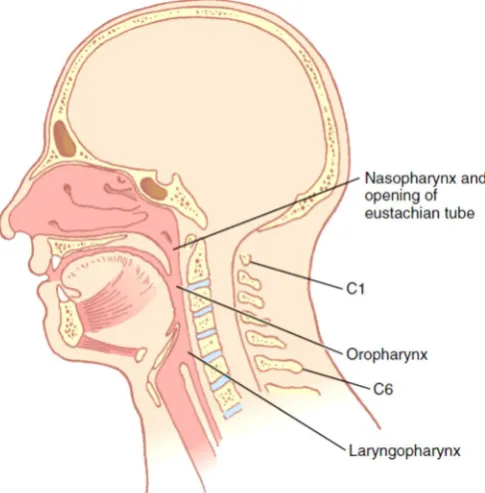
The pharynx is a passage 12-15 cms long that extends from base
of skull to cricoid cartilage and C6. It can be divided into nasopharynx,
oropharynx and laryngopharynx (Figure 3).
The nasopharynx extends from point where turbinates end till the
soft palate. From the soft palate oropharynx extends till superior edge of
epiglottis. The laryngopharynx or hypopharynx extends from C4-C6
superiorly from edge of epiglottis till lower border of cricoid cartilage
OROPHARYNX, NASOPHARYNX, LARYNGOPHARYNX
Figure 4
[image:55.595.155.466.536.752.2]The airway obstruction during sedation and anesthesia was
attributed to the loss of tone of genioglossus muscle and hence the
posterior displacement of tongue on the airway. Recently the focus has
shifted to velopharyngeal segment of airway adjacent to soft palate.
Understanding of the distance relationship between the structures
from oropharynx and trachea is very important while placing the ETT to
avoid complications like endobronchial intubation and cuff leak.
LARYNX:
The larynx is also called the “watchdog” of the airway which
allows only air to pass into the trachea but not food, secretions, and
foreign bodies. It extends from level of C3 – C6.
The laryngeal aperture is bound superiorly by epiglottis, laterally
by aryepiglottic folds, posteriorly by corniculate cartilage and
interarytenoid notch. The part of larynx below the laryngeal inlet is
called laryngeal cavity and one above is called vestibule. The ventricular
folds or false vocal cords are the superior most structure of the laryngeal
cavity. True vocal cords are seen below the ventricular folds and
commissure and posteriorly to arytenoid cartilage. The space between
the true vocal cords is called glottis (Figure 4).
The visualization of the structures in glottis during direct
ASSESSMENT OF AIRWAY AND
PREDICTORS OF DIFFICULT AIRWAY:
The ASA practice guidelines mentions guidelines /
recommendations for airway assessment and management.
Guidelines for Airway assessment: History
A detailed history taking should obtain details of the patient
characteristics like age, h/o snoring, h/o obstructive sleep apnea,
previous h/o difficult intubation or laryngoscopy, disease states like
ankylosing spondylitis, degenerative osteoarthritis, lingual tonsillar
hypertrophy, Treacher Collin syndrome, Pierre Robin syndrome.
Examination
Physical features suggestive of upper airway anatomical
abnormality or pathology should be looked for.
Additional
History and physical examination may give indication to
additional diagnostic testing like radiological imaging, CT scan, and
Recommendations for Airway assessment:
History
To detect medical, surgical, anesthetic factors that may indicate a
difficult airway. A history of previous difficult intubation /
laryngoscopy / mask ventilation may be the most valuable clue to
difficult airway. History and records should be verified with this regard.
Physical examination
Look for anatomical abnormalities of face and neck, ability to
extend neck, long upper incisors, relationship between upper and lower
incisors during jaw closure, protrusion of mandible, interincisor
distance, visibility of uvula, palate characteristics, thyromental distance,
mandibular space, length and thickness of neck, range of motion of head
and neck.
Preoperative airway assessment by specific test / index of
Difficult Airway should follow a general examination. If time permits,
more than one assessment method should be done to increase the
accuracy of airway assessment, as no single test predicts DA accurately
The specific tests include:
1. Interincisor gap (< 3 cms indicates DL)
2. Protrusion of mandible (class B and C indicates DL)
3. Modified mallampati test (class 3 and 4 indicates DL)
4. Extension of upper cervical spine (<90° indicates DL)
5. Thyromental distance (<6 cms indicates DL)
6. Sternomental distance (<12.5 cms indicates DL)
7. WILSON’s score ( 2 indicates DL)
Ideal Predictor:
The ideal test (predictor) should have the following characters:
1. The test should be painless as patients will not tolerate discomfort
for Difficult Airway screening.
2. The test should be simple, consume little time and should require
nil or simple equipments.
3. If any calculation involved, it should be easy to perform.
5. The test should be objective with nil or minimal inter observer
variability and should be reproducible.
6. The test should be economical too.
7. Higher Sensitivity and positive predictive values are desirable.
Syndromes associated with Difficulty Airway:
Congenital Acquired
Pierre Robin syndrome Croup
Treacher Collins syndrome Ludwig’s angina
Goldenhar syndrome Intraoral or retropharyngeal abscess
Downs syndrome Rheumatoid arthritis
Klippel Feil syndrome Ankylosing spondylosis
Alpert syndrome Cystic hygroma / adenoma / goiter
Beckwith syndrome Distortion of airway
Cretinism Carcinoma tongue / thyroid / larynx
Cri du chat syndrome Trauma – Head / facial
Von Reckinghauswen disease Morbid obesity
Hurler / Hunter syndrome Acromegaly
Airway
AIRWAY MANAGEMENT
The airway can be secured by either an invasive (tracheostomy)
or less invasive (endotracheal tubes, LMA) among which endotracheal
tube intubation remains the preferred method even after the advent of
supraglottic devices.
The laryngoscopy and intubation may be easy or difficult. The
understanding of the technique of intubation requires thorough
knowledge of the airway anatomy.
While doing a direct laryngoscopy or endotracheal intubation, one
have to encounter various structures like teeth, tongue, epiglottis, vocal
folds and many. Hence the anatomical abnormality of these structures
may contribute to difficult intubation.
Moreover understanding the orientation of oral, pharyngeal and
laryngeal axis is important when it applies to bringing all 3 axes in line.
The process of neck flexion and extension at atlanto occipital joint
during the positioning undoubtedly helps to bring them in line and
factors altering or limiting these movements may contribute to difficult
GENERAL ANESTHESIA
General Anesthesia (GA) comprises reversible loss of
consciousness, amnesia, and analgesia with or without muscle relaxant.
The GA may be characterized by impairment of ventilatory function,
need for assistance in maintenance of a patent airway, need for positive
pressure ventilation, drug induced skeletal muscle relaxation, and
impairment of cardiovascular function.
The drugs commonly used for GA may be categorized into
premedication drugs, intravenous (IV) induction agents, inhalational or
volatile agents, analgesics, muscle relaxants and reversal agents.
Premedication drugs are mainly given to reduce anxiety, reduce
secretions, decrease volume and acidity of gastric content, act as
analgesics, and anti emetics. It includes anticholinergics
(glycopyrrolate), anxiolytics (midazolam), opioids (morphine, fentanyl),
anti histamines (ranitidine), anti emetics (metoclopromide).
Induction agents may be IV or Inhalational. IV induction agents
are thiopentone sodium, propofol, ketamine and etomidate. inhalational
or volatile induction agents are halothane and sevoflurane. The volatile
agents (halothane, sevoflurane, isoflurane and desflurane) are commonly
Commonly used analgesics are opioids like morphine, fentanyl,
pethidine and others.
Muscle relaxants commonly used are classified into two
categories. They are depolarizing relaxants like Succinylcholine and non
depolarizing agents like vecuronium, rocuronium, atracurium. all
muscle relaxants except succinylcholine are used for maintenance
through the intraoperative period.
Reversal agents include anti cholinesterase (neostigmine) and anti
cholinergic drugs (glycopyrrolate, atropine).
Succinylcholine:
Succinylcholine is the only depolarizing muscle relaxant in
clinical use. It is chemically known as ‘Diacetylcholine’ (2 molecules of
acetylcholine joined together) or Suxamethonium.
Ultra rapid onset (30-60 seconds) and ultra short duration (less
than 10 minutes) are the most important advantages of succinylcholine.
Hence, traditionally succinylcholine was the drug of choice for tracheal
intubation.
It is metabolized by plasma cholinesterase (also called as pseudo
The intubating dose is 1-1.5 mg/kg IV.
The adverse effects are hyperkalemia, fasciculations,
rhabdomyolysis, trigger for malignant hyperthermia, increased
intracranial tension (ICT) and intraocular tension (IOT). Repeat dose or
higher dose of succinyl choline may lead to phase 2 blockade.
In view of the above adverse effects, non depolarizing muscle
relaxants have slowly replaced succinylcholine for the purpose of
elective tracheal intubation.
However, the advantage of ultrashort action and rapid recovery
from muscle paralysis gives succinyl choline an edge over non
depolarizing muscle relaxants for intubation in case of CICV situations.
Direct Laryngoscopy
Direct Laryngoscopy involves direct visualization of the glottis
using a laryngoscope. It facilitates endotracheal intubation and is the
most common technique used for it.
During direct laryngoscope, the anatomic axes – oral, pharyngeal
and laryngeal axes are distorted, brought into a single axis and tongue is
displaced to produce a direct line of visibility form operator’s eye to the
ORAL PHARYNGEAL LARYNGEAL AXES IN DIFFERENT POSITIONS
NORMAL POSITION
NECK FLEXION
NECK FLEXION &
HEAD EXTENSION OA
OA
OA PA
LA
PA
PA LA
[image:69.595.115.428.157.712.2]The normal angle between oral and pharyngeal axis is 90°.
Maximal extension of atlanto occipital joint increases the angle by 35°
and makes it 125°.
The 3 axes can be brought together near a single line by SP
(sniffing position) as described by Sir Ivan Magill. Sniffing position is
described as a neck flexion of 35° (by placing pillow under occiput) and
head extension 15° (by extension of atlanto occipital joint).
An alternate position which is useful especially in obese people is
EAM-SN (External auditory meatus – sternal notch) position.
All the efforts are oriented towards creating an in line space
towards laryngeal aperture for tracheal intubation, the end point being
visualization of glottis. A complete visualization of glottis leads to
successful tracheal intubation.
After positioning the patient, the head is fixed in extended
position by anesthesiologist’s dominant hand and using finger the lower
jaw is opened (if not passively open) to increase the inter incisor
The Macintosh curved blade is commonly used for adult patients.
The blade is introduced into the mouth and the tongue is displaced to
opposite side with the flange of the blade to create space. The tip of the
Macintosh blade is placed in the vallecula and the blade is lifted anterior
- caudad direction (Figure 6).
The operator should carefully avoid any rotating the wrist and
laryngoscope blade in a cephalad direction. This will cause the blade to
injure the upper incisors which is not desirable. Extending blade too
deeply can place the blade tip to rest under the larynx and forward
pressure may lift entire larynx away from the view.
An alternate to Macintosh blade is a Miller’s straight blade.
During laryngoscopy, if satisfactory view is not obtained, an
“optimal external laryngeal manipulation” (OLEM) as described in
Benumof and Hagberg’s textbook of Airway management37 can be used.
It involves applying pressure posteriorly and in the cephalad direction
over the thyroid, hyoid, and cricoid cartilages. BURP
(Backward-Upward-Rightward pressure) maneuver is the typically the most useful
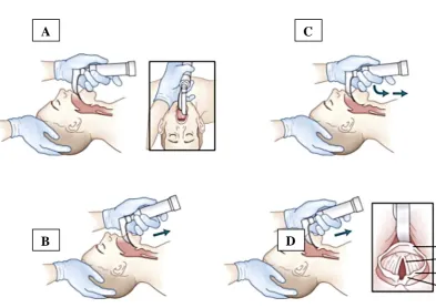
CONVENTIONAL DIRECT LARYNGOSCOPY WITH MACINTOSH BLADE
A – Laryngoscopy blade inserted into right side of mouth sweeping the tongue to the left side of flange
B – Blade is advanced in midline towards base of tongue by rotating wrist so that laryngoscope handle becomes more vertical
C – The laryngoscope is lifted at 45’ as tip of the blade is placed in the vallecula
D – Continued lifting of the handle until visualization of laryngeal aperture
A
B
C
[image:72.595.105.499.210.488.2]When the glottis is visible the endotracheal tube is inserted into it
under vision of the operator. Care should be taken not to obscure the
view of larynx when inserting the endotracheal tube. The tube should be
inserted to a depth of 2 cms after disappearance of the cuff. This allows
the tip of the ETT to be positioned in mid trachea.
Commonly a size 7 8 ID ETT is used in adult female and size 8
-9 ID ETT in adult male. Typically the length at which ETT is fixed (at
upper incisors) is 20-22 cms and 18-20 cms in adult male and female
respectively.
Confirmation of a successful ETT placement is done by various
methods. The gold standard methods are placement of ETT under direct
vision and capnography.
Five point auscultation of the chest, visualization of chest
expansion, observation of tube condensation, self inflated bulbs, lighted
stylet, fibreoptic devices, ultrasonography and chest x-ray are other
methods.
The alternate to direct laryngoscopy is image guided
laryngoscopy which may herald the future in the art of tracheal
MATERIALS AND METHODS
The study was done in the Department of Anesthesiology, ESIC
MC and PGIMSR, K.K. Nagar, Chennai from October 2014 to August
2015.
Study design : Prospective Observational double blind study.
Participants : Patients undergoing elective surgery requiring GA with endotracheal intubation.
Sample : Sample size of 173 is calculated by using nMaster 1.0 software
(PPV - 33, precision - 7%, confidence level - 95%)
The aim, objectives, materials and methods were submitted to the
Institutional ethics committee and approval was obtained. 173 patients
were selected in accordance with inclusion and exclusion criteria for the
study.
Inclusion criteria:
1. 18 – 50 years of either sex
2. ASA physical status I, II
3. Patient undergoing elective surgeries under General anesthesia
Exclusion criteria:
1. Consent not given.
2. Obvious anatomical abnormality of face, head, neck and shoulder.
3. Upper airway abnormality (e.g., tongue tumor, maxillofacial
tumor, or fracture)
4. Recent head and neck surgery
5. Inability to open mouth
Pre Anesthetic Assessment:
As per the departmental protocol the patients posted for elective
surgery were investigated for pre-operative biochemical tests (renal
function tests and liver function tests), hematological tests (hemoglobin,
total count, differential count, platelet count), Chest x-ray (PA view) &
12 lead Electrocardiograph and assessed in the pre-anesthetic
assessment clinic.
The patients on arrival to the operating theatre complex were
Airway assessment:
All patients underwent airway examination prior to surgery
during which Acromio axillo suprasternal notch index (AASI) and
Modified Mallampati test (MMP) were assessed by an anesthesiologist
and recorded in the proforma.
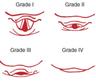
Modified mallampati test: The standard test
MMP score (the oropharyngeal view) was measured while
patients were sitting, with a fully protruded tongue without saying “ah”.
MMP classification(Figure 7) is as follows:
I : Full view of soft palate, uvula, tonsillar pillars
II : Soft palate and upper portion of uvula
III : Soft palate
IV : Hard palate only
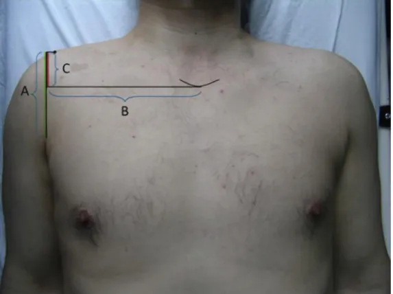
Acromio Axillo Suprasternal Notch Index (AASI):
With the patients lying in a supine position and their upper
MODIFIED MALLAMPATI CLASSIFICATION
Figure 8
[image:78.595.163.450.483.697.2]vertical line was drawn from the top of the acromion process to the
superior border of the axilla at the pectoralis major muscle (line A); (2)
a second line was drawn perpendicular to line A from the suprasternal
notch (line B); and (3) the portion of line A that lay above the point at
which line B intersected line A was line C. AASNI was calculated by
dividing the length of line C by that of line A (AASNI = C/A).
Preparation:
After airway assessment patients shifted into operating room and
minimum mandatory monitors such as pulse oximetry (SpO2), non
invasive blood pressure (NIBP), and electrocardiogram (ECG) were
attached.
Baseline pulse rate, blood pressure and oxygen saturation were
recorded.
An intravenous (IV) line was secured and Ringer lactate / Normal
saline (depending on the diabetic status of patient) started before the
procedure.
Standard preparations and precautions were taken for general
anesthesia with endotracheal tube intubation and controlled mechanical
The airway cart was kept ready which consists of manual
resuscitator bag, anatomical masks of all sizes, oropharyngeal and
nasopharyngeal tubes of all sizes, suction canula of all sizes,
laryngoscope handle with blades of all sizes, McCoy blade, ETT of all
sizes, LMA of all sizes, ILMA (intubating LMA), stylet, ventilating
bougie, emergency cricothyrotomy set, and emergency tracheostomy
set.
The availability of fibreoptic bronchoscope was ensured
whenever we encountered a suspected case of Difficult airway.
Premedication and Induction:
All patients received premedication with Glycopyrrolate (0.2 mg),
midazolam (2 mg) and fentanyl (2 mcg/kg) intravenously. After
pre-oxygenation (100% O2 for 3 minutes) patients induced with propofol
(2.5 mg/kg) and paralysed with succinylcholine (1.5 mg/kg)
intravenously.
After ventilation for 1 minute with 100 % O2, with a 10cm pillow
under the head and the head in the sniffing position, direct laryngoscopy
was done by an experienced anesthesiologist. Direct laryngoscopy was
performed with a Mackintosh blade (No. 3) and Cormack Lehane grade