Copyright © 1998, American Society for Microbiology. All Rights Reserved.
A Third-Generation Lentivirus Vector with a Conditional
Packaging System
TOM DULL,
1ROMAIN ZUFFEREY,
2MICHAEL KELLY,
1R. J. MANDEL,
1MINH NGUYEN,
1DIDIER TRONO,
2ANDLUIGI NALDINI
1*
Cell Genesys, Foster City, California,
1and Department of Genetics and Microbiology,
University of Geneva Medical School, Geneva, Switzerland
2Received 1 June 1998/Accepted 21 July 1998
Vectors derived from human immunodeficiency virus (HIV) are highly efficient vehicles for in vivo gene
de-livery. However, their biosafety is of major concern. Here we exploit the complexity of the HIV genome to
pro-vide lentivirus vectors with novel biosafety features. In addition to the structural genes, HIV contains two
regu-latory genes, tat and rev, that are essential for HIV replication, and four accessory genes that encode critical
virulence factors. We previously reported that the HIV type 1 accessory open reading frames are dispensable
for efficient gene transduction by a lentivirus vector. We now demonstrate that the requirement for the tat gene
can be offset by placing constitutive promoters upstream of the vector transcript. Vectors generated from
con-structs containing such a chimeric long terminal repeat (LTR) transduced neurons in vivo at very high
effi-ciency, whether or not they were produced in the presence of Tat. When the rev gene was also deleted from the
packaging construct, expression of gag and pol was strictly dependent on Rev complementation in trans. By the
combined use of a separate nonoverlapping Rev expression plasmid and a 5
*
LTR chimeric transfer construct,
we achieved optimal yields of vector of high transducing efficiency (up to 10
7transducing units [TU]/ml and
10
4TU/ng of p24). This third-generation lentivirus vector uses only a fractional set of HIV genes: gag, pol, and
rev. Moreover, the HIV-derived constructs, and any recombinant between them, are contingent on upstream
elements and trans complementation for expression and thus are nonfunctional outside of the vector producer
cells. This split-genome, conditional packaging system is based on existing viral sequences and acts as a
built-in device against the generation of productive recombinants. While the actual biosafety of the vector will
ultimately be proven in vivo, the improved design presented here should facilitate testing of lentivirus vectors.
Lentiviruses have attracted the attention of gene therapy
investigators (45) for their ability to integrate into nondividing
cells (8, 15, 16, 25, 26). We previously developed
replication-defective vectors from the lentivirus human immunodeficiency
virus (HIV) and showed that they transduce target cells
inde-pendent of mitosis (32). The vectors proved highly efficient for
in vivo gene delivery and achieved stable long-term expression
of the transgene in several target tissues, such as the brain (5,
33), the retina (31), and the liver and muscle of adult rats (21).
A major concern, however, is the biosafety of vectors derived
from a highly pathogenic human virus.
The complexity of the lentivirus genome may be exploited to
build novel biosafety features in the design of a retrovirus
vec-tor. In addition to the structural gag, pol, and env genes
com-mon to all retroviruses, HIV contains two regulatory genes, tat
and rev, essential for viral replication, and four accessory
genes, vif, vpr, vpu, and nef, that are not crucial for viral growth
in vitro but are critical for in vivo replication and pathogenesis
(27).
The Tat and Rev proteins regulate the levels of HIV gene
expression at transcriptional and posttranscriptional levels,
re-spectively. Due to the weak basal transcriptional activity of the
HIV long terminal repeat (LTR), expression of the provirus
initially results in small amounts of multiply spliced transcripts
coding for the Tat, Rev, and Nef proteins. Tat increases
dra-matically HIV transcription by binding to a stem-loop structure
(transactivation response element [TAR]) in the nascent RNA,
thereby recruiting a cyclin-kinase complex that stimulates
tran-scriptional elongation by the polymerase II complex (46). Once
Rev reaches a threshold concentration, it promotes the
cyto-plasmic accumulation of unspliced and singly spliced viral
tran-scripts, leading to the production of the late viral proteins. Rev
accomplishes this effect by serving as a connector between an
RNA motif (the Rev-responsive element [RRE]), found in the
envelope coding region of the HIV transcript, and components
of the cell nuclear export machinery. Only in the presence of
Tat and Rev are the HIV structural genes expressed and new
viral particles produced (27).
In a first generation of HIV-derived vectors (32), viral
par-ticles were generated by expressing the HIV type 1 (HIV-1)
core proteins, enzymes, and accessory factors from
heterolo-gous transcriptional signals and the envelope of another virus,
most often the G protein of the vesicular stomatitis virus (VSV
G) (9) from a separate plasmid. In a second version of the
system, the HIV-derived packaging component was reduced to
the gag, pol, tat, and rev genes of HIV-1 (51). In either case, the
vector itself carried the HIV-derived cis-acting sequences
nec-essary for transcription, encapsidation, reverse transcription,
and integration (2, 4, 22, 24, 29, 30, 32, 35). It thus
encom-passed, from the 5
9
to 3
9
end, the HIV 5
9
LTR, the leader
sequence and the 5
9
splice donor site, approximately 360 bp of
the gag gene (with the gag reading frame closed by a synthetic
stop codon), 700 bp of the env gene containing the RRE and
a splice acceptor site, an internal promoter (typically the
im-mediate-early enhancer/promoter of human cytomegalovirus
[CMV] or that of the phosphoglycerokinase gene [PGK]), the
transgene, and the HIV 3
9
LTR. Vector particles are produced
by cotransfection of the three constructs in 293T cells (32). In
this design, significant levels of transcription from the vector
* Corresponding author. Mailing address: Cell Genesys, 342
Lake-side Dr., Foster City, CA 94404. Phone: (650) 425-4474. Fax: (650)
358-8636. E-mail: [email protected].
8463
on November 9, 2019 by guest
http://jvi.asm.org/
LTR and of accumulation of unspliced genomic RNA occur
only in the presence of Tat and Rev.
Here, we demonstrate that the trans-acting function of Tat
becomes dispensable if part of the upstream LTR in the
trans-fer vector construct is replaced by constitutively active
pro-moter sequences. Furthermore, we show that the expression of
rev in trans allows the production of high-titer HIV-derived
vector stocks from a packaging construct which contains only
gag and pol. This design makes the expression of the packaging
functions conditional on complementation available only in
producer cells. The resulting gene delivery system, which
con-serves only three of the nine genes of HIV-1 and relies on four
separate transcriptional units for the production of transducing
particles, offers significant advantages for its predicted
bio-safety.
MATERIALS AND METHODS
Transfer vector constructs.pHR9CMV-LacZ and pHR9CMV-Luciferase have been described elsewhere (32). pHR2 is a lentivirus transfer vector in which the polylinker and downstream nef sequences up to the KpnI site of pHR9have been replaced with a ClaI/SpeI/SnaBI/SmaI/BamHI/SacII/EcoRI polylinker. pHR2 was generated by replacing the 3.7-kb ClaI-SacI fragment of pHR9CMVlacZ with a 607-bp ClaI-SacI fragment generated by PCR using pHR9CMVlacZ as the tem-plate with oligonucleotide primers 59-CCATCGATGGACTAGTCCTACGTA TCCCCGGGGACGGGATCCGCGGAATTCCGTTTAAGACCAATGAC-39and 59-TTATAATGTCAAGGCCTCTC-39, followed by digestion with ClaI and SacI.
pHR2PGK-NGFR, pHR2CMV-NGFR, and pHR2MFG-NGFR are lentivirus transfer vectors in which the truncated low-affinity nerve growth factor receptor (NGFR) (6) transgenes under the control of the murine PGK, human CMV, and Moloney leukemia virus (MLV) promoters, respectively, have been inserted into the polylinker of pHR2. The pHR2PGK-NGFR transgene encodes no intron se-quences, the pHR2CMV-NGFR vector includes the intron from plasmid pMD (34), and the pHR2MFG-NGFR vector contains the MLV intron from MFG-S (34). pRRL, pRLL, pCCL, and pCLL are lentivirus transfer vectors containing chimeric Rous sarcoma virus (RSV)-HIV or CMV-HIV 59 LTRs and vector backbones in which the simian virus 40 polyadenylation and (enhancerless) origin of replication sequences have been included downstream of the HIV 39
LTR, replacing most of the human sequence remaining from the HIV integra-tion site. In pRRL, the enhancer and promoter (nucleotides2233 to21 relative to the transcriptional start site; GenBank accession no. J02342) from the U3 region of RSV are joined to the R region of the HIV-1 LTR. In pRLL, the RSV enhancer (nucleotides2233 to250) sequences are joined to the promoter region (from position278 relative to the transcriptional start site) of HIV-1. In pCCL, the enhancer and promoter (nucleotides2673 to21 relative to the transcriptional start site; GenBank accession no. K03104) of CMV were joined to the R region of HIV-1. In pCLL, the CMV enhancer (nucleotides2673 to2220) was joined to the promoter region (position278) of HIV-1. Exact sequences and details of construction are available on request.
pHR2hPGK-GFP, pCCLhPGK-GFP, pCLLhPGK-GFP, pRRLhPGK-GFP, and pRLLhPGK.GFP are lentivirus transfer vectors containing the enhanced green fluorescent protein (eGFP) (750-bp BamHI-NotI fragment from pEGFP-1; Clontech) coding region, under the control of the human PGK promoter (nucleotides 5 to 516; GenBank accession no. M11958), inserted into the poly-linker region of each parental vector. pRRLGFP was obtained by deletion of the
XhoI-BamHI fragment containing the PGK promoter from pRRLhPGK-GFP.
pRRLhPGK.GFP.SIN-18 is a vector in which 39LTR sequences from2418 to
218 relative to the U3/R border have been deleted from pRLLhPGK.GFP (52).
Packaging constructs. The tat-defective packaging construct pCMVDR8.93 was obtained by swapping an EcoRI-SacI fragment from plasmid R7/pneo(2) (12) with the corresponding fragment of pCMVDR8.91, a previously described plasmid expressing Gag, Pol, Tat, and Rev (51). This fragment has a deletion affecting the initiation codon of the tat gene and a frameshift created by the insertion of an MluI linker into the Bsu36I site as described previously. pCMVDR8.74 is a derivative of pCMVDR8.91 in which a 133-bp SacII fragment, containing a splice donor site, has been deleted from the CMV-derived region upstream of the HIV sequences to optimize expression.
pMDLg/p is a CMV-driven expression plasmid that contains only the gag and
pol coding sequences from HIV-1. First, pkat2Lg/p was constructed by ligating a
4.2-kb ClaI-EcoRI fragment from pCMVDR8.74 with a 3.3-kb EcoRI-HindIII fragment from pkat2 (14) and a 0.9-kb HindIII-NcoI fragment from pkat2 along with an NcoI-ClaI linker consisting of synthetic oligonucleotides 59-CATGGGT GCGAGAGCGTCAGTATTAAGCGGGGGAGAATTAGAT-39and 59-CG ATCTAATTCTCCCCCGCTTAATACTGACGCTCTCGCACC-39. Next, pMDLg/p was constructed by inserting the 4.25-kb EcoRI fragment from pkat2Lg/p into the EcoRI site of pMD-2. pMD-2 is a derivative of pMD.G (34) in which the pXF3 plasmid backbone of pMD.G has been replaced with a
minimal pUC plasmid backbone and the 1.6-kb VSV G-encoding EcoRI frag-ment has been removed.
pMDLg/pRRE differs from pMDLg/p by the addition of a 374-bp RRE-con-taining sequence from HIV-1 (HXB2) immediately downstream of the pol cod-ing sequences. To generate pMDLg/pRRE, the 374-bp NotI-HindIII RRE-con-taining fragment from pHR3 was ligated into the 9.3-kb NotI-BglII fragment of pVL1393 (Invitrogen) along with a HindIII-BglII oligonucleotide linker consist-ing of synthetic oligonucleotides 59-AGCTTCCGCGGA-39and 59-GATCTCC GCGGA-39to generate pVL1393RRE (pHR3 was derived from pHR2 by the removal of HIV env coding sequences upstream of the RRE sequences in pHR2). A NotI site remains at the junction between the gag and RRE sequences. pMDLg/pRRE was then constructed by ligating the 380-bp EcoRI-SstII fragment from pV1393RRE with the 3.15-kb SstII-NdeI fragment from pMD-2FIX (pMD-2FIX is a human factor IX-containing variant of pMD-2 which has an SstII site at the 39end of the factor IX insert), the 2.25-kb NdeI-AvrII fragment from pMDLg/p, and the 3.09-kb AvrII-EcoRI fragment from pkat1Lg/p (14).
pRSV-Rev and pTK-Rev (generous gifts of T. Hope, Salk Institute) are rev cDNA-expressing plasmids in which the joined second and third exons of HIV-1
rev are under the transcriptional control of the RSV U3 and herpes simplex virus
type 1 thymidine kinase (TK) promoters, respectively. Both expression plasmids utilize polyadenylation signal sequences from the HIV LTR in a pUC118 plas-mid backbone.
Vector production and assays.Vectors were produced by transient transfec-tion into 293T cells as previously described (33), with the following modificatransfec-tions. A total of 53106293T cells were seeded in 10-cm-diameter dishes 24 h prior to
transfection in Iscove modified Dulbecco culture medium (JRH Biosciences) with 10% fetal bovine serum, penicillin (100 IU/ml), and streptomycin (100
mg/ml) in a 5% CO2incubator, and the culture medium was changed 2 h prior
to transfection. A total of 20mg of plasmid DNA was used for the transfection of one dish: 3.5mg of the envelope plasmid pMD.G, 6.5mg of packaging plasmid, and 10mg of transfer vector plasmid. The precipitate was formed by adding the plasmids to a final volume of 450ml of 0.13TE (13TE is 10 mM Tris [pH 8.0] plus 1 mM EDTA) and 50ml of 2.5 M CaCl2, mixing well, then adding dropwise
500ml of 23HEPES-buffered saline (281 mM NaCl, 100 mM HEPES, 1.5 mM Na2HPO4[pH 7.12]) while vortexing and immediately adding the precipitate to
the cultures. The medium (10 ml) was replaced after 14 to 16 h; the conditioned medium was collected after another 24 h, cleared by low-speed centrifugation, and filtered through 0.22-mm-pore-size cellulose acetate filters. For in vitro ex-periments, serial dilutions of freshly harvested conditioned medium were used to infect 105cells in a six-well plate in the presence of Polybrene (8mg/ml). Viral
p24 antigen concentration was determined by immunocapture (Alliance; Du-Pont-NEN). Vector batches were tested for the absence of replication-compe-tent virus by monitoring p24 antigen expression in the culture medium of trans-duced SupT1 lymphocytes for 3 weeks. In all cases tested, p24 was undetectable (detection limit, 3 pg/ml) once the input antigen had been eliminated from the culture. Transducing activity was expressed in transducing units (TU).
Northern blot analysis.Total RNA was isolated from 13107to 23107cells
harvested at confluence by using RNAsol B as suggested by the manufacturer; 10 to 20mg of RNA was loaded per well on 1% agarose gels, using NorthernMax (Ambion, Austin, Tex.) reagents as described by the manufacturer. Transfer was to Zetabind membranes (Cuno Inc., Meridien, Conn.) by either capillary transfer or pressure blotting (Stratagene).32P-labeled probes were made by random
prim-ing.
Intracerebral injection of vectors.Twelve Fischer 344 male rats weighing approximately 220 g were obtained from Harlan Sprague-Dawley (Indianapolis, Ind.). The rats were housed with access to ad libitum food and water on a 12-h light/dark cycle and were maintained and treated in accordance with published National Institutes of Health guidelines. All surgical procedures were performed with the rats under isoflurane gas anesthesia, using aseptic procedures. After a rat was anesthetized in a sleep box, it was placed in a small animal stereotaxic device (Kopf Instruments, Tujunga, Calif.) using the earbars, which do not break the tympanic membrane. The rats were randomly divided into one control and four treatment groups. After the rats were placed in the stereotaxic frame, 2ml of lentivirus vector concentrated by ultracentrifugation at 50,0003g for 140 min
at 20°C (33) in phosphate-buffered saline (PBS) was injected consecutively into the striatum in both hemispheres over 4 min at a rate of 0.5ml/min (coordinates, AP 0.0, LAT63.0, DV25.5,24.5,23.5 with the incisor bar set at 3.3 mm below the intra-aural line [36]), using a continuous-infusion system as described previously in detail (28). During the injection, the needle was slowly raised 1 mm in the dorsal direction every 40 s (3-mm total withdrawal). One minute after cessation of the injection, the needle was retracted an additional 1 mm and then left in place for an additional 4 min before being slowly withdrawn from the brain.
Histology.One month after vector injection, each animal was deeply anesthe-tized with intraperitoneal pentobarbital and perfused through the aorta with sterile PBS, followed by ice-cold 4% paraformaldehyde perfusion. The brains were removed from the skulls, postfixed in 4% paraformaldehyde by immersion for 24 h, and then transferred into a 30% sucrose–PBS solution for 3 to 4 days, until the brains sank to the bottom of their containers. The brains were then frozen on dry ice, and 40-mm-thick coronal sections were cut on a sliding micro-tome. Sections were collected in series in microtiter well plates that contained a glycerin-based antifreeze solution, and they were kept at230°C until further processing. Immunocytochemistry was performed according to the general
on November 9, 2019 by guest
http://jvi.asm.org/
cedure described previously (44). After several PBS rinses and an incubation in 3% hydrogen peroxide, the sections were placed in a 3% normal goat serum. The sections were then incubated in the primary anti-GFP antibody (1:1,000; Clon-tech, Palo Alto, Calif.) in 1% normal goat serum–0.1% Triton X-100 overnight at room temperature. After rinsing, the sections were incubated in the biotin-ylated rabbit anti-goat secondary antibody (Vector, Burlingame, Calif.) for 3 h. After rinsing, the sections were incubated with horseradish peroxidase-strepta-vidin and then reacted by using a purple chromagen kit (VIP; Vector), mounted, dried, dehydrated, and coverslipped.
RESULTS
Tat is required to produce a vector of efficient transducing
activity.
To investigate the role of Tat in the production of
transducing particles, expression from lentivirus vectors was
first examined by Northern analysis (Fig. 1). The patterns of
RNAs induced by transfer vectors in which the transgene was
driven by an internal PGK, CMV, or retrovirus MFG promoter
were studied in both producer and target cells. In transfected
293T cells, expression occurred mainly from the internal
pro-moter. When a packaging construct expressing both Tat and
Rev was cotransfected, a dramatic enhancement of
transcrip-tion from the LTR was observed, with an accumulatranscrip-tion of
unspliced vector RNA. In cells transduced with the vectors,
that is, in the absence of Tat and Rev, transcription from the
LTR was almost completely suppressed, the residual
tran-scripts underwent splicing, and the internal promoter was
re-sponsible for most of the expression.
A packaging plasmid carrying two mutations in tat (pCMV
D
R
8.93) was then constructed. The first mutation is a deletion of
the T in the ATG initiation codon of the tat gene; the second
is an insertion of a MluI linker producing a translation stop
codon after residue 46 of the Tat protein. These changes
con-fer a tat-defective phenotype to HIV-1 (12). After transfection
of the control or tat-defective packaging constructs into 293T
cells, comparable yields of vector particles were recovered in
the culture medium, as assayed by using the Gag p24 antigen
(see Table 3). Such Tat independence was expected from the
replacement of the HIV LTR by the constitutive CMV
moter in the packaging construct. However, the particles
pro-duced in the absence of Tat had a dramatically repro-duced
trans-ducing activity (Table 1): 5 to 15% of that of particles
produced by the control Tat-positive packaging construct.
We also tested whether the Tat-defective phenotype could
be rescued by complementation in target cells (Table 1).
HeLa-tat cells, a cell line expressing Tat from the HIV-1 LTR (13),
were transduced by vectors produced with or without Tat. The
expression of Tat in target cells did not compensate for the loss
in transduction efficiency of vector produced without Tat.
As expected from the Northern analysis, functional
inacti-vation of the tat gene resulted in a lower abundance of vector
RNA in producer cells. This was indicated by the decrease in
luciferase activity in cells producing a luciferase vector without
an internal promoter. In this case, transgene expression
[image:3.612.143.453.68.197.2]di-FIG. 1. Northern analysis of the RNA expression from lentivirus vectors. Three pHR2 vectors carrying an expression cassette for the same transgene (truncated low-affinity NGFR) and driven by three different promoters (PGK, CMV, and retroviral MFG) were analyzed in producer and transduced cells. Total RNA was extracted and analyzed by Northern blotting with a probe specific for the transgene sequence. (A) Schematic of the vector construct depicts the species of RNA driven by the internal promoter (Prom.; broken arrow, shorter transcript) and the viral LTR (solid arrows, longer transcripts; the two species differ for the splicing of the viral intron). The splice donor and acceptor sites (SD and SA), the packaging sequence (C), the truncated gag sequence (GA), and the RRE are indicated. (B) The vector constructs were transfected in 293T cells without or with the packaging construct. (C) Vector particles produced by the 293T transfectants were used to transduce HeLa cells. In the absence of the viral transactivators, supplied by the core packaging construct only in the producer cells, vector expression occurs mainly from the internal promoter. Note the dramatic enhancement of the upstream transcription and the accumulation of unspliced RNA (carrying theCsequence) in the presence of the packaging construct. In the transduced cells, the LTR is silenced. Note that the three expression cassettes differ in the size of the promoters and 59untranslated sequence. In each case, the smallest RNA species represents transcripts initiated from the internal promoter, while the intermediate-size and larger species correspond to spliced and unspliced LTR-driven RNAs, respectively.
TABLE 1. Transducing activities of lentivirus vectors made with
and without a functional tat gene in the packaging construct
aTransfer vector Targetcells
Mean transducing activity (TU/ng of p24)6SEb With tat in
packaging construct
Without tat in packaging construct
pHR
9
CMV-LacZ
293T
1,056
6
54
152
6
26
pHR2PGK-eGFP
HeLa
5,666
384
pHR
9
CMV-Luciferase
HeLa
3,000
6
152
152
6
26
HeLa-tat
3,777
6
348
486
6
59
pHR
9
Luciferase
cHeLa
46
6
1
0.3
6
0.003
HeLa-tat
3,296
6
276
174
6
75
aVectors were produced by transfection of the indicated transfer vector, a packaging construct either with (pCMVDR8.91) or without (pCMVDR8.93) a functional tat gene, and plasmid pMD.G into 293T cells. Serial dilutions of transfectant conditioned medium were incubated with the indicated cells, and the cultures were scored after 3 days. For calculating transduction activity, sam-ples were selected from the linear portion of the vector dose-response curve.bLacZ transduction was measured by 5-bromo-4-chloro-3-indolyl-b-D -galac-topyranoside (X-Gal) staining and by expression of the number of blue cell colonies as a function of the amount of p24 antigen in the inoculum. eGFP transduction was measured by FACS analysis, multiplying the fraction of fluo-rescent cells by the number of infected cells, and expressing the result as a function of the amount of p24 antigen in the inoculum. Luciferase transduction was measured by luminescence in RLU above background of 50ml of culture extract and dividing the number of RLU31022by the number of nanograms of
p24 antigen in the inoculum. Means of duplicate (pHR2 PGK-eGFP) or tripli-cate (all other constructs) determinations are shown.
cWithout internal promoter.
on November 9, 2019 by guest
http://jvi.asm.org/
[image:3.612.308.551.475.587.2]rectly reflects the abundance of transcripts originating from the
LTR. 293T cells producing luciferase vectors without Tat had
only 5% of the luciferase content of cells producing the same
vector with Tat ([1.0
6
0.2]
3
10
9relative light units [RLU]/
dish without Tat; [20.2
6
0.7]
3
10
9RLU/dish with Tat). This
ratio corresponded very closely to that observed in cells
trans-duced by either type of vector in the course of the same
ex-periment (Table 1), suggesting that the abundance of vector
RNA in producer cells is a rate-limiting factor in the
transduc-tion by lentivirus vectors.
One could thus conclude that Tat is required in producer
cells to activate transcription from the HIV LTR and to
gen-erate vector particles with a high transducing activity.
The tat requirement is offset by placing a constitutive
pro-moter upstream of the transfer vector.
If the only function of
Tat is trans activation of vector transcription from the LTR, the
tat-defective phenotype should be rescued by placing a strong
constitutive promoter upstream of the vector transcript. Three
transcriptional domains have been identified in the HIV
pro-moter in the U3 region of the LTR: the core or basal domain,
the enhancer, and the modulatory domain (27). Transcription
starts at the U3/R boundary, the first nucleotide of R being
numbered 1. The core promoter contains binding sites for the
TATA-binding protein (
2
28 to
2
24) and SP-1 (three binding
sites between
2
78 to
2
45). The enhancer contains two binding
sites for NF-
k
B which overlap with a binding site for NFATc
(
2
104 to
2
81). The modulatory domain contain binding sites
for several cellular factors, including AP-1 (
2
350 to
2
293),
NFAT-1 (
2
256 to
2
218), USF-1 (
2
166 to
2
161), Ets-1 (
2
149
to
2
141), and LEF (
2
136 to
2
125). A panel of 5
9
chimeric
transfer constructs carrying substitutions of either all or part of
the U3 region of the 5
9
LTR was generated. All substitutions
were made to preserve the transcription initiation site of HIV.
Partial substitutions joined new enhancer sequences to the
core promoter of the HIV LTR (
2
78 to 1), while full
substi-tutions replaced also the promoter. pRLL and pRRL vectors
carried the enhancer and the enhancer/promoter, respectively,
of RSV; pCLL and pCCL vectors carried the enhancer and the
enhancer/promoter of human CMV.
[image:4.612.51.295.62.365.2]Control pHR2 and 5
9
chimeric transfer constructs carrying a
PGK-eGFP expression cassette were tested by transfection of
293T cells with control or tat-defective packaging constructs,
and the expression of the eGFP transgene was analyzed by
fluorescence-activated cell sorting (FACS). The RRL chimeric
construct yielded a higher level of eGFP expression than the
pHR2 vector, reflecting the constitutive transcriptional activity
of the new sequence (Fig. 2A). Interestingly, the chimeric
vector also displayed upregulation by Tat, as shown by the
increased eGFP expression of cells cotransfected with the
con-trol packaging construct. Tat upregulation was proven to be
a direct effect by transfecting a pRRL-eGFP vector lacking
an internal promoter with control or tat-defective packaging
constructs and analyzing GFP expression by FACS (Fig. 2B).
Comparable results were obtained with the other chimeric
LTR vectors (not illustrated). Vector particles were then
col-lected from the transfected producer cells and assayed for
transduction of eGFP into HeLa cells and human primary
lymphocytes (peripheral blood lymphocytes [PBL]). As shown
in Table 2, all vectors had efficient transducing activity, as
assessed by endpoint titration on HeLa cells or maximal
trans-duction frequency of PBL. The vector produced by the pRRL
chimera was as efficient as that produced by the pHR2
con-struct and was selected to test transduction independent of
Tat. As shown in Table 3, the pRRL construct yielded a vector
of only slightly reduced transducing activity (60%) when the
packaging construct was tat defective. The residual effect of
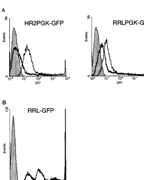
FIG. 2. Transcriptional activities of wild-type and 59 chimeric vector con-structs in the absence and presence of Tat. (A) Control pHR2 and the 59
chimeric pRRL transfer construct carrying a PGK-eGFP expression cassette were transfected into 293T cells with a packaging construct having a functional (pCMVDR8.91; grey line) or inactive (pCMVDR8.93; black line) tat gene. GFP expression was analyzed by FACS. The filled area represents nontransfected cells. In the absence of Tat, the chimeric construct yielded a level of GFP expression higher than that achieved by the pHR2 construct. Both constructs were further upregulated by Tat. (B) A pRRL construct carrying the eGFP gene without an internal promoter was transfected with a packaging construct carrying a functional (grey line, open area) or inactive (black line, open area) tat gene. Direct upregulation of the chimeric promoter by Tat was observed. The filled area represents nontransfected cells.
TABLE 2. GFP transduction by lentivirus vectors made by transfer
constructs with a wild-type or 5
9
chimeric LTR
Transfer construct
Endpoint titer on HeLa cells
(TU/ml)a
Transduction efficiency on human lymphocytes
(% positive cells)b
pHR2
2.3
3
10
730
pCCL
4.6
3
10
614
pCLL
7.9
3
10
618
pRRL
1.8
3
10
729
pRLL
8.9
3
10
618
aDetermined by multiplying the percentage of fluorescent cells for the vector dilution and the number of infected cells. Samples were selected from the linear portion of the vector dose-response curve.
bPercentage of fluorescent human PBL after infection of 106cells with 1 ml of
vector containing medium. Primary human T lymphocytes were isolated and transduced as previously described (14). Vectors carrying a PGK-eGFP expres-sion cassette were produced by transfection of the indicated transfer construct, the packaging plasmid pCMVDR8.91, and the envelope plasmid pMD.G into 293T cells. Fluorescent cells were scored by FACS analysis 6 days after trans-duction. Data are averages of duplicate determinations for a representative experiment of three performed.
on November 9, 2019 by guest
http://jvi.asm.org/
[image:4.612.309.546.555.637.2]Tat on transduction was in agreement with the ability of Tat to
upregulate transcription from the chimeric LTR.
The use of the chimeric LTR construct allowed removal of
Tat from the packaging system with a minimal loss in the
transduction efficiency of the vector in vitro. To test vector
performance in the more challenging setting of in vivo delivery
into brain neurons, high-titer vector stocks were generated
from the pHR2 and pRRL constructs with and without Tat.
The four stocks of eGFP vector were matched for particle
content by p24 antigen and injected bilaterally in the neostriata
of groups of three adult rats. The animals were sacrificed after
1 month, and serial sections of the brain were analyzed for
eGFP fluorescence (not shown) and immunostained by
anti-bodies against eGFP (Fig. 3). The results obtained in vivo
matched the in vitro data. Vector produced by the pHR2
construct only achieved significant transduction of the neurons
when packaged in the presence of Tat. Vector produced by the
pRRL chimera was as efficient when made with or without Tat.
The transduction extended throughout most of the striatum
and reached a very high density of positive cells in the sections
closest to the injection site. No signs of pathology were
detect-able in the injected tissue, except for a small linear scar
mark-ing the needle track, by hematoxylin and eosin stainmark-ing of the
sections (data not shown).
These results provide evidence that Tat is dispensable for
efficient transduction by a lentivirus vector.
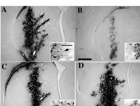
[image:5.612.64.537.314.674.2]FIG. 3. In vivo transduction of eGFP into brain cells by lentivirus vectors produced with and without Tat. Vectors carrying a PGK-eGFP expression cassette were produced by the pHR2 (A and B) or the 59chimeric pRRL (C and D) transfer construct and a packaging construct with (pCMVDR8.91; A and C) or without (pCMVDR8.93; B and D) a functional tat gene, concentrated by ultracentrifugation, and normalized for particle content prior to injection into the corpora striata of adult rats. One month after injection, brain sections were stained for immunoreactivity to the GFP protein. While both types of vectors transduced neurons very efficiently when made with Tat, only the vector made by the chimeric transfer construct worked as well when produced without Tat. Representative sections close to the injection site are shown for one of six striata injected per each type of vector. The bar in panel B represents 1 mm; that in the inset in panel A represents 100mm.
TABLE 3. GFP transduction into HeLa cells by lentivirus vectors
made by transfer constructs with a wild-type or 5
9
chimeric LTR
and packaging constructs with or without a functional tat gene
aTransfer construct
tat gene in
packaging construct
Endpoint titer
(TU/ml) p24 antigen(ng/ml)
Transduction efficiency (TU/ng of p24)
pHR2
1
4.1
3
10
6297
13,805
pHR2
2
2.4
3
10
5545
440
pRRL
1
1.3
3
10
7546
23,810
pRRL
2
4.9
3
10
6344
14,244
aVectors carrying a PGK-eGFP expression cassette were produced by trans-fection of the indicated transfer and packaging plasmid plus plasmid pMD.G into 293T cells. Serial dilutions of transfectant conditioned medium were incubated with HeLa cells, and the cultures were scored after 6 days. For calculating endpoint titers, samples were selected from the linear portion of the vector dose-response curve. Data are averages of duplicate determinations for a rep-resentative experiment of five performed.
on November 9, 2019 by guest
http://jvi.asm.org/
A new split-genome conditional packaging system.
The
pos-sibility of deleting the tat gene prompted us to explore a new
design of the packaging component of the HIV vector system,
in which two separate nonoverlapping expression plasmids,
one for the gag and pol genes and the other for the rev gene,
were used. The gag and pol reading frames were expressed
within the context of the MD cassette, which employs the
CMV promoter and intervening sequence and the human
b
-globin poly(A) site (34). All HIV sequences upstream of the
gag initiation codon were removed, and the leader was
modi-fied for optimal fit to the Kozak consensus for translation. This
construct, however, expressed almost no p24 antigen when
transfected alone in 293T cells. This observation is in
agree-ment with the previously reported presence of cis-repressive or
inhibitory sequences in the gag and pol genes (40, 41). The HIV
RRE was then inserted downstream of the pol gene, and the
resulting plasmid was cotransfected with a rev expression
vec-tor (Table 4). High levels of p24 antigen production were
observed in this case, the highest yields being obtained when
rev was driven by an RSV promoter. When the gag-pol and the
rev constructs were cotransfected with the pRRL chimeric
transfer vector and the VSV G-expressing plasmid, high-titer
vector was obtained in the culture medium. Both the yield of
particles and their transducing efficiency were similar to those
obtained with previous versions of the system. Northern
anal-ysis of producer cells confirmed that unspliced vector genomic
RNA accumulated only in the presence of Rev (data not
shown). Thus, both the expression of the gag and pol genes and
the accumulation of packageable vector transcripts are
depen-dent on trans complementation by a separate Rev expression
construct. Such a conditional packaging system provides an
important safety feature unavailable to oncoretrovirus vectors.
DISCUSSION
The predicted biosafety of a viral vector depends in part on
how much segregation of the cis- and trans-acting functions of
the viral genome is achieved by the vector design and is
main-tained during vector production. A vector particle is assembled
by viral proteins expressed in the producer cell from a
con-struct(s) stripped of the cis-acting sequences required for the
transfer of the viral genome to target cells (packaging
con-struct). These cis-acting sequences are instead linked to the
transgene in the transfer vector. As the vector particle
pack-ages only the genetic information contained in this latter
con-struct, the infection process is limited to a single round without
spreading. Through recombination, it is possible that
se-quences encoding viral proteins rejoin the cis-acting elements
of the transfer vector. If the resulting recombinant expresses all
required functions, it is able to replicate (i.e., it is a
replication-competent retrovirus [RCR]) and presents a risk to the
recip-ient. The formation of heterozygous vector particles containing
RNAs from both the packaging and transfer vectors, followed
by homologous recombination during reverse transcription, is
the mechanism most often incriminated in the emergence of
RCR during the production of retroviral vectors. The
likeli-hood of this type of recombination is dependent on residual
cis-acting sequences in the packaging plasmid, allowing some
level of encapsidation, and on the extent of homology between
packaging and vector constructs (10).
A first strategy to improve the biosafety of a vector is to
use nonoverlapping split-genome packaging constructs that
require multiple recombination events with the transfer
vec-tor for RCR generation. Earlier studies described several
ap-proaches to generate replication-defective HIV vectors (7, 35,
38, 42). However, these vectors could be produced only to
low infectious titers, were restricted to CD4-positive cellular
targets, and carried the risk of generating wild-type HIV
by recombination of the components. A major advance was
achieved when an improved vector design was combined with
the use of the envelope of another virus (32, 33, 39). The
lentivirus vector that we describe here is packaged by three
nonoverlapping expression constructs, two expressing HIV
proteins and the other expressing the envelope of a different
virus. Moreover, all HIV sequences known to be required for
encapsidation and reverse transcription (2, 22, 24, 27, 29, 30,
35) are absent from these constructs, with the exception of the
portion of the gag gene that contributes to the stem-loop
struc-ture of the HIV-1 packaging motif (29).
A second strategy to improve vector biosafety took
advan-tage of the complexity of the lentivirus genome. The minimal
set of HIV-1 genes required to generate an efficient vector was
identified, and all other HIV reading frames were eliminated
from the system. As the products of the removed genes are
important for the completion of the virus life cycle and for
pathogenesis, no recombinant can acquire the pathogenetic
features of the parental virus. We previously demonstrated
that all four accessory genes of HIV could be deleted from the
packaging construct without compromising gene transduction
(51). In this work, we went further by deleting another factor
crucial for HIV replication, the tat gene. Its product is one of
the most powerful transcriptional activators known and plays a
pivotal role in the exceedingly high replication rates that
char-acterize HIV-induced disease (18, 19, 47).
[image:6.612.50.291.98.196.2]It was found that Tat was required in producer cells to
gen-erate vector of efficient transducing activity but that this
re-quirement was offset by inducing constitutive high-level
expres-sion of vector RNA. Due to the low basal transcription from
the HIV LTR, Tat was necessary to increase the abundance of
vector transcripts and allow their efficient encapsidation by
the vector particles. When made in the absence of Tat, vector
particles had 10- to 20-fold-reduced transducing activity.
How-ever, when strong constitutive promoters replaced the HIV
sequence in the 5
9
LTR of the transfer construct, vectors made
without Tat exhibited a less than twofold reduction in
trans-ducing activity. As Tat strongly upregulated transcription from
the chimeric LTR, the transducing activity of the output
par-ticles must reach saturation. The abundance of vector RNA
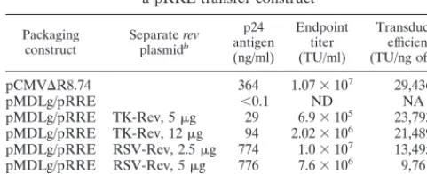
TABLE 4. GFP transduction into HeLa cells by lentivirus vectors
made by linked or split packaging constructs and
a pRRL transfer construct
aPackaging
construct Separate revplasmidb
p24 antigen (ng/ml)
Endpoint titer (TU/ml)
Transduction efficiency (TU/ng of p24)
pCMVDR8.74 364 1.073107 29,436
pMDLg/pRRE ,0.1 ND NA
pMDLg/pRRE TK-Rev, 5mg 29 6.93105 23,793 pMDLg/pRRE TK-Rev, 12mg 94 2.023106 21,489 pMDLg/pRRE RSV-Rev, 2.5mg 774 1.03107 13,495 pMDLg/pRRE RSV-Rev, 5mg 776 7.63106 9,761 pMDLg/pRRE RSV-Rev, 12mg 565 4.83106 8,495
aVectors carrying a PGK-eGFP expression cassette were produced by the transfection of a self-inactivating pRRL transfer construct (with a deletion in the 39LTR [53]), the indicated packaging and rev plasmids, and plasmid pMD.G into 293T cells. Serial dilutions of transfectant conditioned medium were incubated with HeLa cells, and the cultures were scored after 6 days. For calculating endpoint titers, samples were selected from the linear portion of the vector dose-response curve. Data are averages of duplicate determination for a repre-sentative experiment of three performed. ND, none detected (the detection limit of the assay was 102TU/ml); NA, not applicable.
bThe promoter driving the expression of a synthetic rev cDNA and the amount of plasmid transfected are indicated.
on November 9, 2019 by guest
http://jvi.asm.org/
in producer cells thus appears to be a rate-limiting factor for
transduction until it reaches a threshold. Conceivably, an
up-per limit is set by the total output of particles available to
en-capsidate vector RNA. As the total particle output varied with
the types of vector and internal promoter used, this may
ex-plain the quantitative differences obtained in response to tat
deletion.
Successful deletion of the tat gene was unexpected in view of
a reported additional role for Tat in reverse transcription (17,
20). While the reasons for this discrepancy are not obvious, it
should be noted that the transduction pathway of the lentivirus
vector mimics only in part the infection pathway of HIV. The
vector is pseudotyped by the envelope of an unrelated virus
and contains only the core proteins of HIV, without any
ac-cessory gene product. The VSV envelope targets the vector to
the endocytic pathway, and it has been shown that redirection
of HIV-1 from its normal route of entry by fusion at the plasma
membrane significantly changes the biology of the infection.
For example, Nef and cyclophilin A are required for the
opti-mal infectivity of wild-type HIV-1 but not of a (VSV G) HIV
pseudotype (1). It is also possible that the kinetics of reverse
transcription are more critical for the establishment of viral
infection than for gene transduction, given the differences in
size and sequence between the virus and vector genome.
Tat-independent transduction by an HIV-based vector was
recently reported by Kim et al. for in vitro cellular targets (23).
In the vector designed by these authors, however, Tat and Rev
were expressed from the transfer vector and thus were also
present in target cells. A CMV-HIV hybrid LTR was used; this
construct yielded vector titers approximately 30% of that
ob-tained with an intact LTR. When the tat gene was inactivated,
the titer did not change. Srinivasakumar et al. (43) previously
reported a rather low (5- to 10-fold) dependence on Tat of an
HIV-based vector produced by cells stably expressing the HIV
structural proteins. In this case, titers of 5
3
10
3TU/ml with
Tat and 7
3
10
2TU/ml without Tat were obtained on
HeLa-CD4 cells. Although these titers are much lower than those
reported here, the vector particles carried the HIV envelope,
an indication that Tat is not absolutely required for
transduc-tion by vector particles which in that case mirror more closely
the wild-type virus. It remained possible, however, that a
depen-dence on Tat may be revealed in more challenging gene
deliv-eries into the body tissues that are the actual targets of gene
therapy. This could have been due to a stricter Tat
require-ment for optimal transduction efficiency or for the production
of high-titer vector stocks or to differences in cell-type-specific
factors. Our results now establish that Tat is fully dispensable
for lentivirus vector transduction even when high titers are
achieved and, most importantly, for gene delivery in vivo into
terminally differentiated neurons of an adult rat brain.
The Northern analysis of producer and target cells shows
that the Tat dependence of LTR-driven expression restricts the
production of vector genomic RNA to producer cells. This
applies as well to vectors made by the 5
9
chimeric constructs, as
the U3 sequences of both LTRs of the resulting provirus are
derived from the vector 3
9
LTR. However, the functional
re-placement of the tat gene in the packaging construct by
pro-moter sequences upstream of the transfer construct makes
the generation of a transcriptionally active recombinant much
more unlikely. This will be even more significant in stable
producer cell lines that avoid the risk of plasmid recombination
during cotransfection.
We also exploited the Rev dependence of gag-pol expression
and of the accumulation of unspliced, packageable transcripts.
Yu et al. (50) previously showed that the dependence on Rev
can be used to make expression of HIV genes inducible. We
describe a core packaging system split in two separate
nonover-lapping expression constructs, one for the gag and pol reading
frames optimized for Rev-dependent expression and the
oth-er for the rev cDNA. This third-genoth-eration packaging system
matches the performance of its predecessors in terms of both
yield and transducing efficiency. However, it increases
signifi-cantly the predicted biosafety of the vector. It has been
sug-gested that the Rev-RRE axis could be replaced by the use of
constitutive RNA transport elements of other viruses, although
at the price of decreased efficiency (11, 23, 43). We would
sug-gest that maintaining the Rev dependence of the system allows
for an additional level of biosafety through the splitting of the
HIV-derived components of the packaging system.
The conditional packaging system described here can be
combined with a self-inactivating vector construct carrying a
major deletion in the 3
9
LTR (52). This vector design (Fig. 4)
offers significant biosafety features. The contribution of HIV
is reduced to a fraction of cis-acting sequences in the vector,
leaving out in particular most of the LTR, and to only three
genes, gag, pol, and rev, in the packaging constructs, compared
with the nine genes necessary for the in vivo replication and
pathogenesis of wild-type HIV-1 (3, 18, 27, 49). The actual
bio-safety of a vector must be proven in vivo. However, given the
serious limitations of the available animal models of
HIV-induced disease, the biosafety of HIV-derived vectors will
ul-timately be proven only in human hosts. Therefore, the vector
design must ensure the highest predictable biosafety for
clini-cal testing to be acceptable.
[image:7.612.309.549.69.214.2]It is noteworthy that the fraction of the HIV-1 genome that
is left in the vector is probably smaller than could be achieved
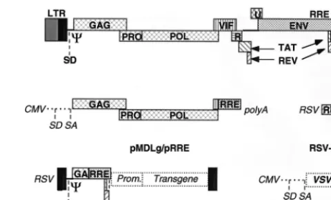
FIG. 4. Schematic drawing of the HIV provirus and the four constructs used to make a lentivirus vector of the third generation. The viral LTRs, the reading frames of the viral genes, the major 59splice donor site (SD), the packaging sequence (C), and the RRE are boxed and indicated in bold type. The condi-tional packaging construct, pMDLg/pRRE, expresses the gag and pol genes from the CMV promoter and intervening sequences and polyadenylation site of the humanb-globin gene. As the transcripts of the gag and pol genes contain cis-repressive sequences, they are expressed only if Rev promotes their nuclear export by binding to the RRE. All tat and rev exons have been deleted, and the viral sequences upstream of the gag gene have been replaced. A nonoverlapping construct, RSV-Rev, expresses the rev cDNA. The transfer construct, pRRL. SIN-18, contains HIV-1 cis-acting sequences and an expression cassette for the transgene. It is the only portion transferred to the target cells and does not contain wild-type copies of the HIV LTR. The 59LTR is chimeric, with the enhancer/promoter of RSV replacing the U3 region (RRL) to rescue the tran-scriptional dependence on Tat. The 39LTR has an almost complete deletion of the U3 region, which includes the TATA box (from nucleotides2418 to218 relative to the U3/R border). As the latter is the template used to generate both copies of the LTR in the integrated provirus, transduction of this vector results in transcriptional inactivation of both LTRs; thus, it is a self-inactivating vector (SIN-18). The fourth construct, pMD.G, encodes a heterologous envelope to pseudotype the vector, here shown coding for VSV G. Only the relevant parts of the constructs are shown.
on November 9, 2019 by guest
http://jvi.asm.org/
with any of the nonprimate lentiviruses, the genomic
complex-ity of which is lower than that of HIV-1 (37). Also, the risks
associated with the introduction in humans of a recombinant
arising from a nonprimate lentivirus, even in a form that in its
cognate animal species appears to be attenuated, are very
difficult to assess, as illustrated by the ongoing debate on
xeno-transplantation (48). In contrast, the almost two decades spent
studying a virus that has now spread in tens of millions of
people worldwide have revealed a considerable amount of
information on the pathogenic features of HIV-1, in particular
on the dependence of virulence on a crucial set of viral genes.
Based on these data, we would like to suggest that the
HIV-based vectors described here are good candidates for the
clin-ical trial of lentivirus vectors in human gene therapy.
ACKNOWLEDGMENTS
We are indebted to Tom Hope for providing the Rev expression
plasmids, to Melinda Van Roey and Heidi Oline for help with the
animal experiments, and to Jennifer Davis and Mitch Finer for
sug-gestions and critical reading of the manuscript.
This work was partly supported by a grant and by a fellowship from
the Swiss National Science Foundation to D.T. and R.Z., respectively.
REFERENCES
1. Aiken, C. 1997. Pseudotyping human immunodeficiency virus type 1 (HIV-1) by the glycoprotein of vesicular stomatitis virus targets HIV-1 entry to an endocytic pathway and suppresses both the requirement for Nef and the sensitivity to cyclosporin A. J. Virol. 71:5871–5877.
2. Aldovini, A., and R. A. Young. 1990. Mutations of RNA and protein se-quences involved in human immunodeficiency virus type 1 packaging result in production of noninfectious virus. J. Virol. 64:1920–1926.
3. Aldrovandi, G. M., and J. A. Zack. 1996. Replication and pathogenicity of human immunodeficiency virus type 1 accessory gene mutants in SCID-hu mice. J. Virol. 70:1505–1507.
4. Berkowitz, R. D., M. L. Hammarskjo¨ld, C. Helga-Maria, D. Rekosh, and
S. P. Goff.1995. 59regions of HIV-1 RNAs are not sufficient for encapsida-tion: implications for the HIV-1 packaging signal. Virology 212:718–723. 5. Blo¨mer, U., L. Naldini, T. Kafri, D. Trono, I. M. Verma, and F. H. Gage.
1997. Highly efficient and sustained gene transfer in adult neurons with a lentivirus vector. J. Virol. 71:6641–6649.
6. Bordignon, C., C. Bonini, S. Verzeletti, N. Nobili, D. Maggioni, C.
Traver-sari, R. Giavazzi, P. Servida, E. Zappone, E. Benazzi, F. Porta, G. Ferrari, F. Mavilio, S. Rossini, R. M. Blaese, and F. Candotti.1995. Transfer of the HSV-tk gene into donor peripheral blood lymphocytes for in vivo modula-tion of donor anti-tumor immunity after allogeneic bone marrow transplan-tation. Hum. Gene Ther. 6:813–819.
7. Buchschacher, G. L. J., and A. T. Panganiban. 1992. Human immunodefi-ciency virus vectors for inducible expression of foreign genes. J. Virol. 66: 2731–2739.
8. Bukrinsky, M. I., S. Haggerty, M. P. Dempsey, N. Sharova, A. Adzhubel, L.
Spitz, P. Lewis, D. Goldfarb, M. Emerman, and M. Stevenson.1993. A nuclear localization signal within HIV-1 matrix protein that governs infec-tion of non-dividing cells. Nature 365:666–669.
9. Burns, J. C., T. Friedmann, W. Driever, M. Burrascano, and J.-K. Yee. 1993. Vesicular stomatitis virus G glycoprotein pseudotyped retroviral vectors: concentration to very high titer and efficient gene transfer into mammalian and non-mammalian cells. Proc. Natl. Acad. Sci. USA 90:8033–8037. 10. Coffin, J. M. 1996. Retroviridae: the viruses and their replication, p. 1767–
1846. In B. N. Fields, D. M. Knipe, P. M. Howley, R. M. Chanock, J. L. Melnick, T. P. Monath, B. Roizman, and S. E. Straus (ed.), Fields virology, 3rd ed. Lippincott-Raven Publishers, Philadelphia, Pa.
11. Corbeau, P., G. Kraus, and F. Wong-Staal. 1998. Transduction of human macrophages using a stable HIV-1/HIV-2-derived gene delivery system. Gene Ther. 5:99–104.
12. Feinberg, M. B., D. Baltimore, and A. L. Frankel. 1991. The role of Tat in the human immunodeficiency virus life cycle indicates a primary effect on tran-scriptional elongation. Proc. Natl. Acad. Sci. USA 88:4045–4049. 13. Felber, B. K., C. M. Drysdale, and G. N. Pavlakis. 1990. Feedback regulation
of human immunodeficiency virus type 1 expression by the Rev protein. J. Virol. 64:3734–3741.
14. Finer, M. H., T. J. Dull, L. Qin, D. Farson, and M. R. Roberts. 1994. kat: a high efficiency retroviral transduction system for primary human T lympho-cytes. Blood 83:43–50.
15. Gallay, P., D. Chin, T. J. Hope, and D. Trono. 1997. HIV-1 infection of nondividing cells mediated through the recognition of integrase by the im-port/karyopherin pathway. Proc. Natl. Acad. Sci. USA 94:9825–9830.
16. Gallay, P., S. Swingler, C. Aiken, and D. Trono. 1995. HIV-1 infection of nondividing cells: C-terminal tyrosine phosphorylation of the viral matrix protein is a key regulator. Cell 80:379–388.
17. Harrich, D., C. Ulich, L. F. Garcia-Martinez, and R. B. Gaynor. 1997. Tat is required for efficient reverse transcription. EMBO J. 16:1224–1235. 18. Haynes, B. F., G. Pantaleo, and A. S. Fauci. 1996. Toward an understanding
of the correlates of protective immunity to HIV infection. Science 271:324– 328.
19. Ho, D. D., A. U. Neumann, A. S. Perelson, W. Chen, J. M. Leonard, and M.
Markowitz.1995. Rapid turnover of plasma virions and CD4 lymphocytes in HIV-1 infection. Nature 373:123–126.
20. Huang, L. M., A. Joshi, R. Willey, J. Orenstein, and K. T. Jeang. 1994. Human immunodeficiency viruses regulated by alternative trans-activators: genetic evidence for a novel non-transcriptional function of Tat in virion infectivity. EMBO J. 13:2886–2896.
21. Kafri, T., U. Blo¨mer, D. A. Peterson, F. H. Gage, and I. M. Verma. 1997. Sustained expression of genes delivered directly into liver and muscle by lentiviral vectors. Nat. Genet. 17:314–317.
22. Kaye, J. F., J. H. Richardson, and A. M. L. Lever. 1995. cis-acting sequences involved in human immunodeficiency virus type 1 RNA packaging. J. Virol.
69:6588–6592.
23. Kim, V. N., K. Mitrophanous, S. M. Kingsman, and A. J. Kingsman. 1998. Minimal requirement for a lentivirus vector based on human immunodefi-ciency virus type 1. J. Virol. 72:811–816.
24. Lever, A., H. Gottlinger, W. Haseltine, and J. Sodroski. 1989. Identification of a sequence required for efficient packaging of human immunodeficiency virus type 1 RNA into virions. J. Virol. 63:4085–4087.
25. Lewis, P. F., M. Hensel, and M. Emerman. 1992. Human immunodeficiency virus infection of cell arrested in the cell cycle. EMBO J. 11:3053–3058. 26. Lewis, P. F., and M. Emerman. 1994. Passage through mitosis is required for
oncoretroviruses but not for the human immunodeficiency virus. J. Virol. 68: 510–516.
27. Luciw, P. A. 1996. Human immunodeficiency viruses and their replication, p. 1881–1975. In B. N. Fields, D. M. Knipe, P. M. Howley, R. M. Chanock, J. L. Melnick, T. P. Monath, B. Roizman, and S. E. Straus (ed.), Fields virology, 3rd ed. Lippincott-Raven Publishers, Philadelphia, Pa.
28. Mandel, R. J., K. G. Rendahl, K. S. Spratt, R. O. Snyder, L. K. Cohen, and
S. E. Leff.Characterization of intrastriatal recombinant adeno-associated virus mediated gene transfer of human tyrosine hydroxylase and human GTP-cyclohydroxylase I in a rat model of Parkinson’s disease. J. Neurosci., in press.
29. McBride, M. S., and A. Panganiban. 1996. The human immunodeficiency virus type 1 encapsidation site is a multipartite RNA element composed of functional hairpin structures. J. Virol. 70:2963–2973.
30. McBride, M. S., M. D. Schwartz, and A. Panganiban. 1997. Efficient encap-sidation of human immunodeficiency virus type 1 vectors and further char-acterization of cis elements required for encapsidation. J. Virol. 71:4544– 4554.
31. Miyoshi, H., M. Takahashi, F. H. Gage, and I. M. Verma. 1997. Stable and efficient gene transfer into the retina using an HIV-based lentiviral vector. Proc. Natl. Acad. Sci. USA 94:10319–10323.
32. Naldini, L., U. Blo¨mer, P. Gallay, D. Ory, R. Mulligan, F. H. Gage, I. M.
Verma, and D. Trono.1996. In vivo gene delivery and stable transduction of nondividing cells by a lentiviral vector. Science 272:263–267.
33. Naldini, L., U. Blo¨mer, F. H. Gage, D. Trono, and I. M. Verma. 1996. Efficient transfer, integration, and sustained long-term expression of the transgene in adult rat brains injected with a lentiviral vector. Proc. Natl. Acad. Sci. USA 93:11382–11388.
34. Ory, D. S., B. A. Neugeboren, and R. C. Mulligan. 1996. A stable human-derived packaging cell line for production of high titer retrovirus/vesicular stomatitis virus G pseudotypes. Proc. Natl. Acad. Sci. USA 93:11400–11406. 35. Parolin, C., T. Dorfman, G. Palu, H. Gottlinger, and J. Sodroski. 1994. Analysis in human immunodeficiency virus type 1 vectors of cis-acting se-quences that affect gene transfer into human lymphocytes. J. Virol. 68:3888– 3895.
36. Paxinos, G., and C. Watson. 1987. The rat brain in stereotaxic coordinates. Academic Press, San Diego, Calif.
37. Poeschla, E., F. Wong-Staal, and D. J. Looney. 1998. Efficient transduction of nondividing human cells by feline immunodeficiency virus lentiviral vec-tors. Nat. Med. 4:354–357.
38. Poznansky, M., A. Lever, L. Bergeron, W. Haseltine, and J. Sodroski. 1991. Gene transfer into human lymphocytes by a defective human immunodefi-ciency virus type 1 vector. J. Virol. 65:532–536.
39. Reiser, J., G. Harmison, S. Kluepfel-Stahl, R. O. Brady, S. Karlsson, and M.
Schubert.1996. Transduction of nondividing cells pseudotyped defective high-titer HIV type 1 particles. Proc. Natl. Acad. Sci. USA 93:15266–15271. 40. Schneider, R., M. Campbell, G. Nasioulas, B. K. Felber, and G. N. Pavlakis. 1997. Inactivation of the human immunodeficiency virus type 1 inhibitory elements allows Rev-independent expression of Gag and Gag/protease and particle formation. J. Virol. 71:4892–4903.
41. Schwartz, S., M. Campbell, G. Nasioulas, J. Harrison, B. K. Felber, and
G. N. Pavlakis.1992. Mutational inactivation of an inhibitory sequence in
on November 9, 2019 by guest
http://jvi.asm.org/
human immunodeficiency virus type 1 results in Rev-independent gag ex-pression. J. Virol. 66:7176–7182.
42. Shimada, T., H. Fujii, A. Mitsuya, and W. Nienhuis. 1991. Targeted and highly efficient gene transfer into CD41cells by a recombinant human im-munodeficiency virus retroviral vector. J. Clin. Investig. 88:1043–1047. 43. Srinivasakumar, N., N. Chazal, C. Helga-Maria, S. Prasad, M.
Hammar-skjold, and D. Rekosh.1997. The effect of viral regulatory protein expression on gene delivery by human immunodeficiency virus type 1 vectors produced in stable packaging cell lines. J. Virol. 71:5841–5848.
44. Sternberger, L. A., P. H. Hardy, J. J. Cuculis, and H. G. Meyer. 1970. The unlabelled antibody-enzyme method of immunohistochemistry. Preparation and properties of soluble antigen-antibody complex (horseradish peroxidase-antihorseradish peroxidase) and its use in the identification of spirochetes. J. Histochem. Cytochem. 18:315–333.
45. Verma, I. M., and N. Somia. 1997. Gene therapy promises, problems and prospects. Nature 389:239–242.
46. Wei, P., M. E. Garber, S.-M. Fang, W. H. Fischer, and K. A. Jones. 1998. A novel CDK9-associated C-type cyclin interacts directly with HIV-1 Tat and
mediates its high-affinity, loop-specific binding to TAR RNA. Cell 92:451– 462.
47. Wei, X., S. K. Ghosh, M. E. Taylor, V. A. Johnson, E. A. Emini, P. Deutsch,
J. D. Lifson, S. Bonhoeffer, M. A. Nowak, B. H. Hahn, M. S. Saag, and G. M. Shaw.1995. Viral dynamics in human immunodeficiency virus type 1 infec-tion. Nature 373:117–122.
48. Weiss, R. A. 1998. Transgenic pigs and virus adaptation. Nature 391:327–328. 49. Wyand, M. S., K. H. Manson, A. A. Lackner, and R. C. Desrosiers. 1997. Resistance of neonatal monkeys to live attenuated vaccine strains of simian immunodeficiency virus. Nat. Med. 3:32–36.
50. Yu, H., A. B. Rabson, M. Kaul, Y. Ron, and J. P. Dougherty. 1996. Inducible human immunodeficiency virus type 1 packaging cell lines. J. Virol. 70:4530– 4537.
51. Zufferey, R., D. Nagy, R. J. Mandel, L. Naldini, and D. Trono. 1997. Multiply attenuated lentiviral vector achieves efficient gene delivery in vivo. Nat. Biotechnol. 15:871–875.
52. Zufferey, R., T. Dull, R. J. Mandel, A. Bukovsky, D. Quiroz, L. Naldini, and
D. Trono.Self-inactivating lentivirus vector for safe and efficient in vivo gene delivery. J. Virol., in press.
on November 9, 2019 by guest
http://jvi.asm.org/