D
EVELOPMENT
AND
A
PPLICATION
OF
L
ABEL
F
REE
Q
UANTITATIVE
P
ROTEOMIC
M
ETHODS
Thesis submitted in accordance with the requirements of the University of Liverpool for the degree of Doctor of Philosophy
by
A
CKNOWLEDGEMENTS
I firstly would like to thank every member, past and present of the protein function group, who have
all individually played a role in creating the scientist and person I am today. They have all influenced
me in some way and helped me to grow so much since I met them four years ago. I would especially
like to thank Jenny Rivers who guided me so smoothly through the transition from undergraduate to
Ph.D. student and Philip Brownridge who has rescued me on numerous occasions, and been a
constant fountain of knowledge that has allowed me to transform into a scientist. Special thanks go
to my supervisor Rob Beynon for providing me with a conducive environment to perform my
research and for his continuous essential guidance and knowledge.
I also want to thank the many housemates and friends who have endured this time with me, who
have supported, encouraged and ultimately who have guided me through this journey. Especially
Amy Claydon, who unfortunately for her has lived and worked with me every single day of my PhD,
so has been an amazing pillar of support and friendship throughout.
I am very grateful to the BBSRC and Genus PLC for funding this research project and a special thank
you goes to Stuart Revell who trained me during my first few years as a Ph.D. student and has always
been on hand with invaluable knowledge. I also want to thank both Marj Faust and Alan Mileham
who have all provided essential guidance throughout my Ph.D.
I also need to thank my parents for their never wavering support, for always believing in me and for
constantly supporting and encouraging me. I also want to thank them for listening and advising me,
A
BSTRACT
TITLE:
DEVELOPMENT
ANDAPPLICATION
OFLABEL
FREE
QUANTITATIVE
PROTEOMIC
METHODS
A
UTHOR:
R
EBECCAC
UMMINGSThe aim of this Ph.D. was to develop advanced methods for quantitative proteomics and use these methods to
investigate the presence of protein biomarkers of sperm performance, differential expression of sperm
membrane proteins and differential expression of E.coli proteins.
Quantitative analysis of E.coli generated analytical samples that were analysed with multiple mass
spectrometers and with multiple software packages. Through these samples an optimal label free quantitative
proteomic workflow was generated and software was thoroughly tested to determine the optimal software to
be used for data analysis on varying biological questions. Identification of protein(s) that correlate with
increased or decreased fertility would be economically beneficial. Currently semen samples are subject to
quality control where general movement and morphological defects are studied, but this does not always
correlate with the ejaculate passing a post cryopreservation quality control check or that specific bull
generating offspring. Identification of a protein or set of proteins with abundance variation in bulls of known
high or low fertility would allow lower fertility bulls to be removed from the breeding programme at an early
age, reducing rearing costs, and would allow longitudinal health monitoring of individual bulls. Discovery of
differentially expressed proteins in the membrane of sperm with the X or Y chromosome would allow the
generation of a method to separate the two sperm populations. This will be beneficial as most livestock
farmers would prefer offspring of a specific sex, either to sell or replenish animal stock.
Quantitative analysis of proteins present in bovine seminal plasma led to the identification and quantitative
comparison of the seminal plasma proteins present in two breeds of bull, Holstein and Belgian Blue and a
quantitative comparison of the seminal plasma from two domestic farm animal species, bovine and porcine.
Intra species comparisons determined no quantitative variation between the two breeds, while the inter
species comparison determined variation between the proteins present in both species seminal plasma and
the corresponding amounts of proteins present in both species. A quantitative comparison was performed to
determine the expression of proteins from two strains of E.coli, a wild type strain (MG1655) and a genome
depleted strain (MDS66), this led to the confirmation of gene deletions in the genome depleted strain due to
their lack of protein products in mass spectrometric analysis, and the identification of proteins that were
differentially expressed due to pleiotropic effects of these genome deletions. To investigate the proteins
expressed in the sperm membrane a mass spectrometer compatible enrichment method was generated and
membrane proteins were identified, quantified and compared between sperm expressing X and Y
chromosomes. This study did not lead to the determination of any proteins with differential expression in the
T
ABLE
OF
C
ONTENTS
List of Figures………i
List of Tables……….v
List of Appendices………vi
1. Introduction
... 1
1.1. Reproduction Industry Challenges ... 2
1.2. Biomarker Discovery ... 4
1.3. Spermatogenesis and Fertilisation ... 6
1.4. Proteomics ... 21
1.5. Mass Spectrometry ... 33
1.6. Hypotheses and General Methodology ... 40
2. Materials and Methods
... 42
Materials ... 42
Sample Collection and Preparation 2.1. Semen Collection ... 42
2.2. BoviPure™ sperm separation ... 42
2.3. Sperm Cell Counting ... 42
2.4. Sperm Viability Quality Control ... 43
2.5. Sperm Lysis ... 43
2.6. Culturing of Escherichia Coli and Lysis ... 43
2.7. Protein Assay ... 44
Sample Separation 2.8. One Dimensional SDS‐Poly Acrylamide Gel Electrophoresis (1D SDS PAGE) ... 44
2.9. Two Dimensional SDS‐Poly Acrylamide Gel Electrophoresis (2D SDS PAGE) ... 45
2.11. StrataClean Resin ... 46
2.12. In‐Gel Proteolysis ... 46
2.13. In‐Solution Proteolysis ... 46
Peptide Separation 2.14. Reverse Phase High Performance Liquid Chromatography (RP‐HPLC) ... 47
2.15. Reverse Phase Ultra Performance Liquid Chromatography (RP‐UPLC) ... 47
Mass Spectrometry 2.16. MALDI‐TOF MS ... 47
AXIMA ToF2 (Shimadzu)………47
UltraFlex (Bruker)……….48
2.17. Electrospray Ionisation MS ... 48
ESI LTQ (Thermo)………..48
ESI LTQ Orbitrap (Thermo)……….48
ESI QToF Synapt™ G1 (Waters)...………..48
ESI QToF Synapt™ G2 (Waters)……… ……….………..49
ESI Xevo® TQ (Waters)………..49
Analysis and Database Searching 2.18. MASCOT ... 49
Peptide Mass Fingerprinting (PMF) ... 49
MS/MS ... 49
2.19. Protein Lynx Global Server (PLGS) ... 50
Processing Data Acquired Through MSE ... 50
Processing Data Acquired Through HDMSE ... 50
Database Searching ... 50
2.20. ISOQuant ... 51
2.21. Progenesis LC‐MS ... 51
2.22. Statistical Analysis ... 52
3. Proteomic Comparison of Wild Type and Genome Depleted
Escherichia
Coli
... 53
Objectives ... 53
3.1. Introduction ... 54
Escherichia Coli………..54
Quantitative proteomics……….58
3.2. Overview of experimental workflow ... 60
3.3. Results and discussion ... 61
Technical and biological replication………61
Method development………..64
Deletion of proteins through the deletion of their coding genes………..93
Pleiotropic effects of the gene deletions……….98
3.4. Conclusions ... 106
4. Global Profiling of Bovine and Porcine Seminal Plasma
... 107
Aims ... 107
Objectives ... 107
4.1. Introduction ... 107
Seminal plasma………107
Roles of seminal plasma proteins………..108
Proteomic markers of fertility………..113
Seminal plasma protein comparisons……….113
4.2. Overview of experimental workflow ... 115
4.3. Results and discussion ... 117
Bovine seminal plasma protein profile………..117
Comparison of bovine seminal plasma proteins……….129
Porcine seminal plasma protein profile……….142
Comparison of bovine and porcine seminal plasma proteins………149
4.4. Conclusions ... 156
5. Spermatozoa Membrane Proteomics
... 158
Objectives ... 158
5.1. Introduction ... 158
Sperm plasma membrane structure………158
Roles of the membrane in fertilisation………..161
Sex sorting semen……….161
Proteomic markers indicating whether the sperm has an X or Y chromosome………...165
Methods for membrane proteomics………166
Egg yolk extender………..168
Selected reaction monitoring (SRM) mass spectrometry……….169
5.2. Overview of experimental work ... 170
5.3. Results and discussion ... 173
Extender removal………..173
Membrane enrichment method development……….181
Candidate list generation and SRM quantification of sperm membrane proteins………..198
Analysis of X Y sorted sperm……….202
5.4. Conclusions ... 210
6. General Discussion
... 212
6.1. Analytical challenges ... 212
6.2. Experimental challenges ... 215
6.3. Further work ... 216
6.4. Final conclusions ... 216
References
... 218
L
IST
OF
F
IGURES
1.
Introduction
1.1. Sire progeny information for farmers ... 5
1.2. Anatomy of the testis ... 7
1.3. Spermatogenesis ... 8
1.4. Anatomy of a spermatozoon ... 11
1.5. Examples of common morphological problems of spermatozoa ... 16
1.6. Anatomical diagram of the reproductive organs of the bull ... 17
1.7. 1D and 2D SDS PAGE ... 23
1.8. Reduction and alkylation of protein cysteine‐cysteine double bonds ... 26
1.9. Peptide mass fingerprinting ... 27
1.10. A typical example of MS/MS analysis of a peptide ... 29
1.11. Overlapping b and y ion sequence ... 30
3.
Proteomic
Comparison
of
Wild
Type
and
Genome
Depleted
Escherichia
Coli
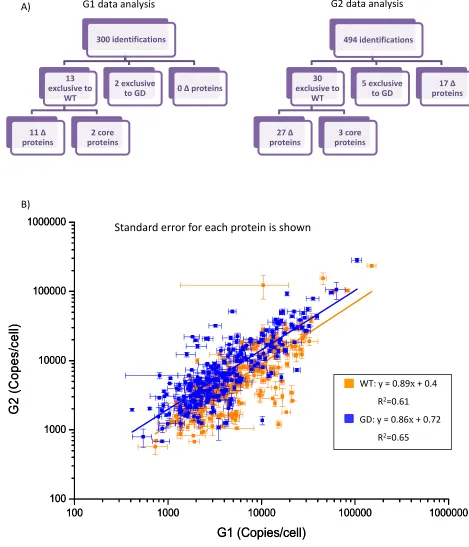
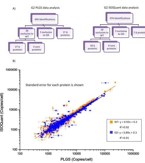
3.1. PaxDB abundance data from the 875 ∆ proteins in the GD strain ... 56
3.2. E.coli flagella complex ... 57
3.3. PaxDB abundance data from the flagellar Δ proteins in the GD strain ... 59
3.4. 1D SDS PAGE protein profile of E.coli biological replicates ... 62
3.5. Validation of chromatographic performance using base peak chromatograms ... 63
3.6. Label free quantitative verification of an ∆ protein ... 65
3.7. Label free quantitative verification of a non ∆ protein ... 67
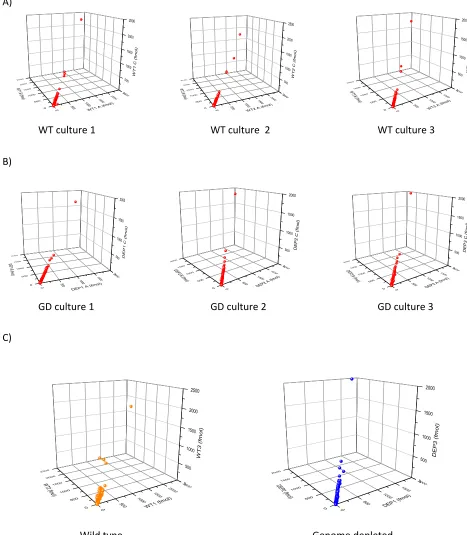
3.8. Synapt™ G1 MSE technical and biological reproducibility, following RP‐UPLC and PLGS analysis ... 69
3.9. Label free mass spectrometric verification of WT and GD E.coli proteomes, facilitated by a first generation Synapt™ QuanToF instrument ... 70
3.12. Comparison of Synapt™ QuanToF first and second generation mass spectrometers... 74
3.13. Synapt™ G2 MSE technical and biological reproducibility, following RP‐UPLC with PLGS and ISOQuant analysis ... 76
3.14. Label free mass spectrometric verification of WT and GD E.coli proteomes, facilitated by a second generation Synapt™ QuanToF instrument, quantification performed using ISOQuant ... 77
3.15. Comparison of quantitation software ... 78
3.16. Synapt™ G2 HDMSE technical and biological reproducibility, following RP‐UPLC with PLGS and ISOQuant analysis ... 80
3.17. Label free mass spectrometric verification of WT and GD E.coli proteomes, facilitated by a Synapt™ G2 an additional ion mobility separation, quantification perfromed with ISOQuant ... 81
3.18. Comparison of MSE and HDMSE data acquisition ... 83
3.19. LTQ Orbitrap Velos technical and biological reproducibility, following RP‐UPLC with Progenesis LC‐MS and MASCOT analysis ... 84
3.20. Relative label free mass spectrometric verification of WT and GD E.coli proteomes, facilitated by an LTQ Orbitrap Velos ... 85
3.21. Relative label free mass spectrometric verification of WT and GD E.coli proteomes, facilitated by an LTQ Orbitrap Velos including gas phase fractionation………..87
3.22. Relative label free quantitative comparison of instrument acquisition methods, gas phase fractionation or none………..88
3.23. Label free quantitative comparison of instruments, Synapt™ G2 and LTQ Orbitrap Velos.….90 3.24. Label free quantitative comparison of instruments, Synapt™ G2 and LTQ Orbitrap Velos.….91 3.25. Comparison of abundnce of all proteins………..92
3.26. Identification of ∆ proteins………94
3.27. Comparison of abundance of Δ proteins in WT strain……….99
3.28. Pleiotropic down regulation of pyrimidine biosynthesis proteins in GD strain………..101
3.29. Network interaction data for pleiotropic down regulated proteins in GD strain………….…….102
3.30. Network interaction data for pleiotropic up regulated proteins in GD strain……….104
3.31. Pleiotropic up regulation of pyrimidine biosynthesis proteins in GD strain……….…105
4.
Global
Profiling
of
Bovine
and
Porcine
Seminal
Plasma
4.1. Interaction of epididymal and seminal vesicle proteins with sperm membrane……….109
4.2. Function of the BSP seminal vesicle proteins………111
4.4. Protein profile of bovine seminal plasma………118
4.5. MALDI‐ToF PMF protein identifications in bovine seminal plasma……….. 119
4.6. 2D SDS PAGE protein profile of bovine seminal plasma……… 121
4.7. Individual bull seminal plasma protein variation……… 122
4.8. 2D SDS PAGE protein identifications……….. 124
4.9. ProSpectrum™ beads mechanism……… 125
4.10. ProSpectrum™ bead analysis………. 126
4.11. Total number of seminal plasma proteins identified by each method……… 128
4.12. Number of unique seminal plasma proteins identified by each method………128
4.13. Comparison of bovine MASCOT identifications after LTQ Orbitrap Velos analysis………. 130
4.14. Bull producing and not producing sperm; seminal plasma protein concentration and profile………. 131
4.15. Bulls with high and low total protein; seminal plasma concentration and profile………..132
4.16. Bulls with high and low sperm motility; seminal plasma concentration and profile…………..134
4.17. Bulls with increased and decreased critical osmolarity post thaw; seminal plasma protein concentration and profile……….135
4.18. Visualisation of the seminal plasma profile of different bovine breeds……….137
4.19. Comparison of Holstein and Belgian Blue bull seminal plasma protein composition, quantitation performed with PLGS…… ………138
4.20. Verification of bovine seminal plasma proteins found in one breed only……….139
4.21. Comparison of Holstein and Belgian Blue bull seminal plasma protein composition, quantitation performed with ISOQuant..………..141
4.22. Protein profile of porcine seminal plasma…. ... 143
4.23. MALDI‐ToF PMF identifications of proteins in porcine seminal plasma ... 144
4.24. Comparison of porcine MASCOT identifications after LTQ Orbitrap Velos analysis ... 147
4.25. Visualisation of different breed and species seminal plasma protein profiles ... 148
4.26.Comparison of bull and boar seminal plasma protein composition, quantitation performed with IOSQuant ... 150
4.27. Comparison of bull and boar seminal plasma protein concentration………151
4.28. Comparison of bull and boar seminal plasma protein composition, quantitation performed with PLGS………. 152
5.
Spermatozoa
Membrane
Proteomics
5.1. Sperm membrane components ... 160
5.2. Selected reaction monitoring ... 171
5.3. Protein profile of egg yolk extender ... 174
5.4. Protein profile of egg yolk extender and subsequent PBS washes of sperm ... 177
5.5. Total proteins identified following extender removal from sperm with sequential PBS washes ... 179
5.6. Total proteins identified following extender removal from sperm with a BoviPure™ gradient ... 180
5.7. Multiple sonication steps to enrich for membrane proteins ... 182
5.8. Functional analysis of proteins from multiple sonication steps ... 183
5.9. Multiple homogenisation steps to enrich for membrane proteins ... 186
5.10. Functional analysis of multiple homogenisation steps to enrich for membrane proteins .... 187
5.11. Multiple homogenisation steps plus deoxycholate detergent solubilisation to enrich for membrane proteins ... 189
5.12. Functional analysis of multiple homogenisation plus deoxycholate solubilsation to enrich for membrane proteins ... 190
5.13. Multiple lysis steps and buffers plus ultracentrifugation and a high concentration salt wash to enrich for membrane proteins ... 192
5.14. Comparison of membrane proteins identified with different methodologies ... 194
5.15. Ultracentrifugation of cyropreserved semen to enrich for membrane proteins ... 195
5.16. 1D SDS PAGE of lysed X and Y sorted sperm cells ... 203
5.17. Sperm lysate X and Y base peak chromatograms ... 205
5.18. Sperm membrane X and Y analysis with a triple quadrupole MS in SRM mode, examples of transition extracted ion chromatograms ... 207
5.19. Sperm membrane X and Y analysis with a triple quadrupole MS in SRM mode, examples of transition extracted ion chromatograms ... 208
5.20. SRM anaysis of proteins found in the sperm membrane ... 209
L
IST
OF
T
ABLES
3.
Proteomic
Comparison
of
Wild
Type
and
Genome
Depleted
Escherichia
Coli
3.1. Confirmed Δ proteins ... 95
3.2. Δ proteins that cannot be confirmed as deleted ... 97
3.3. Proteins exclusive to the WT strain ... 100
3.4. Proteins excluisve to the GD strain ... 100
4.
Global
Profiling
of
Bovine
and
Porcine
Seminal
Plasma
4.1. Bovine 1D SDS PAGE MALDI‐ToF protein identifications ... 120
4.2. MALDI ToF PMF identifications of proteins in porcine seminal plasma ... 145
5.
Spermatozoa
Membrane
Proteomics
5.1. Membrane protein extraction and solubilisation methods ... 167
5.2. Protein identifications in the egg yolk extender ... 175
5.3. Sperm washes to remove extender ... 178
5.4. BoviPure™ gradient to remove extender... 178
5.5. Ultracentrifugation of cryopreserved semen in straws to enrich for membrane proteins .... 196
5.6. Membrane proteins generated by Synapt™ G2 HDMSE analysis of an ultracentrifuged membrane preparation... 200
5.7. Selected candidate membrane proteins ... 201
5.8. Cell count and relative Synpat™ G2 analsis of X and Y sorted cells ... 206
L
IST
OF
A
PPENDICES
1. E.coli identifications (Orbitrap Progenesis)
2. E.coli identifications (G2 HDMSE ISOQuant)
3. Bovine 1D SDS PAGE identifications (LTQ) 4. Bovine 2D SDS PAGE
5. Bovine 2D SDS PAGE identifications (LTQ) 6. Bovine ProSpectrum bead identifications (LTQ) 7. Bovine in‐solution identifications (LTQ)
8. Bovine identifications (Orbitrap MASCOT) 9. Bovine identifications (G2 PLGS)
10. Bovine identifications (G2 ISOQuant) 11. Porcine identifications (Orbitrap MASCOT) 12. Porcine identifications (G2 PLGS)
13. Porcine identifications (G2 ISOQuant)
14. Method‐multiple sonication steps (Orbitrap MASCOT) 15. Method‐multiple homogenisation steps (Orbitrap MASCOT) 16. Method‐multiple homogenisation plus DOC (Orbitrap MASCOT) 17. Method‐homogenisation plus ultracentrifuge x2 (Orbitrap MASCOT) 18. Method‐homogenisation plus ultracentrifuge x2 (G2 HDMSE ISOQuant) 19. Sorted sperm (G2 HDMSE ISOQuant)
Found on disc attached to back cover of thesis
1.
I
NTRODUCTION
The main aim of this Ph.D. programme was to use advanced proteomic methods to investigate the
presence of biomarkers of sperm performance; through the identification of proteins in sperm or
seminal plasma that have the potential to correlate with a fertility parameter, through quantitative
variation in protein(s). The identification of proteomic health markers, such as a pattern of protein
abundance changes correlating with increased or decreased fertility in an individual, could allow
longitudinal health monitoring of individual bulls. This type of monitoring would be beneficial as
although every semen sample is subjected to quality control, sometimes visualisation and
morphological techniques cannot explain why a sample of semen that looks ‘good’ on initial
inspection, fails a post cryopreservation quality control check, or fails to generate offspring.
Identifying proteins that correlate with increased or decreased fertility could be utilised
commercially to identify animals with differential fertility at an early age, allowing the removal of
low fertility animals from the breeding programme. This would reduce costs as only animals with a
higher fertility would be reared to the age of four. Health markers that allow longitudinal monitoring
of individual animals could be utilised commercially to identify batches of semen that have lower
fertility than usual for that animal and discarded prior to processing. Identification of either fertility
and/or health markers would have a positive economic impact within a breeding/reproduction
company such as Genus PLC.
Another aim of this Ph.D. programme was to investigate the possibility that sperm membrane
proteins could be utilised to differentiate between the X and Y DNA bearing sperm populations,
within an ejaculate. This could be then taken forward into the creation of a method that could be
utilised commercially to separate the X and Y sperm, contributing an economic impact for the
company, as they would no longer need to outsource their sperm sorting to another company
(Cogent Breeding Ltd) and this reduction in cost could be passed to farmers. Sex markers could
impact the whole agricultural industry economy in a positive way, as breeders of the majority of
livestock make use of specific gender offspring. This research could also have a negative impact on
society due to ethical questions raised if sexing markers were isolated, and were able to be
transferred into human reproduction. This would allow people to choose the sex of their child, which
has long been an issue for humans as myths and social beliefs of ways to influence your child’s sex
have lasted for centuries, such as eating certain foods, or timing intercourse with phases of the
moon, but in cases of genetic sex linked diseases choosing the sex of your child can be medically
during fertility treatment, for example, isolation of the most motile sperm and does not involve any
embryo death. This research could impact on society as people would query if this research became
widely used, how long it would be before couples can choose not just specific sex of their child, but
also the colour of their eyes, and other genetic traits.
The X and Y bearing sperm differentiation component to this thesis could have led to results that
were commercially valuable and have high intellectual property (IP) value, which in turn would lead
to the creation of patents for any technologies created on the back of this research. Commercially
sensitive results would have been prevented from publication, both in scientific journals and at
conferences. The possibility of this censorship meant another robust platform was chosen to
develop the use of proteomic methodology, workflows, instrumentation and data analysis, plus
facilitating attendance at national and international conferences. This platform was Escherichia coli.
1.1.
REPRODUCTION
INDUSTRY
CHALLENGES
Genus PLC/ABS Global are a biotechnology company that has helped to advance the farming
industry initially in the UK, USA and now worldwide. They use the advances in artificial insemination
and semen storage, along with progeny testing, to deliver semen from the ‘best’ bulls to farms all
over the world.
Ejaculates are collected from bulls in a large collecting area with a deep wood chip floor. Teaser
animals are used for the bulls to mount as stimulation; they mount three times, on the fourth mount
the artificial vagina is placed over the penis and the bull immediately thrusts and ejaculates. The
bulls are rested for 20‐30min before a second collection is made. For some particularly fertile or in
demand animals a third collection is also made. After collection the semen is immediately weighed
(ml), and antibiotics are added. The viability of the cells is then tested; general movement (wave) is
examined by trained company staff members through visualisation of raw semen on a warm
microscope slide using a microscope (x10 magnification) and graded 1‐5. The quality of the sperm
movement which corresponds to roughly the percentage of the sperm that are progressively motile
are also examined though visualising a sample of diluted semen on a warm microscope slide under a
coverslip using a microscope (x100 magnification). If the sperm cells in the ejaculate are graded over
3 for wave movement and over 60% for progressively motile, they are taken forward for processing.
Sperm cells are counted using a NucleoCounter® SP‐100™ (ChemoMetec, Denmark). Sperm diluted
in phosphate buffered saline (PBS) as per manufacturer’s instructions are mixed with Reagent S100
which breaks down the cell membranes and are then taken up by a NucleoCassette™. Within the
DNA, the NucleoCounter® emits a green light and a red fluorescent signal is detected corresponding
to the number of cell nuclei, therefore the number of sperm cells. This is used to calculate the cell
number in the original solution, depending on the initial dilution. The sperm morphology is
examined using an eosin/nigrosin stain; raw sperm is mixed with warm eosin/nigrosin stain before
smearing onto a microscope slide and assessed under oil immersion (x1000 magnification). For each
assessment, 100 sperm are visualised and the proportion of morphological problems, such as bent
tails, deformed heads, and detached heads are counted, if the percentage of abnormalities is over
25% the ejaculate is discarded.
All of this data are recorded, along with the final dilution of sperm per straw required from the
individual bull. This varies between 8‐15million sperm cells per ml, a particularly fertile bull will be
diluted to 8million sperm/ml while samples from young bulls (usually less fertilie) are usually diluted
to 15million sperm/ml. Software calculates how much egg yolk extender is to be added to the
ejaculate to generate the correct sperm/ml. The egg yolk extender contains Tris buffer, citrate,
fructose and glycerol, with egg yolk used as a source of nutrients for the sperm. First the semen
samples are diluted with a 1:1 ratio and left to chill at 4°C for 2h. After 2h the remaining extender is
added. Once the samples have been chilled for a further 2h (4h after collection) the samples can be
processed. Usually multiple samples from each bull are combined and injected by machine into
straws of either 0.25 or 0.5ml depending on where the straws are being sent to; both North and
South America plus Canada use 0.5ml straws, while most other countries use 0.25ml. The straws are
then placed on racks of 150 and put into a chest freezer. The freezer has a starting temperature of
4°C and freezes the straws to ‐140°C over a 7min period, the straws are then stored at ‐195°C in
liquid nitrogen. The day after freezing, sperm are examined for post thaw motility; a small number of
straws are defrosted and Computer Assisted Semen Analysis (CASA) is used to determine the
number of sperm in the sample that are progressively motile. If the sample has less than 3million
sperm/ml progressively motile the ejaculate is discarded. The straws are then stored in quarantine
for a month before shipping. Once at their destination straws are defrosted instantly in water at
37°C and used to artificially inseminate (AI) cows in the uterus (intrauterine insemination). This
insemination can be performed by an approved AI technician, a vet or a farmer (if they feel
confident about getting the placement correct), facilitated by a catheter.
Progeny testing is used in the farming industry to determine the value of a bull in terms of the sex
limited characteristics of its offspring, for example milk production. The performance of a bull’s
female offspring is determined by measurement of characteristics that can be inherited. Progeny
daughters of this mating (F1) are then mated with a bull that has proven performance and the
average performance of this offspring (F2) is then calculated, giving a measure of the male's
respective value to the farmer. The main traits measured by Genus/ABS are; milk (kg), calving ease
(%), lifespan, somatic cell count (%), daughter fertility index, type merit, mammary and legs and feet.
These traits are then reported for farmers to view and assist in their decision of which bulls sperm to
purchase (Figure 1.1). To conduct these thorough progeny tests, semen samples are taken from the
bulls at the age of one year. These samples are used to generate 1500 straws to artificially
inseminate a number of proven cows, on many farms across the UK, or any other specific country.
The daughters of this mating (F1) are then artificially inseminated at the age of one with semen from
a proven sire. Any daughters from this second mating (F2) are then studied for the traits mentioned
above after the age of one. This process is time consuming as each gestation period is 11 months
plus the years (three in total) while the bull and the daughters are maturing to one year old. The
bulls are kept throughout this testing process which incurs costs, and any male offspring are
unnecessary/cannot be used in the progeny testing, so sold during the process, plus nine in every
ten bulls are removed from the breeding programme due to poor scores for the necessary traits,
making the progeny testing un‐economical.
1.2.
BIOMARKER
DISCOVERY
Protein biomarkers are usually accessed from bodily fluids and can confer a biological state,
pathogenic process or pharmacologic response to therapeutic intervention. They are commonly
used in medicine to indicate a disease, for example a protein can indicate cancer metastasis, such as
oestrogen receptor positive breast cancers are predisposed to metastasize in bone [1]. Genetic
biomarkers are routinely used to locate genes that confer a susceptibility to a specific health risk,
such as the BRCA 1 and 2 gene that predispose breast cancer [2]. Biomarkers of fertility are
uncommon because infertility is caused by multiple factors. Sperm infertility can be compensable or
un‐compensable. Compensable fertility, such low sperm number or low number of progressively
motile sperm can be overcome by increasing insemination sperm number. Un‐compensable fertility,
such as morphological defects that do not affect motility, chromatin defects, defects in molecular
mechanisms, plus any defects that effect blastocyst development, cleavage rate or other embryo
development steps cannot be overcome and the cause of these is much harder to specify [3].
A known fertility biomarker in humans is protein SP22, this is highly correlated (r2=0.83) with the
fertility of sperm. If a sperm cell has a high level of membrane damage it will have reduced levels of
SP22 [4]. A panel of markers have been suggested that can monitor the expression of multiple
Figure 1.1. Sire progeny information for farmers.
post vasectomy obstructive azoospermia (azoospermia being a complete absence of sperm in the
ejaculate) and thus avoiding the need for a testicular biopsy, but further work is needed to confirm
the effectiveness of this panel [5]. Many studies have identified candidate biomarkers [3, 6], but few
have been taken forward and proved to be clinically useful biomarkers. Prostate specific antigen is a
seminal plasma protein that has been used as a marker for prostate cancer now for over 20 years [7]
but it is not always viewed as a marker that works, as there is currently no evidence that the use of
PSA screening has reduced the death rate of men with prostate cancer. The main problem with PSA
appears to be with generating ‘normal’ values. Currently a value of under 4ng/ml is deemed normal,
but 15% of men with PSA under 4.1ng/ml have prostate cancer so the ‘normal’ value of 4.1ng/ml
detects 20.5% of cancers. Reducing the normal value to 1.1ng/ml would detect 83.4% of prostate
cancer, but 61.1% of men without cancer would be subject to a painful and dangerous biopsy [8].
Currently sperm are examined for motility, morphology and the sperm number counted, but these
parameters cannot definitively predict sperm fertility. A protein biomarker specific for fertility could
provide a more sensitive test for fertility parameters, such as cryopreservation survival or the ability
of the sperm to undergo the individual molecular events/mechanisms that determine the fertilising
potential of sperm, such as capacitation or the acrosome reaction. Identification of a protein marker
for any fertility parameter would increase reproductive efficiency and ensure lower cost and time‐
loss by breeder, as currently in the USA for example, more than 70% of cows are bred by AI but only
50% of these inseminations result in successful full‐term pregnancy [3]. A differentially expressed
protein biomarker could also be used to differentiate between X and Y bearing sperm (covered in
chapter 5).
1.3.
SPERMATOGENESIS
ANDFERTILISATION
Sperm are produced in the testis in the seminiferous tubule (Figure 1.2) through the process of
spermatogenesis; where spermatogonial stem cells become spermatozoon cells (Figure 1.3). Bulls
have two types of spermatogonia stem cells, A0 and A1, found in the basal compartment of the
seminiferous tubule [9], A0 are the reserve stem cells that divide to create A1 stem cells when
necessary. There are six generations of spermatogonia generated through mitosis, named A0 to B2
[10]. These stages of mitosis are synchronised and coordinated with the seminiferous epithelial cell
cycle [9]. B2 cells then divide via mitosis to produce two primary spermatocytes. These
spermatocytes move to the adluminal compartment of the seminiferous tubule, where they undergo
meiosis, dividing to form two secondary spermatocytes. Between primary and secondary
spermatocytes there are four intermediate cell types; the leptoprene, zygoteone, pachylene and
Figure 1.2. Anatomy of the testis.
Spermiogenesis (Differentiation)
Spermatids (haploid)
Spermatozoa (haploid) Spermatogonial stem cell
(diploid)
Primary spermatocyte (diploid)
Secondary spermatocyte (haploid)
Spermatocytogenesis (Mitotic division)
Spermatocytogenesis (Meiotic division)
[image:22.595.78.503.66.718.2]Spermatidogenesis (Meiotic division)
Figure 1.3. Spermatogenesis.
The pathway from stem cell to sperm cell. Stem cells undergo mitotic division to become spermatocytes that undergo meiosis to become spermatids, spermatids are then differentiated to form spermatozoa. Each division step has many different stages and cell intermediates.
Golgi phase
cap phase
acrosome phase
maturation phase
Leptoprene
zygoteone
pachylene
diplotene A0
A1
A2
A3
In
B1
spermatocytogenesis [9]. These secondary spermatocytes undergo meiosis again to form haploid
spermatids (spermatidogenesis), in the seminiferous epithelia [11].
At this stage of the division of the spermatocytes from the original spermatogonium (stem cell) is
not fully complete as intracellular bridges remain between cells from the same spermatogonium
that can connect up to 50 cells [12] until late in spermatogenesis [13]. Spermatids share transcripts
and proteins across these cytoplasmic bridges [14], as a mechanism for the cells to maintain
homogeneity and remain synchronised, in an effort to gain a large number of high quality,
functionally equivalent gametes [13]. Spermatids become spermatozoa through a complex series of
morphological changes, in six main stages, to change from being rounded to elongated cells with a
tail, in a process known as spermiogenesis. The first stage is the Golgi stage; the nucleus is spherical
and central to the cell and the Golgi apparatus forms a vesicle containing the first acrosome
granules. Next is the cap phase; the acrosome spreads over the nucleus and continues to fill, plus the
striated columns of the connecting piece and tail begin to form. The transition between the cap and
acrosomal phase contains nucleus and acrosome reorientation with the forming tails pointing
towards the lumen. The acrosome phase is where the nucleus elongates and flattens, plus the
chromatin becomes condensed, the acrosome becomes more electron dense and begins to swell
from the anterior of the cell and microtubules appear in the cytoplasm. The connecting piece also
becomes more developed with the striated columns forming around the distal centriole. The
transition between the acrosome and maturation phase occurs when the microtubules and
perinuclear ring disappear, and the chromatin is fully condensed. A post acrosomal sheath is created
and a small spindle shaped body of microtubules form, creating the axoneme (central core) proximal
to the fibrous sheath. During the maturation stage the acrosome formation is completed, the
equatorial segment is formed, mitochondria gather in the mid piece and the cytoplasm is removed
to a droplet on the tail, taking with it the intracellular bridges. Plus the outer dense fibres and the
fibrous sheath are fully developed. Sperm are then released from the Sertoli cells into the lumen of
the seminiferous tubule in a process called spermiation [11]. The process of spermatogenesis is a
continuous cycle of events, at any one time many generations of each cell type are present in the
seminiferous epithelium and in the lumen of the testis [15].
Sertoli cells are somatic cells that extend from the basal cells into the lumen of the seminiferous
tubules. They interact with the other Sertoli cells and germ cells, the Sertoli‐Sertoli cell junctions are
what divide the seminiferous epithelium into the basal and adluminal compartments and also forms
the blood testes barrier (BTB). The BTB is important as it prevents the germ cell antigens from
the forming sperm cells and phagocytose the cytoplasm when it has been removed from the
spermatid, they also secrete inhibin which generates androgen binding protein and initiates
spermatogenesis. They also perform all the transportation of cells from the basal epithelium to the
adluminal epithelium and into the lumen. Also present in the testes is interstitial tissue that contains
connective tissue, blood, lymphatic vessels and Leydig cells that produce testosterone in the
presence of luteinising hormone [11].
Sperm cells have three main sections, the head, mid piece and tail; the tail can be divided again into
a principal and end piece [16](Figure 1.4); in the bull sperm cells are approximately 60µm long [17].
The head predominantly contains condensed DNA in the nucleus [18] along with the acrosomal
region that contains the granules and enzymes for zona pellucida penetration [19]. The DNA is
condensed and quiescent due to interactions with protamine and histone proteins, the majority of
the histones are replaced with protamines during spermatogenesis, through post translational
modification (acetylation [20], methylation [21], phosphorylation [22], ubiquitination [23]) of the
histones. The presence of the varying condensation methods; histone, protamine, nucleosome(DNA
wound around 4 histones) indicates epigenetic control of the sperm DNA, which may be important
after fertilisation [18].
The mid piece contains the mitochondria that create ATP to provide the sperm with energy [24]. The
mitochondria are wrapped around outer dense fibres, which in turn are wrapped around the
axoneme [16]. The mid piece and head are separated by a connecting piece that contains the
centrioles [16] which are used in mitotic division [25], and are also important in tail development
[26]. The mitochondria also produce reactive oxygen species at low rates, through electron leakage
from the electron transport chain that may damage defective spermatozoa [27]. This may play a role
in labelling defective sperm for apoptosis, the mitochondria also store calcium ions for reasons
currently unknown [24].
The axoneme continues through the tail, surrounded by a fibrous sheath. This sheath not only
surrounds the filaments but branches of the sheath insert into the tail to create two longitudinal
columns that run along opposite sides of the tail (central microtubules) [16]. The fibrous sheath ends
just before the end of the tail, with the end piece containing only the axoneme. The tail propels the
sperm and controls the direction of movement. Movement is generated by the microtubules of the
axoneme; the axoneme contains the two central microtubules surrounded by nine doublet
microtubules, dynein arms extend from the microtubules which produce a force through ATP
hydrolysis, causing the microtubule doublets to slide against each other, causing the tail to bend as a
Figure 1.4. Anatomy of a spermatozoon.
The sperm consists of three main sections, head, mid piece and tail. The head contains the DNA and acrosome region, the mid piece contains a large number of mitochondria to create energy through ATP production, while the tail creates the power to move the sperm and controls the direction of the sperm.
Head
Mid/connecting
piece
Principal
piece
End
piece
Acrosome
Nucleus
Acrosomal
membrane
Mitochondria
Axial
filaments,
surrounded
by
outer
dense
fibres
(axoneme)
Fibrous
sheath
regulation of the movement [28]. ATP can also be generated in the principal piece of the tail through
glycolysis [29] making sperm both anaerobic and aerobic.
Spermatogenesis is under endocrine control; with the follicle stimulating hormone (FSH) targeting
Sertoli cells, the luteinising hormone (LH) targeting Leydig cells, and testosterone produced by the
Leydig cells targeting the Sertoli and germ cells [30]. Both FSH and LH are controlled by
Gonadotropin‐releasing hormone (GnRH) from the hypothalamus. Once differentiated the sperm
cells must mature prior to ejaculation as they are incapable of fertilisation. This occurs during their
transit along the epididymis [31]. Sperm are transported to the epididymis through secretion of fluid
from the Sertoli cells and rete testes [32], plus ciliated epithelial cells [30]. Epididymal transit time
for a bull is around 10 days [33].
The epididymis comprises of a connective tissue and smooth muscle wall, separated from the lumen
lining epithelial cells by a basement membrane. The epithelium contains basal cells, apical cells,
principal cells, clear cells, narrow cells and intraepithelial lymphocytes [34]. The principal cells
secrete nutrients, glycoproteins and phospholipids and are involved in fluid reabsorption [34]. They
also form tight junctions with each other to create the blood epididymis barrier which maintains the
epididymis as an immunologically privileged site [35]. The principal cells also protect the sperm from
reactive oxygen species damage [36]. The basal cells act to preserve the epididymis immune
privileged state, through the phagocytosis of antigenic material from the sperm cells [37] and they
may also have a role in maintaining epididymal pH [38]. Apical cells regulate sperm quiescence
through the control of the epididymal pH [38] and may also endocytose epididymal contents [35].
The clear cells are responsible for removing any proteins and cytoplasmic contents from the
epididymal lumen [35]. The narrow cells secrete protons to maintain pH [39] and lymphocytes are
part of the immune system [40].
During their time in the epididymis the sperm are transported from the testis to the deferent duct by
continuous peristaltic contractions originating from the smooth muscles present in the wall of the
epididymis [33], they are concentrated, undergo maturation, any degenerate sperm are removed
and finally sperm are stored in a quiescent state ready for fertilisation [33]. The epididymis is around
50m long in the bull [41] and divided into four sections; the initial segment, caput (head), corpus
(body) and cauda (tail) (Figure 1.2) [35]. Each section is slightly different to the last, allowing them
to have varying physiological roles [42]. Concentration of the sperm occurs in the initial segment
through the reabsorption of up to 95% of the fluid from the rete testis [43], with no change in
manner similar to that of the kidney, using active sodium reabsorption as a facilitator to the water
reabsorption [45].
The sperm undergo maturation in the caput and corpus [46], here the lumen of the epididymis is
predominantly lined with principal epithelial cells that secrete proteins [35]. These secreted proteins
can interact with the sperm in one of many ways. Some proteins bind the sperm loosely and are
involved in maintaining quiescence, while other proteins bind tightly as they have a role in
fertilisation once in the female tract. Proteins that bind to the sperm are hypothesised to be
transferred via secreted apical blebs that disintegrate in the epididymal lumen leaving the proteins
free to bind the sperm. There are also proteins that insert into the sperm membrane modifying the
membrane proteome [46]. These proteins are transferred to the sperm via epididymosomes (high
density or low density membrane bound vesicles [47]), through a currently unknown method [46].
Changes in the sperm plasma membrane proteins are suggested to occur through changes in
glycosylation, by glycotransferases and glycohydrolases in the epididymosomes [42], allowing
proteins to become either masked or unmasked [46] (covered in full detail in chapter 4).
Sperm quiescence is maintained by the basal and apical epithelial cells in the cauda and sperm are
stored here ready for ejaculation [35]. Sperm are maintained in a quiescent state through the
production of sorbitol [48], the continuing acidic pH of the epididymis [46] and the decreased
temperature of the sperm in the scrotum [49]. It is also thought that sperm are processed and the
degenerate sperm removed in the cauda. This would prevent damage to the remaining sperm
occurring as a consequence of enzymes released by the defective sperm [50]. Without this
maturation the sperm would never fertilise an oocyte, sperm that have not undergone the full
epididymal maturation are immotile [51], cannot recognise or bind to the zona pellucida [52] and
cannot undergo the acrosome reaction [53]. Many studies have shown that sperm from the caput
are incapable of fertilisation [31, 54].
Morphological abnormalities of the sperm cells can be caused by defects at any stage of
spermatogenesis or the maturation process in the epididymis. Originally sperm defects were
classified as primary and secondary defects; primary defects being caused by faulty spermatogenesis
and secondary defects arising once spermatogenesis had been completed. They are now classified as
major or minor defects, with major defects being predominantly primary defects present in 10‐15%
of a sperm population, causing infertility or sterility and are heritable. Minor defects can be removed
easily from the sperm population or have no impact on fertility [55]. For this introduction the defects
Common defects in the sperm head can cause sterility, embryo death or low fertility. A knobbed
acrosome, characterised by the acrosome appearing dented, can be caused by malfunction in
spermiogenesis during the late acrosome phase; this defect causes the bull to be sterile. When the
sperm head is narrow post acrosome it is known as a pyriform head while tapered heads are narrow
in both the acrosome and post acrosome regions. Microcephalic and marcocephilic sperm which
have small and large heads respectively, are caused by a malfunction in spermiogenesis during
chromatin condensation, all these defects cause low fertility. Nuclear crests cause the sperm to look
as if the head has been rolled and are caused by malfunction in spermiogenesis during the nuclear
elongation stage. Many of these defects may have a genetic component. Nuclear vacuoles are
craters in the nucleus. Both nuclear crests and vacuoles can be caused by stress, leaving the sperm
capable of fertilisation but the resulting embryo will not survive. Large numbers of detached sperm
heads can be caused by testicular hypoplasia or degeneration and through over sampling of the
animal [30].
Mid piece defects are not as serious as head defects, causing only a decrease in fertility. A distal
midpiece reflex abnormality is caused by a defect in epididymal maturation and results in a bent mid
piece with a cytoplasmic droplet in the bend. This abnormality is due to the sperm being unable to
regulate their osmolarity. A bend in the mid piece due to a gap in the mitochondrial sheath, a dag
defect, is again caused by a defect in epididymal maturation, but could also have a genetic
component. Segmental aplasia of mitochondrial sheath occurs when there is a gap in the
mitochondrial sheath, usually leading to a break. Pseudodroplets are gaps at the bottom of the
mitochondrial sheath that allows the release of vesicles that are caused by a genetic defect. The
corkscrew defect (where the midpiece is corkscrewed) is caused by toxic shock created by a genetic
defect. A bowed midpiece is where the midpiece is bent like a bow [30].
Tail defects can cause sterility, embryo death or low fertility. Sperm with a bent principal piece, a
coiled principal piece or accessory tails that are short and stumpy with a cytoplasmic droplet
adhered to the mid piece, all have genetic defects causing a malfunction in spermatogenesis that
leads to the sperm having low fertility. Abaxial tails are attached to the head incorrectly; this is
caused through a genetic defect during spermatogenesis where centriole replication is suppressed,
although sperm with this defect are perfectly capable of fertilising an oocyte. A tail stump attached
to the nucleus is created by a genetic defect during spermatogenesis where the axoneme has not
formed properly, leading to sterile sperm. Proximal droplets are cytoplasmic droplets found on the
mid piece, distal droplets are found on the tail, both are caused by incorrect epididymal maturation,
on caudal sperm. The droplets should fall off on ejaculation and can indicate over sampling and
shortened maturation times [30].
Teratoid sperm are barely recognisable as sperm and are often mistaken for white blood cells, the
mid piece can be bent over the head and the principal piece coiled over the head. This is caused by a
severe defect in spermatogenesis such as testicular degeneration, testicular fibrosis or vacuoles in
seminiferous epithelia [30] (Figure 1.5).
Once maturation is complete, and sperm have been stored in the epididymis, sperm are ejaculated
on sexual stimulation. On ejaculation the sperm cells and epididymal proteins move along the vas
deferens and ampulla where they are joined by accessory gland fluid and contents (Figure 1.6). The
accessory glands present in bulls are the seminal vesicle, prostate and bulbourethral glands with the
seminal vesicle glands producing the majority of the ejaculate [56]. This mixture is known as seminal
plasma, which as well as functioning to facilitate sperm survival and transport to the female
reproductive tract, includes some components such as individual proteins and Ca2+ ions that directly
influence sperm function [57]. Several components of seminal plasma, such as sorbitol, fructose and
glycerylphosphocholine are energy reserves for the spermatozoa. Also present in the seminal plasma
are ions (Na+, K+, Zn+, Ca2+, Mg2+ Cu2+ and Cl‐), organic compounds such as lipids, hormones,
cytokines, amino acids, peptides, proteins including enzymes and citric acid and nitrogenous
compounds such as urea, ammonia, uric acid and creatinine plus reducing substances such as
ascorbic acids and hypotaurine [58]. Fructose is synthesised from blood glucose in the seminal
vesicles stimulated by testosterone [58]; sperm use the fructose to generate ATP while sorbitol is
converted to fructose [59]. Sperm depend on an ionic environment and need to maintain an osmotic
balance, the ions in seminal plasma balance the environment and are components of enzymes [60].
For example Ca2+ is important in sperm motility, the acrosome reaction and sperm egg fusion [61],
Cu2+ is a component of enzymes [58]. Zn+ has antioxidant properties [62], antibacterial ability [58],
and stabilises the DNA assembly [63].
Hormones like oestrogen, testosterone, progesterone, luteinising hormone, prolactin and
prostaglandins are also present in seminal plasma. The hormones are added to the seminal plasma
by the Leydig cells, epididymis, seminal vesicle glands, prostate and the sperm themselves [58].
Insulin‐like growth factor I (IGF‐I) is a hormone added by Sertoli and Leydig cells to the sperm
acrosome region. It may have a role in membrane reorganisation and maintains sperm mobility
through increasing sperm tail straight line velocity (speed in a forward moving straight line), possibly
through energy metabolism or antioxidant activity [64]. Prostaglandin E has an immunosuppressive
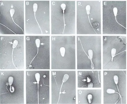
Figure 1.5. Examples of common morphological problems of spermatozoa. Common morphological problems seen in sperm cells.
A. Knobbed acrosome (common form)
B. Knobbed acrosome (beaded form)
C. Pyriform head (severe)
D. Pyriform head (moderate)
E. Pyriform head (slight)
F. Nuclear vacuoles
G. Diadem defects
H. Detached head
I. Distal reflex
J. Dag‐like defect (broken midpiece)
K. Dag‐like defect (severely bent midpiece)
L. Proximal droplet
M. Distal droplet
N. Teratoid (severe) O. Teratoid (moderate)
P. Normal spermatozoa
Images of sperm modified from Barth, A.D., Oko, R. J., Abnormal Morphology of Bovine Spermatozoa. 1 ed.
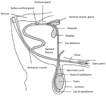
Figure 1.6. Anatomical diagram of the reproductive organs of the bull.
Sperm are generated in the testis, mature during travel through the epididymis, move into the vas deferens and outviathe penis on ejaculation. At this point, seminal plasma is added by the seminal vesicle gland, prostate gland and bulbo urethral gland.
Bulbo‐urethral gland
Prostate gland
Seminal vesicle gland
Ampulla
Bladder
Vas Deferens
Penis
Glans penis
Tail of epididymis
Scrotum
Head of epididymis
Testis
Spermatic cord
Retractor muscle
Sigmoid
Enzymes are thought to be added to the sperm by the testes and during epididymal transport. Some
of the known enzymes are glutamic oxaloacetic transaminase (GOT), glutamic pyruvate
transaminase (GPT), alanine aminotransferase (ALT) all found in the mid piece [58]. Lactate
dehydrogenase (LDH) is found in the cytosol and mitochondria [66] and may drive glycolysis when
oxygen is limited, by facilitating NADH‐mediated reduction of pyruvate to lactate [67]. Alkaline
phosphatase (AP), found on the sperm head, midpiece and tail, regulates phosphorylation of
proteins by cAMP dependent protein kinase [68]. Phospholipase A2 is an immunosuppressant in the
female tract [69]. Another enzyme is platelet activating factor acetylhydrolase which prevents the
sperm undergoing oxidative damage by the neutrophils in the female reproductive tract [70].
Seminal plasma also contains antioxidant enzymes like superoxide dismutase (SOD), glutathione
reductase (GR), glutathione peroxidases (GPx) and catalase (CAT) to prevent damage to the sperm
by reactive oxygen species and free radicals [58].
Lipids play a role in the sperm structure, metabolism, capacitation and fertilisation. They could be
used as an energy source if no carbohydrates were available [71]. The cytokines interleukin 6 (IL‐6),
10 (IL‐10) and tumour necrosis factor alpha (TNFα) are all present in seminal plasma [72]. All three
have an anti‐inflammatory action [72], with IL‐10 and TNFα also possessing immunosuppressive
abilities [65]. Amino acids are added by the testes and epididymis and act as substrates for energy
yielding reactions [73]. The most abundant amino acid is L‐glutamate [58]. L‐arginine increases
spermatogenesis and acts as an energy source in the form of arginine phosphate [74]. It is also
thought that seminal plasma contains an ovulation inducing factor, but studies demonstrating the
effect of seminal plasma inducing ovulation are limited [58].
Nitrogenous compounds like urea, ammonia, uric acid and creatinine have arrived in the seminal
plasma from the blood stream [75], their function is unknown. Many proteins from the accessory
glands have direct roles in sperm quiescence, the formation of the oviductal reservoir [76],
capacitation and sperm‐egg binding [77] (the protein component of seminal plasma is covered in full
detail in chapter 4). This mixture of sperm and accessory gland contents is deposited into the female
reproductive tract.
The sperm cells travel through the vagina, cervix, uterus and into the oviduct, where some will
transiently bind to the oviductal epithelium creating a sperm reservoir. The oviduct is split into three
main sections; isthmus, ampulla and infundibulum, with the connecting areas; uterotubal junction‐
isthmus (UTJ) between the uterus and the isthmus of the oviduct and the ampullary‐isthmic junction
(AIJ) between the isthmus and ampulla of the oviduct [78]. The sperm reservoir is formed in the UTJ