Studies of High Molecular Weight
Systems by NMR Spectroscopy
Shereen Jabar
Research School of Chemistry
October 2018
Declaration
This thesis is composed of my original work and does not contain material previously published or written by another person, except where due reference has been made in the text. The content of this thesis is the result of work that I have carried out since the commencement of my research degree candidature and has not been previously submitted for another degree or diploma in any university or tertiary institution.
Acknowledgements
First and foremost, I would like to express my sincere gratitude to my supervisor Professor Gottfried Otting. I am really thankful for his excellent guidance, support, patience, enthusiasm and invaluable knowledge, without which this work could have not been completed.
I would like to thank Professor Thomas Huber for his support and guidance regarding computational modeling. I am profoundly grateful to our collaborators Associate Professor Bim Graham and his group at Monash University for providing all the chemical tags and unnatural amino acids used in my projects and Professor Daniella Goldfarb and her group at the Weizmann Institute of Science for the EPR measurements.
My thanks also go to all the present and past members of Otting and Huber groups for their support and friendship.
My sincere thanks to the Research School of Chemistry, the Australian National University and the Australian Research Council for their generous funding.
Abstract
Proteins are essential components for all living cells. For better understanding of proteins and how they work, it is essential to know their 3D structure as well as how they interact with other molecules and their dynamics. Nuclear magnetic resonance (NMR) spectroscopy is a versatile technique to study biomolecules, most notably proteins, in solution. NMR provides structural information at atomic resolution and, compared to the other techniques with similar resolution, the measurements can be carried out under near-physiological conditions that are meaningful for the investigation of dynamics.
A great number of unnatural amino acids can now be incorporated into proteins
both in vivo and by cell-free protein synthesis at genetically encoded positions.
Isotopically labelled unnatural amino acids present outstanding probes for site-specific
studies of proteins by NMR. In this thesis, the unnatural amino acid 13C-O-tert
-butyltyrosine (13C-Tby) was incorporated as an NMR probe to study the proteins IMP-1
and the E. coli single-stranded DNA binding protein (SSB).
IMP-1 is a clinically important metallo-β-lactamase which catalyzes the
hydrolysis of almost all β-lactam antibiotics. In order to develop inhibitors against this
enzyme it is important to understand its structure and dynamics. Pseudocontact shifts (PCSs) provide long-range structural information on biological macromolecules. In Chapter 2, PCSs were measured to study the conformation of an active site loop of IMP-1 and its change upon binding of a ligand. This loop is important as it lines the substrate binding site and contributes to substrate specificity. The results suggest that the binding of an inhibitor induces two different conformations in the loop which are in slow equilibrium between each other.
E. coli SSB is a homotetramer of molecular mass 76 kDa. It binds single-stranded DNA (ssDNA) with high affinity and little sequence specificity in two main
binding modes, named (SSB)65 and (SSB)35. Chapter 3 describes studies of ssDNA-SSB
on SSB. The results obtained are in broad agreement with the unusual binding polarity
reported by the crystal structure of a complex between E. coli SSB and a
poly-deoxycytidine oligomer.
In addition to using the tert-butyl group of a Tby residue as an NMR probe, the
present thesis introduced a novel chemical tag containing a trimethylsilyl (TMS) group.
Chapter 4 demonstrates the ability of the tert-butyl and TMS groups to deliver
List of publications
1. Pearce, B.J.G., Jabar, S., Loh, C.-T., Szabo, M., Graham, B. and Otting, G.
(2017) Structure restraints from heteronuclear pseudocontact shifts generated by
lanthanide tags at two different sites. J. Biomol. NMR 68, 19-32.
I contributed in preparation of isotope labelled protein samples for NMR measurements.
2. Jabar, S., Adams, L., Wang, Y., Aurelio, L., Graham, B. and Otting, G. (2017)
Chemical tagging with tert-butyl and trimethylsilyl groups for measuring
intermolecular nuclear Overhauser effects in a large protein-ligand complex.
Chem. Eur. J. 23, 13033-13036.
I carried out all the protein sample preparations including protein expression, purification and ligation with the TMS tag and the NMR measurements and analysis. The chemical synthesis of the tag molecules was performed by the group of A/Prof. Bim Graham at Monash University, including Drs Luke Adams and Luigi Aurelio.
Table of contents
Declaration ... I
Acknowledgements ... II
Abstract ... III
List of publications ... V
General Introduction ... 1
1.1 Paramagnetism in NMR ... 2
1.2 Pseudocontact shifts ... 4
1.3 Introducing the paramagnetic metal ion ... 5
1.4 Protein production ... 9
1.5 Isotopic labelling of proteins ... 10
1.6 Site-specific incorporation of unnatural amino acids ... 11
1.7 Chemical probes for protein NMR ... 14
1.8 Outline of the thesis ... 15
1.9 References ... 17
Study of IMP-1 metallo-β-lactamase active site loop flexibility by NMR spectroscopy ... 25
2.1 Introduction ... 25
2.1.1 β-lactam antibiotics ... 25
2.1.2 β-lactamase enzymes ... 26
2.1.3 Classification of β-lactamases ... 27
2.1.4 Metallo-β-lactamases ... 27
2.1.5 IMP-1 metallo-β-lactamase ... 31
2.1.6 Research objectives ... 33
2.2 Materials and Methods ... 34
2.2.1 Plasmid constructs ... 34
2.2.2 Protein expression ... 35
2.2.3 Protein purification ... 37
2.2.4 Sample preparation for ICP-OES analysis ... 37
2.2.6 NMR spectroscopy ... 38
2.2.7 Δχ-tensor fitting ... 38
2.2.9 Isothermal calorimetry measurements ... 39
2.3 Results and discussion ... 41
2.3.1 ICP-OES analysis of IMP-1 C158* mutants ... 41
2.3.2 Study of active site loop flexibility by NMR spectroscopy ... 43
2.3.3 Captopril binding to IMP-1 ... 63
2.3.4 Study of the flexibility of loop 2 by NMR spectroscopy ... 65
2.4 Conclusion ... 68
2.5 Future work ... 68
2.6 References ... 69
Study of oligonucleotide binding by the E. coli single-stranded DNA binding protein using NMR spectroscopy ... 73
3.1 Introduction ... 73
3.1.1 Single-stranded DNA binding proteins ... 73
3.1.2 E. coli SSB and its binding modes ... 74
3.1.3 ssDNA binding polarity on SSB ... 78
3.14 Research objectives ... 80
3.2 Materials and Methods ... 81
3.2.1 Expression and purification of p-cyanophenylalanyl-tRNA synthetase (pCNF-RS) ... 81
3.2.2 Preparation of total tRNA with optimized suppressor tRNA ... 81
3.2.3 Cell-free protein expression ... 82
3.2.4 Purification of SSB Y9713C-Tby protein ... 82
3.2.5 Oligonucleotides ... 82
3.2.6 NMR sample preparation ... 83
3.2.7 NMR spectroscopy ... 83
3.2.8 EPR sample preparation ... 83
3.3 Results and discussion ... 85
3.3.1 SSB expression and purification ... 85
3.3.2 Modelling ... 85
3.3.3 NMR spectroscopy ... 86
3.3.5 Binding of (dT)35 to SSB at high salt concentration ... 88
3.3.6 Binding of (dT)35 to SSB at low salt concentration ... 91
3.3.7 ssDNA binding polarity of SSB ... 93
3.4 Conclusion ... 98
3.5 Future work ... 98
3.6 References ... 100
Chemical tagging with tert-butyl and trimethylsilyl groups for measuring intermolecular nuclear Overhauser effects in a large protein–ligand complex .... 105
4.1 Introduction ... 105
4.2 Research objectives ... 107
4.3 References ... 108
CHAPTER 1
General Introduction
Proteins are essential for life, carrying out many important functions. In order to understand how proteins work, their three-dimensional (3D) structure must be known at atomic resolution. Currently, this can be achieved by techniques like X-ray crystallography, nuclear magnetic resonance (NMR) spectroscopy and cryo-electron microscopy (cryoEM). Most 3D structures of proteins and protein-ligand complexes have been solved by X-ray crystallography, while an increasing number of very large complexes are being determined by cryoEM. NMR spectroscopy is unique for delivering high-resolution structural information in solution and at ambient temperature, i.e. under conditions close to the physiological state. This enables detailed studies of protein dynamics and their interactions with binding partners under biologically relevant conditions.
NMR spectroscopy offers a large number of local probes, as each proton on the macromolecule results in an individual resonance in the spectrum, which can be resolved using higher-dimensional (2D, 3D and 4D) techniques. The first step in protein
structure determination by NMR involves chemical shift assignment of the 1H, 15N and
13C NMR resonances of the backbone atoms. Nowadays, this process can be carried out
quite rapidly and often it is automated (Atreya and Szyperski, 2005; Freeman and Kupce, 2003). The chemical shifts determined carry information on structural features of backbone and side chain conformations and secondary structure elements of the protein (Wagner et al., 1983; Williamson and Asakura, 1993; Case, 1995).
More recently, residual dipolar couplings (RDCs) have been introduced which are short-range effects (bond orientation relative to an alignment tensor) containing long-range structure information (as the alignment tensor is a global property of the protein; Tjandra et al., 1997). RDCs depend on some alignment of the molecules in the magnetic field. This can be achieved by an anisotropic magnetic susceptibility, which is usually small for diamagnetic proteins, by the use of dilute liquid crystals that spontaneously orient in a magnetic field (Prestegard and Kishore, 2001), or by labelling the protein with a paramagnetic metal ion endowed with an anisotropic magnetic susceptibility tensor (Otting, 2010). Paramagnetic metal ions also generate direct long-range effects, such as paramagnetic relaxation enhancements (PRE; Furuita et al., 2015) or pseudocontact shifts (PCS; Otting, 2010).
PCSs are unique in not only providing long-range distance restraints but also angular information regarding the polar angles of nuclear spins in the tensor frame of the paramagnetic centre. It has been shown that PCS data alone are sufficient to determine the 3D structure of proteins with the help of structure prediction software (Yagi et al., 2013; Pilla et al., 2016; Pilla et al., 2017).
1.1 Paramagnetism in NMR
Figure 1.1 Structural information contained in four different paramagnetic effects,
illustrated with a nuclear spin, HN, and a paramagnetic centre denoted e. (a)
Paramagnetic relaxation enhancement depends on the distance r between electron and
nuclear spin. (b) The pseudocontact shift depends on the polar coordinates of the nuclear spin relative to the magnetic susceptibility tensor c. (c) The residual dipolar
coupling between a 1H and a 15N spin depends on the internuclear distance r
HN and the
polar angles between the internuclear vector and the c tensor. (d) The cross-correlation
effect between Curie spin and 1H-15N dipole-dipole relaxation depends on the
internuclear distance rHN, the distance between nuclear spin and paramagnetic centre
(r), and the angle between rHN and r. Figure reproduced from Otting (2010).
All these effects can be measured in heteronuclear single-quantum coherence (HSQC) spectra recorded of the paramagnetic state and the reference diamagnetic state of the biomolecule. The paramagnetic effects can be described by the magnetic
susceptibility tensor χ, which is defined by three principal axes (χx,χy andχz). Only the
PRE effect is observed for systems with isotropic χ tensors, while all four effects occur
in systems with anisotropic χ tensors. The anisotropy of the χ tensor (Δχ tensor) can be
characterized by axial and rhombic components (Bertini et al., 2002; Otting, 2010):
Δχax = χz – (χx + χy)/2 (1.1)
Δχrh = χx−χy (1.2)
Except for RDCs, the magnitude of the paramagnetic effects depends on the
distance r between the metal ion and nuclear spins. PREs lead to broadening of the
proportional to r-6, PREs decay rapidly compared with PCSs and the CCR effect, which
show a r-3 distance dependence. Therefore, even though nuclear spins near the
paramagnetic centre get broadened beyond detection, nuclear spins that are sufficiently far from can give rise to measurable PCSs (Otting, 2010). The present thesis focuses on the measurement of PCSs to study the conformational dynamics of the protein IMP-1 described in Chapter 2.
1.2 Pseudocontact shifts
PCSs originate from the magnetic dipole field generated at the site of a nuclear spin by rapidly relaxing unpaired electrons associated with a metal ion. This is a through-space effect, which can be described by the following equation (Bertini et al., 2002)
ΔδPCS = 1/(12πr3) [Δχax (3 cos2θ− 1) + 1.5 Δχrh sin2θ cos 2φ] (1.3)
where r is the distance between the unpaired electron and the nuclear spin, Δχax is the
axial component and Δχrh is the rhombic component of the Δχ tensor, and θ and φ are
the polar angles of the nuclear spin with respect to the principal axes of the Δχ tensor.
PCSs are independent of the magnetic field when measured in parts per million (ppm) and can be observed as far as 40 Å from the paramagnetic centre (Allegrozzi et al., 2000).
In 15N-HSQC spectra of proteins, PCSs can be observed as chemical shift
changes of the cross-peaks along diagonal lines as both the proton and the nitrogen of
each 1H-15N group experience similar PCSs. Small deviations from the diagonal shifts
occur due to residual anisotropic chemical shifts, which arise for nuclear spins with large chemical shift anisotropy (CSA) in molecules that are subject to weak paramagnetic alignment in the magnetic field (John et al., 2005). In practice, this
correction is significant for 15N chemical shifts of backbone amides and 13C chemical
shifts of carbonyl groups, but small for other backbone atoms. Therefore PCSs are usually determined as the difference between the chemical shifts of the paramagnetic
and diamagnetic states of 1H rather than heteronuclear spins, eliminating the need for
If the 3D structure of the protein carrying the paramagnetic metal ion is known,
the experimentally measured PCSs can be used to fit the Δχ tensor. Eight parameters
need to be determined, including the x, y and z coordinates defining the location of the
metal ion, three Euler angles α, β and γ defining the orientation of the Δχ tensor with
respect to the protein coordinates, and the axial and rhombic components of the Δχ
tensor. The Δχ tensor can be depicted in the form of PCS isosurfaces overlaid onto the
structure of the protein (Figure 1.2). This gives a graphical representation of the
principal axes of the Δχ tensor and its orientation with respect to the protein (Otting,
2010).
[image:15.595.237.392.288.484.2]
Figure 1.2 Isosurfaces depicting the PCSs induced by a Dy3+ ion plotted on a ribbon
representation of the crystal structure of the protein ε186. Blue and red surfaces identify
the spatial locations of positive and negative PCSs (±3, ±1.5 and ±0.5 ppm),
respectively. The PCS isosurfaces constitute a representation of the Δχ tensor.
Reproduced from Otting (2010).
1.3 Introducing the paramagnetic metal ion
Among the many paramagnetic transition metal ions, high-spin Co2+ stands out for
d-orbitals of transition metals, the delocalization of the unpaired electron density onto the ligand atoms is minimal (Nitsche and Otting, 2017).
The chemical properties of all lanthanide ions are very similar. Therefore, once a lanthanide binding site has been established, it is suitable to bind any lanthanide ion
with similar affinity. La3+, Y3+ and Lu3+ ions are suitable diamagnetic ions to measure
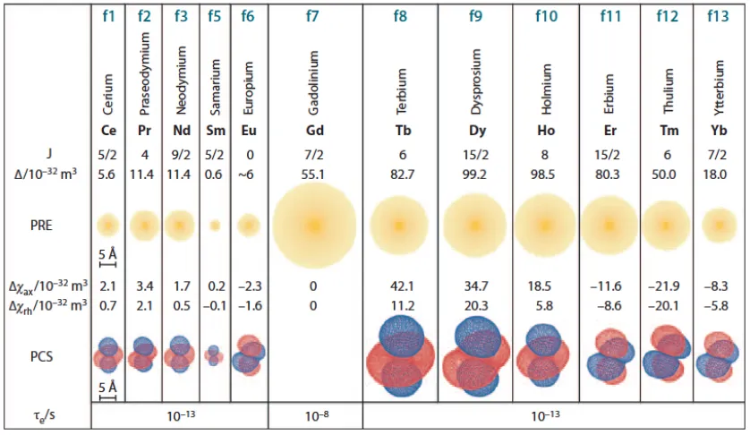
[image:16.595.116.535.322.565.2]the diamagnetic state. The radii of these ions are comparable to those of the paramagnetic lanthanides at the start, centre and end of the lanthanide series, respectively, which makes them good choices for diamagnetic references as required for PCS and PRE measurements. Figure 1.3 gives an overview of the paramagnetic properties of paramagnetic lanthanide ions.
Figure 1.3 Paramagnetic properties of lanthanides. The radii of the yellow spheres
indicate the distance from the metal ion where the 1H-NMR signals of a protein with a
rotational correlation time of 15 ns are expected to broaden by 80 Hz due to PRE on an 800 MHz NMR spectrometer. Isosurfaces represent PCSs of ±5 ppm based on the
tensors reported for calbindin D9k (Bertini et al., 2001). Typical electronic relaxation
Since lanthanides have no known biological function, natural proteins rarely display strong binding sites for lanthanide ions. It has been shown that lanthanide
binding sites can be generated by displacing a native Ca2+ ion from calcium-binding
proteins. This has been demonstrated, for example, for the Ca2+-binding proteins
calbindin D9k (Allegrozzi et al., 2000) and calmodulin (Bertini et al., 2003). Most
proteins, however, do not contain natural metal binding sites. Therefore, other methods for introducing lanthanide ions into proteins have been developed.
It has been shown that the EF-hand motif of a calcium-binding protein can be optimized to create a lanthanide binding peptide (LBP; Ma and Opella, 2000; Franz et al., 2003; Nitz et al., 2003; Nitz et al. 2004). LBPs generate PCSs following attachment to a cysteine residue via formation of a disulfide bond (Su et al., 2006; Su et al., 2008), but mobility of the linker between LBP and protein can decrease the PCSs substantially by averaging between PCSs of different magnitude and sign. In order to obtain sizeable paramagnetic effects it is important to have less mobile LBPs attached onto protein. This has been achieved by anchoring the LBP to the protein at two points, which greatly reduces its mobility and leads to much larger PCSs in the protein. Two-point attachments have been achieved by inserting LBPs into loop regions of proteins as, e.g.,
in the protein interleukin-1β (Barthelmes et al., 2011) or the immunoglobulin G binding
protein (Barb et al., 2012; Barb and Subedi, 2016), or by combining fusion to the N- or C-terminus of the target protein with an additional disulfide bond to a cysteine residue as demonstrated for, e.g., the B1 immunoglobulin-binding domain of protein G (GB1; Saio et al., 2009).
tag and its opposite enantiomer, the C2 tag, have successfully been used in different conformational studies of proteins (Graham et al., 2011; de la Cruz et al., 2011, Yagi et al., 2013, Chen at al., 2014; Chen et al., 2016; Pearce et al., 2017; Mahawaththa et al., 2017). In this thesis, the C2 tag has been used in Chapter 2 to study the active-site loop
conformation of the metallo-β-lactamase IMP-1 by PCS measurements.
Figure 1.4 Chemical structures of the C2 tag and its lanthanide complex. The tag contains three chiral amide pendants contributing to the lanthanide binding site and a nonchiral pendant for attachment to a cysteine residue on the target protein.
A very elegant solution would be presented by site-specific incorporation of a lanthanide binding unnatural amino acid. To date, a genetic encoding system has been reported only for a single unnatural amino acid, 2-amino-3-(8-hydroxyquinolin-3-yl)propanoic acid (HQ-Ala), that can bind lanthanides with considerable affinity (Lee et al., 2009). Unfortunately, proteins containing HQ-Ala precipitate upon titration with lanthanide ions (Jones et al., 2010).
PCSs produced by lanthanides attached to proteins have been used for 3D structure refinement of proteins as well as 3D structure determination. For example,
PCSs and 1H-15N RDCs measured for the calmodulin N60D mutant, where a Ca2+ ion
can be substituted by a lanthanide ion, have been used to refine the 3D structures of two calmodulin-peptide complexes. The PCS data proved particularly useful for the structural refinement of the overall fold of the protein (Bertini et al., 2003). Using PCSs
for determining the 3D structures of proteins is more difficult as Δχ tensor fits rely on
PCS-Rosetta and GPS-Rosetta by generating model structures to fit the Δχ tensors and refining only the best-fitting models (Schmitz et al., 2012; Yagi et al., 2013). A particularly attractive application of PCSs is for determining the structures of
protein-ligand complexes, where Δχ tensors can be fitted to the known 3D structure of the
protein and PCS data of the ligand can be used to determine its binding mode (ligand binding site, relative orientation of the ligand with respect to the protein and ligand conformation; John et al., 2006). In the same way, PCSs can be used to study the dynamics of local parts of proteins.
1.4 Protein production
Structural studies of proteins by NMR spectroscopy require relatively large amounts of pure protein. The required proteins are most frequently produced by over-expression
systems in Escherichia coli. In vivo protein expression is ideal for making large
quantities of protein, but is less suitable for producing selectively isotope-labelled samples (due to “isotope scrambling”, i.e. the shuffling of isotope-labelled chemical groups between different amino acids effected by the natural amino acid metabolism) or samples containing expensive unnatural amino acids (due to protein yields that are quite low relative to the amino acid provided). For these situations, cell-free protein synthesis can be more suitable.
Cell-free protein synthesis (CFPS) systems employ cell extracts to produce proteins from amino acids. The crude lysates obtained from microbial, plant or animal cells contain the necessary elements for transcription, translation, protein folding and energy metabolism such as ribosomes, aminoacyl-tRNA synthetases, translation initiation and elongation factors, ribosome release factors, nucleotide recycling enzymes, metabolic enzymes, chaperones, foldases, etc. (Nirenberg and Mattaei, 1961). The cell extract can thus be used to synthesize the target protein in a coupled transcription/translation system, where the DNA encoding the target protein is provided together with T7 RNA polymerase and nucleotide triphosphates to produce mRNA, and amino acids and a source of ATP are provided to drive the protein translation reaction
(Carison et al., 2012). In this thesis, an E. coli CFPS system established in-house
Apponyi et al., 2008; Jia et al., 2009; Su et al., 2011; Ozawa et al., 2012; Loscha et al.,
2012) was used to produce the E. coli single-stranded DNA-binding protein samples
discussed in Chapters 3 and 4. E. coli cell extracts are relatively easy to prepare and
high protein yields can be obtained from relatively small amounts of amino acids (Torizawa et al., 2004)
1.5 Isotopic labelling of proteins
The main isotopes usually used in protein NMR spectroscopy includes, 1H, 2H, 13C
and 15N. Amongst these isotopes, except for 1H, which has natural abundance of greater
than 99.9%, others must be introduced into proteins by other methods.
E. coli is the most commonly used bacterial host for expressing recombinant
proteins with isotopic labelling, as E. coli cell cultures are readily grown with 15NH4
salts as the only nitrogen source and 13C-glucose as the only carbon source, both of
which are relatively inexpensive (Markley et al., 1968; Marley et al., 2001). Costs can be saved further by growing the cells initially in an unlabelled rich medium to obtain a high cell density followed by a transfer of the cells to a labelled medium before
over-expression (McIntosh and Dahlquist, 1990). In Chapter 2, the metallo-β-lactamase
IMP-1 was uniformly labelled with 15N by in vivo expression, using a M9 medium with 15
N-ammonium chloride as the sole 15N source.
CFPS is an excellent method to produce selectively labelled proteins from a mixture of 20 amino acids, where a specific amino acid type can be supplied in
isotopically labelled form. Compared to in vivo protein expression, isotope scrambling
is strongly suppressed in CFPS because the amino acid metabolism is no longer fully functional in cell extracts.
In the same way as proteins can be produced from amino acid mixtures and their metabolic precursors, a protein can also be site-specifically labelled chemically by incorporating an unnatural amino acid, which may also be labelled isotopically.
which was incorporated into a protein to obtain site-specific information by NMR spectroscopy without having to assign all other NMR resonances of the protein.
1.6 Site-specific incorporation of unnatural amino acids
Site-specific introduction of unique functional groups into proteins presents a powerful tool for structure/function studies. This can be accomplished either by attachment of a chemical probe onto a reactive amino-acid side-chain as in cysteine residues or by incorporating unnatural amino acids (UAAs), which can contain different functional groups. Genetic encoding of UAAs is a particularly elegant way to selectively modify and label proteins without adversely affecting the protein structure.
Genetic encoding of a UAA relies ona pair of tRNA/aminoacyl-tRNA
Figure 1.5 Schematic diagram of an orthogonal aminoacyl-tRNA synthetase/tRNA
pair system for the incorporation of an unnatural amino acid into a protein. The
orthogonal synthetase (purple) aminoacylates the orthogonal tRNA (green) with an unnatural amino acid (red sphere) and does not cross-react with any of the endogenous synthetases and tRNA (grey). The aminoacylated orthogonal tRNA then travels to the ribosome (blue) and incorporates the unnatural amino acid in response to a unique codon. Reproduced from Lin et al. (2008).
Four main aaRS enzymes have been developed for UAA incorporation: (i) the
Methanococcus janaschii tyrosyl-tRNA synthetase (MjTyrRS)/tRNACUA pair, which is
orthogonal in E. coli (Xie and Schultz, 2005); (ii) pyrrolysyl-tRNA synthetase
(PylRS)/tRNACUA pairs from certain Methanosarcina organisms, which are orthogonal
in bacteria and eukaryotic cells (Neumann et al., 2008); (iii) the E. coli tyrosyl-tRNA
synthetase (EcTyrRS)/tRNACUA pair and the E. coli leucyl-tRNA synthetase
(EcLeuRS)/tRNACUA pair, which are orthogonal in eukaryotic cells (Chin, 2003; Chin et
al., 2003, Wu et al., 2004).
The TyrRS/tRNACUA pair derived from the archaebacterium M. jannaschii was
the first aaRS/tRNACUA pair reported to be orthogonal with regard to the E.
anticodon of its target tRNA, which makes it possible to mutate the anticodon to CUA for recognition of the amber stop codon. A wide range of variants have been developed to adopt the amino-acid binding pocket of this aaRS to accommodate different UAAs (Neumann et al., 2010a, Young et al., 2011).
A major problem associated with reassignment of the amber codon to incorporate UAAs is the presence of release-factor 1 (RF1), which also recognizes the amber stop codon and results in termination of protein translation. The competition
between tRNACUA and RF1 results in mixtures of full-length and prematurely truncated
target proteins. It has been reported that amber suppression efficiency can be improved
by utilizing E. coli strains with deleted RF1 gene. The first E. coli RF1 knockout strain
was reported by Mukai et al. (2010), where seven essential genes along with the suppressor tRNA were placed on a plasmid. This resulted in an increase in the yield of full-length protein containing UAAs, as translation termination by RF1 was eliminated.
Subsequently, the E. coli strain JX33 was produced, which is independent of RF1. This
strain displayed higher incorporation efficiency of a reporter protein with multiple
amber codon sites (Johnson et al., 2011). In a major effort, the E. coli genome was
re-engineered to replace all 314 naturally occurring amber codons by ochre codons to eliminate the genetic dependence on RF1 and allowed knockout of RF1 (Isaacs et al.,
2011). These strains have shown success both in vivo (Chatterjee et al. 2014; Schmidt et
al. 2014; Lim et al., 2015; Pirman et al., 2015; Wilkins et al., 2015) and in cell-free systems (Hong et al., 2014; Chemla et al., 2015; Li et al., 2015; Sullivan et al., 2015).
1.7 Chemical probes for protein NMR
From the perspective of NMR spectroscopy, it is attractive to incorporate isotopically
labelled UAAs (13C and 15N) or fluorinated UAAs as they allow the recording of greatly
simplified NMR spectra, which are ideal for obtaining site-specific information in large proteins and protein–ligand complexes, where complete resonance assignments by established techniques are time consuming or impossible to obtain. The incorporation of an NMR-active tag at a specific site of the target protein and its selective detection in the NMR spectrum instantly provides a site-specific probe for monitoring changes in chemical environment at this site. For example, by monitoring chemical shift changes of this signal it is possible to study ligand binding, conformational changes and dynamics of proteins. This strategy opens a path to study large proteins and protein complexes, which are difficult to study in other ways due to the complexity of their NMR spectra.
In an early example using a UAA, Deiters et al. (2005) incorporated 15N-labelled
p-methoxyphenylalanine into myoglobin and produced protein amounts (1 mg/L)
sufficient for NMR studies. Similarly, 13C-labelled p-O-methylphenylalanine was
introduced into cytochrome P450 (CYP119) to study local ligand-induced
conformational heterogeneity (Lampe et al., 2008). Three NMR active UAAs, 13C/15
N-p-methoxyphenylalanine, 2-amino-3-(trifluoromethoxy)phenyl) propanoic acid
(4-trifluoromethoxy-phenylalanine) and 15N-O-nitrobenzyltyrosine, were incorporated at
11 sites in fatty acid synthase to study the binding of a target compound (Cellitti et al., 2008).
Previously, 19F-labelled proteins had been synthesised by incorporating
5-fluoro-tryptophan, 3-fluoro-tyrosine, 4-fluoro-phenylalanine and other fluorinated amino acids in place of their natural counterparts (Gerig, 1994; Gakh et al., 2000; Frieden et al., 2004). Unless the respective amino acid only occurs a single time in the entire polypeptide sequence, however, site-specific labelling cannot be achieved in this
way. UAAs solve this problem, and fluorinated UAAs such as p-
trifluoromethyl-phenylalanine and 4-trifluoromethoxy-phenylalanine have been used successfully in
proteins to study ligand binding and structural changes by 19F NMR (Hammill et al.,
2007; Jackson et al., 2007; Cellitti et al., 2008). Due to the large CSA associated with
molecular weight (Chen et al., 2015).
For large proteins, the 1H-NMR signal of a tert-butyl group has been proven to
deliver an excellent NMR probe that can be observed without any isotopic labelling. It
gives a narrow and intense singlet that can be readily identified in 1D 1H-NMR spectra.
In particular, the UAA O-tert-butyltyrosine has been used successfully for the study of
high-molecular-weight systems without any isotope labelling (Chen et al., 2015; Jabar
et al., 2017). Similarly, Loh et al. (2018) reported successful incorporation of p-(tert
-butyl)phenylalanine into proteins ranging from 8 to 54 kDa and observed the 1H
resonance of the tert-butyl group.
The trimethylsilyl (TMS) group is another outstanding NMR probe, as the TMS
signal appears near 0 ppm in the 1H-NMR spectrum. At this chemical shift, signal
overlap with other protein peaks is minimal, which is an important advantage over the
tert-butyl group. The TMS group has been site-specifically introduced into proteins by
chemical ligation onto a cysteine residue (Jabar et al., 2017; Becker et al., 2018). More recently, a UAA with the TMS group was successfully incorporated by CFPS. Although
the 1H-NMR resonance of the TMS group could be observed, the protein yield was low
(Loh et al., 2018).
1.8 Outline of the thesis
Chapter 2 documents an NMR study performed on the metallo-β-lactamase IMP-1,
where pseudocontact shift measurements were performed to investigate conformational changes of an active-site loop in response to a ligand. Chapter 3 describes a study conducted on the interaction between stranded DNA binding protein and
single-stranded oligonucleotides by NMR spectroscopy, using a 13C-tert-butyl group as NMR
Chapter 1. General introduction.
Chapter 2. Study of IMP-1 metallo-β-lactamase active site loop flexibility by NMR spectroscopy.
Chapter 3. Study of oligonucleotide binding by E. coli single-stranded DNA binding protein usingz NMR spectroscopy.
1.9 References
Allegrozzi, M., Bertini, I., Janik, M.B.L., Lee, Y.-M., Liu, G. and Luchinat, C. (2000) Lanthanide-induced pseudocontact shifts for solution structure refinements of
macromolecules in shells up to 40 Å from the metal ion. J. Am. Chem. Soc. 122,
4154–4161.
Anderson, J.C., Magliery, T.J., and Schultz, P.G. (2002) Exploring the limits of codon
and anticodon size. Chem. Biol. 9, 237–244.
Apponyi, M., Ozawa, K., Dixon, N.E. and Otting, G. (2008) Cell-free protein synthesis
for analysis by NMR spectroscopy. Methods in Molecular Biology 426, Structural
proteomics: high-throughput methods B. Kobe, M. Guss, T. Huber Eds., Humana Press, Totowa, USA pp. 257-268.
Atreya, H.S. and Szyperski, T. (2005) Rapid NMR data collection. Methods Enzymol.
394, 78–108.
Barb, A.W., Ho, T.G., Flanagan-Steet, H., Prestegard, J.H., (2012) Lanthanide binding and IgG affinity construct: potential applications in solution NMR, MRI, and
luminescence microscopy. Protein Sci.21, 1456–1466.
Barb, A.W. and Subedi, G.P. (2016) An encodable lanthanide binding tag with reduced size and flexibility for measuring residual dipolar couplings and pseudocontact shifts
in large proteins. J. Biomol. NMR64, 75–85.
Barthelmes, K., Reynolds, A.M., Peisach, E., Jonker, H.R.A., DeNunzio, N.J., Allen, K.N., Imperiali, B. and Schwalbe, H. (2011) Engineering encodable
lanthanide-binding tags into loop regions of proteins. J. Am. Chem. Soc.133, 808–819.
Becker, W., Adams, L.A., Graham, B., Wagner, G.E., Zangger, K., Otting, G. and Nitsche, C. (2018) Trimethylsilyl tag for probing protein-ligand interactions by NMR
J. Biomol. NMR70, 211-218.
Bertini, I., Janik, M.B.L., Lee, Y.M., Luchinat, C. and Rosato, A. (2001) Magnetic susceptibility tensor anisotropies for a lanthanide ion series in a fixed protein matrix.
J. Am. Chem. Soc.123, 4181–4188.
Bertini, I., Luchinat, C. and Parigi, G. (2002)Magnetic susceptibility in paramagnetic
NMR. Prog. Nucl. Magn. Reson. Spec.40, 249–273.
Bertini, I., Gelis, I., Katsaros, N., Luchinat, C. and Provenzani, A. (2003) Tuning the
affinity for lanthanides of calcium binding proteins. Biochemistry42, 8011–8021.
Carison, E.D., Gan, R., Hodgman, C.E. and Jewett, M.C. (2012) Cell-free protein
synthesis: applications come of age. Biotechnol. Adv. 30, 1185-1194.
Case, D.A. (1995) Calibration of ring-current effects in proteins and nucleic acids. J.
Cellitti, S.E., Jones, D.H., Lagpacan, L., Hao, X., Zhang, Q., Hu, H., Brittain, S.M., Brinker, A., Caldwell, J., Bursulaya, B., Spraggon, G., Brock, A., Ryu, Y., Uno, T.,
Schultz, P.G. and Geierstanger, B.H. (2008) In vivo incorporation of unnatural amino
acids to probe structure, dynamics, and ligand binding in a large protein by nuclear
magnetic resonance spectroscopy. J. Am. Chem. Soc. 130, 9268-9281.
Chatterjee, A., Lajoie, M.J., Xiao, H., Church, G.M. and Schultz, P.G. (2014) A bacterial strain with a unique quadruplet codon specifying non-native amino acids.
ChemBioChem15, 1782–1786.
Chemla, Y., Ozer, E., Schlesinger, O., Noireaux, V. and Alfonta, L. (2015) Genetically expanded cell-free protein synthesis using endogenous pyrrolysyl orthogonal
translation system. Biotechnol. Bioeng.112, 1663–1672.
Chen, W.-N., Loscha, K.V., Nitsche, C., Graham, B. and Otting, G. (2014) The dengue virus NS2B-NS3 protease retains the closed conformation in the complex with BPTI.
FEBS Lett.588, 2206-2211.
Chen, W.-N., Kuppan, K.V., Lee, M.D., Jaudzems, K., Huber, T. and Otting, G. (2015)
O-tert-butyltyrosine, an NMR tag for high-molecular-weight systems and
measurements of submicromolar ligand binding affinities. J. Am. Chem. Soc. 137,
4581-4586.
Chen, W.-N., Nitsche, C., Pilla, K.B., Graham, B., Huber, T., Klein, C.D. and Otting, G. (2016) Sensitive NMR approach for determining the binding mode of tightly binding
ligand molecules to protein targets. J. Am. Chem. Soc.138, 4539-4546.
Chin, J.W. (2003) An expanded eukaryotic genetic code. Science301, 964-967.
Chin, J.W., Cropp, T.A., Chu, S., Meggers, E. and Schultz, P.G. (2003) Progress
towards and expanded eukaryotic genetic code. Chem. Biol. 10, 511-519.
de la Cruz, L., Nguyen, T.H.D., Ozawa, K., Shin, J., Graham, B., Huber, T. and Otting, G. (2011) Binding of low-molecular weight inhibitors promotes large conformational changes in the dengue virus NS2B-NS3 protease: fold analysis by pseudocontact
shifts. J. Am. Chem. Soc.133, 19205-19215.
Deiters, A., Geierstanger, B.H., and Schultz, P.G. (2005) Site-specific in vivo labeling
of proteins for NMR studies. ChemBioChem 6, 55–58.
Franz, K.J., Nitz, M. and Imperiali, B. (2003) Lanthanide-binding tags as versatile
protein coexpression probes. ChemBioChem 4, 265–271.
Freeman, R. and Kupce, E. (2003) New methods for fast multidimensional NMR. J.
Biomol. NMR27, 101–113.
Frieden, C., Hoeltzli, S.D. and Bann, J.G. (2004) The preparation of 19F-labeled
Furuita, K., Kataoka, S., Sugiki, T., Hattori, Y., Kobayashi, N., Ikegami, T., Shiozaki, K., Fujiwara, T. and Kojima, C. (2015) Utilization of paramagnetic relaxation enhancements for high-resolution NMR structure determination of a soluble
loop-rich protein with sparseNOE distance restraints. J. Biomol. NMR 61, 55-64.
Gakh, Y.G., Gakh, A.A. and Gronenborn, A.M. (2000) Fluorine as an NMR probe for
structural studies of chemical and biological systems. Magn. Reson. Chem. 38, 551–
558.
Gerig, J.T. (1994) Fluorine NMR of proteins. Prog. Nucl. Magn. Reson. Spectrosco. 26,
293–370.
Graham, B., Loh, C.-T., Swarbrick, J.D., Ung, P., Shin, J., Yagi, H., Jia, X., Chhabra, S., Pintacuda, G., Huber, T. and Otting, G. (2011) A DOTA-amide lanthanide tag for
reliable generation of pseudocontact shifts in protein NMR spectra. Bioconjugate
Chem.22, 2118-2125.
Guignard, L., Ozawa, K., Pursglove, S.E., Otting, G. and Dixon, N.E. (2002) NMR analysis of in vitro-synthesized proteins without purification: a high-throughput
approach. FEBS Lett.524, 159-162.
Hammill, J.T., Miyake-Stoner, S., Hazen, J.L., Jackson, J.C. and Mehl, R.A. (2007)
Preparation of site-specifically labeled fluorinated proteins for 19F-NMR structural
characterization. Nat. Protoc.2, 2601–2607.
Hong, S.H., Ntai, I., Haimovich, A.D., Kelleher, N.L., Isaacs, F.J. and Jewett, M.C.
(2014) Cell-free protein synthesis from a release factor 1 deficient Escherichia coli
activates efficient and multiple site-specific nonstandard amino acid incorporation.
ACS Synth. Biol. 3, 398–409.
Isaacs, F.J., Carr, P.A., Wang, H.H., Lajoie, M.J., Sterling, B., Kraal, L., et al. (2011) Precise manipulation of chromosomes in vivo enables genome-wide codon
replacement. Science333, 348–353.
Jabar, S., Adams, L., Wang, Y., Aurelio, L., Graham, B., and Otting, G. (2017)
Chemical tagging with tert-butyl and trimethylsilyl groups for measuring
intermolecular nuclear Overhauser effects in a large protein-ligand complex. Chem.
Eur. J.23, 13033-13036.
Jackson, J.C., Hammill, J.T. and Mehl, R.A. (2007) Site-specific incorporation of a 19F
amino acid into proteins as an NMR probe for characterizing protein structure and
reactivity. J. Am. Chem. Soc.129, 1160–1166.
Jia, X., Ozawa, K., Loscha, K. and Otting, G. (2009) Glutarate and
N-acetyl-L-glutamate buffers for cell-free synthesis of selectively 15N-labelled proteins J.
Biomol. NMR44, 59-67.
John, M., Park, A.Y., Pintacuda, G., Dixon, N.E. and Otting, G. (2005) Weak alignment of paramagnetic proteins warrants correction for residual CSA effects in
John, M., Pintacuda, G., Park, A.Y., Dixon, N.E. and Otting, G. (2006) Structure
determination of protein-ligand complexes by transferred paramagnetic shifts. J. Am.
Chem. Soc.128, 12910–12916.
Johnson, D.B., Xu, J., Shen, Z., Takimoto, J.K., Schultz, M.D., Schmitz, R.J., Xiang, Z., Ecker, J.R., Briggs, S.P. and Wang, L. (2011) RF1 knockout allows ribosomal
incorporation of unnatural amino acids at multiple sites. Nat. Chem. Biol. 7, 779–
786.
Jones, D.H., Cellitti, S.E., Hao, X., Zhang, Q., Jahnz, M., Summerer, D., Schultz, P.G., Uno, T. and Geierstanger, B.H. (2010) Site-specific labeling of proteins with
NMR-active unnatural amino acids. J. Biomol. NMR46, 89–100.
Lampe, J.N., Floor, S.N., Gross, J.D., Nishida, C.R., Jiang, Y., Trnka, M.J. and de Montellano, P.R.O. (2008) Ligand-induced conformational heterogeneity of cytochrome P450 CYP119 identified by 2D NMR spectroscopy with the unnatural
amino acid 13C-p-methoxyphenylalanine. J. Am. Chem. Soc.130, 16168–16169.
Lee, H.S., Spraggon, G., Schultz, P.G. and Wang, F. (2009) Genetic incorporation of a
metal-ion chelating amino acid into proteins as biophysical probe. J. Am. Chem. Soc.
131, 2481-2483.
Li, J., Lawton, T.J., Kostecki, J.S., Nisthal, A., Fang, J., Mayo, S.L., Rosenzweig, A.C. and Jewett, M.C. (2015) Cell-free protein synthesis enables high yielding synthesis
of an active multicopper oxidase. Biotechnol. J.11, 212-218.
Lim, S.I., Hahn, Y.S. and Kwon, I. (2015) Site-specific albumination of a therapeutic
protein with multi-subunit to prolong activity in vivo. J. Control Rel. 207, 93–100.
Lin, M.Z. and Wang, L. (2008) Selective labelling of proteins with chemical probes in
living cells. Physiology23, 131-141.
Liu, C.C. and Schultz, P.G. (2010) Adding new chemistries to the genetic code. Annu.
Rev. Biochem.79, 413-444.
Loh, C.-T., Ozawa, K., Tuck, K.L., Barlow, N., Huber, T., Otting, G. and Graham, B. (2013) Lanthanide tags for site-specific ligation to an unnatural amino acid and
generation of pseudocontact shifts in proteins. Bioconjugate Chem.24, 260–268.
Loh, C.-T., Adams, L.A., Graham, B., Otting, G. (2018) Genetically encoded amino
acids with tert-butyl and trimethylsilyl groups for site-selective studies of proteins by
NMR spectroscopy. J. Biomol. NMR 71, 287-293.
Loscha, K.V., Herlt, A.J., Qi, R., Huber, T., Ozawa, K. and Otting, G. (2012)
Multiple-site labeling of proteins with unnatural amino acids. Angew. Chem. Int. Ed.51,
2243-2246.
Ma, C. and Opella, S.J. (2000) Lanthanide ions bind specifically to an added “EF-hand”
and orient a membrane protein in micelles for solution NMR spectroscopy. J. Magn.
Mahawaththa, M.C., Pearce, B.J.G., Szabo, M., Graham, B., Klein, C.D., Nitsche, C. and Otting, G. (2017) Solution conformation of a linked construct of the Zika virus
NS2B-NS3 protease. Antiviral Res.142, 141-147.
Markley, J.L., Putter, I. and Jardetzky, O. (1968) High-resolution nuclear magnetic
resonance spectra of selectively deuterated staphylococcal nuclease. Science161,
1249–1251.
Marley, J., Lu, M. and Bracken, C. (2001) A method for efficient isotopic labeling of
recombinant proteins. J. Biomol. NMR20, 71–77.
McIntosh, L.P. and Dahlquist, F.W. (1990) Biosynthetic incorporation of 15N and 13C
for assignment and interpretation of nuclear magnetic resonance spectra of
proteins. Q. Rev. Biophys.23, 1–38.
Mukai, T., Hayashi, A., Iraha, F., Sato, A., Ohtake, K., Yokoyama, S. and Sakamoto, K.
(2010) Codon reassignment in the Escherichia coli genetic code. Nucleic Acids Res.
38, 8188–8195.
Neumann, H., Peak-Chew, S.Y. and Chin, J.W. (2008) Genetically encoding Nε
-acetyllysine in recombinant proteins. Nat. Chem. Biol.4, 232–234.
Neumann, H., Slusarczyk, A.L., and Chin, J.W. (2010a) De novo generation of
mutually orthogonal aminoacyl-tRNA synthetase/tRNA pairs. J. Am. Chem. Soc.
132, 2142-2144.
Neumann, H., Wang, K., Davis, L., Garcia-Alai, M. and Chin, J.W. (2010b) Encoding multiple unnatural amino acids via evolution of a quadruplet-decoding
ribosome. Nature464, 441–444.
Nirenberg, M.W. and Matthaei, J.H. (1961) The dependence of cell-free protein
synthesis in E. coli upon naturally occurring or synthetic polyribonucleotides. Proc.
Natl. Acad. Sci. USA47, 1588–1602.
Nitsche, C. and Otting, G. (2017) Pseudocontact shifts in biomolecular NMR using
paramagnetic metal tags. Prog. Nucl. Magn. Reson. Spec.98-99, 20-49.
Nitz, M., Franz, K.J., Maglathlin, R.L. and Imperiali, B. (2003) A powerful combinatorial screen to identify high-affinity terbium(III)-binding peptides.
ChemBioChem4, 272–276.
Nitz, M., Sherawat, M., Franz, K.J., Peisach, E., Allen, K.N. and Imperiali, B. (2004) Structural origin of the high affinity of a chemically evolved lanthanide-binding
peptide. Angew. Chem. Int. Ed.43, 3682–3685.
Otting, G. (2008) Prospects for lanthanides in structural biology by NMR. J. Biomol.
NMR42, 1–9.
Otting, G. (2010) Protein NMR using paramagnetic ions. Annu. Rev. Biophys. 39, 387–
Ozawa, K., Headlam, M.J., Schaeffer, P.M., Henderson, B.R., Dixon, N.E. and Otting, G. (2004) Optimization of an Escherichia coli system for cell-free synthesis of
selectively 15N-labelled proteins for rapid analysis by NMR spectroscopy.
Eur. J. Biochem.271, 4084-4093.
Ozawa, K., Wu, P.S., Dixon, N.E. and Otting, G. (2006) 15N-labelled proteins by
cell-free protein synthesis: strategies for high-throughput NMR studies of proteins and
protein-ligand complexes. FEBS J. 273, 4154–4159.
Ozawa, K., Loscha, K.V., Kuppan, K.V., Loh, C.T., Dixon, N.E. and Otting, G. (2012) High-yield cell-free protein synthesis for site-specific incorporation of unnatural
amino acids at two sites. Biochem. Biophys. Res. Commun.418, 652-656.
Pearce, B.J.G., Jabar, S., Loh, C.-T., Szabo, M., Graham, B. and Otting, G. (2017) Structure restraints from heteronuclear pseudocontact shifts generated by lanthanide
tags at two different sites. J. Biomol. NMR 68, 19-32.
Pilla, K.B., Otting, G. and Huber, T. (2016) Pseudocontact shift-driven iterative
resampling for 3D structure determinations of large proteins. J. Mol. Biol. 428,
522-532.
Pilla, K.B., Otting, G. and Huber, T. (2017) Protein structure determination by
assembling super-secondary structure motifs using pseudocontact shifts. Structure
25, 559-568.
Pirman, N.L., Barber, K.W., Aerni, H.R., Ma, N.J., Haimovich, A.D., Rogulina, S., Isaacs, F.J. and Rinehart, J. (2015) A flexible codon in genomically recoded
Escherichia coli permits programmable protein phosphorylation. Nat. Commun. 6, 8130.
Pintacuda, G., John, M., Su, X.-C. and Otting, G. (2007) NMR structure determination
of protein-ligand complexes by lanthanide labeling. Acc. Chem. Res.40, 206-212.
Prestegard, J.H. and Kishore, A.I. (2001) Partial alignment of biomolecules: an aid to
NMR characterization. Curr. Opin. Chem. Biol.5, 584–590.
Rackham, O. and Chin, J.W. (2005). A network of orthogonal ribosome x mRNA
pairs. Nat. Chem. Biol. 1, 159–166.
Saio, T., Ogura, K., Yokochi, M., Kobashigawa, Y. and Inagaki, F. (2009) Two-point anchoring of a lanthanide-binding peptide to a target protein enhances the
paramagnetic anisotropic effect. J. Biomol. NMR44, 157–166.
Schmitz, C., Vernon, R., Otting, G., Baker, D. and Huber, T. (2012) Protein structure
determination from pseudocontact shifts using ROSETTA. J. Mol. Biol. 416, 668–
677.
Schmidt, M.J., Borbas, J., Drescher, M. and Summerer, D. (2014) A genetically
encoded spin label for electron paramagnetic resonance distance measurements. J.
Su, X.-C., Huber, T., Dixon, N.E. and Otting, G. (2006) Site-specific labelling of
proteins with a rigid lanthanide-binding tag. ChemBioChem7, 1599–1604.
Su, X.-C., McAndrew, K., Huber, T. and Otting, G. (2008) Lanthanide-binding peptides for NMR measurements of residual dipolar couplings and paramagnetic effects from
multiple angles. J. Am. Chem. Soc. 130, 1681–1687.
Su, X.-C., Loh, C.-T., Qi, R. and Otting, G. (2011) Suppression of isotope scrambling in
cell-free protein synthesis by broadband inhibition of PLP enzymes for selective 15
N-labelling and production of perdeuterated proteins in H2O. J. Biomol. NMR50,
35-42.
Sullivan, C.J., Pendleton, E.D., Sasmor, H.H., Hicks, W.L., Farnum, J.B., Muto, M., Amendt, E.M., Schoborg, J.A., Martin, R.W., Clark, L.G., Anderson, M.J., Choudhury, A., Fior, R., Lo, Y.H., Griffey, R.H., Chappell, S.A., Jewett, M.C., Mauro, V.P. and Dresios, J. (2015) A cell-free expression and purification process
for rapid production of protein biologics. Biotechnol. J.11,238-248.
Tjandra, N., Omichinski, J.G., Gronenborn, A.M., Clore, G.M. and Bax, A. (1997) Use
of dipolar 1H–15N and 1H–13C couplings in the structure determination of
magnetically oriented macromolecules in solution. Nat. Struct. Biol.4, 732–738.
Torizawa, T., Shimizu, M., Taoka, M., Miyano, H. and Kainosho, M. (2004) Efficient production of isotopically labeled proteins by cell-free synthesis: A practical
protocol. J. Biomol. NMR30, 311-325.
Wagner, G., Pardi, A. and Wuthrich, K. (1983) Hydrogen-bond length and 1H NMR
chemical shifts in proteins. J. Am. Chem. Soc. 105, 5948–5949.
Wang, L., Brock, A., Herberich, B. and Schultz, P.G. (2001) Expanding the genetic
code of Escherichia coli. Science292, 498–500.
Wang, K., Schmied, W.H., and Chin, J.W. (2012) Reprogramming the genetic code:
from triplet to quadruplet codes. Angew. Chem. Int. Ed. Engl. 51, 2288–2297.
Wilkins, B.J., Hahn, L.E., Heitmüller, S., Frauendorf, H., Valerius, O., Braus, G.H. and
Neumann, H. (2015) Genetically encoding lysine modifications on histone H4. ACS
Chem. Biol.10, 939–944.
Williamson, M.P. and Asakura, T. (1993) Empirical comparisons of models for
chemical-shift calculation in proteins. J.Magn. Reson. B.101, 63–71.
Wu, N., Deiters, A., Cropp, T.A., King, D. and Schultz, P.G. (2004) A genetically
encoded photocaged amino acid. J. Am. Chem. Soc. 126, 14306-14307.
Wu, P.S.C., Ozawa, K., Lim, S.P., Vasudevan, S., Dixon, N.E., and Otting, G. (2007) Cell-free transcription/translation from PCR amplified DNA for high-throughput
Wüthrich, K. (1990) Protein structure determination in solution by NMR spectroscopy.
J. Biol. Chem.265, 22059-22062.
Xie, J. and Schultz, P.G. (2005) An expanding genetic code. Methods 36, 227-238.
Yagi, H., Pilla, K.B., Maleckis, A., Graham, B., Huber, T. and Otting, G. (2013) Three dimensional protein fold determination from backbone amide pseudocontact shifts
generated by lanthanide tags at multiple sites. Structure21, 883–890.
Young, D.D., Young, T.S., Jahnz, M., Ahmad, I., Spraggon, G. and Schultz, P.G. (2011) An evolved aminoacyl-tRNA synthetase with atypical polysubstrate
CHAPTER 2
Study of IMP-1 metallo-
β
-lactamase active site loop flexibility
by NMR spectroscopy
2.1 Introduction
2.1.1 β-lactam antibiotics
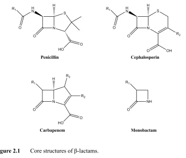
Antibiotics are substances used for the treatment and prevention against bacterial infections. They act by killing or inhibiting the growth of bacteria. There are different classes of antibiotics, yet β-lactam antibiotics are the most widely used group amongall commercially available antibiotics. β-lactam antibiotics include penicillin derivatives (penams), cephalosporins (cephems), monobactams and carbapenems, which contain a β-lactam ring in their core structure.
[image:35.595.124.501.435.755.2]
β-lactam antibiotics kill the bacterial cells by inhibiting peptidoglycan synthesis, which constitutes a major portion of bacterial cell wall synthesis. As a consequence of inhibiting cell wall synthesis, the autolytic system will be activated, which causes bacterial cell death. However, some bacteria can survive against these antibiotics by various mechanisms. These include a) production of another enzyme (β-lactamases) which can inactivate the antibiotic before reaching the target, b) mutation of the active site of the target enzyme, preventing binding of the antibiotic and c) modification of the cell wall to prevent the entry of the drug into the cell or to actively remove the antibiotic via enhanced expression of efflux pumps (Levy and Marshall, 2004).
[image:36.595.190.471.278.576.2]
Figure 2.2 An overall representation of bacterial resistance mechanism. Reproduced from Levy and Marshall (2004).
2.1.2 β-lactamase enzymes
β-lactamases are enzymes that inactivate β-lactam antibiotics by hydrolysing the amide
bond of the β-lactam rings (Essack, 2001; Siu, 2002). Abraham and Chain identified the
first β-lactamase in E. coli already in 1940, even before the widespread use of penicillin
than 1000 of these enzymes have been found in Gram-positive bacteria, Gram-negative bacteria and mycobacteria (Davies and Davies, 2010; Livermore, 1995; Majiduddin et
al., 2002). β-lactamases are either chromosomally encoded or acquired through mobile
genetic elements such as plasmids or transposons (Walsh et al., 2005). As a result,
bacteria are able to acquire β-lactamase genes and become resistant to β-lactams. A few
β-lactamases utilize zinc ions to disrupt the β-lactam ring, but many form an
acyl-enzyme intermediate through an active site serine.
2.1.3 Classification of β-lactamases
β-lactamases are classified according to either their amino acid sequence similarities (Ambler, 1980) or by their functional and biochemical properties (Richmond and Sykes, 1973; Sykes, 2010). According to the Ambler classification, β-lactamases are divided into metallo-β-lactamases (class B) and serine β-lactamases (classes A, C and D) based on amino acid sequence (Ambler, 1980). The Bush-Jacoby classification is based on substrate selectivity and inhibitor profile and separates the enzymes again into four classes, 1 to 4 (Bush et al., 1995).
2.1.4 Metallo-β-lactamases
Unlike serine β-lactamases, which require an active site serine residue for enzymatic
activity, metallo-β-lactamases (MBLs) are metallo-enzymes which require coordination of bivalent metal ions, usually zinc, in their active site.
Subclasses of MBLs
MBLs are further divided into three subclasses, B1, B2 and B3, which are grouped according to substrate selectivity, amino acid sequence (particularly the residues that
chelate the Zn2+ ions) and the Zn2+ ion requirement for catalysis (Rasmussen andBush,
1997).
The MBLs in subclass B1 require the presence of two Zn2+ ions at the active site
for activity and they show broad-spectrum activity towards most β-lactam compounds
B1 compared to subclasses B2 and B3. This subclass harbours MBLs, which are both chromosomally encoded and acquired
Subclass B2 MBLs harbour only a single Zn2+ ion in their active site, which is
required for β-lactam hydrolysis. However, these MBLs are similar to the MBLs in
subclass B1 in size and amino acid sequence (Hall et al., 2003).
Subclass B3 is the least studied of the three subclasses. MBLs in this subclass
harbor two Zn2+ ions at their active site and show a similar substrate profile to subclass
B1 MBLs. Nevertheless, the structure and the amino acid sequence of these enzymes differ significantly from both subclasses B1 and B2. In the present work, the metal binding properties of a B1 MBL were investigated as well as conformational changes of an active site loop in response to ligand binding. In the following, the structural features of the metal binding sites and polypeptide loops near the active site are reviewed.
Three dimensional structure of MBLs
Carfi and co-workers solved the X-ray crystal structure of the mono-zinc form of BcII in 1995, which was the first MBL structure determined (Carfi et al., 1995). Since then crystal structures of MBLs have been determined from all three subclasses. These
structures revealed a common characteristic fold, a αβ/βα quaternary structure, and
catalytic function, indicating that all MBLs evolved from a common ancestor (Daiyasu
et al., 2001). The overall structure comprises two central β-sheets flanked by five
solvent-exposed α-helices with variable loops which connect the α-helices and β
-strands. The Zn2+ ion(s) binding active site is located at the interface of two αβ domains.
Despite common structural features, there are also notable differences between
the 3D structures of each subclass, such as the number of Zn2+ ions in the active site,
different Zn2+ ion(s) coordinating residues and mobile β-hairpin loop regions
contributing to substrate binding. In addition, MBLs in subclass B3 contain intramolecular disulfide bridges, which play an important role in protein folding (Ullah et al., 1998).
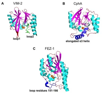
the active site of the enzyme. The crystal structures suggest that this loop interacts with bound substrates or inhibitors, and deletion of loop 1 negatively affects the enzyme activity (Moali et al., 2003). The loop conformation becomes more restricted when an inhibitor is bound (Bebrone, 2007; Concha et al., 1996). In addition, there is a flexible loop 2, which also influences the substrate or inhibitor interactions in subclass B1 MBLs (Dal Peraro et al., 2004).
[image:39.595.149.505.215.540.2]
Figure 2.3 Three-dimensional structures of MBLs. (A) VIM-2, loop 1 and loop 2 are highlighted in red and green respectively (PDB ID: 1KO3, B1 subclass; Garcia-Saez
et al., 2008), (B) CphA, with the elongated α3 helix highlighted in blue (PDB id: 1X8G,
B2 subclass; Garau et al., 2005b) and (C) FEZ-1, where a flexible loop region near the active site is highlighted in blue. (PDB id: 1K07, B3 subclass; Garcia-Saez et al., 2003).
Zn2+ ion(s) are represented as orange spheres.
The loop 1 region is absent in subclass B2 and B3 MBLs (Bebrone, 2007).
specificity for binding carbapenems (Bebrone, 2007; Garau et al., 2005a). The subclass B3 contains a loop region, loop 2, which is formed between helix α3 and β-strand β7 and is in close proximity to the active site (Garcia-Saez et al., 2003).
Active site and Zn binding residues
The active site of MBLs contains two potential catalytic Zn2+ ion(s) coordinating sites known as Zn1 and Zn2. Nevertheless, the number of Zn2+ions at the active site and the
residues that coordinate Zn2+ ion(s) vary between the subclasses B1, B2 and B3. Subclass B1and B3 contains two catalytic Zn2+ ions, whereas subclass B2 acts as a mono-zinc enzyme.
In subclasse B1, the Zn1 site is formed by three histidine residues, His116,
His118 and His 196 (“histidine site”) and a water molecule in a tetrahedral geometry. The Zn2 site is formed by Asp120, Cys221, His263 (“cysteine site”) and two water molecules in a trigonal bipyramidal arrangement. The same water molecule that links both Zn2+ ions also acts as a nucleophile during hydrolysis (Concha et al., 1996; Wang et al.,1999).
The crystal structure of the mono-zinc subclass B2 MBL CphA suggests that the catalytic Zn2+ion resides in the cysteine site. His116 in the histidine site is replaced by Asn116 and shows no direct interaction with the Zn1 site, which explains why MBLs in subclass B2 bind to a single Zn2+ ion via Asp120, Cys221 and His263 (Garau et al.,
Figure 2.4 Zinc sites and metal ligand architecture at the active site. (A) VIM-2 (PDB ID: 1KO3, B1 subclass; Garcia-Saez et al., 2008), (B) CphA (PDB ID: 1X8G, B2 subclass; Garau et al., 2005b) and (C) FEZ-1 (PDB ID: 1K07, B3 subclass; Garcia-Saez et al., 2003). All figures generated with PyMol.
The Zn1 site in the subclass B3 is ahistidine site similar to that of subclass B1. The Zn2 site is formed with His121, which replaces a cysteine residue in subclasses B1 and B2. Hence, the Zn2 site is formed by Asp120, His121, His263 and two water molecules in trigonal bipyramidal geometry (Ullah et al., 1998; Garcia-Saez et al., 2003).
2.1.5 IMP-1 metallo-β-lactamase
Among the families of MBLs, the IMP-type (for “active on imipenem”) β-lactamases
were the first to be identified. They have been detected in many clinically important
Gram-negative bacteria like Pseudomonas spp., Acinetobacter spp. and members of the
Enterobacteriaceae family (Zhao and Hu, 2011).
The MBL IMP-1, which is a subclass B1 β-lactamase, was first identified in a
Pseudomonas aeruginosa isolate in Japan in 1990 (Watanabe et al., 1991).
Subsequently, IMP-1 was found to be prevalent also in Serratia marcescens and in
numerous members of the Enterobacteriaceae family in Japan (Queenan and Bush,
2007). IMP-1 is encoded by the gene blaIMP, which is integrated as a gene cassette into
integrons carried by transferrable plasmids (Arakawa et al., 1995). Hence, the blaIMP
gene can be spread amongst different nosocomial pathogens via horizontal gene transfer.
IMP-1 has the common 3D structure of a MBL. Published IMP-1 crystal
structures show the typical αβ/βα quaternary structural fold and two Zn2+ ions (Concha
et al., 2000). The two Zn2+ ions in the active site of IMP-1 are bound in two distinct
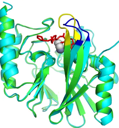
The loop 1 on IMP-1 comprises residues 60–66 (standard numbering defined by
Galleni et al., 2001). None of these residues is fully conserved among subclass B1 β
[image:42.595.214.415.236.451.2]-lactamase (Moali et al., 2003). The X-ray structure of the native enzyme shows an open, disordered conformation of the flap. In the enzyme-substrate complexes, however, the flap is in a closed conformation (Concha et al., 2000). Based on kinetic data by Moali et al. (2003), Trp64 accounts for approximately 50% of the loop effect, probably through hydrophobic interactions.
Figure 2.5 Overlay of crystal structures of native IMP-1 (cyan with the loop 1 highlighted in blue; PDB ID: 1DDK; Concha et al., 2000) and its complex with a mercaptocarboxylate inhibitor (green with loop 1 highlighted in yellow; PDB ID: 1DD6; Concha et al., 2000). Zn atoms are shown as spheres and the inhibitor molecule is shown in red.
In 2014, Carruthers et al. reported the observation of a heterodinuclear form of
IMP-1, [FeZn]-IMP-1. It was shown that when IMP-1 is overexpressed in vivo, the
presence of small amounts (25 µM) of FeCl3 in the expression medium results in
[FeZn]-IMP-1. The formation of [FeZn]-IMP-1 could not be suppressed even with the addition of ten-fold excess of zinc. Moreover, it was determined that the iron bound to
IMP-1 was present as Fe3+ in the Zn1 site (Carruthers et al., 2014). Interestingly, the
2.1.6 Research objectives


![Figure 2.7 Purified [FeZn]-IMP1 samples. Left: wild-type [FeZn]-IMP1. Right:](https://thumb-us.123doks.com/thumbv2/123dok_us/8116857.238391/51.595.222.379.608.727/figure-purified-fezn-imp-samples-left-fezn-right.webp)