0022-538X/05/$08.00
⫹
0
doi:10.1128/JVI.79.1.234–244.2005
Copyright © 2005, American Society for Microbiology. All Rights Reserved.
La Crosse Virus Nonstructural Protein NSs Counteracts the Effects of
Short Interfering RNA
Samantha S. Soldan,
1Matthew L. Plassmeyer,
1,2Meghan K. Matukonis,
1,2and
Francisco Gonza
´lez-Scarano
1*
Departments of Neurology and Microbiology
1and Cell and Molecular Biology Graduate Group,
2University of
Pennsylvania School of Medicine, Philadelphia, Pennsylvania
Received 30 April 2004/Accepted 30 August 2004
Through a process known as RNA interference (RNAi), double-stranded short interfering RNAs (siRNAs)
silence gene expression in a sequence-specific manner. Recently, several viral proteins, including the
non-structural protein NSs of tomato spotted wilt virus (a plant-infecting bunyavirus), the interferon antagonist
protein NS1 of influenza virus, and the E3L protein of vaccinia virus, have been shown to function as
suppressors of RNAi, presumably as a counterdefense against cellular mechanisms that decrease viral
pro-duction. La Crosse virus (LACV), a member of the California serogroup of orthobunyaviruses, has a
triseg-mented negative-stranded genome comprised of large (L), medium (M), and small (S) segments. To develop a
strategy for segment-specific inhibition of transcription, we designed 13 synthetic siRNAs targeting specific
RNA segments of the LACV genome that decreased LACV replication and antigen expression in mammalian
(293T) and insect (C6/36) cells. Furthermore, NSs, a LACV nonstructural protein, markedly inhibited RNAi
directed both against an LACV M segment construct and against a host gene (glyeraldehyde-3-phosphate
dehydrogenase), suggesting a possible role for this viral protein in the suppression of RNA silencing.
Segment-specific siRNAs will be useful as a tool to analyze LACV transcription and replication and to obtain
recom-binant viruses. Additionally, NSs will help us to identify molecular pathways involved in RNAi and further
define its role in the innate immune system.
The
Bunyaviridae
are a large and diverse family of
envel-oped, negative-stranded RNA viruses divided into the
follow-ing five genera:
Orthobunyavirus
,
Hantavirus
,
Nairovirus
,
Phle-bovirus
, and
Tospovirus
(57). The orthobunyaviruses, the first
members of the family that were identified, are maintained in
nature in a transmission and amplification cycle that alternates
between arthropod vectors, predominantly culcine and
anopheline mosquitoes, and small mammals. Within the genus
Orthobunyavirus
, the California serogroup consists of about 14
viruses that are antigenically related to its original member,
California encephalitis virus
(7), which while described in
asso-ciation with a human infection, has since been seldom isolated,
and rarely in relationship to central nervous system disease
(20). Human infections by several other members of the
Cal-ifornia serogroup have been well documented (2, 32, 51, 54, 55,
60), including La Crosse virus (LACV) and Tahyna virus
(TAHV), which have been studied most extensively. LACV is
an important cause of pediatric encephalitis and aseptic
men-ingitis in the Midwestern United States, where its principal
vector,
Aedes triseriatus
, resides (33, 44, 54). TAHV is
associ-ated with an influenza-like illness in central Europe (16, 55).
The bunyavirus genome is composed of three negative-sense
RNA segments designated by their size, as follows: large (L),
medium (M), and small (S). The L segment encodes an
RNA-dependent RNA polymerase (6, 23); the M segment encodes a
polyprotein precursor that is posttranslationally cleaved into
two envelope glycoproteins, G1 and G2, and a third
polypep-tide, NSm, of unknown function (24). All three segments of the
bunyavirus genome are encapsidated by the S
segment-en-coded nucleocapsid (N) protein; this segment also encodes the
12-kDa NSs protein in an overlapping reading frame (21, 22,
28). Interestingly, the NSs protein of
Bunyamwera virus
, the
prototypic member of the family and of the genus
Orthobun-yavirus
, has been reported to decrease RNA synthesis in a
mini-replicon system (62), to act as an interferon antagonist (3,
38, 52, 61), and to play an important role in viral pathogenesis
(3). Although a similar function for the NSs proteins of the
California serogroup viruses has not been described to date, a
recent report demonstrating sequence homology between
Bunyavirus NSs proteins and Reaper, a proapoptotic protein
identified in
Drosophila melanogaster
, suggests a role for
LACV NSs in promoting neuronal apoptosis (12), a function
that has been previously described for the whole virus (47).
Like Reaper, NSs can induce mitochondrial cytochrome
c
re-lease and caspase activation, further suggesting that NSs and
Reaper may be involved in a common mechanism of cell death
(12).
Double-stranded short interfering RNAs (siRNAs) have
been demonstrated to silence gene expression in a
sequence-specific manner through a process known as RNA interference
(RNAi). Naturally occurring RNAi is initiated by the
double-stranded RNA (dsRNA)-specific endonuclease/helicase
Dicer-RDE-1, which cleaves long dsRNA species into 21- to
25-nucleotide fragments called siRNAs (18). siRNAs are
incorporated into a protein complex known as the
RNA-in-duced silencing complex, which recognizes and cleaves target
mRNAs (18). First described for plants, in which it represents
* Corresponding author. Mailing address: Dept. of Neurology,
Uni-versity of Pennsylvania, 3400 Spruce St., Philadelphia, PA 19104-4283.
Phone: (215) 662-3360. Fax: (215) 662-3362. E-mail: scarano
@mail.med.upenn.edu.
234
on November 8, 2019 by guest
http://jvi.asm.org/
an important mechanism for virus resistance (58), RNAi has
been studied intensively in invertebrates and has been shown
to function in antiviral defense and development (8, 25, 36, 56).
Overall, the building blocks of the RNAi-mediated gene
si-lencing pathway have remarkable similarities in otherwise
dis-parate organisms (11, 37, 58), suggesting an ancient origin of
gene silencing in pathogen resistance and organismal
develop-ment. To counteract the RNA silencing mechanism of their
host, plant viruses have developed ways to evade or neutralize
RNAi (4, 48, 53, 58). Recently, NSs of the
Tomato spotted wilt
virus
(TSWV), a plant-infecting
Bunyavirus
of the genus
To-spovirus
, was shown to suppress RNAi, suggesting a role for
this protein in viral pathogenesis (5, 53). While the NSs protein
of TSWV is significantly longer than LACV NSs, these
pro-teins share 33.33% identity and 66.7% similarity within a
27-amino-acid overlap according to the Smith-Waterman
algo-rithm for protein sequence and structural similarity.
While the role of RNAi in plants and invertebrates has been
related to an innate response to pathogens, its role in
mam-malian cells is not entirely clear. Nevertheless, siRNAs have
been shown to decrease viral replication in human
immuno-deficiency virus (HIV), influenza virus, hepatitis C virus, and
several other viral infections (27, 34, 35, 43). Here we describe
13 siRNAs that target individual segments of the tripartite
LACV genome and that inhibit virus replication in both human
and insect cells. These siRNAs may be used as a tool to study
the mechanism of orthobunyavirus replication, as a novel
method for creating reassortants or pseudotypes, or potentially
to develop single-segment reverse genetics. Furthermore, we
describe a role for the LACV NSs protein in the suppression of
RNAi, a function shared by several viral proteins, including
NS1 of influenza virus, the E3L protein of vaccinia virus, NSs
of TSWV, and B2 of flock house virus (5, 40, 41, 53). This study
reinforces the potential physiologic role of siRNAs in viral
resistance in animal cells and suggests that NSs may be
impor-tant in bunyavirus pathogenesis by, among other effects,
coun-teracting the cellular response to viral transcription.
MATERIALS AND METHODS
siRNA transfection.LACV is a negative-strand RNA virus in which the viral
genome (⫺strand) is transcribed into a positive-strand mRNA that serves as a
template for the synthesis of viral protein and a cRNA that is the template for the generation of additional viral RNAs. siRNAs were designed for either the genomic RNA (negative strand) or the antigenomic RNA (positive strand) of LACV by use of the Dharmacon (Lafayette, Colo.) siRNA design center, which selects optimal siRNA sequences based on the Tuschl rules, an empirical algo-rithm that predicts siRNA sequences that are likely to be effective for gene silencing (19). While either the negative- or positive-strand sequence was used to design effective siRNAs, it is unclear which strand is targeted in this system because of the complementarity of the siRNA duplex (27). All LACV siRNAs
were supplied in a 2⬘-deprotected, annealed, and desalted form with 3⬘dTdT
overhangs (Dharmacon). LACV siRNAs (Table 1) were transfected into a hu-man embryonic kidney cell line, 293T, and an epithelial cell-like mosquito cell line, C6/36. The 293T cells were maintained in Dulbecco’s modified Eagle’s medium (DMEM) (Gibco, Grand Island, N.Y.) enriched with 10% heat-inacti-vated fetal calf serum (FCS) (Atlanta Biologicals, Norcross, Ga.) and 2 mM
L-glutamine (Gibco). The C6/36 cells were maintained at 28°C in 5% CO2in
minimal essential medium (Gibco) supplemented with nonessential amino acids (Gibco), 1.5 g of sodium bicarbonate/liter, 1 mM sodium pyruvate (BioWhit-taker, Walkersville, Md.), and 10% heat-inactivated FCS (Atlanta Biologicals). Antibiotic-free medium was used for the duration of the siRNA experiments.
Approximately 24 h before transfection, 8⫻104
293T cells or C6/36 cells were
plated in a 24-well plate in a volume of 500l/well. Chemically synthesized
siRNAs (Dharmacon) were mixed with the amine siPORT (293T) or lipid si-PORT (C6/36) siRNA transfection reagent (Ambion, Austin, Tex.) and Opti-MEM (Gibco) per the kit’s instructions for 30 min before being added to the cells at a concentration of 100 nM siRNA per well. Forty-eight hours after siRNA transfection, the cells were infected for 1 h with either the LACV/original (54) or TAHV/Bardos 92 (42) strain at a multiplicity of infection (MOI) of 0.01 to 0.0001 PFU/cell in DMEM supplemented with 3% FCS, after which the virus was removed and the cells were resuspended in growth medium. Cells and cell culture supernatants were harvested at 12, 24, 48, and 72 h postinfection. All experiments were performed in triplicate.
Plaque assays.Plaque assays for LACV and TAHV were performed in Vero cells, an African green monkey kidney cell line. Vero cells were plated overnight
at 3⫻105
cells/well in a six-well plate, and serial dilutions of cell culture
supernatants (10⫺2to 10⫺6) and uninfected control supernatants were applied in
a volume of 500l for 1 h. The diluted culture supernatants were then removed,
and a 1:1 mixture of 2% sea plaque agarose (Cambrex, Rockland, Maine) and
2⫻DMEM (Gibco) containing 4% FCS was added. Vero cells were incubated
for 72 h at 32°C in 10% CO2before the agarose overlay was removed. Plaques
[image:2.585.43.545.81.255.2]were then visualized by staining with 0.1% crystal violet.
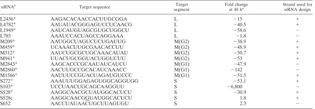
TABLE 1. Change in LACV PFU/ml in siRNA-versus mock-transfected 293T cells
siRNAb Target sequence Target
segment
Fold change
at 48 ha
Strand used for siRNA design
L2436*
AAGACACAACCACUUGCGGA
L
⫺
15
⫹
L4782*
AAUAUACGGGAGUCCUCAACG
L
⫺
40.5
⫹
L1949*
AAUCAUGUAGCGUGCUGGCU
L
⫺
58.6
⫺
L785
AAAUCCACUAGCCAGGAAA
L
⫺
1.8
⫺
M209*
AAUGGCUAGUCUCUGAUUG
M(G2)
⫺
38.9
⫹
M459*
UCAAACUUGCGAACACCUU
M(G2)
⫺
48.9
⫹
M312*
AAUCUGCGCUGCAAACAUAU
M(G2)
⫺
50.7
⫹
M941*
UUACUGCGGUACUGGUCUU
M(G2)
⫺
53
⫹
M2843*
AAGCACCCGCAAUAUCAUCU
M(G1)
⫺
47.9
⫺
M2860*
AACUUGCCGCACAUCAAACC
M(G1)
⫺
142
⫺
M1566*
AAUUUCCGUACUAGAUGUCCC
M(G1)
⫺
51.5
⫹
S272*
AAAUUUGGAGAGUGGCAGGUGG
S
⫺
53.1
⫹
S103*
UCCUAACUGCAGCAAGGUU
S
⫺
6,800
⫺
S528*
AAGGCAACGCUAUGGCACUCU
S
⫺
30.9
⫹
S528c
AAGGCAACGGUAUGGCACUCU
S
1.8
⫹
S652
AACCUAUAACUGCUUAGUGU
S
2.3
⫺
a(LACV MOI⫽0.0001).
b*, statistically significant decrease in virus titer compared to mock-transfected cells (P⬍0.01; Student’sttest).
on November 8, 2019 by guest
http://jvi.asm.org/
Flow cytometry.293T and C6/36 cells were harvested and fixed for 30 min in a 2% paraformaldehyde solution. Nonspecific binding was reduced by exposure to 10% goat serum (Sigma, St. Louis, Mo.) for 30 min and three washes with fluorescence-activated cell sorter (FACS) staining buffer (phosphate-buffered saline with 1% FCS and 0.1% sodium azide) prior to incubation with a 1:500 dilution of mouse immunoglobulin G (IgG) specific for either LACV G1 (807.31, 807.33, 813.13, or 807.35) or TAHV G1 (813.48) (31) for 30 min at room temperature. For each experiment, an aliquot of cells was also treated with isotype-matched control antibodies to establish background staining. The cells were then washed three times with FACS staining buffer and incubated at room temperature with a 1:100 dilution of fluorescein isothiocyanate-conjugated anti-mouse IgG (Sigma) for 30 min. After incubation with the fluorescein isothiocya-nate-conjugated secondary antibody, the cells were washed three times with
FACS staining buffer and resuspended in 400l of FACS staining buffer before
acquisition and analysis on a FACScalibur instrument (Becton Dickinson, Sunny-vale, Calif.) using CellQuest software (Becton Dickinson) (University of Penn-sylvania Cancer Center).
Total RNA extraction and sequencing of virus harvested at 72 h postinfection of siRNA-pretreated cells.Total RNAs were isolated from 293T cells according to the instructions in the Rneasy handbook (Qiagen, Santa Clarita, Calif.).
Approximately 1g of total RNA was reverse transcribed to cDNA by the use
of random hexamers and the SuperScript first-strand synthesis system for RT-PCR (Invitrogen, Carlsbad, Calif.) per the manufacturer’s instructions. The resulting cDNA was used for sequencing (Cell Center, DNA Sequencing Facility, Department of Genetics, University of Pennsylvania).
Molecular cloning of LACV NSs.The LACV S segment was PCR amplified from a pRB322 vector containing a complete double-stranded DNA copy of the LACV/original virus S segment (pLAC 4C-26) (6) and was then inserted into pGEM7Z by the use of Vent DNA polymerase. Briefly, specific primers for the
5⬘and 3⬘ends of the S segment (XhoI-LACS [GCGCTCGAGATTAGTGTAT
CCACTTGAATACTTTGA] and HindIII-LACS [GCGAAGCTTAGTAGTGT GCTCCACTGAATACATTTTA ], respectively; underlining indicates restric-tion sites) were used to amplify the S segment. The resulting fragment was digested with both XhoI and HindIII, inserted into a linearized pGEM7Z vector (Promega, Madison, Wis.), and subsequently sequenced by use of the following
primers: LACS79F (5⬘-GTGATGTCGGATTTGGTG), LACS270R (5⬘-AGGG
TTAGCCTTCCTCTCTGG), LACS370F (5⬘-GGGTATTTAGCCAGATGG),
and LACS652F (5⬘-GCAGATAAGTGGATGTCAC). La Crosse NSs clones
were amplified by PCRs using rTth DNA polymerase XL (PE Biosystems, Foster City, Calif.), with the pGEM-S segment as a template and with specific primers
to introduce restriction enzyme cut sites at the 5⬘and 3⬘ends of NSs. Briefly, NSs
was amplified by PCRs with thermal cycling conditions consisting of a 2-min denaturing step at 94°C followed by 30 cycles of 94°C for 30 s, 55°C for 45 s, and 72°C for 1 min and an extension step of 72°C for 10 min. The forward primer
NSsBgl II-F was complementary to the 5⬘end of NSs and contained a BglII
restriction site (underlined) (GGAAGATCTTCCGATTTGGTGTTTTATGAT
GTCGCAT). The reverse primer NSsEcoRI-R corresponded to the 3⬘end and
contained an EcoRI restriction site (underlined) (GGGGAATTCGGGATCTG GCCAAATACCCAGATA). The resulting fragment was digested with both BglII and EcoRI and inserted into linearized pIRES2-EGFP (BD Biosciences, San Diego, Calif.) that had been digested with BglII and EcoRI. In addition to cloning NSs into pIRES2-EGFP in the correct orientation, we also cloned it into pIRES2-EGFP in the reverse orientation by PCR amplification with the forward
primer NSsEcoRI-F, corresponding to the 5⬘ end of NSs and containing an
EcoRI restriction site (underlined) (GGGGAATTCGGGGATTTGGTGTTTT ATGATGTCGCAT), and the reverse primer NssBglII-R, corresponding to the
3⬘end of NSs and containing a BglII restriction site (underlined) (GGAAGAT
CTTCCCATTCTCGTTATACTGATCAAGGACCC).
To detect NSs expression in the absence of a LACV NSs-specific antibody, we engineered a vector to express an NSs-FLAG fusion protein. The LACV NSs was
cloned into p3XFLAG-CMV-13 (Sigma) in frame with the 3⫻FLAG sequence
by PCR amplification with primers corresponding to the 5⬘and 3⬘ends of NSs
and containing restriction sites for EcoRI and BglII. Two primer sets were used to clone NSs into two different sites in the multiple cloning region of p3XFLAG-CMV-13. Primer set A consisted of NSsEcoRI-F (described above) and
NSsB-glII-R2 (GGAAGATCTTCCATCTGGCGCAATACCCAGATA).
NSsBg-lII-R2 was used to eliminate the NSs stop codon and to add an alanine residue (double underline), allowing the translation of NSs and the FLAG epitope. Successful transformation of the 3X-FLAG vector with the LACV NSs was confirmed by sequencing.
Molecular cloning of the LACV M segment (G1/G2) into pCAGGS. The LACV M segment open reading frame (ORF) was subcloned from pcMORF (44) into the expression vector pCAGGS (kindly provided by Andrew Pekosz).
The LACV M ORF was amplified by PCR from pcMORF (pcDNA3.1-LACV
M) by use of a primer set that introduced restriction cut sites at both the 5⬘and
3⬘ends. Briefly, the LACV M ORF was amplified by the use of rTth DNA
polymerase XL (PE Biosystems). The PCR cycle consisted of a 2-min denaturing step at 93°C followed by 35 cycles of 93°C for 30 s, 48°C for 1 min, and 72°C for 6 min and an extension step of 72°C for 10 min. The forward primer LAC-ClaIF
was complementary to the 5⬘end and contained a ClaI restriction site (CCAT
CGATGGCCAAAGATGATTTGTATATTGGTGC) (underlined), and the
re-verse primer LAC-XhoIR corresponded to the 3⬘end and contained an XhoI
restriction site (CCGCTCGAGCGGCTATCTAATTTTCATCTCTCTCTTAT ATGC) (underlined). The resulting fragment was digested with both ClaI and XhoI and inserted into the linearized pCAGGS expression vector digested with ClaI and XhoI. Transient cotransfection of NSs-FLAG or bacterial alkaline phosphatase (BAP)-FLAG (Sigma) and the LACV M segment mRNA-derived
construct was achieved by calcium phosphate transfection (Promega) of 1g of
DNA per well of a 24-well plate.
Western blotting.Western blot analysis was performed after the cells were
harvested by centrifugation at 600⫻gfor 10 min. The cell supernatants were
discarded, and the pellets were lysed in 150l of immunoprecipitation assay
buffer (50 mM NaCl, 1% NP-40, 0.5% deoxycholic acid, 50 mM Tris [pH 7.4], 1 mM EGTA, 10 mM sodium orthovanadate, 10 mM NaF, and a protease inhibitor cocktail [one tablet per 50 ml]) for 10 min at 4°C. Each sample was then
centrifuged at 14,000⫻gfor 10 min at 4°C. After a rapid freeze (⫺80°C)-thaw
cycle, protein levels in the whole-cell lysates were determined (DC protein assay; Bio-Rad, Hercules, Calif.). The lysates were then mixed with an equal amount of
2⫻sample buffer (0.125 M Tris, 4% sodium dodecyl sulfate, 20% [vol/vol]
glycerol, 0.01% [wt/vol] bromophenol blue, 10% 2-mercaptoethanol) and boiled for 5 min. Ten micrograms of each cell lysate was resolved by gel electrophoresis
with an 8 to 16% Tris-glycine gel (Cambrex) and then transferred to a 0.2-
m-pore-size nitrocellulose membrane (Bio-Rad). Western blot analysis for glycer-aldehyde-3-phosphate dehydrogenase (GAPDH) was performed with anti-GAPDH (1:3,000) rabbit IgG (Abcam, Cambridge, United Kingdom), and the protein was visualized by incubation with horseradish peroxidase (HRP)-conju-gated donkey anti-rabbit IgG (Amersham Biosciences, Piscataway, N.J.) at a 1:10,000 dilution and subsequent detection by an enhanced chemiluminescence assay (SuperSignalWest Pico chemiluminescence substrate; Pierce, Rockford,
Ill.). Western blot analysis for-actin was performed with anti--actin (1:3,000)
goat IgG (Santa Cruz Biotechnology, Santa Cruz, Calif.), and the protein was visualized with HRP-conjugated mouse anti-goat IgG at a dilution of 1:10,000 (Santa Cruz Biotechnology). Western blot analysis for FLAG was performed
with 0.1g of anti-FLAG antibody (Sigma)/ml, and the protein was visualized by
incubation with HRP-conjugated rabbit anti-mouse IgG (Sigma) at a 1:10,000 dilution and detection by an enhanced chemiluminescence assay.
NSs inhibition of RNAi.NSs and control constructs were transfected into 293T
cells (1.0g of DNA/well) 24 h prior to treatment with either a GAPDH or
LACV M siRNA. As controls, 293T cells were also (i) mock DNA transfected, (ii) transfected with the 3X-FLAG vector alone, and (iii) transfected with a control FLAG fusion protein, bacterial alkaline phosphatase (BAP-FLAG). The silencing of GAPDH was assessed by Western blotting and TaqMan quantitative PCR. LACV glycoprotein expression was measured in cells that were cotrans-fected with the LACV M segment construct by FACS analysis with a mixture of LACV G1-specific antibodies (807.31, 807.33, 813.13, and 807.35).
Real-time PCR detection of GAPDH.RNAs were extracted from NSs-FLAG-, BAP-FLAG-, and mock-transfected 293T cells 72 h after GAPDH siRNA treat-ment. Total RNAs were isolated from cells by use of an Rneasy kit (Qiagen) and reverse transcribed to cDNA by the use of random hexamers and the SuperScript first-strand synthesis system (Invitrogen). Quantitative real-time PCRs were per-formed on an ABI Prism 770 sequence detection system (Perkin-Elmer, Foster City, Calif.). The amplification protocol followed the instructions of a TaqMan Gold RT-PCR kit. A normalization experiment was performed simultaneously for each sample with 18S RNA primers and probe. CT (threshold) values were calculated for the target (GAPDH CT) and the standard (18S RNA CT) by determining the points at which the fluorescence exceeded a threshold limit (10
times the standard deviation of the baseline). The results are expressed as⌬CT
(mean GAPDH CT⫺mean 18S RNA CT) (9).
RESULTS
Inhibition of LACV infection by use of siRNAs.
RNAi can be
induced by the introduction of synthetic
⬇
21- to 23-nucleotide
siRNA duplexes into cells, bypassing the requirement for
pro-cessing of a long dsRNA mediated by Dicer-RDE-1 (45).
on November 8, 2019 by guest
http://jvi.asm.org/
These siRNAs confer transient interference of gene expression
in a sequence-specific manner and are generally thought to be
too short to induce an alpha/beta interferon response in
mam-malian cells (39, 45). 293T cells were pretreated with siRNAs
targeting the LACV L, M, and S segments (Table 1). Of 15
siRNAs synthesized, 13 reduced LACV replication (
⬎
35%
inhibition, as measured by the virus titer, 48 h after infection at
an MOI of 0.0001 PFU/cell) (Table 1). Twelve of the siRNAs
were specific for LACV; one siRNA (M2860) also inhibited
replication of the closely related TAHV (data not shown).
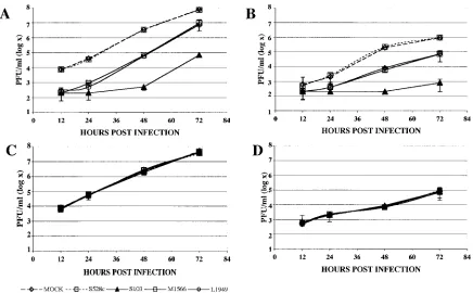
LACV and TAHV growth curves were generated for
mam-malian and insect cell lines (293T and C6/36 cells,
respec-tively), with and without prior siRNA transfections. siRNAs
targeting the LACV L, M, and S segments were transfected
into cells prior to infection with LACV or TAHV, and the
efficacies of the siRNAs were assessed by a plaque assay 12 to
72 h after infection (Fig. 1A and B). S103, an siRNA targeting
the S segment and the most effective siRNA of the panel
generated, inhibited LACV up to 6,800-fold in both 293T and
C6/36 cells. All siRNAs that inhibited LACV were effective in
both 293T and C6/36 cells, indicating that siRNA inhibition of
LACV is equally potent in mammalian and insect cells.
Moover, LACV-directed siRNAs decreased LACV replication
re-gardless of whether the L, M, or S segment was targeted.
For most of the siRNAs, maximal inhibition occurred at
48 h, with a trend toward a rebound of virus replication at 72 h
(Fig. 1). No inhibition of LACV infection was observed with a
control siRNA designed to target the LACV S segment
(LACS528c) that had a 1-bp nucleotide substitution (Fig. 1A
and B) or with an siRNA targeting GAPDH (data not shown).
With one exception, TAHV replication was not inhibited by
pretreatment with LACV siRNAs in either 293T or C6/36
cells. The outlier siRNA, LACM2860, inhibited TAHV
repli-cation
⬇
138-fold 48 h after infection in 293T cells and
⬇
110-fold in C6/36 cells, although its sequence is not present in the
TAHV strain used for these experiments.
To determine the effect of different MOIs on inhibition by
siRNAs, we also performed plaque assays with supernatants
from cultures that were infected with LACV at MOIs ranging
from 0.01 to 0.0001. Whereas the pretreatment of 293T cells
with LACV siRNAs resulted in 99% or more inhibition of
virus replication at a low MOI (0.0001), the efficiency
de-creased markedly when the transfected cells were challenged
with increasingly higher concentrations of virus (Fig. 2). To
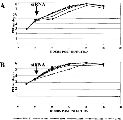
determine if siRNAs could clear the virus when they were
delivered after virus infection and viral gene expression, we
infected 293T and C6/36 cells with LACV at an MOI of 0.0001
PFU/cell and transfected them with LACV L1949, S103, and
M1566 segment siRNAs or control siRNAs (LAC528c and
LACM1566c) at 24 h postinfection. While these siRNAs were
highly effective at decreasing LACV replication when they
were used before virus infection, the ability of these siRNAs to
inhibit LACV replication was significantly impaired when they
were delivered after virus infection (Fig. 3). The
best-perform-FIG. 1. Specific inhibition of LACV replication. 293T and C6/36 cells were pretreated with LACV L, S, and M segment siRNAs. LACV (A and
B) and TAHV (C and D) replication was measured in cell culture supernatants obtained from 293T (A and C) and C6/36 (B and D) cells 12, 24,
48, and 72 h after infection with LACV or TAHV at an MOI of 0.0001 PFU/cell. This representative experiment demonstrates virus replication
in mock-transfected cells, cells that were pretreated with LAC528c (a control for the LACV S segment), and cells that were treated with LACS103,
LACM1566, and LACL1949 siRNAs, designed for the S, M, and L segments of LACV, respectively. Error bars represent the variabilities (standard
errors of the means [SEM]) between triplicate samples for each time point. TAHV replication was not inhibited by pretreatment with LACV
siRNAs, except for LACM2860, which inhibited TAHV replication 138-fold at 48 h postinfection of 293T cells (data not shown).
on November 8, 2019 by guest
http://jvi.asm.org/
[image:4.585.75.511.68.338.2]ing siRNAs for the LACV S and M segments decreased virus
replication when they were applied after virus infection and
viral gene expression, albeit to a far lesser extent than when
they were transfected prior to LACV infection. The LACV L
segment siRNA that we tested did not significantly decrease
virus replication when it was transfected after LACV infection
(Fig. 3).
To confirm that the inhibition of viral replication in
siRNA-pretreated cells was due to a decrease in gene expression, we
measured antigen accumulation in 293T and C6/36 cells by
FACS with a mixture of LACV G1-specific monoclonal
anti-bodies (807.31, 807.33, 813.13, and 807.35) 12, 24, and 48 h
after siRNA transfection (31). Glycoprotein expression was
significantly reduced in both 293T and C6/36 cells that were
pretreated with 13 of the 15 siRNAs designed for the LACV L,
M, and S segments. Figure 4 shows the results for two siRNAs
(LACM1566 and LACS103). The control siRNA 528c did not
affect glycoprotein expression, and with the exception of
M2860, the LACV siRNAs did not decrease TAHV
glycopro-tein expression in either 293T or C6/36 cells (data not shown).
LACV siRNAs also did not inhibit GAPDH expression (data
not shown), indicating that the effects of LACV siRNAs are
indeed highly specific.
siRNA-resistant viruses.
Others have reported that
[image:5.585.64.265.67.209.2]RNAi-resistant viruses can emerge in siRNA-treated cells by either
mutation or deletion of the siRNA-targeted sequence (14, 29,
30). To determine whether RNAi-resistant viruses emerged at
FIG. 2. siRNA-mediated inhibition of LACV replication. The
per-cent decrease in LACV titer in LACV siRNA-treated cells was
com-pared to that in mock-transfected cells at 48 h for infections with
various MOIs (0.01, 0.001, and 0.0001 PFU/cell). The pretreatment of
293T cells with LACV siRNAs resulted in an up to 99% inhibition of
virus replication when the cells were challenged with LACV at a low
MOI (0.0001). The efficiency of LACV siRNAs decreased as cells were
challenged with increasing MOIs (0.001 and 0.01). Error bars
repre-sent variabilities (SEM) between triplicate samples for each time
point.
[image:5.585.304.545.71.387.2]FIG. 3. Inhibition of LACV replication is reduced when siRNAs
are delivered after virus infection. 293T (A) and C6/36 cells (B) were
infected with LACV at an MOI of 0.0001 PFU/cell and were
trans-fected 24 h later (arrow) with LACV L1949, S103, and M1566 segment
siRNAs or control siRNAs (LAC528c and LACM1566c). Error bars
represent variabilities (SEM) between triplicate samples for each time
point.
FIG. 4. LACV glycoprotein expression is decreased in 293T cells
that are pretreated with LACV siRNAs. LACV antigen accumulation
in 293T (A to C) and C6/36 (D to F) cells was measured by FACS with
a panel of LACV G1-specific antibodies 48 h after siRNA transfection.
As shown in these representative plots, LACV-specific siRNAs,
includ-ing M1627 (A and D) and S356 (B and E), inhibited LACV G1
expression. The LACV control siRNA 528c (C and F) did not affect
LACV glycoprotein expression in 293T cells. LACV siRNAs did not
decrease TAHV glycoprotein expression in either 293T or C6/36 cells,
with the exception of M2860 (data not shown).
on November 8, 2019 by guest
http://jvi.asm.org/
[image:5.585.44.286.421.657.2]points after infection when inhibition was no longer evident,
we used supernatants harvested 72 h after LACV infection of
293T cells that had been pretreated with LACV siRNAs to
infect a second round of cells that were pretreated with the
same LACV siRNAs (Fig. 5). Assays were performed after
infection with either LACV alone or LACV cultured for 72 h
in 293T cells that were pretreated with the LACM1566 (Fig.
5A), LACL1949 (Fig. 5B), or LACS103 (Fig. 5C) siRNA. A
slight increase (approximately 1 log) in LACV titer was
ob-served for viruses that were treated with a siRNA and had been
previously exposed to the same inhibitor (LACM1566 [Fig.
5A] and LACL1949 [Fig. 5B]) compared with wild-type LACV
(Fig. 5A and B). However, the differences were negligible at
later time points. In contrast, the virus that was previously
exposed to LACS103 demonstrated a highly significant
(Stu-dent’s
t
test;
P
⬍
0.01) increase in titer, particularly at later
time points (Fig. 5C). When the M and S segments from virus
supernatants harvested 72 h after LACM1566, LACS103, or
mock transfection were sequenced, both wild-type LACV and
viruses with single or double nucleotide changes were observed
(Table 2). At least 30 cDNA clones were sequenced for each
group. Significantly fewer (
2⫽
10.8;
P
⫽
0.01) wild-type S
segment sequences (26.6%) were observed for S segment
clones obtained from siRNA LACS103-treated cells than for
wild-type S segment clones (66.6%) derived from
mock-trans-fected cells (Table 2). While fewer wild-type LACV M segment
clones were obtained from the siRNA LACM1566-pretreated
progeny virus (38.7%) than from mock-transfected cells
(61.3%), this trend did not reach statistical significance (
2⫽
3.84;
P
⫽
0.1).
NSs inhibition of RNAi.
The NSs protein of TSWV, a
Bun-yavirus
of the genus
Tospovirus
, was shown to suppress RNA
silencing (5, 53). To determine whether a similar function
could be associated with LACV NSs, we cloned the gene for
this protein into a pIRES2-EGFP vector in both the correct
(NSs-CO) and the opposite (NSs-WO) orientation and into a
3X-FLAG vector in the correct orientation (NSs-FLAG).
These NSs constructs were transfected into 293T cells (1.0
g
of DNA/well), and interference with silencing of the
endoge-nous gene GAPDH was determined. As controls, 293T cells
were also (i) mock transfected, (ii) transfected with the
3X-FLAG vector alone, and (iii) transfected with a control 3X-FLAG
fusion protein, BAP-FLAG. Silencing of GAPDH was
ob-served for 293T cells that were transfected with a GAPDH
siRNA (GAPDH
⫹) compared with mock-transfected cells or
cells that were transfected with a negative control for the
GAPDH siRNA (GAPDH
⫺) (Fig. 6). GAPDH silencing was
observed for 293T cells that were transfected with a vector
containing NSs in the incorrect (opposite) orientation
(NSs-WO) or with the 3X-FLAG vector. In contrast, GAPDH
ex-pression was not decreased in 293T cells that were transfected
with NSs in the correct orientation (NSs-CO) or with the
NSs-FLAG fusion protein. Similar results were observed with
NIH 3T3 cells (data not shown). The failure of the GAPDH
⫹siRNA to decrease the expression of GAPDH in the presence
of NSs suggests that the NSs protein of LACV may function in
part as an RNA silencing suppressor.
[image:6.585.76.516.69.232.2]To confirm the results observed at the protein level, we used
quantitative real-time TaqMan PCR to examine the effects of
the GAPDH siRNA on GAPDH mRNA expression. RNAs
were extracted from NSs-FLAG-, BAP-FLAG-, and
mock-transfected 293T cells 72 h after GAPDH siRNA treatment.
Quantitative real-time PCRs for GAPDH were performed in
conjunction with normalization for 18S RNA. Cycle threshold
(CT) values were calculated for the target (GAPDH CT) and
the standard (18S RNA CT), and the results were analyzed as
FIG. 5. RNAi resistance of LACV. Supernatants harvested 72 h after LACV infection of 293T cells that had been pretreated with LACV
siRNAs were used to infect a second round of 293T cells that were pretreated with the same LACV siRNAs (LACM1566 [A], LACL1949 [B], and
LACS103 [C]). Plaque assays were performed 12, 24, 48, and 72 h after infection with either LACV or LACV cultured for 72 h in 293T cells that
were pretreated with LACM1566. In all instances, green lines represent mock siRNA-treated wild-type LACV while red lines represent LACV
siRNA-treated wild-type LACV (A), LACL1949 (B), or LACS103 (C). (A) Green, mock siRNA-treated wild-type LACV; purple, mock
siRNA-treated, pretreated LACV; red, LACM1566 siRNA-treated wild-type LACV; blue, LACM1566 siRNA-treated,
LACM1566-pretreated LACV. (B) Green, mock treated LACV; purple, mock treated, LACL1949-LACM1566-pretreated LACV; red, LACL1949
treated LACV; blue, LACL1949 treated, LACL1949-pretreated LACV. (C) Green, mock treated LACV; purple, mock
siRNA-treated, LACS103-pretreated LACV; red, LACS103 siRNA-treated LACV; blue, LACS103 siRNA-siRNA-treated, LACS103-pretreated LACV. A
significant (
P
⬍
0.01 by Student’s
t
test) increase in titer was observed at 12 to 72 h for viruses that were previously exposed to LACS103 compared
to naı¨ve, wild-type LACV.
on November 8, 2019 by guest
http://jvi.asm.org/
⌬
CTs (mean GAPDH CT
⫺
mean 18S RNA CT). Because
high CT values are equated with low copy numbers, low
⌬
CTs
correlate with high GAPDH expression levels and high
⌬
CTs
correlate with low GAPDH expression levels. GAPDH mRNA
expression was significantly reduced in 293T cells that were
transfected with the GAPDH siRNA (GAPDH
⫹) compared to
mock-transfected cells and cells transfected with a GAPDH
siRNA negative control (GAPDH
⫺). In addition, the GAPDH
mRNA level was significantly increased in GAPDH
⫹siRNA-transfected 293T cells that were cosiRNA-transfected with NSs-FLAG
compared to mock- and BAP-FLAG-transfected 293T cells (
P
⬍
0.01 by analysis of variance [ANOVA]). The increased
GAPDH mRNA expression in GAPDH siRNA-treated cells in
the presence of NSs-FLAG supports a role for LACV NSs as
a suppressor of RNAi.
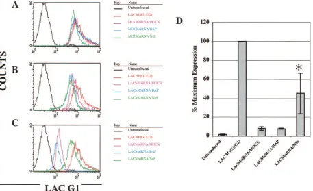
We next examined the ability of NSs to inhibit RNAi of the
LACV G1 glycoprotein. 293T cells that had been pretreated
with the LACM1566 siRNA or a scrambled LACM1566
con-trol siRNA (AACCGTTTCATTAGAGTTCCC) were
co-transfected with the LACV M segment (G1/G2) and either
NSs-FLAG or the BAP-FLAG control vector (Fig. 7A). LACV
glycoprotein expression was measured by FACS with a mixture
of LACV G1-specific antibodies (807.31, 807.33, 813.13, and
807.35). Transfection with NSs-FLAG and BAP-FLAG did not
affect LACV glycoprotein expression in LACM
(G1/G2)-co-transfected cells (Fig. 7A). LACV glycoprotein expression was
also not inhibited significantly in 293T cells that were
pre-treated with the M segment control siRNA (Fig. 7B). As
ex-pected, LACV glycoprotein expression was downregulated in
mock-transfected or BAP-FLAG-cotransfected 293T cells that
were pretreated with the LACM1566 siRNA (Fig. 7C).
How-ever, LACV M expression was partially recovered in 293T cells
that were cotransfected with NSs (Fig. 7C). This experiment
was performed on three separate occasions, and the data were
analyzed as percentages of maximum expression of the mean
fluorescence intensities (Fig. 7D). LACV glycoprotein
expres-sion was significantly increased in 293T cells that were
pre-treated with the LACM1566 siRNA and transfected with
NSs-FLAG compared with BAP-NSs-FLAG-transfected 293T cells or
cells that were transfected only with the siRNA (ANOVA;
P
⬍
0.05 compared to BAP-FLAG- and mock-transfected cells)
(Fig. 7D). The inability of the LACM1566 siRNA to maximally
inhibit LACV glycoprotein expression in the presence of NSs
supports the potential role of NSs as an RNA silencing
sup-pressor. However, NSs-FLAG did not affect siRNA inhibition
of LACV infection itself when NSs was transfected into cells
prior to siRNA transfection and LACV infection (data not
shown), indicating that the maximal NSs effect may occur in
infected cells that express this protein.
DISCUSSION
We have demonstrated that siRNAs targeting the L, M, and
S segments of LACV inhibit virus replication for up to 72 h
after infection. A siRNA treatment directed against the M
segment was associated with diminished expression of the G1
glycoprotein, reinforcing the proposed mechanism for its
ef-fects. Collectively, these results indicate that siRNAs targeting
the LACV genome may be used to confer intracellular
immu-nization. More importantly, these findings suggest that siRNAs
may be a useful tool for studying the mechanisms of LACV
replication and pathogenesis and, at least theoretically, for the
creation of orthobunyavirus segment-specific genome
reassor-tants. Moreover, the ability to induce RNAi of LACV
replica-TABLE 2. LACV clones recovered at 72 hours postinfection of LACV siRNA-pretreated 293T cells
siRNA mRNA target (⫹strand)a Mutation No. of wild-type or mutant clones/
total no. of clones sequenced (%)
S103
TCACTC
AACCTTGCTGCAGTTAGGA
TCTTCTT
LAC wild type
8/30 (26.6)
bTCACTC
AACTTTGCTGCAGTTAGGAT
CTTCTT
Thr
3
Thr (NSs), Leu
3
Phe (N)
5/30 (16.6)
TCACTC
AACCTTGCTGCAATTAGGA
TCTTCTT
Gln
3
Gln (NSs), Val
3
Ile (N)
6/30 (20)
TCACTC
AACCTTACTGCAATTAGGA
TCTTCTT
Leu
3
Leu (NSs), Aln
3
Thr (N)
1/30 (3.3)
TCACTC
AACCTTGCAGCAGTTAGGA
TCTTCTT
Leu
3
Gln (NSs), Aln
3
Aln(N)
8/30 (26.6)
TCACTC
AGCCTTGCTGCAGTTAGGA
TCTTCTT
Thr
3
Ala (NSs), Asn
3
Ser
1/30 (3.3)
TCACTC
AACCTGTCTGCAGGTTAG
TCTTCTT
Frame shift (NSs, N)
1/30 (3.3)
Mock
TCACTC
AACCTTGCTGCAGTTAGGA
TCTTCTT
LAC wild type
22/33 (66.6)
TCACTC
AACCTTGCAGCAGTTAGGA
TCTTCTT
Leu
3
Gln (NSs), Aln
3
Aln(N)
7/33 (21.2)
TCACTC
AACCTTGCTGCAATTAGGA
TCTTCTT
Gln
3
Gln (NSs), Val
3
Ile (N)
4/33 (12.1)
M1566
AAGGC
AATTTCCGTACTAGATGTCCC
TATAA
LAC wild type
12/31 (38.7)
cAAGGC
AATTACCGTACTAGATGTCCC
TATAA
Ser
3
Thr (G1)
2/31 (6.5)
AAGGC
AATTTCCGTGCTAGATGTCCC
TATAA
Val
3
Val (G1)
5/31 (16.1)
AAGGC
AATTTCCGTACTGGATGTCCC
TATAA
Leu
3
Leu (G1)
6/31 (19.4)
AAGGC
AATTTCCGTACTGGATGTTCC
TATAA
Leu
3
Leu and Val
3
Val (G1)
3/31 (9.6)
AAGGC
AATTTCCGTACTAGATGAACC
TATAA
Val
3
Glu (G1)
2/31 (6.5)
AAGGC
GGTTTCCGTACTAGATGTCCC
TATAA
Ala
3
Ala and ile
3
Val (G1)
1/31 (3.2)
Mock
AAGGC
AATTTCCGTACTAGATGTCCC
TATAA
LAC wild type
19/31 (61.3)
M1566
AAGGC
AATTTCCGTGCTAGATGTCCC
TATAA
Val
3
Val (G1)
5/31 (16.1)
AAGGC
AATTTCCGTACTGGATGTCCC
TATAA
Leu
3
Leu (G1)
4/31 (12.9)
AAGGC
AATTACCGTACTAGATGTCCC
TATAA
Ser
3
Thr (G1)
2/31 (6.5)
AAGGC
AATTTCCGTACTAGATGAACC
TATAA
Val
3
Glu (G1)
1/31 (3.2)
aBold letters indicate the target (positions 103 to 122 for the S segment and positions 1627 to 1646 for the M segment). Underlining indicates mutations.
bSignificantly fewer wild-type LACV clones were obtained from LAC S103 siRNA-pretreated cells than from mock-transfected cells (2⫽10.8,P⫽0.01).
cFewer wild-type LACV clones were obtained from LACM1566 siRNA-pretreated cells than from mock-transfected cells. However, this trend did not reach
statistical significance (2⫽3.84,P⫽0.10).
on November 8, 2019 by guest
http://jvi.asm.org/
[image:7.585.46.540.81.309.2]tion in both human and insect cells indicates that these LACV
siRNAs may be exploited to study arthropod-borne
transmis-sion and may potentially be used for prophylaxis and therapy of
LACV infections. The reduced susceptibility of LACV to a
second round of siRNA treatment and the emergence of
RNAi-resistant viral isolates after only 72 h in culture may
have resulted from mutant viruses present in the initial swarm
inoculum or from the acquisition of mutations during siRNA
treatment (14, 30) and will make any therapeutic applications
challenging. However, when similar patterns of RNAi-resistant
viruses were described for poliovirus and for HIV, it was
sug-gested that a successful approach to siRNA-mediated antiviral
therapies for RNA viruses will need to include multiple
siR-NAs, a strategy that is theoretically possible given our results
(14, 29, 30, 34).
We propose that LACV segment-specific siRNAs may be
adapted for protocols for reverse genetics, a strategy used to
engineer specific mutations into viral genomes and for the
generation of recombinant viruses. If successful, the use of
siRNAs in reverse genetics for the genus
Orthobunyavirus
will
be less cumbersome than the approaches that are currently
available (26, 46). Moreover, the use of siRNAs in reverse
genetics can be applied to other viruses with segmented RNA
genomes, such as influenza virus.
Antiviral RNA silencing has been demonstrated for both
plants and insects as a natural defense mechanism for antiviral
protection (1, 10, 13, 40). Plant viruses have been shown to
counteract RNA silencing defense systems (5, 17, 48, 50, 53,
59). Tospoviruses have life cycles in both plants and arthropod
vectors (thrips) (63), and thus these bunyaviruses confront
RNA silencing defense mechanisms of both plant and insect
hosts (4, 49). Likewise, arboviruses that replicate in both
ver-FIG. 6. NSs inhibition of GAPDH RNAi. (A) Expression of NSs FLAG in 293T cells and inhibition of RNA silencing. LACV NSs-FLAG (1.0
g of DNA/well) was transfected into 293T cells by use of the ProFection mammalian calcium phosphate transfection system (Promega) 24 h prior
to GAPDH and control siRNA treatment. GAPDH expression in 293T cells was assessed 72 h after treatment with GAPDH siRNA (
⫹
) and
controls, as measured by Western blotting (see Materials and Methods). Effective silencing of GAPDH was observed for 293T cells that were
transfected with a GAPDH siRNA (
⫹
) (Ambion) (lane 2) compared to mock-transfected cells (lane 1) or cells that were transfected with a negative
control for GAPDH siRNA (
⫺
) (lane 3) (Ambion). A similar inhibition of GAPDH expression was observed for 293T cells that were transfected
with a vector containing NSs in the wrong orientation (NSs-WO; lane 6) or with the BAP-FLAG vector (lane 10). GAPDH expression was not
silenced in 293T cells that were transfected with NSs in the correct orientation (NSs-Co; lane 4) or with the NSs-FLAG fusion protein (lane 8).
(B) Quantitative real-time TaqMan PCR was used to examine the effects of GAPDH siRNA on GAPDH expression. RNAs were extracted from
NSs-FLAG-, BAP-FLAG-, and mock-transfected 293T cells 72 h after GAPDH siRNA treatment. Quantitative real-time PCRs for GAPDH were
performed on an ABI Prism 770 sequence detection system (Perkin-Elmer). A normalization 18S RNA experiment was performed simultaneously
for each sample by the use of 18S RNA primers and probe. CT values were calculated for the target (GAPDH CT) and the standard (18S RNA
CT), and the results are expressed as
⌬
CTs (mean GAPDH CT
⫺
mean 18S RNA CT). High CT values are equated with low copy numbers.
Therefore, low
⌬
CTs correlate with high GAPDH expression levels and high
⌬
CTs correlate with low GAPDH expression levels. GAPDH mRNA
expression was significantly reduced in 293T cells that were transfected with the GAPDH siRNA (GAPDH
⫹) compared to mock-transfected cells
and cells that were transfected with a GAPDH siRNA negative control (GAPDH
⫺). In addition, GAPDH mRNA was significantly increased in
GAPDH
⫹siRNA-transfected 293T cells that were cotransfected with NSs-FLAG compared to mock- and BAP-FLAG-transfected 293T cells
(asterisk;
P
⬍
0.01 by ANOVA). (C) BAP-FLAG and NSs-FLAG expression. As controls, 293T cells were mock transfected (lane 2), transfected
with the 3X-FLAG vector alone (lane 3), and transfected with a control FLAG fusion protein, BAP-FLAG (lane 1). With an anti-FLAG
monoclonal antibody, NSs FLAG was detected (lane 4) at approximately 16 kDa, the predicted size for the NSs-FLAG fusion protein.
on November 8, 2019 by guest
http://jvi.asm.org/
[image:8.585.90.503.72.346.2]tebrates and invertebrates, such as the orthobunyaviruses, may
need to compensate for both the complex immune systems of
the vertebrate host and the RNA silencing defense
mecha-nisms of the insect vector. Here we describe the apparent
diminution of siRNA-mediated silencing of GAPDH and
LACM (G1/G2) by LACV NSs. The ability to inhibit
suppres-sion of both a host (GAPDH) and a viral (LACM) gene in
293T cells suggests that the NSs of LACV may function in part
as an RNA silencing suppressor.
Recently, it was demonstrated that the interferon antagonist
proteins of influenza virus and vaccinia virus, named NS1 and
E3L, respectively, are also suppressors of RNAi (40, 41). These
suppressors of RNAi may function by binding and sequestering
dsRNA. A similar mechanism has been implicated for the p19
protein of tombusvirus, which also functions as an RNA
silenc-ing suppressor (50). There is little structural or sequence
sim-ilarity between these proteins (5, 15, 41). However, the
sup-pression of RNAi by both E3L and NS1 requires the
N-terminal dsRNA binding domain, which is also required for
the inhibition of innate antiviral immunity (64). Therefore, it
cannot be ruled out that the inhibition of RNAi is unrelated to
the targeting of innate antiviral immunity. Accordingly, LACV
NSs-mediated inhibition of silencing may be due to a direct
effect or to indirect effects related to its putative function as an
interferon antagonist, a function demonstrated for the NSs
protein of the closely related Bunyamwera virus (3, 38, 52, 61).
The ability of LACV NSs to suppress RNAi in mammalian
cells adds to the growing body of evidence suggesting that
RNAi also plays an antiviral role in mammalian cells, but its
capacity to bind and sequester dsRNA and to act as an
inter-feron antagonist has yet to be determined. Future studies are
required to determine the mechanisms of suppression of RNAi
by LACV NSs and whether the interferon system is involved.
ACKNOWLEDGMENTS
This work was supported by Public Health Service grants NS-30606
and NS-07180 (training grant for S.S.S.). M.K.M. was funded by NIH
grant T32 GM-07229.
We thank Julio Martin-Garcia, Robin Vos, and other members of
the Gonza
´lez-Scarano laboratory for their many helpful comments and
suggestions. In addition, we thank Karen Stachelek and Thomas
Har-vey for their technical help.
REFERENCES
1.Al-Kaff, N. S., S. N. Covey, M. M. Kreike, A. M. Page, R. Pinder, and P. J. Dale.1998. Transcriptional and posttranscriptional plant gene silencing in
response to a pathogen. Science279:2113–2115.
[image:9.585.66.523.69.348.2]2.Arunagiri, C. K., L. P. Perera, S. B. Abeykoon, and J. S. Peiris.1991. A
FIG. 7. Inhibition of LACV M segment RNAi by NSs. 293T cells were transfected with either NSs-FLAG or BAP-FLAG, were treated 24 h
later with the LACM1566 siRNA, and were cotransfected with the LACV M segment (G1/G2). LACV glycoprotein expression was measured by
FACS with a cocktail of LACV G1-specific antibodies (807.31, 807.33, 813.13, and 807.35). Representative FACS plots from one of three
experiments are shown (A to C). (A) Transfection with NSs-FLAG and BAP-FLAG did not affect LACV glycoprotein expression in LACM
(G1/G2)-cotransfected cells. (B) LACV glycoprotein expression was not inhibited significantly in 293T cells that were pretreated with an M
segment control siRNA. (C) LACV glycoprotein expression was downregulated in mock-transfected or BAP-FLAG-cotransfected 293T cells that
were pretreated with the LACM1566 siRNA. Importantly, LACM expression was partially recovered in 293T cells that were cotransfected with
NSs. (D) This experiment was performed on three separate occasions, and the data were analyzed as percentages of maximum expression of the
mean fluorescence intensities. LACV glycoprotein expression was significantly increased in NSs-FLAG-transfected 293T cells that were treated
with the LACM1566 siRNA compared to mock- and BAP-FLAG-transfected 293T cells (asterisk;
P
⬍
0.05 by ANOVA).
on November 8, 2019 by guest
http://jvi.asm.org/
serologic study of California serogroup bunyaviruses in Sri Lanka. Am. J.
Trop. Med. Hyg.45:377–382.
3.Bridgen, A., F. Weber, J. K. Fazakerley, and R. M. Elliott.2001. Bunyam-wera bunyavirus nonstructural protein NSs is a nonessential gene product
that contributes to viral pathogenesis. Proc. Natl. Acad. Sci. USA98:664–
669.
4.Brigneti, G., O. Voinnet, W. X. Li, L. H. Ji, S. W. Ding, and D. C. Baulcombe.
1998. Viral pathogenicity determinants are suppressors of transgene
silenc-ing in Nicotiana benthamiana. EMBO J.17:6739–6746.
5.Bucher, E., T. Sijen, P. De Haan, R. Goldbach, and M. Prins.2003. Negative-strand tospoviruses and tenuiviruses carry a gene for a suppressor of gene
silencing at analogous genomic positions. J. Virol.77:1329–1336.
6.Cabradilla, C. D., Jr., B. P. Holloway, and J. F. Obijeski.1983. Molecular
cloning and sequencing of the La Crosse virus S RNA. Virology128:463–468.
7.Calisher, C. H.1996. History, classification, and taxonomy of viruses in the
familyBunyaviridae, p. 1–17.InR. M. Elliot (ed.), TheBunyaviridae. Plenum
Press, New York, N.Y.
8.Caplen, N. J., Z. Zheng, B. Falgout, and R. A. Morgan.2002. Inhibition of viral gene expression and replication in mosquito cells by dsRNA-triggered
RNA interference. Mol. Ther.6:243–251.
9.Carcelain, G., R. Tubiana, A. Samri, V. Calvez, C. Delaugerre, H. Agut, C. Katlama, and B. Autran.2001. Transient mobilization of human immuno-deficiency virus (HIV)-specific CD4 T-helper cells fails to control virus rebounds during intermittent antiretroviral therapy in chronic HIV type 1
infection. J. Virol.75:234–241.
10.Carrington, J. C., and S. A. Whitham.1998. Viral invasion and host defense:
strategies and counter-strategies. Curr. Opin. Plant Biol.1:336–341.
11.Cogoni, C., and G. Macino.2000. Post-transcriptional gene silencing across
kingdoms. Curr. Opin. Genet. Dev.10:638–643.
12.Colon-Ramos, D. A., P. M. Irusta, E. C. Gan, M. R. Olson, J. Song, R. I. Morimoto, R. M. Elliott, M. Lombard, R. Hollingsworth, J. M. Hardwick, G. K. Smith, and S. Kornbluth.2003. Inhibition of translation and induction of apoptosis by bunyaviral nonstructural proteins bearing sequence similarity
to reaper. Mol. Biol. Cell14:4162–4172.
13.Covey, S. N., and N. S. Al-Kaff.2000. Plant DNA viruses and gene silencing.
Plant Mol. Biol.43:307–322.
14.Das, A. T., T. R. Brummelkamp, E. M. Westerhout, M. Vink, M. Madiredjo, R. Bernards, and B. Berkhout.2004. Human immunodeficiency virus type 1
escapes from RNA interference-mediated inhibition. J. Virol.78:2601–2605.
15.Delgadillo, M. O., P. Saenz, B. Salvador, J. A. Garcia, and C. Simon-Mateo.
2004. Human influenza virus NS1 protein enhances viral pathogenicity and
acts as an RNA silencing suppressor in plants. J. Gen. Virol.85:993–999.
16.Demikhov, V. G., V. G. Chaitsev, A. M. Butenko, M. S. Nedyalkova, and T. N. Morozova.1991. California serogroup virus infections in the Ryazan region
of the USSR. Am. J. Trop. Med. Hyg.45:371–376.
17.Dunoyer, P., S. Pfeffer, C. Fritsch, O. Hemmer, O. Voinnet, and K. E. Richards.2002. Identification, subcellular localization and some properties of a cysteine-rich suppressor of gene silencing encoded by peanut clump
virus. Plant J.29:555–567.
18.Elbashir, S. M., J. Harborth, W. Lendeckel, A. Yalcin, K. Weber, and T. Tuschl.2001. Duplexes of 21-nucleotide RNAs mediate RNA interference in
cultured mammalian cells. Nature411:494–498.
19.Elbashir, S. M., J. Martinez, A. Patkaniowska, W. Lendeckel, and T. Tuschl.
2001. Functional anatomy of siRNAs for mediating efficient RNAi in
Dro-sophila melanogaster embryo lysate. EMBO J.20:6877–6888.
20.Eldridge, B. F., C. Glaser, R. E. Pedrin, and R. E. Chiles.2001. The first reported case of California encephalitis in more than 50 years. Emerg. Infect.
Dis.7:451–452.
21.Elliott, R. M.1989. Nucleotide sequence analysis of the small (S) RNA segment of Bunyamwera virus, the prototype of the family Bunyaviridae.
J. Gen. Virol.70:1281–1285.
22.Elliott, R. M., and A. McGregor.1989. Nucleotide sequence and expression
of the small (S) RNA segment of Maguari bunyavirus. Virology171:516–524.
23.Endres, M. J., D. R. Jacoby, R. S. Janssen, F. Gonzalez-Scarano, and N. Nathanson.1989. The large viral RNA segment of California serogroup
bunyaviruses encodes the large viral protein. J. Gen. Virol.70:223–228.
24.Fazakerley, J. K., F. Gonzalez-Scarano, J. Strickler, B. Dietzschold, F. Ka-rush, and N. Nathanson.1988. Organization of the middle RNA segment of
snowshoe hare Bunyavirus. Virology167:422–432.
25.Fjose, A., S. Ellingsen, A. Wargelius, and H. C. Seo.2001. RNA interference:
mechanisms and applications. Biotechnol. Annu. Rev.7:31–57.
26.Flick, R., K. Flick, H. Feldmann, and F. Elgh.2003. Reverse genetics for
Crimean-Congo hemorrhagic fever virus. J. Virol.77:5997–6006.
27.Ge, Q., M. T. McManus, T. Nguyen, C. H. Shen, P. A. Sharp, H. N. Eisen, and J. Chen.2003. RNA interference of influenza virus production by di-rectly targeting mRNA for degradation and indidi-rectly inhibiting all viral
RNA transcription. Proc. Natl. Acad. Sci. USA100:2718–2723.
28.Gentsch, J. R., and D. H. Bishop.1978. Small viral RNA segment of
bun-yaviruses codes for viral nucleocapsid protein. J. Virol.28:417–419.
29.Gitlin, L., and R. Andino.2003. Nucleic acid-based immune system: the
antiviral potential of mammalian RNA silencing. J. Virol.77:7159–7165.
30.Gitlin, L., S. Karelsky, and R. Andino.2002. Short interfering RNA confers
intracellular antiviral immunity in human cells. Nature418:430–434.
31.Gonzalez-Scarano, F., R. E. Shope, C. E. Calisher, and N. Nathanson.1982. Characterization of monoclonal antibodies against the G1 and N proteins of
LaCrosse and Tahyna, two California serogroup bunyaviruses. Virology120:
42–53.
32.Grimstad, P. R., C. L. Shabino, C. H. Calisher, and R. J. Waldman.1982. A case of encephalitis in a human associated with a serologic rise to Jamestown
Canyon virus. Am. J. Trop. Med. Hyg.31:1238–1244.
33.Griot, C., F. Gonzalez-Scarano, and N. Nathanson.1993. Molecular deter-minants of the virulence and infectivity of California serogroup bunyaviruses.
Annu. Rev. Microbiol.47:117–138.
34.Jacque, J. M., K. Triques, and M. Stevenson.2002. Modulation of HIV-1
replication by RNA interference. Nature418:435–438.
35.Kapadia, S. B., A. Brideau-Andersen, and F. V. Chisari.2003. Interference of hepatitis C virus RNA replication by short interfering RNAs. Proc. Natl.
Acad. Sci. USA100:2014–2018.
36.Kennerdell, J. R., and R. W. Carthew.2000. Heritable gene silencing in
Drosophila using double-stranded RNA. Nat. Biotechnol.18:896–898.
37.Ketting, R. F., S. E. Fischer, E. Bernstein, T. Sijen, G. J. Hannon, and R. H. Plasterk.2001. Dicer functions in RNA interference and in synthesis of small
RNA involved in developmental timing in C. elegans. Genes Dev.15:2654–
2659.
38.Kohl, A., R. F. Clayton, F. Weber, A. Bridgen, R. E. Randall, and R. M. Elliott.2003. Bunyamwera virus nonstructural protein NSs counteracts in-terferon regulatory factor 3-mediated induction of early cell death. J. Virol.
77:7999–8008.
39.Kumar, M., and G. G. Carmichael.1998. Antisense RNA: function and fate of duplex RNA in cells of higher eukaryotes. Microbiol. Mol. Biol. Rev.
62:1415–1434.
40.Li, H., W. X. Li, and S. W. Ding.2002. Induction and suppression of RNA
silencing by an animal virus. Science296:1319–1321.
41.Li, W. X., H. Li, R. Lu, F. Li, M. Dus, P. Atkinson, E. W. Brydon, K. L. Johnson, A. Garcia-Sastre, L. A. Ball, P. Palese, and S. W. Ding.2004. Interferon antagonist proteins of influenza and vaccinia viruses are
suppres-sors of RNA silencing. Proc. Natl. Acad. Sci. USA101:1350–1355.
42.Malkova, D.1971. Virulence of Tahyna virus in mice and its relation to thermosensitivity and character of plaque population markers. Acta Virol.
15:473–478.
43.McCown, M., M. S. Diamond, and A. Pekosz.2003. The utility of siRNA transcripts produced by RNA polymerase in down regulating viral gene expression and replication of negative- and positive-strand RNA viruses.
Virology313:514–524.
44.McJunkin, J. E., E. C. de los Reyes, J. E. Irazuzta, M. J. Caceres, R. R. Khan, L. L. Minnich, K. D. Fu, G. D. Lovett, T. Tsai, and A. Thompson.2001. La
Crosse encephalitis in children. N. Engl. J. Med.344:801–807.
45.McManus, M. T., and P. A. Sharp.2002. Gene silencing in mammals by
small interfering RNAs. Nat. Rev. Genet.3:737–747.
46.Pekosz, A., B. He, and R. A. Lamb.1999. Reverse genetics of negative-strand
RNA viruses: closing the circle. Proc. Natl. Acad. Sci. USA96:8804–8806.
47.Pekosz, A., J. Phillips, D. Pleasure, D. Merry, and F. Gonzalez-Scarano.
1996. Induction of apoptosis by La Crosse virus infection and role of neu-ronal differentiation and human bcl-2 expression in its prevention. J. Virol.
70:5329–5335.
48.Pfeffer, S., P. Dunoyer, F. Heim, K. E. Richards, G. Jonard, and V. Ziegler-Graff.2002. P0 of beet Western yellows virus is a suppressor of
posttran-scriptional gene silencing. J. Virol.76:6815–6824.
49.Pruss, G., X. Ge, X. M. Shi, J. C. Carrington, and V. Bowman Vance.1997. Plant viral synergism: the potyviral genome encodes a broad-range patho-genicity enhancer that transactivates replication of heterologous viruses.
Plant Cell9:859–868.
50.Silhavy, D., A. Molnar, A. Lucioli, G. Szittya, C. Hornyik, M. Tavazza, and J. Burgyan.2002. A viral protein suppresses RNA silencing and binds si-lencing-generated, 21- to 25-nucleotide double-stranded RNAs. EMBO J.
21:3070–3080.
51.Srihongse, S., M. A. Grayson, and R. Deibel.1984. California serogroup viruses in New York State: the role of subtypes in human infections. Am. J.
Trop. Med. Hyg.33:1218–1227.
52.Streitenfeld, H., A. Boyd, J. K. Fazakerley, A. Bridgen, R. M. Elliott, and F. Weber.2003. Activation of PKR by Bunyamwera virus is independent of the
viral interferon antagonist NSs. J. Virol.77:5507–5511.
53.Takeda, A., K. Sugiyama, H. Nagano, M. Mori, M. Kaido, K. Mise, S. Tsuda, and T. Okuno.2002. Identification of a novel RNA silencing suppressor, NSs
protein of Tomato spotted wilt virus. FEBS Lett.532:75–79.
54.Thompson, W. H., B. Kalfayan, and R. O. Anslow.1965. Isolation of Cali-fornia encephalitis group virus from a fatal human illness. Am. J. Epidemiol.
81:245–253.
55.Traavik, T., R. Mehl, and R. Wiger.1978. California encephalitis group viruses isolated from mosquitoes collected in Southern and Arctic Norway.
Acta Pathol. Microbiol. Scand. B86:335–341.
56.Tuschl, T., P. D. Zamore, R. Lehmann, D. P. Bartel, and P. A. Sharp.1999.
on November 8, 2019 by guest
http://jvi.asm.org/
Targeted mRNA degradation by double-stranded RNA in vitro. Genes Dev.
13:3191–3197.
57.van Regenmortel, M. H. V., C. M. Fauquet, D. H. L. Bishop, E. B. Carstens, M. K. Estes, S. M. Lemon, J. Maniloff, M. A. Mayo, D. J. McGeoch, C. R. Pringle, and R. B. Wickner.2000. Family Bunyaviridae, p. 35–43.InR. B. Wickner (ed.), Virus taxonomy: 7th report of the International Committee on Taxonomy of Viruses. Academic Press, San Diego, Calif.
58.Voinnet, O.2001. RNA silencing as a plant immune system against viruses.
Trends Genet.17:449–459.
59.Voinnet, O., C. Lederer, and D. C. Baulcombe.2000. A viral movement protein prevents spread of the gene silencing signal in Nicotiana
benthami-ana. Cell103:157–167.
60.Watts, D. M., J. W. LeDuc, C. L. Bailey, J. M. Dalrymple, and T. P. Gargan
II.1982. Serologic evidence of Jamestown Canyon and Keystone virus
in-fection in vertebrates of the DelMarVa Peninsula. Am. J. Trop. Med. Hyg.
31:1245–1251.
61.Weber, F., A. Bridgen, J. K. Fazakerley, H. Streitenfeld, N. Kessler, R. E. Randall, and R. M. Elliott.2002. Bunyamwera bunyavirus nonstructural protein NSs counteracts the induction of alpha/beta interferon. J. Virol.
76:7949–7955.
62.Weber, F., E. F. Dunn, A. Bridgen, and R. M. Elliott.2001. The Bunyamwera virus nonstructural protein NSs inhibits viral RNA synthesis in a
minirepli-con system. Virology281:67–74.
63.Wijkamp, I., J. van Lent, R. Kormelink, R. Goldbach, and D. Peters.1993. Multiplication of tomato spotted wilt virus in its insect vector, Frankliniella
occidentalis. J. Gen. Virol.74:341–349.
64.Xiang, Y., R. C. Condit, S. Vijaysri, B. Jacobs, B. R. Williams, and R. H. Silverman.2002. Blockade of interferon induction and action by the E3L
dou-ble-stranded RNA binding proteins of vaccinia virus. J. Virol.76:5251–5259.
on November 8, 2019 by guest
http://jvi.asm.org/
![FIG. 5. RNAi resistance of LACV. Supernatants harvested 72 h after LACV infection of 293T cells that had been pretreated with LACVsiRNAs were used to infect a second round of 293T cells that were pretreated with the same LACV siRNAs (LACM1566 [A], LACL1949 [B], and](https://thumb-us.123doks.com/thumbv2/123dok_us/185315.54091/6.585.76.516.69.232/resistance-supernatants-harvested-infection-pretreated-lacvsirnas-pretreated-sirnas.webp)