Int. J. Electrochem. Sci., 11 (2016) 3086 - 3094
International Journal of
ELECTROCHEMICAL
SCIENCE
www.electrochemsci.org
A New Electrochemical HbA
1cBiosensor Based on Flow
Injection and Screen-Printed Electrode
Ang Liu1, Shaorui Xu1, Hongyu Deng2,and Xiaochun Wang1, *
1
Department of Laboratory Medicine, Xiangya School of Medicine, Central South University, Changsha, Hunan 410013, PR China
2
Department of Clinical Laboratory, Hunan Cancer Hospital & The Affiliated Cancer Hospital of Xiangya School of Medicine, Central South University, Changsha, 410013, PR China
*
E-mail: [email protected]
Received: 15 January 2016 / Accepted: 22 February 2016 / Published: 1 March 2016
In this paper a novel elctrochemical HbA1c biosensor based on flow injection and screen-printed
electrode was developed. The reticulated vitreous carbon (RVC) electrode was modified with 3-aminophenylboronic acid, chitosan (CS), and tetraenthoxyl silica (TEOS), which entrapped carbon nanotubes to form a sol-gel film at the surface of the screen-printed electrode. The electrochemical behavior of the biosensor was investigated using cyclic voltammograms and amperometric method. Then the reusability of RVC electrode was investigated at various pH values. Furthermore, the selectivity and stability of the HbA1c biosensor was studied. The biosensor was also employed to detect
the real samples, and the results were compared to automatic biochemical analysis.
Keywords: HbA1c; screen printed electrode; electrochemical biosensor; reusability
1. INTRODUCTION
Hemoglobin A1c (HbA1c), a specific glycated hemoglobin, is formed irreversibly through a
nonenzymatic reaction of glucose and the N-terminal valine of the β-chain in hemoglobin[1]. The level of HbA1c reflects the average blood glucose concentration over the preceding 2 to 3 months. So it is the
most significant index for diabetes or related diseases. The normal level of HbA1c is lower than 6%,
whereas diabetes can be diagnosed when the level of HbA1c is higher than 6.5%[2]. Thus it is
important to control the level of HbA1c, which is a diagnostic criterion for diabetes.
There are several methods to detect the ratio of HbA1c to total hemoglobin (%HbA1c), such as
chromatography. Most of those methods have common disadvantages including long time of analysis and high cost for each test[4-6]. So it is the trend to search for a method with simplicity, low-cost, and sensitivity. The diol group of boronic acid can bind with the cis-diol group of the sugar from HbA1c
under alkaline conditions[1,2,7]; thus it is used to capture glycated protein and the HbA1c can be
separated from hemoglobin. Whereas Hb contains four iron heme groups, Hb or HbA1c can be oxidized
at a special electrode. Baccarin et al. developed a poly(amidoamine)-dendrimer-modified glassy carbon electrode to realize direct electron transfer from Hb[8].
The electrochemical biosensor, especially disposable electrochemical sensors based on screen-printed electrode (SPE) technology, has the advantages of low-cost, simplicity, high sensitivity, and high efficiency[9-14]. It has been applied in various fields such as clinically, environmentally and in food production. The aim of this work is introducing of a rapid, simple and sensitive HbA1c biosensor
based on flow injection and screen-printed electrode. Reticulated vitreous carbon (RVC), with low electrical resistance and high current density, is used as the porous electrode[15-17]. The RVC electrode is modified with 3-aminophenylboronic acid. When the sample is injected, HbA1c can bind
with the RVC electrode, and Hb can be separated and oxidized at the SPE. On account of the biocompatibility and electrocatalyzing potential of carbon nanotubes (CNT)[18], the material was employed and entrapped to the SPE by chitosan (CS) and tetraenthoxyl silica (TEOS), forming a sol-gel film. Judged by investigation of the electrochemical behaviors of the HbA1c biosensor, the method
can have clinical application.
2. EXPERIMENTAL
2.1 Materials
The reticulated vitreous carbon (RVC) foam was purchased from Aerospace (Oakland, CA, USA). Ascorbic acid, uric acid, acetaminophen, 3-aminophenylboronic acid, chitosan(CS), tetraenthoxyl silica(TEOS), hemoglobin(Hb), N-hydroxysuccinimide (NHS), and 1-ethyl-3-(3-dimethylaminopropyl) carbodiimide hydrochloride (EDC) were purchased from Sigma-Aldrich (St. Louis, MO, USA). HbA1c calibrators were obtained from Primus (Kansas, MO, USA).
2.2 Preparation of boronate-modified RVC electrode
The reticulated vitreous carbon (RVC) electrode was prepared as follows: the RVC foam was compressed 10 times and designed as the working electrode with a diameter of 3 mm and a length of 5 mm, and then the RVC electrode was immersed in the mixed solution of HNO3 and H2SO4 with a
modified RVC electrode was set into homemade cylindrical PEEK to form the flow cell. The inlet of the RVC electrode assembly was connected to a six-port rotary valve.
2.3 Preparation of Hb-detecting electrode based on the screen-printed electrode
The hemoglobin (Hb) detecting electrode was prepared by using chitosan(CS) and tetraenthoxyl silica(TEOS) to entrap carbon nanotubes, forming a sol-gel film on the screen-printed electrode. The ratio of CS with TEOS was 1:1. The screen-printed electrode was dipped into the mixed solution (10 μL) and dried at 45 °C for 10 min. Then the direct electrochemical behavior of Hb was investigated using cyclic voltammetry. The potential was in the range from -0.65 V to 0.15 V, and the scan rate was 100 mV/s. The chronoamperometric detection of Hb was done under a potential of -0.2 V. The modified screen-printed electrode was mounted to the thin-layer cell body and connected with the RVC electrode with a nut.
2.4 Electrochemical flow injection assay system of HbA1c
All the electrochemical measurements were carried out with a model CHI832C Electrochemical Workstation (CH Instruments, Austin, TX, USA). The flow rate was 1.0 mL/min. The potential at the screen-printed electrode was set at -0.2 V. The carrier solution was PBS solution with pH of 8.0. 10 µL of sample or whole blood was added into 10 µL red cell lysis buffer and reacted 30 s, and then the solution was injected through a valve by a syringe pump. After the signal at the screen-printed electrode decreased, a potential of 0.2 V was set at the RVC electrode. Based on calculation of the ratio of electric charge at the RVC electrode and the screen-printed electrode, the %HbA1c can be
obtained. After amperometric measurements in PBS solution, the RVC electrode can be reused after 1-min replacement of the carrier solution with acetate buffer with a pH of 4.5. Then the RVC electrodes were kept dry at room temperature. All experiments were carried out at room temperature unless otherwise stated.
2.5 Real sample preparation and analysis
Whole blood samples (2.0 mL each) were collected into vacuum tubes containing EDTA. Then the samples were added to 2 mL red cell lysis buffer. %HbA1c of the blood samples can be obtained by
calculating the ratio of the electric charges at the RVC electrode and the screen-printed electrode. The results were compared to those determined with an standard HbA1c analyzer which used the HPLC
method (Bio-Rad VARIANT™ II Hemoglobin Testing System, USA).
2.6 Reference method for HbA1c determination
HPLC is the reference method for HbA1c determination developed by the International
United States. The method requires anticoagulated blood which is diluted with a hemolysis reagent. Apart from the hemolysis process, samples usually need pretreatment by incubation at 37 °C for 30 minutes, whereby an interfering substance is removed. Then a series of three phosphate buffers (of increasing ionic strength) was passed through the column. Light absorbance is read at two different wavelengths, a curve is obtained, and integration is performed to calculate the area under the peaks[2].
3. RESULTS AND DISCUSSION
3.1 The electrochemical behavior of Hb at screen-printed electrode
Cyclic voltammetry (CV) was employed to investigate the direct electrochemical behavior of hemoglobin (Hb). Chitosan (CS) and tetraenthoxyl silica (TEOS) was employed to entrap CNT to the screen-printed electrode. And the ratio of CS to TEOS was also investigated. When the ratio was 1.0, Hb can be oxidized directly at the screen-printed electrode. The oxidation potential and the reduction potential were -0.21 V and -0.34 V respectively as shown in Figure 1. The results revealed that CNT can facilitate the electron transfer between Hb and the screen-printed electrode.
-0.6 -0.4 -0.2 0.0 0.2
-2 -1 0 1 2
C
u
rr
en
t
(
A
)
Potential (V) a
[image:4.596.170.426.386.596.2]b
Figure 1. Cyclic voltammograms of PBS solution containing 0(a) or 100 mg/mL (b) Hb at the screen-printed electrode. The scan rate was 100 mV/s.
0 50 100 150 200 250
0.6 1.2 1.8
[HB](mg/mL)
C
h
a
r
g
e
(
10
-4
C
)
0 1 2
0.6 0.9 1.2 1.5
C
h
a
r
g
e
(
10
-4
C
)
Flow Rate (mL/min)
[image:5.596.140.419.71.297.2]Figure 2. The relationship between flow rate with electric charge when Hb concentration was 100 mg/mL and the potential was -0.2 V.
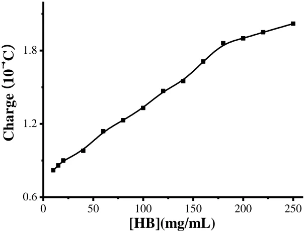
Figure 3. The relationship between Hb concentration (from 10-250 mg/mL) and electric charge of the chronoamperometry curve when the potential was -0.2V.
[image:5.596.141.445.361.594.2]
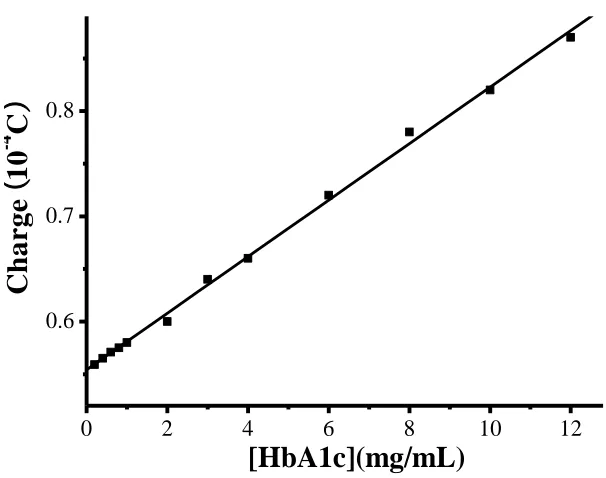
0 2 4 6 8 10 12
0.6 0.7 0.8
[HbA1c](mg/mL)
C
h
a
r
g
e
(
10
-4
C
)
3.2 The electrochemical behavior of HbA1c at RVC electrode
As known, the diol group in boronic acid can bind with the cis-diol group in sugar from HbA1c
under alkaline conditions[1,2,7], so 3-aminophenylboronic acid was employed to modify the reticulated vitreous carbon (RVC) electrode. When the sample was injected into the porous RVC electrode, HbA1c can bind with the group in boronic acid at the modified RVC electrode, thus HbA1c
can be separated from the sample. When the potential was set at 0.2 V, HbA1c can be oxidized at the
RVC electrode because of the low electrical resistance and high current density of RVC. As shown in Figure 4, there was a good linear relationship between HbA1c concentration and electric charge with
HbA1c concentrations between 0.2 mg/mL and 12.0 mg/mL, and the linear regression equation was Q(c)
=0.027 c(HbA1c)(mg/mL) + 0.55. The limit of detection as calculated by multiplying the background
noise by 3 was 89 μg/mL. The final result is %HbA1c,which is the ratio of charges at the RVC and the
SPE.
Figure 4. The calibration curve of HbA1c concentration (from 0.2-12.0 mg/mL) with oxidization
current of the chronoamperometry curve.
Our method attained a lower limit of detection than other tests of HbA1c. For example, Chawla
and Pundir developed a amperometric biosensor of HbA1c based on immobilization of fructosyl amino
acid oxidase onto a hybrid film. It had a detection limit of 50 μmol/L fructosyl valine (a proteolysis product of HbA1c)[5], which corresponded to 1.6 mg/mL HbA1c. The better performance of our test is
[image:6.596.145.448.308.547.2]
3.3 Effects of pH on RVC electrode reusability
To investigate the reusability of the RVC electrode, various pH was employed. Given the electric charge determined at the RVC electrode, we can calculate the HbA1c remaining at RVC
electrode. It is easier for HbA1c to bind with boronic acid in alkaline conditions, so to reuse the RVC
electrode, HbA1c can be dissociated with pH lower than 5.0. Thus the best pH for RVC electrode
reusability was 4.0-5.0. We chose pH of 4.5 in this research, and the washing time was 1 min.
3.4 Selectivity and stability of the HbA1c biosensor
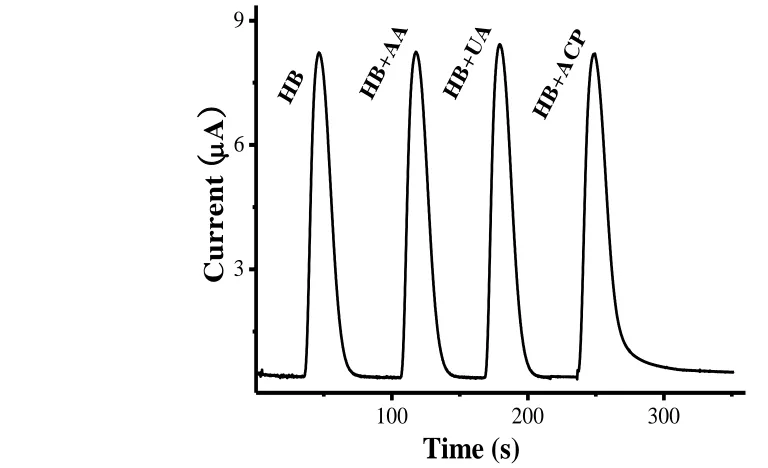
The electrochemical biosensor is often subject to interference by the oxidization or reduction of redox-active species such as uric acid (UA), ascorbic acid (AA), and acetaminophen (ACP) [19-21]. So it is necessary to study the selectivity and specificity of the HbA1c biosensor. As shown in Figure 5,
[image:7.596.51.435.356.590.2]when the potential at the screen-printed electrode was -0.2 V, the electrochemically active species caused no interference in Hb detection because those species cannot be oxidized at such a low potential, so the results demonstrated that the HbA1c biosensor had good selectivity and specificity.
Figure 5. Selectivity of the HbA1c biosensor for Hb in the absence or presence of redox-active species
(AA, ACP and UA), the concentration being 100 mg/mL.
Some other studies also dealt with the interference issue in HbA1c measurements. For example, Chawla and Pundir’s amperometric biosensor had a signal decrease of 8.85% with ascorbic acid and 21.28% with uric acid[5]. They considered those fractions negligible while we excluded the possibility of interference by using the low potential.
To investigate the stability of the HbA1c biosensor, 2.5 mg/mL HbA1c for the RVC electrode
and 50 mg/mL Hb for the screen-printed electrodes (same bath) were employed. After each
100 200 300
3 6 9
HB
HB +A
CP HB
+U A
HB +A
A
C
u
r
r
e
n
t
(
A
)
measurement of HbA1c at the RVC electrode, the electrode was washed with acetate buffer with the pH
of 4.5 for 1 min. The RVC electrode lost 4.2% of its initial activity after 100 times used, which demonstrated that the RVC electrode had good stability. Due to the stability of the sol-gel film on the screen-printed electrodes[22], the same batch of electrodes had good repeatability for determination of Hb. Thus the HbA1c biosensor can be used for real samples.
3.5 Real sample measurement
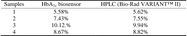
Whole blood samples (2.0 mL each) were collected into vacuum tubes containing EDTA. 10 µL sample was added into 10 µL red cell lysis buffer and reacted 30 s, the solution was injected through a valve by a syringe pump at a flow rate of 1.0 mL/min, then the potential at the screen-printed electrode was set at -0.2 V, and the chronoamperometry curve was recorded to detect Hb. When the signal kept constant, the potential at the RVC electrode was changed to 0.2 V to detect HbA1c, and then
we could calculate %HbA1c based on the ratio of electric charge at the RVC electrode and the
screen-printed electrode. The results were compared to those determined with an HPLC-based standard HbA1c
analyzer (Bio-Rad VARIANT™ II Hemoglobin Testing System, USA). As shown in Table 1, the results based on our method were in excellent agreement with the commercial automatic clinical analyzer. Compared with the clinical analyzer, there was no significant difference in accuracy. Our HbA1c biosensor based on flow injection and screen-printed electrode had lower cost, was easier to
prepare, and the results could be acquired in less than 5 min, which enabled clinical application.
Table 1. Comparison of %HbA1c determined by our HbA1c biosensor and HPLC, the reference
method (example data)
Samples HbA1c biosensor HPLC (Bio-Rad VARIANT™ II)
1 2 3 4
5.58% 7.43% 10.12.%
8.67%
5.62% 7.55% 9.94% 8.82%
4. CONCLUSIONS
In this paper we developed a novel electrochemical HbA1c biosensor based on flow injection and
screen-printed electrode. The RVC electrode was modified with 3-aminophenylboronic acid. Carbon nanotubes were entrapped by chitosan (CS) and tetraenthoxyl silica (TEOS) to form a sol-gel film at the surface of the screen-printed electrode. Hb could undergo direct electrochemical oxidation at the surface of the screen-printed electrode. There was good relationship between the concentrations of Hb (from 20 to 200 mg/mL) and electric charge of the chronoamperometry curve when the potential was -0.2 V at the screen-printed electrode. There was also good relationship between the HbA1c
[image:8.596.108.488.484.558.2]
The RVC electrode was good. Furthermore, the biosensor exhibited good reusability when pH was 4.5, and also selectivity and stability. This method was also successfully employed to test blood samples as compared with the automatic clinical analyzer. Those results revealed that the method could have clinical application.
References
1. K.M. Hsieh, K.C. Lan, W.L. Hu, MK Chen, LS Jang, and WH Wang, Biosens. Bioelectron., 49 (2013) 450.
2. D.B. Sacks, in: C.A. Burtis and D.E. Bruns, Tietz Fundamentals of Clinical Chemistry and Molecular Diagnostics, 7e, p 608. Published by Elsevier (2014), Amsterdam, Netherlands. 3. R.R. Little and C.L. Rohlfing, Clin. Chim. Acta, 418 (2013) 63.
4. Y. Zhou, H. Dong, L. Liu, Y. Hao, Z. Chang, and M. Xu, Biosens. Bioelectron., 64 (2015) 442. 5. S. Chawla and C.S. Pundir, Anal Biochem., 430 (2012) 156.
6. B.W. Bode, B.R. Irvin, J.A. Pierce, M. Allen, and A.L. Clark, J. Diabetes Sci. Technol., 1 (2007) 405.
7. Y.C. Chuang, K.C. Lan, K.M. Hsieh, L.S. Jang, and M.K. Chen, Sens Actuators B Chem., 171-172 (2012) 1222.
8. M. Baccarin, B.C. Janegitz, R. Berté, F.C. Vicentini, C.E. Banks, O. Fatibello-Filho, and V. Zucolotto, Mat. Sci. Eng. C-Mater., 58 (2016) 97.
9. D. Lowinsohn, P.T. Lee, and R.G. Compton, Int. J. Electrochem. Sci., 9 (2014) 3458. 10. A. Liu and X. Wang, Int. J. Electrochem. Sci., 10 (2015) 9342.
11. T. Shimomura, T. Sumiya, M. Ono, T. Ito, and T.A. Hanaoka, Anal. Bioanal. Chem., 405 (2013) 297.
12. V. Gubala, L.F. Harris, A.J. Ricco, M.X. Tan, and D.E. Williams, Anal. Chem., 84 (2012) 487. 13. J. Chen, Z. Jiang, J.D. Ackerman, M. Yazdani, S. Hou, S.R. Nugen, and V.M. Rotello, Analyst,
140 (2015) 4991.
14. N. Bunyakul and A.J. Baeumner, Sensors (Basel), 15 (2014) 547.
15. L.M. Abrantes, in: G. Kreysa, K. Ota, and R.F. Savinell, Encyclopedia of Applied Electrochemistry, p 2077. Published by Springer (2014), New York, USA.
16. K. Lenghartova, L. Lauko, F. Cachob, and E. Beinrohr, Acta Chim. Slov., 62 (2015) 152. 17. X. Mo, Z.H. Yang, H.Y. Xu, G.M. Zeng, J. Huang, X. Yang, P.P. Song, and L.K. Wang, J.
Hazard. Mater., 286 (2015) 493.
18. J. Zhang, X. Liu, R. Blume, A. Zhang, R. Schlögl, and D.S. Su, Science, 322 (2008) 5898.
19. P. D’Orazio and M.E. Meyerhoff, in: C.A. Burtis and D.E. Bruns, Tietz Fundamentals of Clinical Chemistry and Molecular Diagnostics, 7e, p 151. Published by Elsevier (2014), Amsterdam, Netherlands.
20. J.Z. Tsai, C.J. Chen, K. Settu, Y.F. Lin, C.L. Chen, and J.T. Liu, Biosens. Bioelectron., 77 (2016) 1175.
21. S. Azzouzi, L. Rotariu, A.M. Benito, W.K. Maser, M. Ben Ali, and C. Bala, Biosens. Bioelectron., 69 (2015) 280.
22. T. Venckus, R. Celiešiūtė, A. Radzevič, T. Rakickas, Š. Vaitekonis, Ž. Ruželė, and R. Pauliukaite, Electroanalysis, 26 (2014) 2273.