City, University of London Institutional Repository
Citation
:
Fahey, B., Barlow, S., Day, J. S. and O'Mara, S. M. (2008). Interferon-alpha-induced deficits in novel object recognition are rescued by chronic exercise. Physiology and Behavior, 95(1-2), pp. 125-129. doi: 10.1016/j.physbeh.2008.05.008This is the accepted version of the paper.
This version of the publication may differ from the final published
version.
Permanent repository link:
http://openaccess.city.ac.uk/5755/Link to published version
:
http://dx.doi.org/10.1016/j.physbeh.2008.05.008Copyright and reuse:
City Research Online aims to make research
outputs of City, University of London available to a wider audience.
Copyright and Moral Rights remain with the author(s) and/or copyright
holders. URLs from City Research Online may be freely distributed and
linked to.
City Research Online: http://openaccess.city.ac.uk/ [email protected]
Interferon-α-induced deficits in novel object recognition are rescued by chronic
exercise
Fahey, Bríana, Barlow, Sally, Day, Jennifer S., O’Mara, Shane M.*.
Trinity College Institute of Neuroscience,
Trinity College Dublin,
Dublin 2,
Ireland.
*Corresponding author – Shane M. O’Mara,
Trinity College Institute of Neuroscience,
Lloyd Building,
Trinity College Dublin,
Dublin 2,
Ireland
E-mail address: [email protected]
Telephone: +353-1-8968447
Abstract
The anti-viral drug interferon-alpha (IFN-α) is widely-known to induce psychiatric
and cognitive effects in patients. Previous work has shown that physical exercise can
have a positive effect against brain insult. We investigated the effects of a
clinically-comparable treatment regime of IFN-α on cognitive function in male Wistar rats and
assessed the impact of chronic treadmill running on the deficits generated by IFN-α.
We found that IFN-α induced significant impairments in performance on both spatial
novelty and object novelty recognition. Chronic forced exercise did not protect
against IFN-α-induced learning deficits in reactivity to spatial change, but did restore
the capacity for novel object recognition in IFN-α-treated animals.
Introduction
Interferons are a family of multi-functional, pleiotropic proteins belonging to the
cytokine family of proteins. They are produced by the body in response to viral
infections and tumors; key functions include antiviral activities, inhibition of cell
growth and control of apoptosis. While IFN-α is currently a mainstay of treatment for
a variety of viral diseases, cancers and other disorders (comprehensively reviewed in
[1]), there are considerable problems regarding the emergence of psychiatric adverse
events during patient treatment. Although systemic effects such as myalgia and chills
normally regress after about two weeks of treatment and do not tend to interfere with
treatment compliance [2], serious psychiatric and cognitive phenomena emerge later
on in the course of treatment and can even necessitate treatment cessation [3].
Clinical studies have documented a wide range of IFN-α-related psychiatric
disturbances including depression [4-7], psychoses [8] and aggressive tendencies [9].
Few have investigated the effects of IFN-α on neurocognitive function, but significant
findings have been made: both numerical working memory function [10] and verbal
recall abilities [11] have been found to be significantly impaired in IFN-α patients,
independent of depressive symptoms. Further, prefrontal cortical hypometabolism
has been detected using PET imaging in patients receiving low-dose IFN-α treatment
[12]. In contrast, Capuron et al., [13], found no effect of four weeks high-dose IFN-α
on spatial working memory accuracy or spatial planning ability.
Suggestions of IFN-α-generated cognitive dysregulation are also found in the animal
literature. Mice that over-express IFN-α in astrocytes have significantly impaired
spatial learning abilities and deficits in the induction of hippocampal LTP [14]. One
might hypothesize that systemic IFN-α administration could exert similar effects on
There has been a lack of consensus in the literature on the ability of systemic
administration of human IFN-α in rodents to provoke a CNS syndrome that mimics
the patient condition [15-19]. However, our laboratory has recently described a
clinically-relevant animal model of IFN-α affective dysregulation [20]: our procedure
involves sustained (>3 weeks) systemic administration of IFN-α with a dose and
regime similar to that used in humans. Here, we first set about investigating cognitive
effects of IFN-α in this model using the object recognition (OE) task which has long
been used to investigate animals’ ability to encode spatial representations [21].
Chronic physical activity has been found to confer significant benefits in terms of
brain wellbeing. In both clinical and analogous animal populations, exercise may act
as a neuroprotectant in aging, Parkinson’s disease (PD) traumatic brain injury (TBI),
cerebral ischemia/stroke and kainic acid-induced neurodegeneration (for recent
examples and discussions see [22-30]). Even in pathology-free animals, exercise can
alter several cognition-related parameters, as indexed by variety of behavioral
(watermaze [31, 32], object recognition [32], DNMS [33], radial arm maze [34]) and
neurobiological (LTP [32], neurogenesis [35]) measures. On a molecular level,
alterations in a number of plasticity-related, synaptic, anti-apoptotic and immune
response gene expression profiles are seen following chronic wheel-running in rats
[36]. In the study presented here, we also evaluated the efficacy of a chronic, forced
treadmill running regime in preventing predicted IFN-α-induced learning deficits.
Materials and Methods
Animals
Thirty-two male Wistar rats (215-330g at the beginning of the experiment,
three (standard hard-bottomed, polypropylene cages, cage dimensions - 44 x 28 x
18cm) or four (cage dimensions - 55 x 30 x 25cm) in a temperature-controlled
vivarium (20 to 22°C), with a 12:12h light:dark cycle, and allowed free access to food
and water. Animals were given at least 5 days to acclimate to their environment and
receive gentle handling before any experimental manipulation began. All experiments
were performed under a license issued by the Department of Health (Ireland), in strict
adherence to national laws and international recommendations for the use and care of
experimental animals (European Community; 86/609/EEC).
Exercise Protocol
The protocol was a modified version of that described previously, which was shown
to have a beneficial impact on cognitive performance in other spatial learning tasks
[32]. Animals were familiarized to motorized treadmills (Exer 3/6 treadmill,
Columbus Instruments) over two consecutive days. Briefly, animals were gently
placed on the moving treadmill (at a speed of 0.78km/h) and belt speed was gradually
increased to the test speed of 1.02 km/h over a 15–30 minute period. The treadmill
was equipped with wire loops at one end of the belt through which a mild electric
shock could be delivered; this induced the rats to run continuously and were activated
at low levels (on average an intensity of 2 on a scale of 0–10; this represents a current
of 0.7 mA with an inter-pulse interval of 2 s) throughout all habituation and exercise
sessions. During habituation, animals were carefully monitored for signs of fatigue or
distress.
Animals were then assigned to treatment groups: saline sedentary, SALSED; IFN-α
sedentary, IFNSED; saline exercised, SALRUN; IFN-α exercised, IFNRUN; n = 8 in
each initially, but two animals from the saline exercised group were excluded during
significantly after approximately 20 minutes running; therefore, each exercise session
consisted of two 20-minute running sessions (belt speed 1.02 km/h), with a 20-minute
inter-session rest interval. Exercise sessions were carried out every second day for a
total of seven weeks. The exercise sessions took place towards the end of the light
period (usually between 18.30 and 20.00h), as near as possible to waking time to
introduce minimal disturbance to the animals’ sleep cycle. Rats were carefully
monitored while exercising to ensure they ran continuously and were not unduly
stressed. Control (sedentary) rats were placed in stationary treadmills for the same
duration, with shock loops activated as for exercised animals.
Animals were exercised for two weeks prior to initiation of IFN-α treatment to
prevent the formation of a negative association between IFN-α administration and
exercise.
Drug Treatment
Animals were treated with either approximately 170,000 IU (international units)/kg
Roferon-A (human recombinant interferon-alpha 2a, Roche Pharmaceuticals, USA;
0.2 mL, subcutaneously), or vehicle (0.9% NaCl, 0.2 mL, s.c.) three times per week
(rat doses if IFN-α were calculated using the mean weight of rats in the IFN-α group;
weight was monitored once a week and IFN-α doses adjusted accordingly with weight
gain). Roferon-A was supplied in 0.5 mL syringes, containing 3,000,000 IU (3MIU)
interferon-alpha 2a (11.5 µg/0.5 mL). Previous work in our laboratory has shown that
treatment with this dose for four weeks reliably induces behavioral effects in rats [20],
and is in line with a mid-range human dose (12MIU); IFN-α doses can range from, for
example, 3MIU, s.c. three times per week for chronic Hepatitis C virus to 30MIU/m2
Behavioral Procedures
Object Exploration Task
The OE task was performed towards the end of week six of IFN-α treatment. OE
testing for each treatment group began approximately 14 hours after the last exercise
session of that group and 24 hours after the last IFN-alpha injection to ensure that the
acute effects of the drug did not interfere with behavioral assessment. Each group was
tested on separate days, testing was thus carried out across four days.
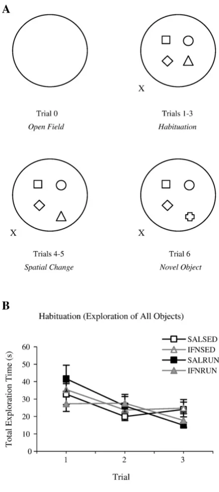
Habituation and reactions to spatial change and novel object introduction were
evaluated by the exploration of distinct objects placed in an open field using a task
design modified from Gobbo et al., [39]. The apparatus consisted of a black, circular
fiberglass arena (diameter = 200 cm, height = 35 cm) surrounded by black curtains
with several extra-maze cues attached. The experimenter stood at the south-eastern
side of the arena during testing. Four objects were placed in a square formation at the
centre of the arena, approximately 30 cm apart. The objects used included a concrete
pillar, a cardboard rectangular box, a metal jug, a white translucent plastic bottle and
brown glass bottle; all objects were of similar dimensions and their weight was such
that they could not be displaced by the rats.
Animals were initially placed in the centre of the empty arena and allowed to explore
freely for six minutes (open field exploration, trial 0). Data for this trial were recorded
using the image analyzing software system EthoVision (Noldus Information
Technology, Wageningen, Netherlands). Parameters measured were: distance traveled
(cm), mean velocity (cm/s) and time spent in a virtually-defined thigmotactic
boundary region (15 cm from the arena walls). Animals were then removed from the
experimental area and returned to their home cage, while objects were placed in the
minutes duration with three-minute inter-trial intervals (ITI’s). In trial 4, one of the
objects was moved to a new location towards the periphery of the open field (spatial
novelty recognition trial). This spatial configuration was maintained for trial 5. In trial
6, the displaced object was replaced by a novel object (object novelty recognition
trial); see figure 1A for a pictorial representation of the task protocol. To eliminate
any bias of olfactory cues, all objects were manipulated before trials 4 and 6, and the
arena and all objects were cleaned with a lightly-scented disinfected between each
animal’s set of trials.
Object exploration was evaluated by the time spent sniffing the objects, defined as
directed head movement whisker twitching with nose proximity to objects within
2-3cm. Both time spent exploring the moved or novel object specifically and time spent
exploring other objects were recorded manually by the experimenter.
Statistical Analyses
Statistics were carried out using Statistical Package for the Social Sciences (SPSS)
version 11. Data were assessed for normality prior to analyses using the
Shapiro-Wilks’ W test. Data are expressed as means ± standard errors; * p < 0.05, * p < 0.01,
* p < 0.001.
Object recognition task Open field exploration (trial 0) was analyzed using one-way
ANOVA’s to examine the effect of treatment group. Habituation to the objects (trials
1-3) was assessed by means of a mixed-factorial ANOVA with exploration time of all
objects (3 levels) as within-subjects factor and treatment group as between-subjects
factor. Subsequent one-way ANOVA’s were carried out to examine the effect of
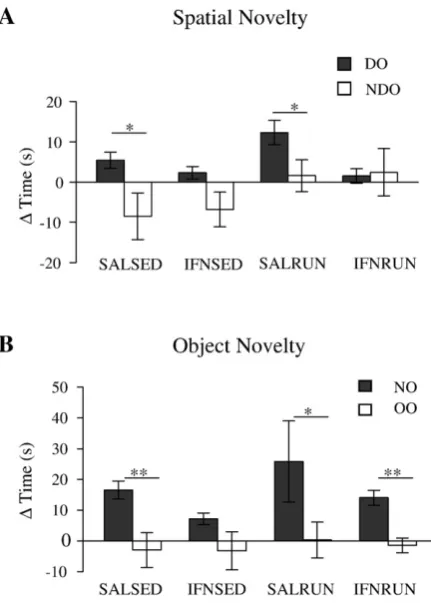
Response to spatial change was assessed by calculating the mean time in contact with
either displaced, DO, or non-displaced, NDO objects in trial 4 minus the mean time
spent in contact with the same object category in trial 3. Response to introduction of a
novel object was assessed by calculating the mean time in contact with either novel,
NO, or old, OO objects in trial 6 minusthe mean time spent in contact with the same
object category in trial 5 (since the novel object was not present in trial 5, the time
spent exploring the object to be replaced by the new object was used for calculations).
Planned paired samples t-tests (paired variables: DO-NDO or NO-OO) were
subsequently performed to assess object-location recognition and novel object
recognition performance for each group.
Results
Open Field Exploration
One-way ANOVA analyses did not reveal any significant effect of treatment group on
locomotion in the open field (trial 0) in terms of distance traveled, velocity or
thigmotaxis; data not shown.
Object Recognition Task
Habituation Over trials 1 – 3 of this task, all groups showed habituation to their
environment as expected, with total exploration time decreasing across trials (Fig.
1B); a mixed-factorial ANOVA revealed a significant effect of trial (F(2, 52) = 9.41, p
< 0.001), but no significant trial x group interaction (F(2, 52) = 1.36, p = 0.247), and
no significant effect of group (F(1, 26) = 0.428, p = 0.735). In addition, no significant
effect of treatment group was seen on any of the habituation trials following one-way
Recognition of a spatial change For object location recognition paired samples t-tests
(DO-NDO) indicated that both saline sedentary (t = 2.99, df = 7, p < 0.05) and saline
exercised (t = 4.00, df = 5, p < 0.05) animals showed significant recognition of a
spatial change, while neither group of IFN-α animals (sedentary: t = 1.83, df = 7, p =
0.11; exercised: t = -0.17, df = 7 p = 0.872) discriminated between DO and NDO
(Figure 2A).
Recognition of a novel object For object recognition paired samples t-tests (NO-OO)
revealed that saline sedentary (t = 4.6, df = 7, p < 0.01), saline exercised (t = 2.73, df
= 5, p < 0.05) and IFN-α exercised (t = 4.88, df = 7, p < 0.01) animals showed
significant preference for the novel object compared to old objects (Figure 2B). IFN-α
sedentary animals did not show this preference (t = 1.82, df = 6, p = 0.12).
Discussion
The aims of this study were to evaluate the impact of chronic, systemic IFN-α
treatment on cognitive function in the rat, and to assess the potential of chronic forced
exercise to prevent the induction of IFN-α-induced cognitive deficits. Previous data
indicate the positive prophylactic effects of exercise against neuronal insult (see
Introduction). We found that our treatment regime of IFN-α (which has previously
been shown to induce an affective-like syndrome in rodents, [20]) had a significant
negative impact on spatial learning and novel object recognition performance.
Further, exercise training in IFN-α animals rescued the deficits in novel object
recognition.
Both groups of IFN-α-treated animals spontaneously explored the open field
normally, in terms of distance traveled and velocity, and did not show increased
initial exploration in the task (trial 1), indicating normal visuomotor function and
exploratory tendencies. In addition, all groups showed normal habituation to their
spatial environment, gradually decreasing exploration of the objects across trials 1-3
(Figure 1A). If such an asymptotic habituation curve reflects normal encoding of the
environmental features and spatial configuration, one may predict that alterations to
the initial situation should elicit increased exploratory behavior. However, previous
studies have shown a dissociation between the capacity for normal habituation and
that for recognition of spatial or object novelty [40-42]. The results presented here
support such a dissociation: while IFN-α sedentary animals show a normal
habituation curve, they fail to detect either a spatial change or the introduction of a
novel object.
The decrease in the ability to detect both spatial change and novelty introduction in
the IFN-α sedentary animals might imply a generalized decrease in the normal,
natural tendency to explore novelty. However, the dichotomous effects of exercise on
these parameters in IFN-α exercised animals point to discrete mechanisms underlying
the two phenomena: while exercise can restore the ability to recognize novel features
of the spatial arrangement, it does not allow the IFN-α animals to notice a
configurational change in the spatial arrangement. This result is in agreement with
previous results [32] where a similar exercise protocol was found to improve novel
object recognition but did not affect spatial learning in the reference memory version
of the watermaze. This idea is also supported by lesions studies. In a task similar to
that used here, hippocampal lesions have been found to impair performance on the
spatial change trial, but not the novel object trial, while entorhinal cortex and
subicular lesions impaired both novel object recognition and spatial change
impair performance in a continuous recognition (spatial location) memory procedure
[43] this behavioral testing procedure is very different from the OE task used here.
The protocol requires several days of training and testing, unlike the short timespan of
the OE protocol. These differences could underlie the conflicting lesion reports
regarding the role of entorhinal cortex in spatial learning and memory. Moreover,
Hargreaves et al., [44] show that spatial information is represented by medial
entorhinal cortex and nonspatial by lateral entorhinal cortex. These results are in
support of the concept that both medial and lateral entorhinal function (representing
spatial and nonspatial information, respectively) could be impaired in IFN-alpha rats.
The data presented here suggest that chronic IFN-α treatment has a negative impact
on the function of the extended hippocampal formation circuit (entorhinal cortex,
subiculum and hippocampus proper) as measured by recognition of spatial and object
novelty in the OE task. Concurrent, chronic forced exercise may ameliorate IFN-α
-induced deficits in recognition of a novel object by somehow modulating entorhinal
cortical and subicular function to a degree. However, the persistent impairments in
recognition of a spatial change in IFN-α exercised animals indicate that, along with
overriding deficits in hippocampus proper, deficits in entorhinal cortical and subicular
function may also contribute. It is possible that the forced exercise regime used here
may not have an entorhinal cortex-/subiculum-specific effect and that the OE task
may only be sensitive to these effects; any ameliorative effects on hippocampal
function may not be strong enough to produce significant results in this task. Any
suggested mechanism underlying an apparent entorhinal cortex-/subiculum-specific
effect of exercise would be purely speculative at this stage. It remains to be seen
on the hippocampal formation or in the in vivo electrophysiological profile/synaptic
plasticity potential of the hippocampus.
It is interesting to note that in clinical studies on the CNS effects of IFN-α
examinations of cognitive status are rarely conducted, with most focusing on the
affective standing of the individual. These novel results from our animal model
suggest that it could be beneficial to patients to incorporate monitoring of cognitive
status during IFN-α treatment into clinical protocol. This is especially important in
light of earlier findings that cognitive impairments in IFN-α-treated individuals seem
to emerge independently of depressive symptoms [10, 11]. The results of this study
strengthen the case for the neuroprotective properties of exercise and provide an
interesting first insight into the cognitive deficits induced by IFN-α in an animal
model.
Acknowledgements
This research was funded by the Programme for Research in Third-Level Institutions
(PRTLI) of the Higher Education Authority, Ireland.
References
[1] Schaefer M, Engelbrecht MA, Gut O, Fiebich BL, Bauer J, Schmidt F, Grunze
H, Lieb K. Interferon alpha (IFNalpha) and psychiatric syndromes: A review.
Prog Neuropsychopharmacol Biol Psychiatry 2002;26(4):731-46.
[2] Dieperink, E., Willenbring M, Ho SB. Neuropsychiatric Symptoms
Associated With Hepatitis C and Interferon Alpha: A Review. Am J
Psychiatry 2000;157(6):867-76.
[3] Kraus MR, Schafer A, Faller H, Csef H, Scheurlen M. Psychiatric symptoms
in patients with chronic hepatitis C receiving interferon alfa-2b therapy. J Clin
Psychiatry 2003;64(6):708-14.
[4] Hauser P, Khosla J, Aurora H, Laurin J, Kling MA, Hill J, Gulati M, Thornton
AJ, Schultz RL, Valentine AD, Meyers CA, Howell CD. A prospective study
of the incidence and open-label treatment of interferon-induced major
depressive disorder in patients with hepatitis C. Mol Psychiatry
2002;7(9):942-7.
[5] Okanoue T, Sakamoto S, Itoh Y, Minami M, Yasui K, Sakamoto M, Nishioji
K, Katagishi T, Nakagawa Y, Tada H, Sawa Y, Mizuno M, Kagawa K,
Kashima K. Side effects of high-dose interferon therapy for chronic hepatitis
[6] Schaefer M, Schmidt F, Folwaczny C, Lorenz R, Martin G, Schindlbeck N,
Heldwein W, Soyka M, Grunze H, Koenig A, Loeschke K. Adherence and
mental side effects during hepatitis C treatment with interferon alfa and
ribavirin in psychiatric risk groups. Hepatology 2003;37(2):443-51.
[7] Musselman DL, Lawson DH, Gumnick JF, Manatunga AK, Penna S, Goodkin
RS, Greiner K, Nemeroff CB, Miller AH. Paroxetine for the prevention of
depression induced by high-dose interferon alpha. N Engl J M 2001;344(13):
961-6.
[8] Fattovich G, Giustina G, Favarato S, Ruol A. A survey of adverse events in
11,241 patients with chronic viral hepatitis treated with alpha interferon. J
Hepatol 1996;24(1):38-47.
[9] Janssen HL, Brouwer JT, van der Mast RC, Schalm SW. Suicide associated
with alfa-interferon therapy for chronic viral hepatitis. J Hepatol 1994;21(2):
241-3.
[10] Kraus MR, Schäfer A, Wissmann S, Reimer P, Scheurlen M. Neurocognitive
changes in patients with hepatitis C receiving interferon alfa-2b and ribavirin.
Clin Pharmacol Ther 2005;77(1):90-100.
[11] Lieb K, Engelbrecht MA, Gut O, Fiebich BL, Bauer J, Janssen G, Schaefer M
alpha (IFNalpha): results from a prospective study. Eur Psychiatry 2006;
21(3): 204-210.
[12] Juengling FD, Ebert D, Gut O, Engelbrecht MA, Rasenack J, Nitzsche EU,
Bauer J, Lieb K. Prefrontal cortical hypometabolism during low-dose
interferon alpha treatment. Psychopharmacology 2000;152(4):383-9.
[13] Capuron L, Ravaud A, Dantzer R. Timing and specificity of the cognitive
changes induced by interleukin-2 and interferon-alpha treatments in cancer
patients. Psychosom Med 2001;63(3):376-86.
[14] Campbell IL, Krucker T, Steffensen S, Akwa Y, Powell HC, Lane T, Carr DJ,
Gold LH, Henriksen SJ, Siggins GR.Structural and functional neuropathology
in transgenic mice with CNS expression of IFN-alpha. Brain Res 1999;
835(1):46-61.
[15] De La Garza R 2nd. Recombinant human interferon-alpha does not alter reward
behavior, or neuroimmune and neuroendocrine activation in rats. Prog
Neuropsychopharmacol Biol Psychiatry 2005;29(5):781-92.
[16] Kentner AC, James JS, Miguelez M, Bielajew C. Investigating the hedonic
effects of interferon-alpha on female rats using brain-stimulation reward.
[17] Makino M, Kitano Y, Hirohashi M, Takasuna K.Enhancement of immobility
in mouse forced swimming test by treatment with human interferon. Eur J
Pharmacol 1998;356(1):1-7.
[18] Sammut S, Bethus I, Goodall G, Muscat R. Antidepressant reversal of
interferon-alpha-induced anhedonia.. Physiol Behav 2002;75(5):765-72.
[19] Sammut S, Goodall G, Muscat R Acute interferon-alpha administration
modulates sucrose consumption in the rat. Psychoneuroendocrinology
2001;26(3):261-72.
[20] Fahey B, Hickey B, Kelleher D, O'Dwyer AM, O'Mara SM.The Widely-Used
Anti-Viral Drug Interferon-alpha (IFN-α) Induces Depressive- and
Anxiogenic-like Effects in Healthy Rats. Behav Brain Res 2007;182(1):80-7.
[21] Wilz KJ, Bolton RL, Exploratory behavior in response to the spatial rearrangement of familiar stimuli. Psychonomic Science 1971;24:117-8.
[22] Gobbo OL, O'Mara SM. Exercise, but not environmental enrichment,
improves learning after kainic acid-induced hippocampal neurodegeneration in
association with an increase in brain-derived neurotrophic factor. Behav Brain
Res 2005;159(1):21-6.
[23] Albeck DS, Sano K, Prewitt GE, Dalton L Mild forced treadmill exercise
[24] Crizzle AM, Newhouse IJ. Is physical exercise beneficial for persons with
Parkinson's disease? Clin J Sport Med 2006;16(5):422-5.
[25] Grealy MA, Johnson DA, Rushton SK Improving cognitive function after
brain injury: the use of exercise and virtual reality. Arch Phys Med Rehabil
1999;80(6): 661-7.
[26] Griesbach GS, Hovda DA, Molteni R, Wu A, Gomez-Pinilla F Voluntary
exercise following traumatic brain injury: brain-derived neurotrophic factor
upregulation and recovery of function. Neuroscience 2004;125(1):129-39.
[27] Kramer AF, Erickson KI, Colcombe SJ, Exercise, cognition, and the aging
brain. J Appl Physiol 2006;101(4):1237-42.
[28] Luo CX, Jiang J, Zhou QG, Zhu XJ, Wang W, Zhang ZJ, Han X, Zhu DY.
Voluntary exercise-induced neurogenesis in the postischemic dentate gyrus is
associated with spatial memory recovery from stroke. J Neurosci Res
2007;85(8):1637-46
[29] Tillerson JL, Caudle WM, Reverón ME, Miller GW. Exercise induces
behavioral recovery and attenuates neurochemical deficits in rodent models of
[30] Wendel-Vos GC, Schuit AJ, Feskens EJ, Boshuizen HC, Verschuren WM,
Saris WH, Kromhout D. Physical activity and stroke. A meta-analysis of
observational data. Int J Epidemiol 2004;33(4): 787-98.
[31] Ang ET, Dawe GS, Wong PT, Moochhala S, Ng YK. Alterations in spatial
learning and memory after forced exercise. Brain Res 2006;1113(1): 186-93.
[32] O'Callaghan RM, Ohle R, Kelly AM. The effects of forced exercise on
hippocampal plasticity in the rat: A comparison of LTP, spatial- and
non-spatial learning. Behav Brain Res 2007;176(2):362-6.
[33] Braszko JJ, Kamiñski KA, Hryszko T, Jedynak W, Brzósko S.Diverse effects
of prolonged physical training on learning of the delayed non-matching to
sample by rats. Neurosci Res 2001;39(1):79-84.
[34] Anderson BJ, Rapp DN, Baek DH, McCloskey DP, Coburn-Litvak PS,
Robinson JK. Exercise influences spatial learning in the radial arm maze.
Physiol Behav 2000;70(5):425-9.
[35] Uda M, Ishido M, Kami K, Masuhara M. Effects of chronic treadmill running
on neurogenesis in the dentate gyrus of the hippocampus of adult rat. Brain
[36] Tong L, Shen H, Perreau VM, Balazs R, Cotman CW. Effects of exercise on
gene-expression profile in the rat hippocampus. Neurobiol Dis 2001;8(6):
1046-56.
[37] Roche, P. Roferon-A Product Information. N.J., U.S.A: Hoffman-La Roche
Company; 2003, p32.
[38] Schering, C. Intron-A Product Information. N.J., U.S.A: Schering
Corporation; 2002, p9.
[39] Gobbo OL, O'Mara SM. Post-treatment, but not pre-treatment, with the
selective cyclooxygenase-2 inhibitor celecoxib markedly enhances functional
recovery from kainic acid-induced neurodegeneration. Neuroscience 2004;
125(2):317-27.
[40] Galani R, Weiss I, Cassel JC, Kelche C. Spatial memory, habituation, and
reactions to spatial and nonspatial changes in rats with selective lesions of the
hippocampus, the entorhinal cortex or the subiculum. Behav Brain Res 1998;
96(1-2):1-12.
[41] Poucet, B. Object exploration, habituation, and response to a spatial change in
rats following septal or medial frontal cortical damage. Behav Neurosci 1989;
[42] Save E, Poucet B, Foreman N, Buhot MC.Object exploration and reactions to
spatial and nonspatial changes in hooded rats following damage to parietal
cortex or hippocampal formation. Behav Neurosci 1992;106(3): 447-56.
[43] Kesner RP, Giles R. Neural circuit analysis of spatial working memory: role
of pre- and parasubiculum, medial and lateral entorhinal cortex. Hippocampus
1998;8(4):416-23.
[44] Hargreaves EL, Rao G, Lee I, Knierim JJ.Major dissociation between medial
and latera entorhinal input to dorsal hippocampus. Science 2005;308(5729):
Figure legends
Figure 1 Neither IFN-α nor exercise affects habituation in the OE task
Pictorial representation of the task protocol (A). X marks the experimenter’s position. All groups
habituate normally to the spatial layout of the environment, decreasing total exploration time across
Figure 2 IFN-α impairs both recognition of spatial novelty and object novelty; exercise prevents
only the deficits in novelty recognition.
Only SALSED and SALRUN animals show reactivity to the spatial change (A; paired t-tests, p < 0.05
for both groups). While IFNSED animals are also impaired in reactivity to object novelty, SALSED,
SALRUN and IFNRUN animals show significant preference for the novel object (B; paired samples
t-test, p < 0.05 for each group). Data are expressed as mean change in time (s) ± SEM; n = 6-8 for each