R E V I E W
Open Access
Environmental exposures during windows
of susceptibility for breast cancer: a
framework for prevention research
Mary Beth Terry
1†, Karin B. Michels
2†, Julia Green Brody
3, Celia Byrne
4, Shiuan Chen
5, D. Joseph Jerry
6,
Kristen M. C. Malecki
7, Mary Beth Martin
8, Rachel L. Miller
9, Susan L. Neuhausen
10, Kami Silk
11,
Amy Trentham-Dietz
12*and on behalf of Breast Cancer and the Environment Research Program (BCERP)
Abstract
Background:
The long time from exposure to potentially harmful chemicals until breast cancer occurrence poses
challenges for designing etiologic studies and for implementing successful prevention programs. Growing evidence
from animal and human studies indicates that distinct time periods of heightened susceptibility to endocrine
disruptors exist throughout the life course. The influence of environmental chemicals on breast cancer risk may be
greater during several windows of susceptibility (WOS) in a woman
’
s life, including prenatal development, puberty,
pregnancy, and the menopausal transition. These time windows are considered as specific periods of susceptibility
for breast cancer because significant structural and functional changes occur in the mammary gland, as well as
alterations in the mammary micro-environment and hormone signaling that may influence risk. Breast cancer
research focused on these breast cancer WOS will accelerate understanding of disease etiology and prevention.
Main text:
Despite the plausible heightened mechanistic influences of environmental chemicals on breast cancer
risk during time periods of change in the mammary gland
’
s structure and function, most human studies of
environmental chemicals are not focused on specific WOS. This article reviews studies conducted over the past few
decades that have specifically addressed the effect of environmental chemicals and metals on breast cancer risk
during at least one of these WOS. In addition to summarizing the broader evidence-base specific to WOS, we
include discussion of the NIH-funded Breast Cancer and the Environment Research Program (BCERP) which
included population-based and basic science research focused on specific WOS to evaluate associations between
breast cancer risk and particular classes of endocrine-disrupting chemicals
—
including polycyclic aromatic
hydrocarbons, perfluorinated compounds, polybrominated diphenyl ethers, and phenols
—
and metals. We outline
ways in which ongoing transdisciplinary BCERP projects incorporate animal research and human epidemiologic
studies in close partnership with community organizations and communication scientists to identify research
priorities and effectively translate evidence-based findings to the public and policy makers.
Conclusions:
An integrative model of breast cancer research is needed to determine the impact and mechanisms
of action of endocrine disruptors at different WOS. By focusing on environmental chemical exposure during specific
WOS, scientists and their community partners may identify when prevention efforts are likely to be most effective.
Keywords:
Breast neoplasms, Puberty, Pregnancy, Menopause, Environment
© The Author(s). 2019Open AccessThis article is distributed under the terms of the Creative Commons Attribution 4.0 International License (http://creativecommons.org/licenses/by/4.0/), which permits unrestricted use, distribution, and reproduction in any medium, provided you give appropriate credit to the original author(s) and the source, provide a link to the Creative Commons license, and indicate if changes were made. The Creative Commons Public Domain Dedication waiver (http://creativecommons.org/publicdomain/zero/1.0/) applies to the data made available in this article, unless otherwise stated.
* Correspondence:[email protected]
†Mary Beth Terry and Karin B. Michels should be considered shared first
authors.
12Department of Population Health Sciences and Carbone Cancer Center,
School of Medicine and Public Health, University of Wisconsin-Madison, 610 Walnut St., WARF Room 307, Madison, WI 53726, USA
Background
Despite the considerable personal and societal burden
from breast cancer, primary prevention efforts encounter
challenges. Unlike other cancers that are linked to a
pre-dominant risk factor (e.g., smoking and lung cancer [
1
],
human papillomavirus, and cervical cancer [
2
]), most
established breast cancer risk factors have modest
asso-ciations; moreover, many risk factors are not conducive
to population-level intervention. The American Cancer
Society guidelines for breast cancer prevention include
limiting alcohol intake, avoiding post-menopausal
hor-mone use, increasing physical activity, and maintaining a
healthy body weight [
3
]. Yet even considering these
fac-tors, estimates indicate that a substantial proportion of
breast cancer risk remains unexplained [
4
,
5
].
Migrant studies, atomic bomb survivor studies, and
experimental model studies reinforce the concept that
exposures during certain periods in a woman
’
s life are
important to later breast cancer risk [
6
–
9
]. These time
intervals represent windows of susceptibility (WOS) and
coincide with landmark events when a woman
’
s breast
tissue changes in structure and function including the
prenatal, pubertal, pregnancy, and menopausal WOS.
Epidemiologic data support that both medications [
10
]
and medical conditions [
11
,
12
] during these WOS may
affect breast cancer risk; more limited evidence
ad-dresses specific environmental chemicals and metals
during these same WOS. In 2003, the National Institute
for Environmental Health Sciences (NIEHS) initiated the
Breast Cancer and the Environment Research Program
(BCERP) with support from the National Cancer
Institute (NCI) to specifically examine whether
environ-mental exposures during the pubertal WOS affect the
timing of puberty, a risk factor for breast cancer. Since
2009, BCERP expanded the WOS to include the
pre-natal, pregnancy, and menopausal transition WOS. In
addition, studies of mammographic breast density
(MBD), breast tissue measurements, and other
inter-mediate biomarkers of the effects of environmental
ex-posures were included. The BCERP consortium unites
basic and population scientists in advancing our
under-standing of the role of environmental chemicals during
WOS in breast cancer risk. Scientific research in BCERP
also builds from community partnerships and
collabora-tions with communication scientists within the
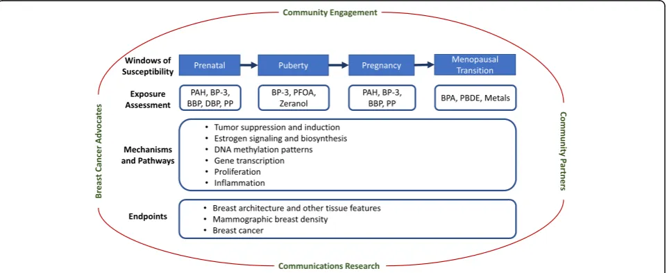
consor-tium to facilitate direct translation to the public (Fig.
1
).
Numerous previous studies examined environmental
chemical exposure and breast cancer risk; however, most
research in humans has not specifically focused on
measuring environmental chemical exposures during
WOS (for review, see [
13
,
14
]). For example, of the 146
epidemiologic reports published in 2006
–
2016 on
envir-onmental chemicals and incident breast cancer, only 16
(11%) report on exposures measured during a specific
WOS [
14
]. In this review, we outline the scientific
evi-dence generated by experimental and epidemiologic
scientists including (but not limited to) those in BCERP
addressing the link between breast cancer risk and
environmental chemicals and metals within four WOS
—
prenatal, puberty, pregnancy, and the menopausal
transi-tion
—
to inform breast cancer etiology and future
interventions.
Windows of susceptibility (WOS)
Breast cancer etiology appears to be driven in part by
perturbations to breast tissue as well as alterations of the
Fig. 1BCERP framework. A model of transdisciplinary community-engaged research by epidemiologists, basic scientists, communication
[image:2.595.59.540.499.696.2]mammary gland micro-environment during critical
win-dows. Here we briefly summarize breast tissue changes
occurring during each WOS, review evidence that
ad-dresses environmental carcinogenesis during each WOS,
and outline the motivation for ongoing research on the
chemicals and metals targeted in BCERP.
Prenatal WOS
The prenatal period is a particularly vulnerable WOS
be-cause breast tissue begins to develop in the embryonic
stage when epidermal cells in concert with embryonic
mesenchyme become breast buds [
15
–
18
]. Faster fetal
growth and greater birth-weight increase breast cancer
risk later in life [
19
–
21
]. Proposed mechanisms by which
chemicals can alter normal mammary development
tra-jectories [
15
,
18
,
19
,
22
,
23
] include changes in maternal
hormone levels regulating development and sex
differen-tiation, high levels of growth factors, potential DNA
damage and mutations in germ cells, and other genetic
or epigenetic processes [
24
].
Pregnancy and birth cohorts reveal possible associations
between environmental chemicals during the prenatal
period and breast cancer. The Child Health and
Develop-ment Studies (CHDS) found high levels of maternal
ex-posure to dichlorodiphenyltrichloroethane (DDT) during
pregnancy increased the daughters
’
later breast cancer risk
to age 52 nearly fourfold compared to daughters of
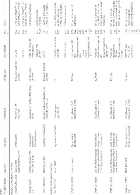
women with low levels of exposure (Table
1
) [
25
].
Al-though production of many of the organochlorine
chemi-cals
—
including dioxins, polychlorinated biphenyls (PCBs),
and pesticides such as DDT
—
stopped in the 1970s, there
is continued exposure to these complex mixtures with
di-verse biological activity. Animal fats and fish from
con-taminated waters are on-going sources of human
exposure as a result of bioaccumulation [
26
]; PCB
expos-ure also persists through inhalation both outdoors and of
indoor air and dust from caulk, building materials, and
floor finishes [
27
]. Organochlorines are hormonally active
and may contribute to breast cancer by altering mammary
gland development or hormone responsiveness early in
life, or by promoting tumor growth [
25
]. Epidemiologic
studies of DDT exposure measured outside of a WOS and
breast cancer risk were less likely to report consistent
findings [
14
,
19
].
Another class of chemical exposures of concern during
the prenatal WOS is polycyclic aromatic hydrocarbons
(PAH). PAH are produced as a result of combustion of
hydrocarbons. Some of the common sources of PAH
ex-posure include consuming grilled meats and certain
other food items [
28
], inhaling cigarette smoke and
motor vehicle exhaust [
29
], and exposure to industrial
processes [
29
–
31
]. PAH are widespread and enter the
body largely through ingestion and inhalation of
sus-pended particulate matter [
32
,
33
]. The International
Agency for Research on Cancer classifies PAH as
probable carcinogens; the US Environmental Protection
Agency lists PAH as possible carcinogens [
34
,
35
].
Like DDT and other organochlorines, PAH are
lipo-philic and stored in fat tissue including breast tissue
[
36
]. Most PAH compounds are weakly estrogenic
and may induce cell proliferation via activation of the
estrogen receptor (ER) [
37
]. Exposure to PAH was
linked to mammary cancer in rodents [
38
]. PAH
ex-posure has been measured directly in both blood [
39
]
and breast tissue [
40
], and higher levels of PAH-DNA
adducts have been found in breast cancer cases
com-pared with women without breast cancer [
41
].
Simi-larly, breast cancer cases reported higher levels of
PAH exposures than controls based on questionnaire
assessments of indirect exposure [
42
–
46
]. For all
these epidemiologic studies, specific WOS were not
investigated. Because experimental and epidemiologic
associations implicate prenatal PAH exposure in
mul-tiple adverse health effects including obesity [
47
–
49
],
one focus of BCERP is the impact of PAH exposure
during the prenatal WOS. BCERP research specifically
addresses how exposure to PAH during the prenatal
and pregnancy WOS may increase the development
of mammary tumors in mice. Concurrent human
studies within BCERP evaluate how prenatal PAH
ex-posure alters breast tissue development and tissue
composition in adolescent girls.
Pubertal window of susceptibility
Table
1
Epidemiologic
studies
investigating
environmental
exposures
during
three
windows
of
susceptibility
in
relation
to
an
intermediate
marker
of
brea
st
cancer
risk
or
breast
cancer
First autho r (Yea r) Exposu re Out come Population Sampl e size Risk estimat e 95% CI Notes Exposu re dur ing pren atal windo w Bonn er (2005 ) [ 33 ] Regiona l total suspende d part iculates Breast cance r Wom en 35 – 79, New York 1166 cases and 2105 contro ls OR 2. 42 0.97 – 6.09 > 140 vs < 84 μ g/m 3TSP, postm enopa usal wom en OR 1. 78 0.62 – 5.10 > 140 vs < 84 μ g/m 3 TSP, preme nopausal wom en Boc skay (2005 ) [ 32 ] Personal airb orne PAH; PAH DNA add ucts Chro mosomal abe rrations from cord blood Newb orns in Nort hern Manhattan; Bronx 60 (32 fem ale, 28 male) Data not shown for PAH add ucts “ No strong associ ation ” Airborn e PAH β = 0.14 p = 0.006 Line ar reg ression line slope Coh n (2015) [ 25 ] Maternal o,p ’ -DDT Daug hter breast cance r Moth ers an d adu lt daughters in Alame da Count y, CA 118 cas es an d 354 contro ls OR 3. 7 1.5 – 9.0 Fou rth vs first qua rtile (> 0.78 vs < 0.27 ng/mL) Exposu re dur ing puberty windo w Tsai (2015 ) [ 82 ] Serum PFOA log-tran sformed SHBG Taiwane se girls aged 12 – 17 65 2.96 (SE 0.34) vs 3.50 (SE 0.24) p < 0.05 Mean PFO A level s 90th vs 50 th percen tile (> 9.80 vs < 3.63 ng/mL) Data not shown p > 0.05 FSH and testoster one Wol ff (2015 ) [ 57 ] Urinary phenol s Age at bre ast de velopm ent US girls aged 6 – 8 followe d for 7 years 1239 girls Enter olactone : HR 0.79 Benzop henone -3: HR0.80 Triclosan: HR
[image:4.595.71.529.92.729.2]various phenols (including bisphenol A [BPA]), parabens,
phthalates, and persistent
organohalogenated
com-pounds, were associated with
delayed
median puberty
endpoints by 5
–
11 months when comparing extreme
categories of exposure (Table
1
) [
56
–
60
].
Epidemiologic and experimental evidence from
investi-gators outside of BCERP suggest environmental
expo-sures during the pubertal WOS are associated with an
increase in breast cancer risk. Human studies have
examined high doses of radiation from medical
treat-ment or atomic bomb exposure [
61
,
62
] and nutritional
exposures during puberty and adolescence [
63
–
66
].
DDT exposure during infancy and puberty was
associ-ated with increased breast cancer risk [
67
,
68
]. In
experi-mental studies of rats, exposure to a carcinogen
(dimethylbenz [a] anthracene, DMBA) resulted in the
highest number of tumors when administered to rodents
during
“
puberty
”
possibly through induction of
proin-flammatory responses [
50
,
51
,
69
–
74
]. Excessive
signal-ing through the ER appears to be another primary
mechanism for mammary carcinogenesis as modest
overexpression of ER
α
in response to endogenous
estro-gen during puberty in transestro-genic mice resulted in
mam-mary hyperplasia and tumors [
75
,
76
].
BCERP members are studying the effect of pubertal
levels of perfluorooctanoic acid (PFOA) and per- and
polyfluoralkyl substances (PFAS) on breast development
and breast density. PFAS are used in many commercial
products because of their non-stick, stain-resistant, and
waterproof characteristics. Sources of human exposure
include production facilities, firefighting training,
con-sumer products, diet, and drinking water. Dietary
sources include seafood [
77
] and food packaging [
78
].
PFAS enhance the estrogenic effects of 17
β
-estradiol in
T47D human breast cancer cells [
79
] and promote the
proliferation, migration and invasion potential of human
breast epithelial cells [
80
]. Animal studies provide
evi-dence that PFOA affects the developing mammary gland
[
81
], although limited human epidemiologic data have
been less conclusive when PFOA and PFAS exposure
was examined in relation to intermediate breast cancer
markers (hormone levels) [
82
] or measured during
adulthood [
83
]. Because environmental chemicals may
influence the timing and duration of the pubertal
trajec-tory, studies including breast tissue biomarkers that can
be reliably measured to provide greater information than
a single event in time, such as age at menarche, are
crit-ical to move the field forward.
Pregnancy window of susceptibility
Pregnancy is another period of rapid breast tissue and
micro-environmental changes during which
susceptibil-ity to environmental exposures may increase the risk of
breast cancer [
8
]. During pregnancy, breast tissue
changes rapidly in size and function to prepare for
lacta-tion. Estrogen, progesterone, and prolactin are the major
drivers of branching and development of the
lobuloal-veolar structures
’
characteristic of the mature breast
[
84
]. Pregnancy also decreases the number of mammary
stem cells [
85
,
86
]. However, the protective pathways
acti-vated during pregnancy can be eroded by prolonged
expos-ure to exogenous 17
β
-estradiol which restores sensitivity to
carcinogen-induced mammary tumors [
87
–
89
]. These
observations may explain why pregnancy is accompanied
by a short-term increase in breast cancer risk [
12
,
90
];
“
pregnancy-associated breast cancer
”
has poorer overall
survival [
91
,
92
]. However, in the long term after a
preg-nancy, breast cells are less sensitive to carcinogenesis with
the lifetime risk of breast cancer reduced by up to 50%
[
93
–
96
]. Thus, the mechanisms mediating the competition
between tumor-promoting and tumor-suppressive effects
of estrogens in the breast provide fundamental insights into
mechanisms underlying risk and resistance in the presence
of environmental chemicals.
In mice, there is a greater than 100-fold increase in the
number of mammary epithelial cells during pregnancy
demonstrating the rapid changes that occur in mammary
tissue. Despite the rapid proliferation, a full-term
preg-nancy renders the mammary epithelium resistant to
tumorigenesis subsequent to the pregnancy. This is
ob-served in studies of exposure to carcinogens [
70
,
97
–
99
]
as well as inherited genetic risk alleles [
100
–
103
].
Admin-istering exogenous estrogen, either alone or in
combin-ation with progesterone to rodents at an early age,
sufficiently mimics the effect of pregnancy in reducing
tu-mors in rodents [
104
–
106
]. Lobuloalveolar structures may
be less susceptible to carcinogens [
107
,
108
], in part,
through more robust p53-dependent responses to DNA
damage [
109
].
Epidemiologic evidence directly linking environmental
exposures during pregnancy and breast cancer risk
arises from the previously mentioned prospective CHDS
which measured PCB and DDT soon after pregnancy
and confirmed breast cancer diagnoses with medical
re-cords. Relative risk estimates for breast cancer
compar-ing upper to lower quartiles of 16 individual PCB
congeners ranged from 0.2 to 6.3; a composite score of
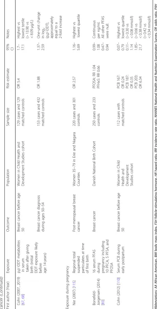
exposure was associated with an odds ratio of 2.8 (95%
CI 1.1
–
7.1) (Table
1
) [
110
]. Other epidemiologic studies
suggest no association between breast cancer and
or-ganochlorine pesticide residues in blood collected near
the time of diagnosis [
111
,
112
], but these
measure-ments may not be representative of exposure to the
parent chemical during the relevant WOS [
113
].
Children Center for Environmental Health [
32
,
114
].
The design of this mother-daughter cohort, similar to
CHDS, facilitates efficient examination of exposure to
PAH during two WOS (pregnancy and prenatal) in
the two generations [
115
]. As a complement to this
epidemiologic study, other BCERP members aim to
elucidate the mechanisms for the dual effect of
preg-nancy on breast cancer risk by examining chemicals
that are found in higher levels among pregnant
women [
116
,
117
] and their potential to impair the
protective pathways associated with breast
develop-ment during pregnancy. These pathways include the
activity of p53 [
109
] and limiting the stem cell
popu-lations [
118
].
Menopausal transition window of susceptibility
Although menopause is often defined as the cessation of
menstrual periods for at least 1 year, the menopausal
transition begins a number of years prior to menopause.
During the menopausal transition, micro-environment
changes occur in the breast tissue along with declining
systemic levels of endogenous estrogen and progesterone
[
119
]. As the majority of breast cancers are responsive to
these two sex steroid hormones, their decline explains
the leveling-off of the age-specific rate curve of breast
cancer after menopause [
120
]. Later age at menopause is
associated with a higher risk of developing breast cancer
due to a longer period of exposure to higher levels of
sex steroid hormones [
121
]. Despite the leveling in the
age-specific rate curve of breast cancer, the vast majority
of breast cancers are diagnosed after menopause, in part
through enhanced hormone receptor sensitivity during
the menopausal transition. Mammary tissue may be more
responsive to lower levels of estrogen and progesterone, as
well as to hormone mimics, by adapting to the abrupt
re-duced production of ovarian hormones [
122
,
123
].
Analyses of data from the Women
’
s Health Initiative
(WHI) showed that the increased incidence of breast
cancer with use of exogenous estrogen and progesterone
[
124
–
127
] was mediated through the change in
mam-mographic breast density that occurred in the first year
of use [
128
]. A biologically based breast tumor growth
rate model [
129
] suggests that hormone therapy
pro-motes growth of pre-existing occult lesions and
minim-ally initiated de novo tumors. EDCs with estrogen-like
and/or progesterone-like activities or those modifying
aromatase expression/activity including polybrominated
diphenyl ethers (PBDE), BPA, or selected metals may act
in a similar manner and promote the growth of occult
disease to clinically detectable tumors during the
meno-pausal transition.
PBDE are a class of over 200 organohalogenated
compounds widely used as flame retardants and may
modulate steroidogenesis including expression of
aro-matase [
130
–
136
]. BPA is an industrial chemical
found in polycarbonate plastics, epoxy resins, dental
sealants, and thermal paper [
137
,
138
]. Both PBDE
[
136
] and BPA [
139
] have been shown to act as
li-gands of ER
α
. While experimental studies suggest that
PBDE and BPA cause breast cancer and
biomonitor-ing studies confirm that women are exposed,
epidemi-ologic studies have not to-date measured exposure
during relevant WOS, used methods that reflect
long-term exposure, or included measures of
mammo-graphic density or other intermediate markers of
breast cancer risk [
138
,
140
,
141
].
as of yet have carefully examined whether
metalloestro-gens may have the greatest impact during the menopausal
transition when endogenous hormone levels are declining.
BCERP members are examining whether exposure to
PBDEs, BPA, or selected metals during the menopausal
transition is associated with breast cancer risk in
humans, and evaluating potential mechanisms to explain
these associations in rodent models.
Strategies to address long latency
The long time between exposures during the early WOS
(prenatal, puberty, pregnancy) and breast cancer
occur-rence has multiple implications for breast cancer
re-search. First, because many environmental exposures are
stored long-term in adipose tissue, even compounds
now banned, such as DDT and PBDE, may continue to
be relevant for breast cancer risk. Bioaccumulation of
lipophilic chemicals and their long-term storage also
means studies incorporating biomarkers in breast tissue
need to consider both the effects on adipose tissue as
well as epithelial and stromal tissues.
Second, because it may be decades after the relevant
windows of exposure before breast cancer is diagnosed,
the examination and validation of intermediate
bio-markers of response, apparent closer to the timing of
ex-posure and before diagnosis, are imperative, particularly
in prospective human studies. BCERP first started as a
cohort study of the environmental exposures that may
accelerate puberty. The main outcome of the cohort
study was based on Tanner Stages [
172
]. As BCERP
ex-panded to include other WOS, additional measures of
breast tissue composition and breast density were added.
BCERP investigators are now using a variety of
inter-mediate markers
—
as both outcomes in relation to
chemical exposures and as predictors of breast and
mammary cancers
—
conducted in parallel human and
rodent studies including epigenetic biomarkers, altered
tumor suppression and induction, and altered estrogen
signaling and biosynthesis (Fig.
1
) [
173
].
One intermediate outcome is mammographic breast
density (MBD), defined as the fraction of connective and
glandular tissue to adipose tissue on a mammogram
[
174
–
181
]. MBD is one of the strongest predictors of
breast cancer risk with a four- to sixfold elevation in risk
comparing
≥
75% MBD to < 5% [
182
], but the mechanisms
explaining how environmental chemicals affect the overall
level and rate of change of MBD are uncertain. While
MBD declines with age in many women, particularly
around the time of menopause [
183
–
185
], this pattern
does not occur uniformly for all women [
8
,
186
,
187
].
Little is known of the drivers of breast tissue changes
across adolescence, early adulthood, and the menopausal
transition and thus the contributors to breast density.
Most of what is known about normal breast tissue
charac-teristics is from mammography data in women over 40
years of age. In women under 40 years, two alternative
im-aging methods have been used to assess breast
compos-ition including three studies of magnetic resonance
imaging (MRI) in women aged 15
–
30 years [
188
–
190
] and
two of dual X-ray absorptiometry (DXA) in girls aged
10
–
16 years [
191
,
192
]. In addition, optical spectroscopy
(OS) provides a compositional view of the breast
captur-ing variation in the amount of water, lipid, hemoglobin,
and collagen, as well as overall cellular and connective
tis-sue density [
174
–
176
]. Collagen density may promote
epi-thelial cell proliferation and increase tumor mobility and
invasion, while hemoglobin is associated with angiogenesis
[
193
–
195
]. OS has been used to measure differences in
adolescent breast tissue across developmental stages as
assessed by Tanner stage [
196
]. Thus, MRI, DXA, and OS
provide novel intermediate outcomes to measure breast
tissue changes across the developmental trajectory of
adolescence and early adulthood and may be important
tools for examining environmental effects during these life
stages. Mammography techniques now include digital
breast tomosynthesis measures as well as the use of
ultra-sound in measuring breast density without radiation
ex-posure [
181
]. While density of the adult breast is highly
correlated with breast cancer risk, longitudinal measures
of pubertal density are currently lacking but are being
col-lected in BCERP.
Conclusions
Given the changes in mammary tissue architecture and
hormone signaling during the prenatal, pubertal,
preg-nancy, and menopausal transition windows, these critical
time periods may reflect windows of heightened risk.
Thus, measuring the impact of environmental chemical
and metal exposures during these WOS is essential to
understand their roles in breast cancer risk; these issues
have not been addressed by the majority of
epidemio-logic studies to date.
Experimental studies in cell lines and animals are
pro-viding causative mechanistic links between
environmen-tal exposures and altered mammary carcinogenesis,
particularly during key WOS. Increasingly,
epidemio-logic studies are able to link the human exposure of
che-micals and metals during relevant WOS through the use
of intermediate breast outcomes including specific breast
tissue characteristics and breast density in adolescence
and adulthood to address the challenge of long latency
time posed in cancer research.
WOS, BCERP integrates basic and population researchers
with communication scientists and representatives of
community-based organizations (Fig.
1
). Community
part-nerships are vital, because both the sources and the
rem-edies for environmental exposures are outside of clinical
settings. Community input also can identify issues of
con-cern to the community, motivate participation in studies,
and translate findings to public audiences. Scientists need
to disseminate research findings to the public to enable
people to make informed choices in their personal lives
and workplaces, and to influence health policies as voters
and community leaders. For example, participation of
community partners in BCERP has led to the development
of strategies to provide reports of personal chemical
expo-sures to individuals who donated biological samples, so
they can learn about environmental health and make
in-formed decisions regarding possible behavioral
modifica-tion in general and with particular reference to WOS
[
197
,
198
]. Digital methods using libraries of vetted
expos-ure and health information and decision rules, set by the
study team, make personalized results practical [
199
]. In
addition, communication scientists within BCERP are
test-ing different messages and channels for future outreach
efforts [
200
]. Scholarship about community-engaged
re-search shows that this approach improves the
“
rigor,
rele-vance, and reach
”
of research [
201
].
Although the median age when women are diagnosed
with breast cancer is 62 years [
202
], primary prevention of
potentially hazardous environmental exposures during
earlier WOS is critical [
13
], particularly when considering
that exposure to environmental chemicals may contribute
to cancer health disparities [
203
–
206
]. Furthermore, just
as family-based studies facilitated the discovery of breast
cancer genes relevant to all women, studies during specific
WOS will facilitate the assessment of the effects from
en-vironmental exposures that will be relevant outside of
these WOS. As evidence from WOS accumulates, the
paradigm for breast cancer needs to expand beyond the
secondary prevention efforts of screening and mid-life risk
assessment to primary prevention efforts with
involve-ment of community partners, educators and school
dis-tricts, families, and primary care providers including
pediatricians for lifelong impact [
207
].
Abbreviations
BCERP:Breast Cancer and the Environment Research Program; BPA: Bisphenol A; CHDS: Child Health and Development Study;
DDT: Dichlorodiphenyltrichloroethane; DMBA: Dimethylbenz [a]anthracene; DNA: Deoxyribonucleic acid; DXA: Dual X-ray absorptiometry;
EDC: Endocrine-disrupting chemical; ER: Estrogen receptor;
MBD: Mammographic breast density; MRI: Magnetic resonance imaging; NCI: National Cancer Institute; NIEHS: National Institute of Environmental Health Sciences; OR: Odds ratio OSoptical spectroscopy; PAH: Polycyclic aromatic hydrocarbons; PBDE: Polybrominated diphenyl ethers; PCB: Polychlorinated biphenyl; PFAS: Per- and poly-fluoroalkyl substances; PFOA: Perfluorooctanoic acid; RR: Relative risk; WHI: Women’s Health Initiative
Acknowledgements
The authors would like to thank Natalia Arroyo, Julie McGregor, and Eileen McGuine for their assistance with this manuscript. The funders had no role in the design of the study, the writing of the manuscript, or the decision to submit the manuscript for publication.
The Breast Cancer and the Environment Research Program includes Mary Pat Berry, Jennifer Bird, Christopher Bradfield, Ronald Gangnon, Michael Gould, John Hampton, Sara Lindberg, Sarah Luongo, Kristen Malecki, Betsy Rolland, and James Shull (University of Wisconsin-Madison); Mia Gaudet (American Cancer Society); Mark Thornquist (Fred Hutchinson Cancer Research Center); Mark D. Aupperlee, Sandra Z. Haslam, Reyhane Hoshyar, Anastasia Kariagina, Juliana R. Lopes, Karen J. Miller, Olena Morozova, Cathy J. Newkirk, Richard C. Schwartz, Brandon Thomas, Daniel Totzkay, and Fang Xie (Michigan State University); Kami J. Silk (University of Delaware); Frank M. Biro (Cincinnati Children’s Hospital); Cecily S. Fassler, Courtney M. Giannini, and Susan Pinney (University of Cincinnati); Melissa A. Troester (University of North Carolina Chapel Hill); Kimberly Burke, Julie Herbstman, Rebecca Kehm, Jasmine McDo-nald, Rachel Miller, Sabine Oskar, Frederica Perera, Debeshish Sahay, Parisa Tehranifar, Mary Beth Terry, Desiree Walker, and Nur Zeinomar (Columbia University); Milagros de Hoz and Peggy Shepard (WeACT); Julia Knight (Lunenfeld-Tanenbaum Research Institute); Lothar Lilge (University Health Network); Alexandra Binder, Karin Michels (University of California Los Angeles); Vincent Bessonneau, Julia Brody, Vanessa De La Rosa, Jennifer Oha-yon, and Ruthann Rudel (Silent Spring Institute); Camila Corvalan, Ana Pereira, and Rosario Toro-Campos (University of Chile); Julia Pereira, Jose Russo, and Su Yanrong (Fox Chase Cancer Center); John Shepherd (University of Hawaii Cancer Center); Lucile Adams-Campbell, Chiranjeev Dash, Bassem Haddad, Rhonda Hamilton, Mary Beth Martin, and Brenda Richardson (Georgetown University); Celia Byrne and Hristina Denic-Roberts (Uniformed Services University of the Health Sciences); Gregory Chang, Shiuan Chen, Yuan Chun Ding, Noriko Kanaya, Susan Neuhausen, Michele Rakoff, Kohei Saeki, and Mayra Serrano (City of Hope); Peggy Reynolds (University of California San Francisco); Karen Dunphy, Joseph Jerry, Anna Symington, and Laura Vandenberg (University of Massachusetts Amherst); Sallie Schneider (Baystate Medical Center); Swann Arp Adams, Heather M. Brandt, Daniela Friedman, and Jamie R. Lead (University of South Carolina); Xiaomei Cai, Gary Kreps, Camella J. Rising, and Kevin B. Wright (George Mason University); Amelia Burke-Garcia (University of Chicago); Dasha Afanaseva (Westat); and Carla L. Fisher and Michaela Devyn Mullis (University of Florida).
Disclosure
Celia Byrne: the views expressed are those of the author(s) and do not necessarily reflect the official views of the Uniformed Services University of the Health Sciences or the Department of Defense.
Authors’contributions
All authors performed parts of the literature review and wrote drafts of the manuscript. MBT, KBM, and ATD conceptualized the paper and provided overall supervision and coordination of the manuscript preparation. ATD generated the table and figures, which were revised by all authors. All authors were involved in writing the manuscript and approved the final manuscript.
Funding
This work was completed as part of the Breast Cancer and the Environment Research Program (BCERP), supported by the National Institute of Environmental Health Sciences and the National Cancer Institute at the National Institutes of Health (grant numbers U01 ES026119, U01 ES026122, U01 ES026127, U01 ES026130, U01 ES026132, U01 ES026137, U01 ES026140, R03 ES027406, R21 ES027417, and R21 ES027418).
Availability of data and materials
Not applicable.
Ethics approval and consent to participate
Not applicable.
Consent for publication
Competing interests
The authors declare that they have no competing interests.
Author details
1Department of Epidemiology, Mailman School of Public Health, Columbia
University, 722 West 168th Street, Room 1611, New York, NY 10032, USA.
2Department of Epidemiology, Fielding School of Public Health, University of
California, 650 Charles E. Young Drive South, CHS 71-254, Los Angeles, CA 90095, USA.3Silent Spring Institute, 320 Nevada St., Newton, MA 02460, USA. 4Department of Preventive Medicine and Biostatistics, Uniformed Services
University of the Health Sciences, 4301 Jones Bridge Road A-1039F, Bethesda, MD 20814, USA.5Department of Cancer Biology, Beckman Research Institute of City of Hope, 1450 E. Duarte Road, Duarte, CA 91010, USA.6Pioneer Valley
Life Sciences Institute and Department of Veterinary & Animal Sciences, University of Massachusetts Amherst, 661 North Pleasant St., Amherst, MA 01003, USA.7Department of Population Health Sciences and the Carbone Cancer Center, School of Medicine and Public Health, University of Wisconsin-Madison, 610 Walnut St., WARF Room 605, Madison, WI 53726, USA.8Departments of Oncology and Biochemistry & Molecular Biology,
Georgetown University Medical Center, E411 New Research Building, Washington, DC 20057, USA.9Departments of Medicine, Pediatrics,
Environmental Health Sciences; Vagelos College of Physicians and Surgeons, Mailman School of Public Health, Columbia University, PH8E-101B, 630 W. 168th St, New York, NY 10032, USA.10Department of Population Sciences, Beckman Research Institute of City of Hope, 1450 E. Duarte Road, 1500 E. Duarte Road, Duarte, CA 91010, USA.11Department of Communication,
University of Delaware, 250 Pearson Hall, 125 Academy St, Newark, DE 19716, USA.12Department of Population Health Sciences and Carbone Cancer Center, School of Medicine and Public Health, University of
Wisconsin-Madison, 610 Walnut St., WARF Room 307, Madison, WI 53726, USA.
References
1. Lee PN, Forey BA, Coombs KJ. Systematic review with meta-analysis of the epidemiological evidence in the 1900s relating smoking to lung cancer. BMC Cancer. 2012;12:385.
2. Walboomers JM, Jacobs MV, Manos MM, Bosch FX, Kummer JA, Shah KV, Snijders PJ, Peto J, Meijer CJ, Munoz N. Human papillomavirus is a necessary cause of invasive cervical cancer worldwide. J Pathol. 1999;189(1):12–9. 3. Breast Cancer Risk and Prevention. Available athttps://www.cancer.org/
cancer/breast-cancer/risk-and-prevention.html. Accessed 3 July 2019. 4. Colditz GA, Bohlke K. Priorities for the primary prevention of breast cancer.
CA Cancer J Clin. 2014;64(3):186–94.
5. Sprague BL, Trentham-Dietz A, Egan KM, Titus-Ernstoff L, Hampton JM, Newcomb PA. Proportion of invasive breast cancer attributable to risk factors modifiable after menopause. Am J Epidemiol. 2008;168(4):404–11. 6. IOM (Institute of Medicine). Breast cancer and the environment: a life course
approach. Washington, D.C.: National Academies Press; 2012.
7. Korenman SG. The endocrinology of breast cancer. Cancer. 1980;46(4 Suppl):874–8. 8. Pike MC, Krailo MD, Henderson BE, Casagrande JT. Hoel DG:‘Hormonal’risk
factors, 'breast tissue age' and the age-incidence of breast cancer. Nature. 1983;303(5920):767–70.
9. Russo J, Russo IH. Toward a physiological approach to breast cancer prevention. Cancer Epidemiol Biomark Prev. 1994;3(4):353–64. 10. Hilakivi-Clarke L. Maternal exposure to diethylstilbestrol during
pregnancy and increased breast cancer risk in daughters. Breast Cancer Res. 2014;16(2):208.
11. Nechuta S, Paneth N, Velie EM. Pregnancy characteristics and maternal breast cancer risk: a review of the epidemiologic literature. Cancer Causes Control. 2010;21(7):967–89.
12. Nichols HB, Schoemaker MJ, Cai J, Xu J, Wright LB, Brook MN, Jones ME, Adami HO, Baglietto L, Bertrand KA, et al. Breast cancer risk after recent childbirth: a pooled analysis of 15 prospective studies. Ann Intern Med. 2018.https://doi.org/10.7326/M18-1323.
13. Interagency Breast Cancer and the Environment Research Coordinating Committee. In.Breast cancer and the environment: prioritizing prevention. Bethesda, MD: National Institutes of Health; February 2013. (http://www.niehs. nih.gov/about/assets/docs/ibcercc_full_508.pdf). (Accessed 1 Dec 2014).
14. Rodgers KM, Udesky JO, Rudel RA, Brody JG. Environmental chemicals and breast cancer: an updated review of epidemiological literature informed by biological mechanisms. Environ Res. 2018;160:152–82.
15. Fenton SE, Reed C, Newbold RR. Perinatal environmental exposures affect mammary development, function, and cancer risk in adulthood. Annu Rev Pharmacol Toxicol. 2012;52:455–79.
16. Russo J, Lynch H, Russo IH. Mammary gland architecture as a determining factor in the susceptibility of the human breast to cancer. Breast J. 2001;7(5): 278–91.
17. Russo J, Russo IH. Development of the human breast. Maturitas. 2004;49(1):2–15. 18. Schmidt CW. IOM issues report on breast cancer and the environment.
Environ Health Perspect. 2012;120(2):a60–1.
19. Brody JG, Rudel RA, Michels KB, Moysich KB, Bernstein L, Attfield KR, Gray S. Environmental pollutants, diet, physical activity, body size, and breast cancer: where do we stand in research to identify opportunities for prevention? Cancer. 2007;109(12 Suppl):2627–34.
20. Michels KB, Xue F, Terry KL, Willett WC. Longitudinal study of birthweight and the incidence of breast cancer in adulthood. Carcinogenesis. 2006; 27(12):2464–8.
21. Warner ET, Hu R, Collins LC, Beck AH, Schnitt S, Rosner B, Eliassen AH, Michels KB, Willett WC, Tamimi RM. Height and body size in childhood, adolescence, and young adulthood and breast cancer risk according to molecular subtype in the Nurses’Health Studies. Cancer Prev Res (Phila). 2016;9(9):732–8.
22. Michels KB, Mohllajee AP, Roset-Bahmanyar E, Beehler GP, Moysich KB. Diet and breast cancer: a review of the prospective observational studies. Cancer. 2007;109(12 Suppl):2712–49.
23. Potischman N, Troisi R. In-utero and early life exposures in relation to risk of breast cancer. Cancer Causes Control. 1999;10(6):561–73.
24. Xue F, Michels KB. Intrauterine factors and risk of breast cancer: a systematic review and meta-analysis of current evidence. Lancet Oncol. 2007;8(12):1088–100. 25. Cohn BA, La Merrill M, Krigbaum NY, Yeh G, Park JS, Zimmermann L, Cirillo
PM. DDT exposure in utero and breast cancer. J Clin Endocrinol Metab. 2015;100(8):2865–72.
26. Mrema EJ, Rubino FM, Brambilla G, Moretto A, Tsatsakis AM, Colosio C. Persistent organochlorinated pesticides and mechanisms of their toxicity. Toxicology. 2013;307:74–88.
27. Rudel RA, Seryak LM, Brody JG. PCB-containing wood floor finish is a likely source of elevated PCBs in residents’blood, household air and dust: a case study of exposure. Environ Health. 2008;7:2.
28. Phillips D. Polycyclic aromatic hydrocarbons in the diet. Mutat Res. 1999; 443(1–2):139–47.
29. Miguel A, Kirchstetter T, Harley R. On-road emissions of particulate polycyclic aromatic hydrocarbons and black carbon from gasoline and diesel vehicles. Environmental Science & Technology. 1998;32(4):450–5. 30. IARC: Polynuclear aromatic hydrocarbons. Part 3, Industrial exposures in
aluminum production, coal gasification, coke production, and iron and steel founding. International Agency for Research on Cancer Monographs on the Carcinogenic Risk of Chemicals to Humans. Lyon; 1984.
31. Pedersen M, Wichmann J, Autrup H, Dang D, Decordier I, Hvidberg M, Bossi R, Jakobsen J, Loft S, Knudsen L. Increased micronuclei and bulky DNA adducts in cord blood after maternal exposures to traffic-related air pollution. Environ Res. 2009;109(8):1012–20.
32. Bocskay KA, Tang D, Orjuela MA, Liu X, Warburton DP, Perera FP. Chromosomal aberrations in cord blood are associated with prenatal exposure to carcinogenic polycyclic aromatic hydrocarbons. Cancer Epidemiol Biomark Prev. 2005;14(2):506–11.
33. Bonner MR, Han D, Nie J, Rogerson P, Vena JE, Muti P, Trevisan M, Edge SB, Freudenheim JL. Breast cancer risk and exposure in early life to polycyclic aromatic hydrocarbons using total suspended particulates as a proxy measure. Cancer Epidemiol Biomark Prev. 2005;14(1):53–60.
34. Agency for Toxic Substances and Disease Registry (ATSDR). Toxicological profile for polycyclic aromatic hydrocarbons (PAHs). Atlanta: U.S. Department of Health and Human Services, Public Health Service; 1995. 35. IARC. Volume 82. Some traditional herbal medicines, some mycotoxins,
naphthalene and styrene. In: IARC Monographs on the Evaluation of Carcinogenic Risks to Humans. Lyon: World Health Organization; 2002. 36. Obana H, Hori S, Kashimoto T, Kunita N. Polycyclic aromatic hydrocarbons in
human fat and liver. Bull Environ Contam Toxicol. 1981;27(1):23–7. 37. Plísková M, Vondrácek J, Vojtesek B, Kozubík A, Machala M.
human breast carcinoma MCF-7 cells reflects both genotoxic and nongenotoxic events. Toxicol Sci. 2004;83(2):246–56.
38. Ambrosone C, Shields P. Molecular epidemiology of breast cancer. In: Aldaz C, Gould M, McLachlan J, Slaga T, editors. Etiology of Breast and
gynecological cancers (Progress in clinical and biologic research). New York: Wiley-Liss, Inc.; 1997. p. 93–9.
39. Gammon MD, Santella RM, Neugut AI, Eng SM, Teitelbaum SL, Paykin A, Levin B, Terry MB, Young TL, Wang LW, et al. Environmental toxins and breast cancer on Long Island. I. Polycyclic aromatic hydrocarbon DNA adducts. Cancer Epidemiol Biomark Prev. 2002;11(8):677–85.
40. Rundle A, Tang DL, Hibshoosh H, Estabrook A, Schnabel F, Cao WF, Grumet S, Perera FP. The relationship between genetic damage from polycyclic aromatic hydrocarbons in breast tissue and breast cancer. Carcinogenesis. 2000;21(7):1281–9.
41. Korsh J, Shen A, Aliano K, Davenport T. Polycyclic aromatic
hydrocarbons and breast cancer: a review of the literature. Breast Care (Basel). 2015;10(5):316–8.
42. White A, Teitelbaum S, Stellman S, Beyea J, Steck S, Mordukhovich I, McCarty K, Ahn J, Rossner P, Santella R, et al. Indoor air pollution exposure from use of indoor stoves and fireplaces in association with breast cancer: a case-control study. Environ Health. 2014;13(1):108.
43. White AJ, Sandler DP. Indoor wood-burning stove and fireplace use and breast cancer in a prospective cohort study. Environ Health Perspect. 2017; 125(7):077011.
44. Mordukhovich I, Beyea J, Herring AH, Hatch M, Stellman SD, Teitelbaum SL, Richardson DB, Millikan RC, Engel LS, Shantakumar S, et al. Vehicular traffic-related polycyclic aromatic hydrocarbon exposure and breast cancer incidence: the Long Island Breast Cancer Study Project (LIBCSP). Environ Health Perspect. 2016;124(1):30–8.
45. Agudo A, Peluso M, Munnia A, Lujan-Barroso L, Barricarte A, Amiano P, Navarro C, Sanchez MJ, Quiros JR, Ardanaz E, et al. Aromatic DNA adducts and breast cancer risk: a case-cohort study within the EPIC-Spain. Carcinogenesis. 2017;38(7):691–8.
46. Lee KH, Shu XO, Gao YT, Ji BT, Yang G, Blair A, Rothman N, Zheng W, Chow WH, Kang D. Breast cancer and urinary biomarkers of polycyclic aromatic hydrocarbon and oxidative stress in the Shanghai Women’s Health Study. Cancer Epidemiol Biomark Prev. 2010;19(3):877–83.
47. Miller RL, Yan Z, Maher C, Zhang H, Gudsnuk K, McDonald J, Champagne FA. Impact of prenatal polycyclic aromatic hydrocarbon exposure on behavior, cortical gene expression and DNA methylation of the Bdnf gene. Neuroepigenetics. 2016;5:11–8.
48. Rundle A, Hoepner L, Hassoun A, Oberfield S, Freyer G, Holmes D, Reyes M, Quinn J, Camann D, Perera F, et al. Association of childhood obesity with maternal exposure to ambient air polycyclic aromatic hydrocarbons during pregnancy. Am J Epidemiol. 2012;175(11):1163–72.
49. Yan Z, Zhang H, Maher C, Arteaga-Solis E, Champagne FA, Wu L, McDonald JD, Yan B, Schwartz GJ, Miller RL. Prenatal polycyclic aromatic hydrocarbon, adiposity, peroxisome proliferator-activated receptor (PPAR) gamma methylation in offspring, grand-offspring mice. PLoS One. 2014;9(10):e110706.
50. Russo J, Tay LK, Russo IH. Differentiation of the mammary gland and susceptibility to carcinogenesis. Breast Cancer Res Treat. 1982;2(1):5–73. 51. Russo J, Wilgus G, Russo IH. Susceptibility of the mammary gland to
carcinogenesis: I Differentiation of the mammary gland as determinant of tumor incidence and type of lesion. Am J Pathol. 1979;96(3):721–36. 52. Mallepell S, Krust A, Chambon P, Brisken C. Paracrine signaling through the
epithelial estrogen receptor alpha is required for proliferation and morphogenesis in the mammary gland. Proc Natl Acad Sci U S A. 2006; 103(7):2196–201.
53. Feng Y, Manka D, Wagner KU, Khan SA. Estrogen receptor-alpha expression in the mammary epithelium is required for ductal and alveolar
morphogenesis in mice. Proc Natl Acad Sci U S A. 2007;104(37):14718–23. 54. Prins GS, Calderon-Gierszal EL, Hu WY. Stem cells as hormone targets that lead to increased cancer susceptibility. Endocrinology. 2015;156(10): 3451–7.
55. Ercan C, van Diest PJ, Vooijs M. Mammary development and breast cancer: the role of stem cells. Curr Mol Med. 2011;11(4):270–85.
56. Mervish NA, Pajak A, Teitelbaum SL, Pinney SM, Windham GC, Kushi LH, Biro FM, Valentin-Blasini L, Blount BC, Wolff MS, et al. Thyroid antagonists (perchlorate, thiocyanate, and nitrate) and childhood growth in a longitudinal study of U.S. girls. Environ Health Perspect. 2016;124(4):542–9.
57. Wolff MS, Teitelbaum SL, McGovern K, Pinney SM, Windham GC, Galvez M, Pajak A, Rybak M, Calafat AM, Kushi LH, et al. Environmental phenols and pubertal development in girls. Environ Int. 2015;84:174–80.
58. Wolff MS, Teitelbaum SL, McGovern K, Windham GC, Pinney SM, Galvez M, Calafat AM, Kushi LH, Biro FM, Breast C, et al. Phthalate exposure and pubertal development in a longitudinal study of US girls. Hum Reprod. 2014;29(7):1558–66.
59. Wolff MS, Teitelbaum SL, Pinney SM, Windham G, Liao L, Biro F, Kushi LH, Erdmann C, Hiatt RA, Rybak ME, et al. Investigation of relationships between urinary biomarkers of phytoestrogens, phthalates, and phenols and pubertal stages in girls. Environ Health Perspect. 2010;118(7):1039–46.
60. Windham GC, Pinney SM, Voss RW, Sjodin A, Biro FM, Greenspan LC, Stewart S, Hiatt RA, Kushi LH. Brominated flame retardants and other persistent organohalogenated compounds in relation to timing of puberty in a longitudinal study of girls. Environ Health Perspect. 2015; 123(10):1046–52.
61. Bhatia S, Robison LL, Oberlin O, Greenberg M, Bunin G, Fossati-Bellani F, Meadows AT. Breast cancer and other second neoplasms after childhood Hodgkin’s disease. N Engl J Med. 1996;334(12):745–51.
62. Land CE. Studies of cancer and radiation dose among atomic bomb survivors. The example of breast cancer. JAMA. 1995;274(5):402–7. 63. Ahlgren M, Melbye M, Wohlfahrt J, Sorensen TI. Growth patterns and the
risk of breast cancer in women. N Engl J Med. 2004;351(16):1619–26. 64. Harris HR, Willett WC, Vaidya RL, Michels KB. An adolescent and early
adulthood dietary pattern associated with inflammation and the incidence of breast cancer. Cancer Res. 2017;77(5):1179–87.
65. Michels KB, Ekbom A. Caloric restriction and incidence of breast cancer. JAMA. 2004;291(10):1226–30.
66. van den Brandt PA, Spiegelman D, Yaun SS, Adami HO, Beeson L, Folsom AR, Fraser G, Goldbohm RA, Graham S, Kushi L, et al. Pooled analysis of prospective cohort studies on height, weight, and breast cancer risk. Am J Epidemiol. 2000;152(6):514–27.
67. Cohn BA, Wolff MS, Cirillo PM, Sholtz RI. DDT and breast cancer in young women: new data on the significance of age at exposure. Environ Health Perspect. 2007;115(10):1406–14.
68. Cohn BA, Cirillo PM, Terry MB. DDT and breast cancer: prospective study of induction time and susceptibility windows. J Natl Cancer Inst. 2019;111(8):djy198.
69. Russo J, Russo IH. Influence of differentiation and cell kinetics on the susceptibility of the rat mammary gland to carcinogenesis. Cancer Res. 1980;40(8 Pt 1):2677–87.
70. Russo J, Russo IH. Susceptibility of the mammary gland to carcinogenesis. II. Pregnancy interruption as a risk factor in tumor incidence. Am J Pathol. 1980;100(2):497–512.
71. Russo J, Russo IH. Biological and molecular bases of mammary carcinogenesis. Lab Investig. 1987;57(2):112–37.
72. Russo J, Tay LK, Ciocca DR, Russo IH. Molecular and cellular basis of the mammary gland susceptibility to carcinogenesis. Environ Health Perspect. 1983;49:185–99.
73. Russo J, Wilgus G, Tait L, Russo IH. Influence of age and parity on the susceptibility of rat mammary gland epithelial cells in primary cultures to 7,12-dimethylbenz(a)anthracene. In Vitro. 1981;17(10):877–84.
74. Tay LK, Russo J. Formation and removal of 7,12-dimethylbenz [a]anthracene--nucleic acid adducts in rat mammary epithelial cells with different susceptibility to carcinogenesis. Carcinogenesis. 1981;2(12):1327–33. 75. Frech MS, Halama ED, Tilli MT, Singh B, Gunther EJ, Chodosh LA, Flaws JA,
Furth PA. Deregulated estrogen receptor alpha expression in mammary epithelial cells of transgenic mice results in the development of ductal carcinoma in situ. Cancer Res. 2005;65(3):681–5.
76. Jones LP, Tilli MT, Assefnia S, Torre K, Halama ED, Parrish A, Rosen EM, Furth PA. Activation of estrogen signaling pathways collaborates with loss of Brca1 to promote development of ERalpha-negative and ERalpha-positive mammary preneoplasia and cancer. Oncogene. 2008;27(6):794–802. 77. Christensen KY, Raymond M, Blackowicz M, Liu Y, Thompson BA, Anderson
HA, Turyk M. Perfluoroalkyl substances and fish consumption. Environ Res. 2017;154:145–51.
78. Schaider LA, Balan SA, Blum A, Andrews DQ, Strynar MJ, Dickinson ME, Lunderberg DM, Lang JR, Peaslee GF. Fluorinated compounds in US fast food packaging. Environ Sci Technol Lett. 2017;4(3):105–11.
estrogenic effects of 17beta-estradiol in T47D human breast cancer cells. J Appl Toxicol. 2016;36(6):790–801.
80. Pierozan P, Jerneren F, Karlsson O. Perfluorooctanoic acid (PFOA) exposure promotes proliferation, migration and invasion potential in human breast epithelial cells. Arch Toxicol. 2018;92(5):1729–39.
81. Rudel RA, Fenton SE, Ackerman JM, Euling SY, Makris SL. Environmental exposures and mammary gland development: state of the science, public health implications, and research recommendations. Environ Health Perspect. 2011;119(8):1053–61.
82. Tsai MS, Lin CY, Lin CC, Chen MH, Hsu SH, Chien KL, Sung FC, Chen PC, Su TC. Association between perfluoroalkyl substances and reproductive hormones in adolescents and young adults. Int J Hyg Environ Health. 2015; 218(5):437–43.
83. Bonefeld-Jorgensen EC, Long M, Fredslund SO, Bossi R, Olsen J. Breast cancer risk after exposure to perfluorinated compounds in Danish women: a case-control study nested in the Danish National Birth Cohort. Cancer Causes Control. 2014;25(11):1439–48.
84. David B, Fenton S. Chapter 61 - Mammary gland. In: Hascheck WM, Rousseaux CG, Wallig MA, Bolon B, Ochoa R, Mahler BW, editors. Hascheck and Rousseaux’s handbook of toxicologic pathology, volume III. 3rd ed. New York: Academic Press; 2013.
85. Meier-Abt F, Bentires-Alj M. How pregnancy at early age protects against breast cancer. Trends Mol Med. 2014;20(3):143–53.
86. Meier-Abt F, Brinkhaus H, Bentires-Alj M. Early but not late pregnancy induces lifelong reductions in the proportion of mammary
progesterone sensing cells and epithelial Wnt signaling. Breast Cancer Res. 2014;16(2):402.
87. Rajkumar L, Arumugam A, Elsayed A, Schecter S, Kotkowski E, Castillo R, de la Torre A, Hernandez C. Long-term hormonal promotion overcomes genetic resistance to mammary cancer. Steroids. 2011;76(1–2):31–7. 88. Swanson SM, Guzman RC, Collins G, Tafoya P, Thordarson G, Talamantes F,
Nandi S. Refractoriness to mammary carcinogenesis in the parous mouse is reversible by hormonal stimulation induced by pituitary isografts. Cancer Lett. 1995;90(2):171–81.
89. Thordarson G, Van Horn K, Guzman RC, Nandi S, Talamantes F. Parous rats regain high susceptibility to chemically induced mammary cancer after treatment with various mammotropic hormones. Carcinogenesis. 2001;22(7): 1027–33.
90. Schedin P. Pregnancy-associated breast cancer and metastasis. Nat Rev Cancer. 2006;6(4):281–91.
91. Azim HA Jr, Santoro L, Russell-Edu W, Pentheroudakis G, Pavlidis N, Peccatori FA. Prognosis of pregnancy-associated breast cancer: a meta-analysis of 30 studies. Cancer Treat Rev. 2012;38(7):834–42.
92. Callihan EB, Gao D, Jindal S, Lyons TR, Manthey E, Edgerton S, Urquhart A, Schedin P, Borges VF. Postpartum diagnosis demonstrates a high risk for metastasis and merits an expanded definition of pregnancy-associated breast cancer. Breast Cancer Res Treat. 2013;138(2):549–59.
93. Albrektsen G, Heuch I, Hansen S, Kvale G. Breast cancer risk by age at birth, time since birth and time intervals between births: exploring interaction effects. Br J Cancer. 2005;92(1):167–75.
94. Chie WC, Hsieh C, Newcomb PA, Longnecker MP, Mittendorf R, Greenberg ER, Clapp RW, Burke KP, Titus-Ernstoff L, Trentham-Dietz A, et al. Age at any full-term pregnancy and breast cancer risk. Am J Epidemiol. 2000;151(7): 715–22.
95. MacMahon B, Cole P, Lin TM, Lowe CR, Mirra AP, Ravnihar B, Salber EJ, Valaoras VG, Yuasa S. Age at first birth and breast cancer risk. Bull World Health Organ. 1970;43(2):209–21.
96. Rosner B, Colditz GA, Willett WC. Reproductive risk factors in a prospective study of breast cancer: the Nurses’Health Study. Am J Epidemiol. 1994; 139(8):819–35.
97. Ronckers CM, Erdmann CA, Land CE. Radiation and breast cancer: a review of current evidence. Breast Cancer Res. 2005;7(1):21–32.
98. Russo IH, Russo J. Developmental stage of the rat mammary gland as determinant of its susceptibility to 7,12-dimethylbenz [a]anthracene. J Natl Cancer Inst. 1978;61(6):1439–49.
99. Sinha DK, Pazik JE, Dao TL. Prevention of mammary carcinogenesis in rats by pregnancy: effect of full-term and interrupted pregnancy. Br J Cancer. 1988;57(4):390–4.
100. Munford RE. Changes in the mammary glands of rats and mice during pregnancy, lactation and involution. 2. Levels of deoxyribonucleic acid, and alkaline and acid phosphatases. J Endocrinol. 1963;28:17–34.
101. Antonio AC, Shenton A, Maher ER, Watson E, Woodward E, Lalloo F, Easton DF, Evans DG. Parity and breast cancer risk among BRCA1 and BRCA2 mutation carriers. Breast Cancer Res. 2006;8(6):R72.
102. Jerry DJ, Kittrell FS, Kuperwasser C, Laucirica R, Dickinson ES, Bonilla PJ, Butel JS, Medina D. A mammary-specific model demonstrates the role of the p53 tumor suppressor gene in tumor development. Oncogene. 2000;19(8):1052–8. 103. Milne RL, Osorio A, Ramon y Cajal T, Baiget M, Lasa A, Diaz-Rubio E, de la
Hoya M, Caldes T, Teule A, Lazaro Cet al: Parity and the risk of breast and ovarian cancer in BRCA1 and BRCA2 mutation carriers. Breast Cancer Res Treat 2010, 119(1):221–232.
104. Medina D, Kittrell FS. p53 function is required for hormone-mediated protection of mouse mammary tumorigenesis. Cancer Res. 2003;63(19):6140–3. 105. Rajkumar L, Guzman RC, Yang J, Thordarson G, Talamantes F, Nandi S.
Short-term exposure to pregnancy levels of estrogen prevents mammary carcinogenesis. ProcNatlAcadSciUSA. 2001;98(20):11755–9.
106. Rajkumar L, Kittrell FS, Guzman RC, Brown PH, Nandi S, Medina D. Hormone-induced protection of mammary tumorigenesis in genetically engineered mouse models. Breast Cancer Res. 2007;9(1):R12.
107. Russo J, Ao X, Grill C, Russo IH. Pattern of distribution of cells positive for estrogen receptor alpha and progesterone receptor in relation to proliferating cells in the mammary gland. Breast Cancer ResTreat. 1999;53(3):217–27. 108. Russo J, Gusterson BA, Rogers AE, Russo IH, Wellings SR, van Zwieten MJ.
Comparative study of human and rat mammary tumorigenesis. Lab Investig. 1990;62(3):244–78.
109. Dunphy KA, Blackburn AC, Yan H, O'Connell LR, Jerry DJ. Estrogen and progesterone induce persistent increases in p53-dependent apoptosis and suppress mammary tumors in BALB/c-Trp53+/−mice. Breast Cancer Res. 2008;10(3):R43.
110. Cohn BA, Terry MB, Plumb M, Cirillo PM. Exposure to polychlorinated biphenyl (PCB) congeners measured shortly after giving birth and subsequent risk of maternal breast cancer before age 50. Breast Cancer Res Treat. 2012;136(1):267–75.
111. Brody JG, Moysich KB, Humblet O, Attfield KR, Beehler GP, Rudel RA. Environmental pollutants and breast cancer: epidemiologic studies. Cancer. 2007;109(12 Suppl):2667–711.
112. Rodgers KM, Udesky JO, Rudel RA, Brody JG. Environmental chemicals and breast cancer: an updated review of epidemiological literature informed by biological mechanisms. Environ Res. 2017;160:152–82.
113. Snedeker SM. Pesticides and breast cancer risk: a review of DDT, DDE, and dieldrin. Environ Health Perspect. 2001;109(Suppl 1):35–47.
114. Whyatt RM, Perzanowski MS, Just AC, Rundle AG, Donohue KM, Calafat AM, Hoepner LA, Perera FP, Miller RL. Asthma in inner-city children at 5-11 years of age and prenatal exposure to phthalates: the Columbia Center for Children’s Environmental Health Cohort. Environ Health Perspect. 2014;122(10):1141–6. 115. Nie J, Beyea J, Bonner MR, Han D, Vena JE, Rogerson P, Vito D, Muti P,
Trevisan M, Edge SB, et al. Exposure to traffic emissions throughout life and risk of breast cancer: the Western New York Exposures and Breast Cancer (WEB) study. Cancer Causes Control. 2007;18(9):947–55.
116. Mortensen ME, Calafat AM, Ye X, Wong LY, Wright DJ, Pirkle JL, Merrill LS, Moye J. Urinary concentrations of environmental phenols in pregnant women in a pilot study of the National Children’s Study. Environ Res. 2014;129:32–8. 117. Philippat C, Wolff MS, Calafat AM, Ye X, Bausell R, Meadows M, Stone J,
Slama R, Engel SM. Prenatal exposure to environmental phenols: concentrations in amniotic fluid and variability in urinary concentrations during pregnancy. Environ Health Perspect. 2013;121(10):1225–31. 118. Meier-Abt F, Milani E, Roloff T, Brinkhaus H, Duss S, Meyer DS, Klebba I,
Balwierz PJ, van Nimwegen E, Bentires-Alj M. Parity induces differentiation and reduces Wnt/Notch signaling ratio and proliferation potential of basal stem/progenitor cells isolated from mouse mammary epithelium. Breast Cancer Res. 2013;15(2):R36.
119. Benz CC. Impact of aging on the biology of breast cancer. Crit Rev Oncol Hematol. 2008;66(1):65–74.
120. Pike MC, Spicer DV, Dahmoush L, Press MF. Estrogens, progestogens, normal breast cell proliferation, and breast cancer risk. Epidemiol Rev. 1993; 15(1):17–35.
121. Trichopoulos D, MacMahon B, Cole P. Menopause and breast cancer risk. J Natl Cancer Inst. 1972;48(3):605–13.