A STUDY ON eGFR, PROTEIN-CREATININE RATIO, THYROID
AND LIPID PROFILE ON ASSESSING RISK FACTOR FOR
RENAL DYSFUNCTION
Dissertation submitted to
THE TAMILNADU DR.M.G.R MEDICAL UNIVERSITY,
CHENNAI – 600032.
In partial fulfillment of the regulations
For the award of the degree
M.D. BIOCHEMISTRY BRANCH-XIII
DEPARTMENT OF BIOCHEMISTRY
TRICHY SRM MEDICAL COLLEGE HOSPITAL AND
RESEARCH CENTRE
(Formerly Chennai Medical College Hospital and Research Centre)
IRUNGALUR,TRICHY – 621 105.
THE TAMILNADU DR.M.G.R MEDICAL UNIVERSITY, CHENNAI – 600032
CERTIFICATE
This is to certify that this dissertation entitled “A STUDY ON eGFR,
PROTEIN-CREATININE RATIO, THYROID AND LIPID PROFILE ON ASSESSING RISK FACTOR FOR RENAL DYSFUNCTION” is a bonafide work done by
Dr.T.JAYAKALA, in partial fulfillment of the requirements for M.D., Branch –XIII (Biochemistry) examination of The Tamil Nadu Dr. M.G.R Medical University,
Chennai to be held in May – 2019. The period of study was from 2016-2019.
Dr.A. JESUDOSS, M.S., DLO., The Dean,
Trichy SRM Medical College
Hospital & Research Centre,
Irungalur,Trichy-621105
Dr. KALAVATHY PONNIRAIVAN,M.D., Professor and Head,
Department of Biochemistry,
Trichy SRM Medical College
Hospital &Research Center,
Irungalur,Trichy-621105
GUIDE CERTIFICATE
GUIDE : Prof. Dr. KALAVATHY PONNIRAIVAN M.D.,
Professor and Head of the Department, Department of Biochemistry,
Trichy SRM Medical College Hospital and Research Centre, Irungalur, Trichy-621105.
CO-GUIDE : Dr.SP.S SUBRAHMANIAN M.D., DM(Nephrology)
Assistant Professor, Department of Nephrology
Trichy SRM Medical College Hospital and Research Centre, Irungalur, Trichy-621105.
Remark of the Guide:
This work by Dr.T.JAYAKALA, on “A STUDY ON eGFR,
PROTEIN-CREATININE RATIO, THYROID AND LIPID PROFILE ON ASSESSING RISK FACTOR FOR RENAL DYSFUNCTION” was done under my supervision
and I assure that this candidate has abided by the rules of the Ethical committee.
Guide
Prof. Dr. KALAVATHY PONNIRAIVAN M.D.,
Professor and Head of the Department, Department of Biochemistry,
DECLARATION BY THE CANDITATE
I, Dr.T. Jayakala, here by solemnly declare that the dissertation titled “A STUDY
ON eGFR, PROTEIN-CREATININE RATIO, THYROID AND LIPID PROFILE ON ASSESSING RISK FACTOR FOR RENAL DYSFUNCTION”
was done by me at Trichy SRM Medical College Hospital and Research Centre,
Irungalur, Trichy under the supervision and guidance of Professor and Head of the
Department Dr.Kalavathy Ponniraivan, M.D., This dissertation is submitted to the TamilNadu Dr. M.G.R. Medical University, Chennai, towards partial fulfillment
required for the award of M.D., Degree (Branch XIII) in Biochemistry.
Place: Irungalur, Trichy Signature of the Candidate
CERTIFICATE II
This is to certify that this dissertation work titled “A STUDY ON
eGFR,PROTEIN-CREATININE RATIO, THYROID AND LIPID PROFILE ON ASSESSING RISK FACTOR FOR RENAL DYSFUNCTION” of the candidate
Dr.T.JAYAKALA with Registration Number 201623401 for the award of M.D. DEGREE in the Branch–XIII BIOCHEMISTRY. I personally verified the urkund.com website for the purpose of plagiarism check. I found that the uploaded
thesis file contains from introduction to conclusion pages and result shows 17%
plagiarism in the dissertation.
Guide & Supervisor
Prof. Dr. KALAVATHY PONNIRAIVAN M.D.,
Professor and Head of the Department, Department of Biochemistry,
ACKNOWLEDGEMENT
I am thankful to the Chairman Dr.R.Shivakumar, M.D., and the Dean Dr.A.Jesudoss
M.S.,DLO., of Trichy SRM Medical College Hospital and Research Centre, Irungalur
for giving permission to do the research work in our hospital.
My sincere gratitude to my guide Prof. Dr. KalavathyPonniraivan M.D., Professor and Head of the Department, Department of Biochemistry, Trichy SRM Medical
College Hospital and Research Centre, for her valuable guidance, novel ideas,
constant support and encouragement since the beginning of the study.
I am thankful to my Co-Guide Dr.SP.S.Subrahmanian M.D., DM(Nephrology),
Assistant Professor, Department of Nephrology, Trichy SRM Medical College
Hospital and Research Centre, Irungalur, for helping me out in the study whenever
needed, besides his busy schedule.
I am thankful to Prof. Dr.H.Geetha PhD, for her support during the study period.
I am grateful to Dr.R.Thamarai M.D., Associate Professor, Department of
Biochemistry, Trichy SRM Medical College Hospital and Research Centre, for her
constructive suggestions and opinions during the study and helping me in bringing up
the work in a complete form.
I am thankful to Dr.A.Velayutharaj M.D., Assistant Professor Department of
Biochemistry, Trichy SRM Medical College Hospital and Research Centre, for his
I am thankful to Dr.M.Rasheedhkhan M.D.,and Dr.T.M.Moonishaa M.D., Assistant
Professors Department of Biochemistry, Trichy SRM Medical College Hospital and
Research Centre for their timely advises, tips and encouragement during the study
period.
I am thankful to Dr.K.Hemalatha M.D., Associate Professor, Department of
Community Medicine,Trichy SRM Medical College Hospital and Research Centre,
Irungalur. She has been patient enough to help me out in Statistical analysis of the
study despite her tight schedule.
I am thankful to Dr.K.Ilavenil M.Sc., Tutor Department of Anatomy for the help
rendered.
My sincere thanks to Dr.Arya M.D., Dr.P.Sivakumar M.D., Dr.P.Lawrence M.D.,
Dr.R.Sowrirajan M.D., and Dr.M.PraveenKumar M.D., Assistant professors of Department of Medicine, for helping me to identify patients in OPD from the very
beginning of my study.
I am thankful to my friends Dr.D.BellaDevaleenal, Dr.S.Kalavathy, Dr.S.Santhini and
Dr.Freethi for their suggestions and for helping me in the dissertation.I am thankful to
all the laboratory staffs of Biochemistry and Pathology, and the outpatient department
staffs who helped me during the study.
I am indebted to my son J.NaveenAdithya for his patience and for encouraging me
during my tough days. I thank my family members for their moral support.
CONTENTS
SL.NO TITLE PAGE NO.
1.
INTRODUCTION
12.
AIMS AND OBJECTIVES
43.
REVIEW OF LITERATURE
54.
MATERIALS AND METHODS
355.
STATISTICAL ANALYSIS AND RESULTS
676.
DISCUSSION
827.
SUMMARY
888.
CONCLUSION
909.
LIMITATIONS
9110.
BIBLIOGRAPHY
11.
ANNEXURE I – MASTER CHART
ANNEXURE II- CASE PROFORMA
ABBREVATIONS
AKI- Acute Kidney Injury
AKD- Acute Kidney Disease
AA- Afferent Arteriole
ADMA- Asymmetric Di Methyl Arginine
ADH- Anti Diuretic Hormone
ANP – Atrial Natriuretic Peptide
AER- Albumin Excretion Rate
ACR – Albumin Creatinine Ratio
AGE- Advanced Glycated End product
AchR- Acetyl Choline Receptor
BMI- Body Mass Index
BNP-B- B type Natriuretic Peptide-B
BP- Blood Pressure
CVD- Cardio Vascular Disease
CKD- Chronic Kidney Disease
CKD- EPI – Chronic Kidney Disease Epidemiology Collaboration Group
CHO- Cholesterol Oxidase
CRP- C-Reactive Protein
DM- Diabetes Mellitus
DNA- Deoxy Ribonucleic Acid
EA- Efferent Arteriole
EPO- Erythropoietin
EC- Extracellular
ECLIA- Electro ChemiluminescenceImmuno Assay
eGFR- Estimated Glomerular Filtration Rate
ESRD- End Stage Renal Disease
FA- Fatty Acid
FT3 – Free Triiodothyronine
FT4- Free Thyroxine
GAPDH- Glyceraldehyde-3-phosphate dehydrogenase
GLDH- Glutamate dehydrogenase
GOD- Glucose Oxidase
GLUT-Glucose Transporter
HT- Hypertension
HDLc- High Density Cholesterol
HGF- Hepatocyte Growth factor
IGF-1- Insulin like Growth Factor-1
IL-6- Interleukine -6
KIM-1-Kidney Injury Marker-1
K-DOQI- National Kidney Foundation Disease Outcomes Quality Initiative
KDIGO- Kidney Diseases Improving Global Outcome
LDLc- Low Density Lipoprotein Cholesterol
MeN- Mesoamerican Nephropathy
MDRD- Modified Diet In Renal Diseases
MAPK-Mitogen Activated Protein Kinase
NAP- Non Albumin Proteinuria
N-GAL-Neutrophil Gelatinase Associated Lipocalin
NSAID-Non Steroidal Anti Inflammatory Drug
NO- Nitric Oxide
Na- Sodium
NKCC2- (NaK2Cl)- Sodium Potassium Chloride pump
OSAS- Obstructive Sleep Apnoea Syndrome
PDGF- Platelet Derived Growth Factor
PCR- Protein Creatinine Ratio
PKC- Phospho Kinase C
PARP- Poly-Adenosine di phosphate-Ribose Polymerase
RAS- Renin Angiotensin System
RAAS Renin Angiotensin Aldosterone System
ROS- Reactive Oxygen Species
SREBP- Sterol Regulatory Element Binding Protein
SEEK- Screening and Early Evaluation of Kidney Disease
TC- Total Cholesterol
TSH- Thyroid Stimulating Hormone
TGL- Triglycerides
TNF-∞- Tumor Necrosis Factor alpha
TZD- Thiazolidinediones
UPCR – Urinary Protein Creatinine Ratio
VEGF- Vascular Endothelial Growth Factor
VSMC-Vascular Smooth Muscle Cell
1
INTRODUCTION
Chronic Kidney Disease (CKD), is a major health problem in India. As per the
Kidney Diseases Improving Global Outcomes (KDIGO) 2012, CKD is defined1 as
abnormalities of kidney structure or function present for more than three months.
Presence of either of the following markers of kidney damage (one or more) present
for > 3 months is considered as criteria for CKD.
1. Decreased Glomerular Filtration Rate (GFR) < 60 ml/min/1.73 m2.
2. Albuminuria (Albumin Excretion Rate (AER) 30 mg/ 24 hrs, Albumin Creatinine
Ratio (ACR) 30 mg/g).
3. Urine Sediment abnormalities.
4. Electrolyte and other abnormalities due to tubular disorder.
5. Abnormalities detected by histology.
6. Structural abnormalities detected by imaging.
7. History of Kidney transplantation.
As per the Screening and Early Evaluation of Kidney Disease (SEEK) India,
study the prevalence of CKD in India is 17.2%(Stage1-5).2 The prevalence of CKD
globally remain up to 13.4%.3As per KDIGO 2012, CKD stages are determined
according to estimated Glomerular Filtration Rate (eGFR) categories as G1-G5 and
Albuminuria Categories as A1-A3, stressing the importance of albuminuria in
predicting the prognosis of CKD.1
An upsurge in CKD has been attributed to the following risk factors age, sex,
race, hypertension, diabetes mellitus, obesity, smoking, cardiovascular disease,
exposure to nephrotoxic drugs, agrochemicals, herbal medication and food
additives.4,5 Serum elevation of excretory products like metabolites of purines, amino
2
There is growing evidence that abnormalities in lipid metabolism may also
contribute to progression of renal dysfunction.9 High level of Triglycerides (TGL) and
low High Density Cholesterol (HDLc) are identified as risk factors of renal
dysfunction.10,11 The ratio of TGL/HDLc and renal dysfunction is under study and a
study among adult Koreans has stated that elevated TG/HDLc ratio is associated with
renal dysfunction.12 Injury induced by cellular TGL accumulation can cause
disruption and damage to the cellular homeostasis. TGL accumulation caused renal
tubular damage in both, in vivo and in vitro models.13 HDLc promote TGL clearance
from circulation and prevents lipid deposition in the arterial walls which may slow the
progression of CKD.14,15
Thyroid hormones were also found to influence GFR, tubular secretion and
absorption.16,17One study has shown that normal to high levels of Thyroid Stimulating
Hormone (TSH) and normal to low levels of free triiodothyronine (FT3) were
associated with increased risk of CKD in euthyroid subjects.18 It is also said that high
level of serum free thyroxine (FT4) was associated with increased risk of CKD rather
than TSH and FT3.There was also associated rapid decline in eGFR.19Many studies
have found that hypothyroidism is associated with dyslipidemia and renal
dysfunction.20,21,22 The association of thyroid dysfunction and dyslipidemia may play
an important role in pathogenesis of renal dysfunction.Heat stress and dehydration are
considered as risk factors for CKD. The pathophysiology is due to dehydration,
decreased renal blood flow and increased level of uric acid.23
Agrochemicals like 2,4 D and paraquat dichlorate, non-steroidal
anti-inflammatory drugs(NSAID), heavy metal exposure are associated with acute kidney
injury.23,24,25 Studies suggest that in obesity, adipose tissue may contribute to renal
3
endothelial dysfunction, inflammation and insulin resistance.26,27 Smoking is
associated with CKD, the possible mechanism includes increased blood pressure and
heart rate, cell proliferation, fibronectin, arteriosclerosis of renal artery and arterioles,
increased production of growth factors like angiotensin II, endothelin-I, transforming
Growth Factor-1, oxidative stress, tubular toxicity, increased aggregation of platelet
and vasopressin mediated antidiuresis.28,29
There is not much of study in India to assess the risk factors and early
detection of renal dysfunction in a heterogeneous group of population. Community
based screening programs for early detection of renal dysfunction will prevent
progression to CKD and decreases morbidity and mortality. Identification of novel
4
AIM OF THE STUDY
To assess the risk factors for renal dysfunction in a heterogeneous population.
OBJECTIVES OF THE STUDY
1. To determine the age and sex wise distribution of eGFR and Urinary spot
Protein- Creatinine ratio (UPCR) in a heterogeneous population.
2. To find out prevalence of various CKD stages based on eGFR .
3. To estimate the lipid profile and thyroid profile and assess their role as risk
factor in renal dysfunction.
4. To find out the association between lipid profile and thyroid profile and
different stages of CKD.
5. To find out the association between the risk factors like Obesity, Diabetes
mellitus, Hypertension and Cardiovascular disease and different stages of
5
REVIEW OF LITERATURE
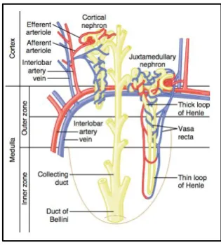
KIDNEY
The kidney is a functionally diverse organ and participates in many of the
body‟s homeostatic mechanisms. Kidneys are a pair of bean shaped organs located in
the retroperitoneal space. Each kidney is divided into an outer cortex and inner
medulla. The Nephron is the functional unit of kidney. Each kidney consists of about
a million nephrons. Each nephron consists of
1. Glomerulus, which is a tuft of capillaries surrounded by expanded end of renal
tubule called Bowman‟s capsule, supplied by an afferent and efferent arteriole.
2. Proximal Convoluted tubules, which are tightly coiled in the beginning called
as pars convoluta, which towards the medulla become straightened to form
pars recta.
3. Loop of Henle, consists of the thin descending limb and the thick ascending
limb.
4. Distal convoluted tubule.
5. Collecting duct, formed by two or more distal convoluted tubules that drains
urine from each nephron. Collecting ducts merge and empty their content into
the renal pelvis.30
Figure: 1shows the anatomy and organization of kidney.
6
Figure130: Organization of kidney30
Figure230: Basic Tubular Segments of aNephron30
[image:21.595.218.376.563.738.2]7
KIDNEY FUNCTION
The basic functions of kidney are30
1. Urine formation
2. Fluid and electrolyte balance
3. Regulation of acid base balance
4. Excretion of waste products of metabolism
5. Secretion of hormones
Renin
Erythropoietin
1,25, dihydroxy Vitamin D3 (Calcitriol) Prostaglandin
KIDNEY DISEASES
The kidney diseases have been classified as
1. Acute Kidney Disease (AKD).
2. Chronic Kidney Disease (CKD).
3. End Stage Renal Disease (ESRD).
PATHOPHYSIOLOGY OF KIDNEY DISEASES
Irrespective of the etiology of the kidney disease, the pathogenesis leading to
renal dysfunction remain the same. The pathological process is characterized by,early
inflammation followed by accumulation and deposition of extracellular matrix,
leading to tubulointerstitial fibrosis andtubular atrophy and finally leading to
8
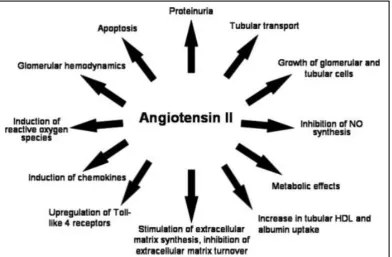
The local Renin Angiotensin Aldosterone System (RAAS) has an important
role in the pathological process of kidney disease.32 Angiotensin II produces toxic
reactive oxygen species (ROS) within the renal cells and affect signal transduction. It
also induces many inflammatory mediators like cytokines, chemokines and growth
factors which activates the lymphocytes and macrophages. The activated cells further
causes lysis of cells and proliferation of interstitial fibroblast which causes increase in
the extracellular matrix and fibrosis of the glomerulus and tubules. Blood supply will
be affected leading to cell death.31,32 The figure 4,gives an outline of the pathological
[image:23.595.92.485.376.633.2]process causing renal disease.
Figure:432
Role of Angiotensin II in renal pathology
The kidneys increase their functional capacity after injury. Symptoms or major
biochemical changes occur occur only if 50-60% of the functioning kidney mass is
lost.There will be increase in workload of the nephron and is considered as an
9
the decline in nephron number is reached, further loss in nephron will lead to
progressive renal disease as a consequence of a common pathway leading to
interstitial fibrosis.31,34
CHRONIC KIDNEY DISEASE
The increasing incidence of lifestyle disorders may cause resource crunch in
economy of our country. One such disease which India faces is Chronic Kidney
Diseases. CKD constitute a major cost burden in health care system worldwide. The
incidence of CKD is increasing worldwide. As per the Screening and Early
Evaluation of Kidney Disease (SEEK) India,study the prevalence of CKD in India is
17.2%.2 The prevalence of CKD globallyremains up to 13.4%.3
Definition of CKD35,36,37
According to Kidney Diseases Improving Global Outcomes (KDIGO) 2012,
Chronic Kidney Disease(CKD) is defined as abnormalities of kidney structure or
function present for more than three months.
Criteria of CKD (either of the following present for > 3 months)
1. Markers of Kidney damage (one or more)35,36,37
Albuminuria (AER 30 mg/24 hours, ACR 30 mg/g / 30 mg/mmol)
Structural abnormalities detected by imaging.
Urine Sediment abnormalities.
Electrolyte and other abnormalities due to tubular disorders.
Abnormalities detected by histology.
History of Kidney transplantation.
2. Decreased GFR35,36,37
10
The criteria GFR <60 ml/min/1.73m2 for CKD is selected because at this value
approximately one half of adult renal function is lost.38,39
RISK FACTORS OF CKD
Risk factors which are associated with renal dysfunction include
hypertension(HT), diabetes mellitus (DM), cardiovascular disease (CVD), proteinuria,
anaemia.40The non-traditional CKD risk factors include nephrotoxin exposure, renal
stones, acute kidney injury (AKI), infections,41 dyslipidemia are associated with renal
disease progression.42Thyroid dysfunction is also a risk factor for progression of renal
dysfunction.43
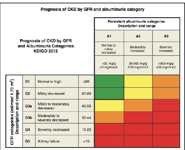
GRADING OF CKD35,36,37
As per KDIGO 2012, CKD is classified based on
1. GFR category and
2. Albuminuria category
1.GFR Category 37
G1 - eGFR 90ml/min/1.73 m2 – Normal or High G2 - eGFR 60-89ml/min/1.73 m2 – Mildly decreased
G3a - eGFR 45-59ml/min/1.73 m2 – Mildly to moderately decreased
G3b - eGFR 30-44ml/min/1.73 m2 – Moderately to severely decreased
G4 - eGFR 15-29ml/min/1.73 m2 – Severely decreased G5- eGFR <15ml/min/1.73 m2 - Kidney failure
In the absence of kidney damage GFR category, neither G1 nor G2 fulfill the criteria
11
[image:26.595.156.464.476.724.2]2. Albuminuria Category Table :137
Predicting prognosis of CKD
For predicting the prognosis of CKD, the cause or aetiology for CKD should
be identified. Then CKD is categorized based on GFR and Albuminuria. Figure 5,
gives an idea of prognosis of various stages of CKD.
12
CKD PROGRESSION
CKD progression is defined based on one or more of the following
Decline in GFR category, a certain drop in eGFR is defined as a drop in GFR
category accompanied by a drop of 25% or greater of drop in eGFR from
baseline.
Rapid progression is defined as a sustained decline in eGFR more than
5ml/min/1.73m2 per year.
The confidence in assessing progression is increased with number of serum
creatinine measurement and duration of follow up.36
The factors associated with CKD progression are GFR, albuminuria, hypertension,
diabetes, smoking, obesity, age, ethnicity etc.
ESTIMATED GLOMERULAR FILTRATION RATE (eGFR)
Serum creatinine is an endogenous marker of Glomerular filtration rate (GFR). In clinical practice, serum creatinine is used as an index of renal function.44 Plasma
concentration of creatinine is affected by muscle mass, diet, age, gender, exercise and
ethnicity, so there will be false estimation of renal function.45 A 24 hours urine
creatinine clearance is considered as a sensitive tool for detection of renal
dysfunction. Creatinine is freely filtered at glomerulus, but there is some tubular
secretion. Also the timed urine collection is difficult. There was a wide(11%) within
subject variability. These factors affected the urine clearance method of GFR
estimation.44
The National Kidney Foundation Disease Outcomes Quality Initiative
(K-DOQI) recommended the use of estimated GFR (eGFR)calculated by using formulas
13
equation based on age, weight, height and plasma creatinine with correction
factors.46The limitation of this formula is that, it was derived from hospitalised
individuals mostly men. Also the need for height and weight measurement for
calculation is a limitation.45
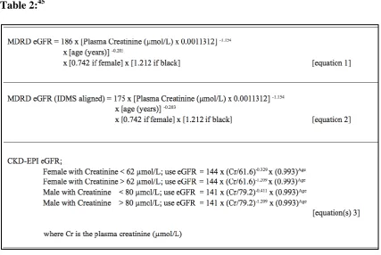
A Modification of Diet in Renal Disease (MDRD) study, evaluated the effect
of protein restricted diet on renal disease progression and created an equation for
calculation of eGFR, which included age, gender, plasma creatinine and race
differentiation (white or black).47MDRD did not require body weight or height. As
MDRD was originally derived from a group of CKD patients, its use in healthy
individuals is unclear. It is not validated for children, pregnant women, obesity and
elderly patients (>70 years).45 At a low creatinine value the performance of MDRD
formula was poor.38,48
Chronic Kidney Diseases-Epidemiology Collaboration group (CKD-EPI)
developed a new formula in 2009, to have greater accuracy even at high GFR. It
includes race, gender, and age.49 Many studies have found cystatin C derived equation
for eGFR is better than creatinine in predicting renal function.50,51 Schwartz formula
developed in 1970 is used to estimate GFR in chidren.
Schwartz Formula for children52
eGFR=0.41x height (cm)/ Scr (mg/dl) (updated Schwartz bedside formula)
The best equation for GFR in children was 52,53
eGFR=39.1X [height(m2)/ Scr (mg/dl)]0.516 X [1.8/cystatin C (mg/L)]0.294X
14
[image:29.595.90.518.101.387.2]Equations for calculating eGFR:45
Table 2:45
PROTEINURIA
Proteinuria is an independent risk factor for renal disease and cardiovascular
disease.54 The proteins of molecular weight of < 20,000 Da can be filtered by the
glomerulus, the negative charge of the glomerular membrane restrict the passage of
albumin through the membrane.55 Normally the filtered protein are reabsorbed and
catabolized in the proximal convoluted tubules. The normal protein excreted in urine
is 150 mg/day. Excretion of albumin 30mg/day to 300mg/day is termed as
microalbuminuria. Protein excretion >300mg/day is known as proteinuria or macro
albuminuria.56 Of the proteins excreted in urine, the high molecular weight
albumin(65,000Da) is about 40%, low molecular weight like light chain
immunoglobulin (20,000 Da) is of 20% and about 40% of Tamm-Horsfall
15
Pathologic Proteinuria can be classified into three types
1. Glomerular proteinuria
2. Tubular proteinuria
3. Overflow proteinuria
Glomerular proteinuria is due to the permeability of glomerulus to high
molecular weight proteins like albumin.56Glomerular proteinuria is the one which is
used for staging of Chronic Kidney Disease. Measurement of albumin in urine
determines the glomerular proteinuria.55
Tubular proteinuria results from decreased reabsorption of the protein present
in the glomerular filtrate or due to the excretion of proteins produced due to tubular
injury due to tubulointerstitial diseases like hypertensive nephrosclerosis, nephrotoxic
drugs like nonsteroidal anti-inflammatory drugs etc.55,56
Overflow proteinuria is due to excess low molecular weight protein, which
when excreted in normal amounts will be reabsorbed. Low molecular weight proteins
that occur in overflow nephropathy are Bence–Jones protein of multiple myeloma,
lysozyme in myelomonocytic leukemia, myoglobin in rhabdomyolysis.55,56
A 24–hour urine collection is considered as the gold standard in determining
the urinary protein excretion. A 24-hour urinary protein collection show variation in
urinary protein due to variation in intake of water, excretion of water, exercise,
posture, diet etc. Nowadays expressing a spot urine protein as ratio to spot creatinine
concentration is used. When the GFR remains constant the protein and creatinine
excretion rates are constant during day54.Currently spot urine protein- creatinine ratio
(UPCR) is widely used to estimate the daily urinary protein excretion.57
Microalbuminuria is an early sign of progression of renal and cardiovascular disease
16
albuminuria and non albumin proteinuria(NAP). Isolated non albumin proteinuria
(NAP) is a condition when urine total protein is elevated without elevation of urine
albumin.55 Non albumin proteinuria include alpha–microglobin, beta2 macroglobulin
and IgG which are associated with renal damage in renal transplant patients.59
Non albumin proteinuria = Total protein – albuminuria.60 Studies advice measurement
of both urine spot albumin and total protein to identify the non albumin protein which
may occur earlier than microalbuminuria.55,60,61
The KDIGO 2012 guidelines suggest early morning spot urine protein
estimation in the following descending order,37
1. Urinary albumin –creatinine ratio (ACR).
2. Urine protein-creatinine ratio (PCR).
3. Reagent strip urinalysis for total protein with automated reading.
4. Reagent strip urinalysis for total protein with manual reading.
KDIGO also states that if non-albumin proteinuria is suspected, assays for
specific urine proteins like Bence Jones protein is done.37
Consequences of proteinuria:
Proteinuria is not just a marker of renal dysfunction but it is also a risk factor
for progression of kidney disease. The proteins accumulate in the tubular lumen
causes inflammation and leads to interstitial structural damage and progression of
kidney disease.62 Also studies state that glomerular filtration of large amounts of
protein by glomerulus causes mesangial cell injury and thus causing
17
DIABETES AND CHRONIC KIDNEY DISEASE
Diabetic nephropathy, one of the microvascular complications of diabetes
mellitus, is one of the most important etiology for CKD and ESRD. Chronic
hyperglycemia is the cause for pathogenesis of the microvascular complication.64 The
pathological finding in advanced diabetic nephropathy are glomerulosclerosis and
tubulointerstitial fibrosis.65 Experimental animal model studies have shown that in
renal glomerulus and tubules there was increase in gene expression for synthesis of
extracellular matrix proteins.66 Hyperglycemia activate the polyol pathway of glucose
metabolism and this leads to collagen accumulation in the renal tubular
interstitium.67,68,69,70 Hyperglycemia induces increased formation of advanced
glycated end products of proteins,71,72 growth factors/ cytokines like transforming
growth factor- (TGF-),73,74 platelet derived growth factors (PDGF),75,76insulin-like growth factor-I (IGF-I),77,78 hepatocyte growth factor (HGF),79 Vasoactive substances
like angiotensin–II,80,81 endothelin-I,82 thromboxane,83 which through autocrine and
paracrine mechanism causes cell proliferation and growth.84
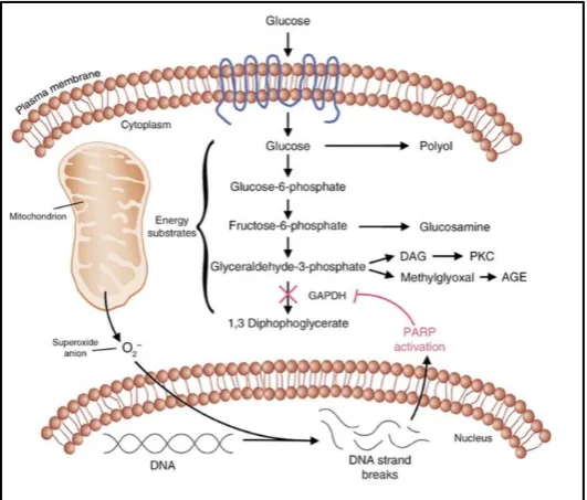
[image:32.595.167.433.543.769.2]Figure 6, explains the mechanism of renal toxicity of hyperglycemia
18
With increase in glucose, the mitochondria generate reactive oxygen species
(ROS), which leads to break in DNA strands and activation of poly ADP ribose
polymerase(PARP). PARP inactivates Glyceraldehyde 3-phosphate
dehydrogenase(GAPDH) and the normal glucose metabolism is altered.
There will be
1. Activation of polyol pathway
2. Activation of Phosphokinase C(PKC) via diacyl glycerol.
3. Activation of Hexosaminase pathway and glucosamine gets accumulated.
4. Formation of Advanced glycated end products due to non enzymatic glycation of
proteins.
All these mechanism produces increased growth factors and causes glomerular
pathology.64,85 Studies have shown that PKC stimulate the mitogen activated protein
kinase (MAPK) pathway. The MAPK pathway, is an intracellular signal transduction
pathway of cell growth, which activate cell growth and mesangial proliferation.86,87
Further studies have proven that advanced glycated end products (AGE) also activate
the MAPK pathway in proximal tubular LLC-PK1 cells.88
Apoptosis causes the tubular atrophy and tubulointerstitial fibrosis.89 Gene
products like bcl2 is protective against apoptosis.90 In a study there was increase in
apoptotic cells in diabetic, db/db mice than in non-diabeticlittermates. Theapoptotic
cells were found in the tubulointerstitium and not in glomeruli. There was increased
19
DYSLIPIDEMIA OF CHRONIC KIDNEY DISEASE
Dyslipidemia is a common finding among CKD patients and is responsible for
associated morbidity and mortality.92 The plasma lipid profile in CKD shows elevated
levels of triglycerides(TGL), low high density lipoprotein(HDLc),93 increased very
low density lipoprotein(VLDL), remnants and intermediate density lipoprotein,94
accumulation of small dense low density lipoprotein(LDLc), glycated and
carbamylated apolipoprotein,95 prolonged persistent chylomicron remnant post
prandially.96 The composition of plasma lipoprotein is also altered in CKD.97,98,99,100
In CKD the cholesterol content of VLDL is increased and there is relative decrease in
TGL content. In LDLc the cholesterol content is decreased and there is relative
increase in TGL content in CKD. In HDLc the cholesterol ester and free cholesterol
content are reduced and TGL content is elevated.54,55,56,57There is hepatic lipase
deficiency in humans and animals in CKD.101 Normally hepatic lipase catalyses
hydrolysis and removal of triglyceride content of HDLc. As there is deficiency of
hepatic lipase there is increase in TGL content of HDL.58 Elevated TG/HDL ratio can
cause renal dysfunction.102 The atherogenic lipoproteins like lipoprotein(a) and
oxidized LDL are present in hemodialysis patients.103
Dyslipidemia causes renal damage and can cause progression of renal
failure.104 Dyslipidemia cause damage to mesangial cells, podocytes and glomerular
capillary endothelial cells. Mesangial cells express LDL and oxidized LDL receptors,
on activation there will be mesangial cell proliferation, increased mesangial matrix
deposition, increase in cytokines like interleukin -6 or growth factors.Oxidized LDL
increases the adhesion of monocytes to glomerular endothelial cells and causes
monocyteinfiltration and causes tubular epithelial damage.42,105 Hypercholesterolemia
20
In vivo studies have shown that there is increased expression of a transcription factor,
sterol regulatory element binding protein(SREBP) which is associated with
lipogenesis and increase in accumulation of lipid in the glomerulus and
tubulointerstitial cell, leading to glomerulosclerosis and proteinuria.107,108
THYROID AND CKD
Thyroid hormones have its direct effect on kidney by causing systemic or local
hemodynamic changes.109Thyroid hormone directly influence renal development and
growth, GFR, renal transport systems and electrolyte homeostasis.16,17,110The presence
of renal abnormalities in congenital hypothyroid children111 indicate that thyroid
hormone has a role in development of fetal kidneys. Animal studies have proved
this.112
Effect of Thyroid Hormone on Renal physiology:
Thyroid hormone influences the renal function by action on cardiovascular
system and there by the blood flow to the kidneys. Thyroid also affects the GFR,
tubular secretion and reabsorption. In the proximal convoluted tubules, Na/K ATPase
is influenced by thyroid hormones and thus the sodium reabsorption.113 Thyroid
hormone also influence the tubular potassium handling.114 The renin angiotensin
aldosterone axis is influenced by thyroid
hormone by regulating renin release,115adrenergic system116and angiotensinase
activity.117Both hyperthyroidism and hypothyroidism has effects on kidney.
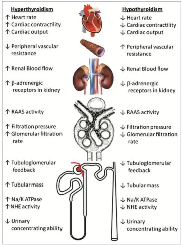
Role of Hyperthyroidism on Kidney function:
In hyperthyroid patients, an increase of 18-25% GFR has been noticed,17 due
to increased renal blood flow and renin angiotensin aldosterone system (RAAS)
21
have shown that thyroid hormone influence, increase in -adrenergic receptors in renal cortex and is the cause for RAAS activation.118 RAAS activity causes dilatation
of afferent arteriole and constriction of efferent arteriole and increase filtration
pressure leading to increase in GFR. Due to efferent arteriolar constriction, there will
be decrease in the perfusion in proximal convoluted tubule (PCT) and there will be
increased sodium and chloride reabsorption in PCT.119
Hyperthyroidism increases heartrate, raises cardiac output, decreases the
systemic vascular resistance and thus increases systolic blood pressure.120
Hyperthyroidism causes CKD by increasing the renal blood flow leading to intra
glomerular hypertension and increased filtration pressure. Associated proteinuria in
hyperthyroidism also causes renal injury.121
Role of Hypothyroidism on Kidney function
In adults and children, primary hypothyroidism is associated with a reversible
increase in serum creatinine and decrease of GFR.122,123,124,125,126,127 It has been
reported that these defects were corrected with thyroid hormone replacement.128 GFR
measurement using Inulin or 51Cr-EDTA has proved that hypothyroidism is associated
with reduced GFR.124 Hypothyroidism causes alteration in hemodynamics and
structure of kidney.129 The decrease in GFR in hypothyroidism is due to decreased
renal blood flow.130 Hypothyroidism also causes decreased cardiac output and low
circulating volume. Decreased activity of renin angiotensin system(RAAS) and
decreased levels of atrial natriuretic factor(ANF) are found in hypothyroidism and
these cause decrease in renal perfusion.109 Decreased expression of vascular
endothelial growth factor(VEGF) and insulin like growth factor(IGF) which are renal
vasodilators is seen in hypothyroidism.131
22
Figure7 :121
Effect of Thyroid dysfunction on Kidney
Chronic Kidney Disease and Thyroid disease
Primary and subclinical hypothyroidism is common among CKD patients132.
Decrease in T3(low T3 syndrome or euthyroid sick syndrome) is noticed in CKD
patients.133This decrease in T3 (mainly total T3 and not free T3) is due to chronic
23
and decrease in T3 binding to protein.134 Also decrease in deiodinase activity in CKD
is due to inflammatory cytokines, which causes decrease in peripheral conversion of
T4 toT3.135There is also decrease in thyroid hormone synthesis due to accumulation
of inorganic iodine in CKD.136
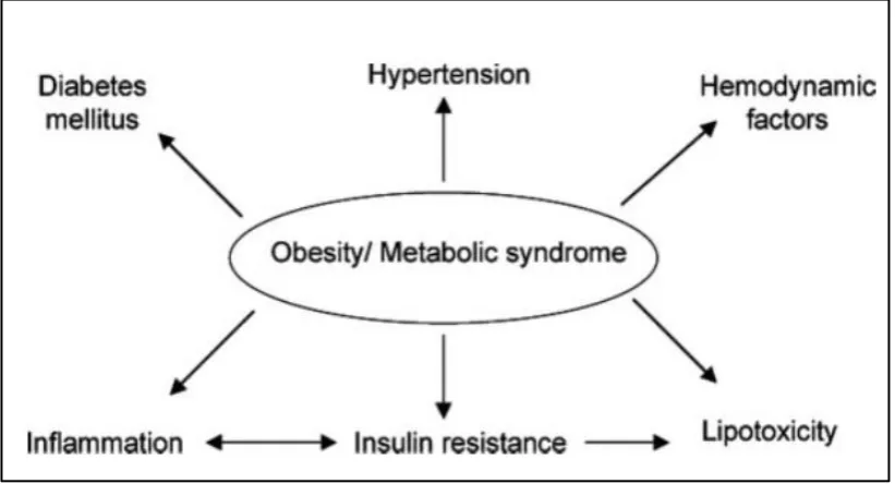
OBESITY AND CKD
Obesity, overweight and metabolic syndrome are emerging as important
independent risk factor for CKD.137,138 Epidemiological studies have shown the
association of obesity with CKD and end stage renal disease (ESRD). Studies showed
that high BMI was associated with ESRD as an independent risk factor, independent
of hypertension, proteinuria139 smoking and diabetes.140 Obesity was found toaffect
the progression of preexisting renal diseases like IgA nephropathy,141 unilateral renal
agenesis142 or after unilateral nephrectomy.143 These studies suggest that obesity
initiates CKD. A study of 6,500 nondiabetic subjects showed that increasing body
mass index (BMI) and waist circumference(WC), had decreased eGFR and increased
CKD.27 Also obesity is closely associated with Type II diabetes and hypertension
which are considered as important risk factors for CKD and ESRD.144,145,146
Mechanism of Obesity associated Kidney disease
Obesity causes renal damage directly by hemodynamic and hormonal
effect147,148 or indirectly by development of Type II diabetes mellitus and
hypertension.144,145,146Figure 8,149 gives an outline of mechanism of renal dysfunction
24
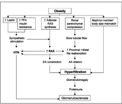
Figure 8:149
Potential Mechanism of renal dysfunction in Obesity
Animal and human studies have shown that obesity causes increased renal
tubular sodium reabsorption which causes renal vasodilatation150 causing glomerular
hyper perfusion and hyper filtration, which may cause focal segmental
glomerulosclerosis and proteinuria.148,151The mechanism for early rise in GFR in
obesity may be due to maculadensa feedback mechanism.152 The visceral obesity
causes compression of kidneys which may increase the sodium reabsorption in the
loop of Henle153 and hence less sodium in the macula densa and causes a tubulo
glomerular feedback and increases renin secretion, increased blood flow and GFR.
The glomerular hyper filtration then gradually subsides and there will be a gradual
decrease in GFR with renal injury associated with prolonged obesity induced
hypertension and diabetes. There will be increased glomerular wall tension and
glomerular hypertrophy that leads to injury, glomerular sclerosis and nephron
loss.153,154 Activation of sympathetic nervous system(SNS)155,156and renin angiotensin
25
Obesity related glomerulopathy has been documented from kidney biopsy
specimens.148 Changes in the structure of kidney is noticed within few weeks of rapid
weight gain. In animal experiments with dogs, enlargement of bowman‟s space,
increase in glomerular cell proliferation, mesangial matrix and basement membrane
thickening were observed. Glomerular transforming growth factor , expression was also increased.148Also the leptin from adipose tissue might cause renal
fibrosis.157Deposition of lipids in non adipose tissue like kidney leads to accumulation
of diacylglycerol and ceramides, which may cause mitochondrial dysfunction,
endoplasmic reticulum stress, apoptosis and renal injury and dysfunction.158,159
Inflammatory changes in Obesity causing CKD
Insulin resistance is present in metabolic syndrome, which is a
proinflammatory state.160,161Visceral fat produce increased level of adipokines like
interleukine-6 (IL-6), tumor necrosis factor, C-reactive protein (CRP) and resistin. A
study has shown that in patients with obesity related glomerulopathy, the glomeruli
showed increased expression of genes related to lipid metabolism, inflammatory
cytokines and insulin resistance.162 Leptin an adipocyte derived hormone, that
suppresses appetite (hypothalamus) and increases insulin sensitivity. In obesity the
hypothalamic effect of leptin is lost.160,163,164The leptin receptor (Ob-Ra) is expressed
in kidney.165 Recombinant leptin stimulate proliferation of cultured glomerular
endothelial cells and also in rats.166 Leptin stimulate type I collagen in cultured
mesangial cells,167 these show that leptin is involved in glomerulosclerosis. Figure
26
Figure 9:149
Hemodynamic consequences of Obesity leading to hyper filtration and hypertension
FFA- Free Fatty Acid;RAS –Renin Angiotensin System; HTN-Hypertension; AA- Afferent Arteriole; EA –Efferent Arteriole;Na –Sodium
CARDIOVASCULAR DISEASE AND CKD
Accelerated cardiovascular disease is a complication of renal disease.
Mortality and morbidity due to cardiovascular disease is common in CKD patients.
Studies have shown that prevalence of CKD is more among cardiovascular disease
patients, CKD is also a risk factor for Cardiovascular disease (CVD),168 and its
27
smoking for CVD and CKD may be the cause for increase in incidence of CVD
among CKD. 170,171The spectrum of cardiovascular disease seen in CKD patients
are172
1. Coronary artery disease (CAD) and myocardial infarction (MI).
2. Congestive heart failure (CHF).
3. Peripheral arterial disease (PAD).
4. Arrhythmias.
5. Cerebrovascular disease (CVA).
6. Sudden cardiac death.
Hypertension (HT) is one of the most common cause for CVD in CKD.
Activation of RAAS and sodium retention is the mechanism for developing HT in
CKD. Increased sympathetic activity and elevated catecholamine concentration is
seen in CKD.173 Renalase, a soluble monooxygenase which degrades catecholamines,
is expressed in renal glomeruli, PCT and cardiomyocytes is absent in CKD, so
increase in catecholamine activity is seen in CKD.174Endothelial dysfunction is
common in glomerular microalbuminuria and causes hypertension. Hypertension
causes left ventricular hypertrophy causing ventricular dysfunction.175 Asymmetric di
methyl arginine (ADMA), which is a competitive inhibitor of nitric oxide synthase is
increased in CKD. So there will be decreased bio availability of NO in CKD, which
causes endothelial dysfunction.176
In CKD there will be pressure overload and volume overload that causes
cardiomyopathy and left ventricular failure. Arterial stiffening due to medial and soft
tissue calcification also contribute to CVD in CKD.177 All these contribute to the
cardio renal syndrome.
28
Figure 10:178Pathophysiology of Cardio-renal Syndrome
ADH-Anti Diuretic Hormone, ANP- Atrial natriuretic peptide, BNP-B type natriuretic peptide, EPO-Erythropoietin, HTN –Hypertension, DM-Diabetes, LV-left ventricular, N-GAL-Neutrophil gelatinase associated lipocalin, KIM -1- Kidney Injury molecule, NSAID- Non steroidal anti inflammatory drug, OSAS-Obstructive sleep apnoea syndrome, PTH-Parathyroid hormone, SNS-Sympathetic nervous system, TNF-- Tumor necrosis factor-, TZD-Thiazolidinediones, VSMC-Vascular smooth muscle cell.
OCCUPATION AND CKD
Several studies in different parts of the world has shown association between
29
by educational status and occupational levels.179,180Low occupational level people are
exposed to lot of nephrotoxins like metals (lead, mercury, silica, copper, silver etc),
organic solvent, grain dust, and welding fumes (glycol ether).181,182,183 Agricultural
labourers are prone for heat stress and hence CKD. Strenuous work done in hot
environment leads to volume depletion and rhabdomyolysis.184 Our body tries to
maintain the temperature at approximately 37oC. Any increase in temperature
stimulate the thermoregulatory center in the hypothalamus and causes vasodilatation
and sweat glands stimulation and there will be regulation of temperature by
convection, conduction and evaporation. There will be loss of electrolytes and
extracellular fluid causing a stress to the kidneys.185When there is heat stress there
will be release of cytokines and heat shock proteins which causes inflammatory
response.25 Persons exposed to heat stress are prone to develop both acute kidney
disease(AKD) and chronic kidney disease(CKD).185,186In a study in El Salvador, about
the Mesoamerican nephropathy (MeN), the renal biopsy showed tubulointerstitial
injury and glomerulosclerosis with minimal vascular changes.187
Agrochemical exposure (commonly used 2,4-Dichlorophenoxyacetic
acid(2,4-D), paraquat dichlorate, glyphosate) is also associated with renal dysfunction.25 In a
study among sugar cane workers for MeN have mentioned that the workers eat
sugarcane and they are exposed to agrochemicals,4 also the high fructose intake
causes metabolically induced inflammatory renal damage.4,188,189Fructose are
transported by the glucose transporters GLUT5 /GLUT2, mainly expressed in the
proximal convoluted tubule. In the liver and kidney fructose is metabolized to
fructose-1-phosphate by the enzyme fructokinase [Ketohexokinase(KHK)].
ThenAldose-B acts on fructose-1-phosphate to form dihydroacetone phosphate and
30
depletion of ATP activates the enzyme of purine metabolism, to yield uric acid
leading to hyperuricemia, oxidants and inflammatory mediators leading to tubular
injury and inflammation.188,189,190 Also when they eat sugarcane they are at risk of
getting exposed to leptospiral infection.24 Leptospirosis produces acute kidney injury,
by a tubulointerstitial process with interstitial oedema and mononuclear cell
infiltration. Associated electrolyte abnormalities (sodium wasting and hypokalemia) is
due to the inhibition of the NaK2Cl (NKCC2) transporter in the ascending Loop of
Henle.191 In some studies, tubular dysfunction persisted and progressed to CKD when
there was associated diseases like hypertension.192Occupational activities involving
metals like lead, cadmium etc contaminate the environment. The source of lead
contamination is from gasoline and paint.193 These metals get accumulated in
proximal tubule producing chronic tubulointerstitial nephritis. Nephritis may be due
to oxidative stress leading to lipid peroxidation, apoptosis and necrosis.194
SMOKING AND CKD
Smoking is a preventable cause for many non-communicable diseases. In a
metaanalysis Xia et al has stated smoking as an independent risk factor for developing
CKD.195 In patients with diabetes and hypertension smoking contributes to the
progression of CKD and CVD.196,197,198 Renal function loss was noticed in persons
with history of current smoking of more than 20 cigarettes a day.199 In diabetic
nephropathy, smoking develops micro and macro albuminuria and progression to end
stage renal disease.196,198
Mechanism of Smoking and renal disease:
Several factors affect the toxic effect of smoking on renal dysfunction. The
genetic susceptibility of the individuals decide the amount of damage. The potential
31
1. Non hemodynamic mechanism
2. Hemodynamic mechanism
Non Hemodynamic mechanism:
Smoking causes endothelial dysfunction, growth factor activation, oxidative
stress, toxic effects on tubules, altered metabolism of lipoprotein and
glycosaminoglycans and insulin resistance.196,198 Studies have shown that nicotine
induces mesangial cell proliferation and fibronectin production.200 There is expression
of nicotinic acetyl choline receptors in the mesangial cells which mediates the
proliferation.201
Figure11,29 explains the non hemodynamic mechanism of smoking causing renal
[image:46.595.92.529.402.722.2]damage.
Figure1129 : Potential Mechanism of Smoking induced Kidney damage
32
Hemodynamic Mechanism:
Smoking increases the blood pressure and heart rate due to the action of
nicotine present in cigarettes.197The increase in concentration of epinephrine and
norepinephrine and direct stimulation of postganglionic nerve ending by the nicotine
causes the increase in BP and heart rate.202 In smokers there is an associated decrease
in BP at night,203which may lead to renal dysfunction.204 Ritz et al in their study has
found that nicotine, increased the activity of sympathetic nervous system causing
increased heart rate, blood pressure, intra glomerular capillary pressure leading to
decrease in GFR, filtration fraction and renal plasma flow.205 John main et al has
stated that smoking causes cholesterol micro embolism causing atherosclerotic
changes in renal artery and progression to end stage renal disease (ESRD).206
Pathological finding in kidney of smokers:
An increase in arteriolar wall thickness due to fibroelastic proliferation and
intimal hyaline thickening is seen.207 There is increased myointimal hyperplasia of
intrarenal arterioles which is absent in hypertension, so smoking is the causative agent
behind this change.208 Idiopathic glomerulosclerosis is seen exclusively in smokers.209
ALCOHOL AND CKD
Chronic excessive alcohol intake is considered as a risk factor for liver
disease, cardiovascular illness and cerebrovascular illness.210The association between
chronic alcohol intake and kidney disease is under controversy. Experimental studies
states that alcohol has an acute nephrotoxic effect.211Chronic alcohol consumption
indirectly causes CKD by causing alcohol induced hypertension.In certain specific
situations chronic alcohol dependency causes renal papillary necrosis, infection
33
traumatic).212,213,214Studies report that an inverse association exist between high
alcohol consumption and CKD risk.215The inverse association may be due to the
increase in high density lipoprotein (HDLc) and plasma endogenous tissue type
plasminogen activator levels and decrease in platelet aggregation by alcohol. HDLc is
less atherogenic and may be responsible for the inverse association of
CKD.216,217Animal studies state that antioxidant property of polyphenol present in red
wine may reduce renal damage by the induction of glutathione peroxidase and
superoxide dismutase.218 Another animal model study has said that alcohol prevent
leukocyte induced endothelial barrier damage and prevents renal ischemia.219
KIDNEY STONE AND CKD
Kidney stone is considered to be a risk factor for CKD. Hippisley et al have
found an increased risk of ESRD among female than male kidney stone formers.220
Some studies state that there is no relationship between renal stone and CKD.221
Junger‟s et al in a study has found that 3.2% of the 1391 patients with ESRD had
renal stones, and stones caused ESRD. They have found that 40% of patients with
stones in the study group had solitary functioning kidney.222 There is association of
development of CKD and the kidney stone type, in a study of community stone
formers struvite stones were found to be a risk factor for CKD.223 Brushite stone type
(CaHPO4) is associated with glomerulosclerosis, tubular atrophy and interstitial
fibrosis.224
Many pathways have been postulated for the development of CKD in kidney
stone patients. Obstruction by stones causing obstructive uropathy can cause acute or
irreversible chronic injury. There can be renal vasoconstriction and inflammation due
34
obstruction, osteopontin mediated interstitial influx and interstitial fibrosis have been
noticed.227Infective struvite stones form chronic pyelonephritis, which can cause
destruction of renal parenchyma.228 In uric acid stones, the hyperuricemia causes uric
35
MATERIALS AND METHODS
STUDY CENTRE:
The study was conducted among the adult population who attended the
out-patient departments at Trichy SRM Medical College Hospital and Research Centre,
Irungalur, Trichy.
STUDY DESIGN:
Cross sectional Observational study
STUDY PERIOD:
One year, June 2017 to May 2018
GEOGRAPHICAL DISTRIBUTION:
Both rural and urban areas in and around Trichy.
SAMPLE SIZE:
The calculated sample size using prevalence of CKD as 17.2%, power of the
study of 80% and significance level as 5% from the SEEK study2 was 463.Atotal of
500 subjects who attended the medicine and allied out-patient department were
included as study participants.
ETHICAL CONSIDERATION:
Ethical clearance for the study was obtained from the Institutional Ethical
Committee(IEC) of Trichy SRM Medical College Hospital and Research Centre,
Irungalur. All study subjects were explained about the study and informed written
consent was obtained and the confidentiality of their results were maintained.
INCLUSION CRITERIA:
36
EXCLUSION CRITERIA:
Patients with Acute Kidney Injury (AKI) were excluded on the basis of
oliguria and rapid increase in creatinine from previous recent records.
STUDY PROTOCOL:
After enrollment in the study a detailed history was taken, which included
history of presenting illness, past illness, occupation, any exposure to agrochemicals,
personal history likesubstance abuse, smoking, alcohol intake was noted. The patient
was enquired about history of any renal stones, cardiovascular illness, thyroid illness,
diabetes mellitusand hypertension. A detailed history of treatment and drugs
takenwere documented
CLINICAL EXAMINATION:
Routine clinical examination of various systems was carried out.
Blood Pressure (BP):
Blood pressure was recorded in the sitting position in the right arm using
mercury sphygmomanometer. Participants whose BP 140/90 mm Hg was considered as hypertensive, JNC-7 guidelines.230 Participants who were already on
anti hypertensives were considered as hypertensive irrespective of the BP
measurement.
Anthropometric measurements:
Height and weight of the study subjects were measured. Body mass index was
calculated from height and weight. Height was measured using a stadiometer in
centimeters. The individual was asked to stand in the upright posture barefoot, heels
kept together in close approximation, with the subject‟s back against the vertical
backboard and eyes directed forward. Weight was measured in kilograms using an
37
Waist circumference(WC) was measured in centimeters using a non-stretchable
measuring tape. WC was measured at the smallest horizontal girth between the costal
margin and the iliac crest at the end of expiration. Body mass index (BMI)was
calculated using the formula
BMI = Weight / height2 (kg/m2)
BMI Categorization WHO Guidelines :231
Under weight: < 18.5 kg/ m2
Normal : 18.5 to 24.9 kg/m2
Over weight : 25- 29.9 kg/m2
Obese : > 30 kg/m2
Systemic Examination was done.
Biochemical Investigation (Estimated Parameters):
Sample Collection:
An overnight 12 hours fasting venous sample was collected in a blood clot
activator (BCA) tube, for analysis of fastingblood glucose, fasting lipid profile [Total
cholesterol(TC), Triglycerides(TGL), High Density Lipoprotein Cholesterol(HDLc),
Low Density Lipoprotein(LDLc)],thyroid function tests [Thyroid Stimulating
Hormone(TSH),Free thyroxine (FT4)], Renal Function test(serum urea, serum
creatinine).
A spot urine sample was collected in a clean 100ml urine collection container
for spot UPCR. The samples were processed within 30 minutes to1 hour of collection.
Thyroid Hormone assay was done by Electro Chemiluminescence Immunoassay
(ECLIA) using Roche, cobas e411 analyzer. All other tests were performed using
38
Diagnosis of DM was made based on WHO criteria of fasting blood glucose level of
more than 126 mg/dl.232Known diabetics on treatment were considered diabetic
irrespective of their glycemic status.
Thyroid Function:
Euthyroid state was diagnosed with TSH – 0.4 – 4.2IU/L and FT4 level 0.8- 2 ng/dl. Overt hypothyroidism was diagnosed with TSH 5IU/L and Free T4 level < 0.8 ng/dl; Subclinical hypothyroidism was diagnosed when TSH 5IU/L and with normal Free T4 levels. Hyperthyroidism was diagnosed when TSH 0.25IU/L and Free T4>2 ng/dl. Subclinical Hyperthyroidism was considered when TSH 0.25IU/L and Free T4 within normal range. Patients who were on treatment with Thyroxine
supplementation were considered as hypothyroid.
Renal Function:
Serum Urea and Serum Creatinine were measured.
Estimated Glomerular Filtration Rate (eGFR) :
Using serum creatinine Estimated Glomerular Filtration Rate (eGFR) was calculated
using Chronic Kidney Diseases- Epidemiology Collaboration group (CKD-EPI)
formula49.
The CKD-EPI equation, expressed as a single equation
GFR = 141*min (Scr/K,1)∞*max(Scr/K,1)-1.209*0.993Age*1.018[if female]*1.159[if black]
Scr is serum creatinine(mg/dl), K is 0.7 for female and 0.9 for males,∞ is -0.329 for
females and -0.411 for males, min indicates the minimum of Scr/K or 1, and max
39
Staging of CKD was done as per GFRcategory based on KDIGO 201239
G1 - eGFR 90ml/min/1.73 m2 – Normal or High
G2 - eGFR 60-89ml/min/1.73 m2 – Mildly decreased
G3a - eGFR 45-59ml/min/1.73 m2 – Mildly to moderately decreased
G3b - eGFR 30-44ml/min/1.73 m2 – Moderately to severely decreased
G4 - eGFR 15-29ml/min/1.73 m2 – Severely decreased
G5- eGFR <15ml/min/1.73 m2 - Kidney failure
CKD was defined as eGFR <60ml/min/1.73 m2
Urine Spot Protein- Creatinine Ratio(UPCR) Categorisation35
1. Normal, P1 - UPCR < 0.15
2. Moderate proteinuria, P2 - UPCR > 0.15-0.5
3. Overt Proteinuria, P3 - UPCR > 0.5
Cardiovascular disease was considered present or absent based on the diagnosis made
40
METHODOLOGY
ESTIMATION OF PLASMA GLUCOSE Method:
Glucose Oxidase Peroxidase method (GOD-POD) (End point)
Principle:
Plasma glucose is first oxidized to Gluconic acid by the enzyme Glucose
Oxidase(GOD)with release of Hydrogen peroxide, which is further converted into
nascent oxygen and water by the enzyme peroxidise (POD). 4-Aminoantipyrine takes
up the oxygen, simultaneously with phenol,forms a pink coloured chromogen,
measured at 505 nm.
Glucose + O2 Gluconic acid + H2O2
2 H2O2 + 4-Aminoantipyrine+Phenol Quinoneimine+ 4 H2O
Reagents:
Glucose Oxidase : ≥10 KU/L
Peroxidase : ≥ 1 KU/L
Phosphate buffer (pH) 7.5 : 250mmol/L
Phenol : 5 mmol/L
4-Aminoantipyrine : 0.5 mmol/L
Standard concentration : 100 mg/dl GOD
41
Procedure:
Take three test tubes and label as Blank (B), Test (T) and Standard (S) as
follows, Incubation period: 10 min,Temperature at 370C.Mix well and read
absorbance of Test (T) and Standard(S) against distilled water at 505 nm
Calculation:
Glucose concentration (mg/dl) =( ∆ Abs-Test / ∆ Abs-Standard) x100 Reference value:
Plasma Fasting Glucose : 70-100 mg/dl
42
ESTIMATION OF SERUM UREA
Method:
Urease –Glutamate Dehydrogenase(“Urease-GLDH”)
Principle:
Urea is hydrolysed into ammonia and carbon dioxide by the presence of water
and urease. The ammonia combines with 2-oxoglutarate and NADH in the presence
of glutamate-dehydrogenase (GLDH) to produce glutamate and NAD+. The GLDH is
the rate limiting enzyme. The rate of decrease in NADH is proportional to the
concentration of urea, within the given time intervals. As the kinetic is very fast, this
test is preferably designed for analyzer application.
Urea+ 2H2O 2NH4+ + 2 HCO3
-2-Oxoglutarate + NH4+ +NADH L-Glutamate + NAD++ H2O
Reagents: 1. Enzymes:
Tris buffer (pH 7.8) : 125 mmol/L
Urease : ≥ 20 kU/L
GLDH : ≥ 0.3 kU/L
ADP : 0.88 mmol/L
Sodium Azide : 0.095 %
2. Substrates:
2-oxoglutrate : 25 mmol/L
NADH : 1.25 mmol/L Urease
43 Sodium Azide : 0.095%
3.Standard:
Urea : 80mg/dl
Sodium Azide : 0.095%
Preparation of reagent:
Reagent is prepared by mixing 4 parts of enzymes with 1 part of substrates.
Specimen:
Serum
Assay:
Wavelength : 334nm, 340nm, 365nm
Temperature : 25°C, 30°C or 37°C
Optical path : 1cm
Measurement : Against reagent blank(RB).
Only one reagent blank, per series is required.
44
Substrate Start Procedure:
Calculation:
C= 80.0× ΔA Sample/ ΔA standard [mg/dl]
Measuring range:
The test has a measuring range of 2 to 300mg/dl or 0.3to 50mmol/l.
Reference values:
Serum: 10-50 mg/dl
Blank Standard
Sample /standard - 10µl
Reagent 1 1000µl 1000 µl
Mix, incubate approximately for 5 minute.
Reagent 2 250µl 250µl
Mix, read absorbance of Sample /standard after 60 seconds and read
after exactly 1 minute (A2).
Calculate the absorbance difference:
45
ESTIMATION OF SERUM CREATININE
Method:
Modified Jaffe‟s kinetic method
Principle:
Creatinine, in the sample reacts with picric acid in alkaline medium to form
creatinine picrate (red coloured complex), measured at 500 nm.
Creatinine + Picricacid Creatinine picrate complex
Specimen:
Serum, Urine
Reagents:
Standard : 2 mg/dl
Reagent-1
Sodium Hydroxide : 0.2 mol / L
Reagent 2
Picric acid : 20 mmol / L
Storage: 2º-8ºC
Preparation of working solution:
The reagents are brought to room temperature. 4 parts of reagent 1 and 1 part of
reagent 2 are mixed.
General system parameters:
Reaction slope : Increasing
Reaction Type : Fixed Time
Flow cell Temp. : 25°C, 30°C or 37°C
Wavelength : 500 nm (490-510 nm)
46 Delay Time : 30 sec.
Reagent Volume : 1.0 ml
Sample Volume : 100 µl
Std. Concentration : 2 mg/dl
Path length : 1 cm
Zero Setting With : Distilled water
The instrument is set with above system general parameters.
Procedure:
The sample and working solution are allowed to attain room temperature, prior
to use Three test tubes are taken and labelled as Standard (S), Blank (B), Test (T). 1
ml of working reagent is added to all three test tubes. 100µl of sample is added to test
tube „T‟ and 100µl of standard is added to test tube „S‟. Then mixed and reading is
taken absorbance A1 after 60 seconds and A2 after further 120 seconds. ΔA = A2-A1.
For urine spot creati