Range of CD4-Bound Conformations of HIV-1 gp120, as Defined
Using Conditional CD4-Induced Antibodies
Gilad Kaplan,a*Anna Roitburd-Berman,aGeorge K. Lewis,bJonathan M. Gershonia
Department of Cell Research and Immunology, George S. Wise Faculty of Life Sciences, Tel Aviv University, Tel Aviv, Israela; Division of Vaccine Research, The Institute of
Human Virology, University of Maryland School of Medicine, Baltimore, Maryland, USAb
ABSTRACT
The HIV envelope binds cellular CD4 and undergoes a range of conformational changes that lead to membrane fusion and
deliv-ery of the viral nucleocapsid into the cellular cytoplasm. This binding to CD4 reveals cryptic and highly conserved epitopes, the
molecular nature of which is still not fully understood. The atomic structures of CD4 complexed with gp120 core molecules (a
form of gp120 in which the V1, V2, and V3 loops and N and C termini have been truncated) have indicated that a hallmark
fea-ture of the CD4-bound conformation is the bridging sheet minidomain. Variations in the orientation of the bridging sheet
hair-pins have been revealed when CD4-liganded gp120 was compared to CD4-unliganded trimeric envelope structures. Hence, there
appears to be a number of conformational transitions possible in HIV-1 monomeric gp120 that are affected by CD4 binding. The
spectrum of CD4-bound conformations has been interrogated in this study by using a well-characterized panel of conditional,
CD4-induced (CD4i) monoclonal antibodies (MAbs) that bind HIV-1 gp120 and its mutations under various conditions. Two
distinct CD4i epitopes of the outer domain were studied: the first comprises the bridging sheet, while the second contains
ele-ments of the V2 loop. Furthermore, we show that the unliganded extended monomeric core of gp120 (core
e) assumes an
inter-mediate CD4i conformation in solution that further undergoes detectable rearrangements upon association with CD4. These
discoveries impact both accepted paradigms concerning gp120 structure and the field of HIV immunogen design.
IMPORTANCE
Elucidation of the conformational transitions that the HIV-1 envelope protein undergoes during the course of entry into CD4
ⴙcells is fundamental to our understanding of HIV biology. The binding of CD4 triggers a range of gp120 structural
rearrange-ments that could present targets for future drug design and development of preventive vaccines. Here we have systematically
interrogated and scrutinized these conformational transitions using a panel of antibody probes that share a specific preference
for the CD4i conformations. These have been employed to study a collection of gp120 mutations and truncations. Through these
analyses, we propose 4 distinct sequential steps in CD4i transitions of gp120 conformations, each defined by antibody
specifici-ties and structural requirements of the HIV envelope monomer. As a result, we not only provide new insights into this dynamic
process but also define probes to further investigate HIV infection.
V
iral tropism is mediated by the specific binding of the viral
spike protein to its corresponding cell surface receptor.
Evo-lution has driven human immunodeficiency virus (HIV) to
elab-orate on this canonical paradigm, introducing a series of
orches-trated sequential events involving two receptors: CD4 as a primary
receptor (
1–3
) and a chemokine receptor (CXCR4 or CCR5) as a
subsequent coreceptor (
4–10
). However, many critical details of
the molecular mechanisms by which CD4 triggers a number of
conformational rearrangements within gp120 to assemble a
core-ceptor binding site and how this ultimately leads to
gp41-medi-ated membrane fusion are still missing. Obviously, it would be
extremely beneficial to have high-resolution atomic structures for
the viral spike before it encounters CD4 and serial snapshots of the
structural transitions that the gp120 subunits undergo until gp41
steps in to drive membrane fusion. However, this has proven
ex-tremely challenging, in part due to the fact that the HIV-1
enve-lope exists in dynamic equilibrium among an ensemble of
confor-mations (
11–17
).
In 1998, the first structure of the monomeric HIV-1 gp120
subunit was solved (
18
) but only when its N and C termini;
vari-able loops V1, V2, and V3; and sugar moieties were removed and
the remaining “core” was further stabilized via binding to CD4
along with a Fab of a gp120-specific monoclonal antibody (MAb)
(MAb 17b). Nonetheless, this tripartite crystal proved extremely
informative and provided the first glimpse of the gp120 structure
in a CD4-bound state. Compared to the atomic structure of an
unliganded simian immunodeficiency virus (SIV) envelope (
19
),
it was proposed that the four-

-stranded “bridging sheet,”
con-sistently found in a variety of HIV-1 gp120/CD4/Fab cocrystal
structures (
16
,
18
,
20–23
) yet absent from the SIV structure, was a
defining structural hallmark of the CD4-bound conformation.
Subsequently, Kwon and collaborators discovered that by
exten-sion of the N terminus of monomeric gp120 core and retention of
Received22 December 2015Accepted14 February 2016
Accepted manuscript posted online17 February 2016
CitationKaplan G, Roitburd-Berman A, Lewis GK, Gershoni JM. 2016. Range of CD4-bound conformations of HIV-1 gp120, as defined using conditional CD4-induced antibodies. J Virol 90:4481– 4493.doi:10.1128/JVI.03206-15.
Editor:G. Silvestri
Address correspondence to Jonathan M. Gershoni, [email protected].
*Present address: Gilad Kaplan, National Cancer Institute, National Institutes of Health, Bethesda, Maryland, USA.
G.K. and A.R.-B. contributed equally to this work.
Copyright © 2016, American Society for Microbiology. All Rights Reserved.
on November 7, 2019 by guest
http://jvi.asm.org/
the base of the V3 loop (yet still V1 to V3 depleted), one could
generate high-quality crystals in the absence of both CD4 and a
stabilizing Fab, thus providing atomic structures for an extended
core version (core
e) of unliganded HIV-1 gp120 (
24
).
Unexpect-edly, the fully assembled four-stranded bridging sheet, previously
taken as the epitome of the CD4-bound conformation, persisted
in all the analyzed coree
structures. This led Kwon et al. to propose
that the default structure of monomeric gp120, depleted of its
variable loops (V1 to V3), assumes an energetically favorable
ground-state “CD4-bound conformation” characterized by a fully
formed bridging sheet. It was postulated that within the trimer,
interactions between the gp120 protomers and the variable loops
lock the envelope into a higher-energy state (
24
). Binding of the
trimer to CD4 triggers a series of conformational changes, shifting
the variable loops to allow each monomer to snap into a preferred
“ground state,” thus opening up the trimer and enabling the
bind-ing of the second coreceptor followed by gp41 transitions and,
ultimately, membrane fusion (
14
,
15
,
17
,
24–28
) (also see
Fig. 5
in
reference
24
). Cryo-electron microscopy (EM) tomography
anal-yses integrated with X-ray structures of stabilized, fully cleaved,
soluble Env trimers have been extremely effective in generating a
detailed model for the HIV spike (clade A BG505 SOSIP.664
gp140 [
17
,
29–31
], the clade B B41 SOSIP.664 trimer [
22
], and the
clade C ZM197M and DU422 SOSIP.664 trimers [
32
]). A
surpris-ing feature of the CD4-unliganded gp120 molecule is the presence
of a four-stranded bridging sheet, in contrast to the SIV structure
discussed above (
29–31
). However, this structure in the trimer is
distinct from bridging sheets seen in the monomeric gp120/CD4
cocrystals (
16
,
18
,
20–23
) and the unliganded gp120 coree
struc-tures (
24
). Whereas the two versions of the bridging sheet have the
same four--strand composition, they differ with regard to the
juxtapositions and relative orientations of these

-strands. Hence,
CD4 still plays a role in inducing conformational reorientations
within the bridging sheet. A better definition of what actually
oc-curs within the gp120 protomer as a result of CD4 binding is
essential to our understanding of the biology of the mechanisms of
HIV infection of CD4 cells.
In a desire to reveal structural elements that discriminate and
delineate the essence of the CD4-induced (CD4i) conformational
rearrangements in the HIV envelope, we and others have
investi-gated gp120 structures using “conditional” probes, MAbs, that
specifically define epitopes associated with CD4i transitions of
gp120 (
33–41
). Here we describe the analysis of gp120 with a select
panel of five well-established CD4i MAbs that target the core outer
domain. Our results illustrate that gp120 undergoes a range of
CD4i conformations that go beyond that of the previously
re-ported structure of monomeric coree.
MATERIALS AND METHODS
Antibodies and reagents.The outer domain-specific CD4i MAbs used in this study are described inTable 1.
MAbs 17b (18,38) and 19e (42,43) were kindly provided by J. Rob-inson (Tulane University Medical Center, USA). MAb N12-i15 was iso-lated by Y. Guan and colleagues, including one of us (G.K.L.), at the Institute of Human Virology, as described previously (41). MAb 21c was kindly provided by R. Diskin (Division of Biology, California Institute of Technology, USA) (23,39). MAbs CG10, CG9, 1B6, and LG4 were iso-lated at Tel Aviv University. The chimeric human IgG version of the mu-rine CG10 CD4i MAb (44–46) was produced by cloning the heavy and light chain variable-domain sequences of this MAb into the pMAZ-IgH and pMAZ-IgL vectors designed for the production of IgG1 antibodies in mammalian cells, as described previously (47). The vectors were then used to transiently transfect HEK 293T cells as described below. MAbs LG4 and 1B6 are both used as capture reagents that bind gp120 (MAb LG4 targets a conserved epitope at the carboxy terminus of gp120, “SGGPLGVAPTKAKRRVVQREKRAD,” while 1B6 binds gp120 with-out interfering with binding by CD4i MAbs or CD4 [48]). MAb CG9 binds to CD4 (46). Soluble CD4 (sCD4) (domains D1 to D4) was a kind gift from GlaxoSmithKline. HIVIg is a pool of purified IgGs from HIV-infected individuals, kindly provided by Nabi, Inc. The m2 pep-tide is a phage-displayed, 14-amino-acid-long, cysteine-constrained peptide (CDRRDLPDWAIRAC). Binding of gp120 by the m2 peptide allosterically induces CD4i epitopes (48).
HIV envelopes.Except for gp120 from the CDC451 strain (which was purchased as soluble monomeric gp120 from Advanced BioScience Labs, USA) and the monomeric and SOSIP forms of BG505 (kindly provided by J. Moore, Weill Cornell Medical College, USA), all of the other gp120s used in this study were produced by transient transfection of HEK 293T cells (see below). For R2, a vector containing a codon-optimized R2 gp120/gp160 gene was kindly provided by G. V. Quinnan and C. C. Broder (Uniformed Services University of the Health Sciences, USA). For JR-FL, a vector containing a codon-optimized JR-FL gp120 gene was kindly provided by R. Pantophlet (Scripps Institute, USA). For BaL, a codon-optimized BaL gp120 gene from the pNGVL-FLSC-RT vector, provided by A. L. DeVico (Institute of Human Virology, USA) was re-cloned into the pCDNA3 expression vector (Invitrogen, USA). For YU2, a vector encoding the YU2 gp160 gene was kindly provided by Joseph G. Sodroski (Dana Farber Cancer Institute, USA). For extended gp120 cores, vectors encoding extended core gp120 genes were kindly provided by Y. D. Kwon and P. D. Kwong (National Institutes of Health, Bethesda, MD, USA). The BaL gp120 truncation mutants were constructed based on the codon-optimized BaL gene by using overlap PCR. The resulting mu-tatedenvgenes were verified by sequencing and cloned into the pCDNA3 expression vector.
[image:2.585.42.547.77.175.2]Amino acid numbering system.In view of the fact that all the genetic manipulations, truncations, and mutations of gp120 reported here are derived from BaL, the numbering system used throughout this study is based on BaL gp120. In order to assist in comparisons with other HIV-1
TABLE 1List of antibodies used in this study
Antibody Biological source Source Description References
17b Human MAb J. E. Robinson, Tulane University
Binds the gp120 bridging sheet, as determined by X-ray structure determination
18,38
21c Human MAb J. E. Robinson Binds a hybrid epitope comprised of gp120 and CD4, as determined by X-ray structure determination
23,39
19e Human MAb J. E. Robinson Specific for the gp120-CD4 complex 42,43
N12-i15 Human MAb Institute of Human Virology Specific for the gp120-CD4 complex 41,65
CG10 Murine MAb Tel Aviv University Specific for the gp120-CD4 complex; a chimeric human version of this MAb was produced and used as part of this study
44–46
on November 7, 2019 by guest
http://jvi.asm.org/
envelope molecules, we have indicated landmark cysteine residues in the figures and text.
Production of gp120 and antibodies by transient transfection.HIV gp120s and chimeric human CG10 were produced by transient transfec-tion of HEK 293T cells with 10g of the appropriate gp120-encoding vector or 10g each of the chimeric CG10 heavy and light chain-encoding vectors per 10-cm plate by the calcium phosphate method. HEK 293T cells were maintained in Dulbecco’s modified Eagle’s medium (DMEM; Gibco) complemented with 10% fetal calf serum (Biological Industries, Beit Haemek, Israel). Spent medium was collected at 48 h posttransfection. The chimeric CG10 MAb was purified from spent me-dium by using protein G affinity chromatography, dialyzed against phos-phate-buffered saline (PBS), and quantified by UV spectrometry. The gp120 proteins were filtered and concentrated by using Amicon Ultracel Centricon with a 50,000-molecular-weight (MW) cutoff (Millipore) ac-cording to the manufacturer’s instructions. The gp120s were then quan-tified by binding to HIVIg against gp120s of a known concentration and tested for functional folding in a quality control enzyme-linked immu-nosorbent assay (ELISA) (ligand overlay of Western blots was also used as described previously [46,49]). In the quality control ELISA, mutated envelopes were tested for binding to the stringent CG10 MAb in the pres-ence of sCD4, showing that the produced envelopes were able to bind sCD4 and undergo CD4i conformational changes. Both of these traits were taken as an indication of proper protein folding. Further confirma-tion of the funcconfirma-tional configuraconfirma-tion of gp120 and its mutants was dem-onstrated by binding of conformation-demanding MAbs, such as b12 (50, 51) and b6 (52,53), as well as the glycomoiety-specific MAb 2G12. SDS-PAGE analyses of the gp120 preparations indicated the presence of both monomeric and oligomeric forms, as previously reported (54–63). The aberrant disulfide-linked oligomers continued to bind CD4 as expected. Moreover, Coutu and Finzi recently compared the binding kinetics of non-fast protein liquid chromatography (FLPC)-purified gp120 with FPLC-purified monomeric gp120 by surface plasmon resonance and con-firmed that the affinity constants for CD4 in both preparations are not statistically different (some differences in antibody on-rates were mea-sured) (55). It is noteworthy that all the conclusions reported here are based exclusively on positive results, i.e., the acquisition of robust binding as a result of CD4 association with the gp120s tested. Nonetheless, a num-ber of the gp120 truncations were FPLC purified according to methods described previously by Coutu and Finzi (55), and the monomers (⬎95% purity) were tested, compared to FPLC-purified wild-type (wt) BaL mo-nomeric gp120 where indicated, and found in all cases to respond the same as non-FPLC-purified gp120. Furthermore, Western blot analyses confirmed that both monomeric gp120 and the -mercaptoethanol-sen-sitive oligomers (54,56–63) continued to bind CD4, which further in-duced both relaxed (e.g., 17b) as well as stringent (e.g., CG10) MAb bind-ing, as previously described (46,49).
Envelope ELISA.ELISA plates were coated overnight at 4°C with 100 l of 7.5g/ml of capture MAb (either LG4 or 1B6) or 5g/ml gp120 diluted in PBS. The wells were blocked for 1 h at 37°C by using 5% nonfat dry milk and 20% horse serum in PBS. Capture of gp120 in MAb-coated wells was carried out by adding 5g/ml gp120 to the wells for 1 h at room temperature (RT). Detecting MAbs were added at 2.5g/ml for 1 h at RT. Horseradish peroxidase (HRP)-conjugated secondary antibodies (e.g., anti-human in the case of the human-derived CD4i MAbs) were then diluted 1:5,000 and added to the wells for 1 h at RT. All antibodies and proteins were added in blocking solution. Between incubations, the wells were washed three times with 0.05% Tween 20 in PBS (PBST). Finally, the wells were reacted with 100 l of the 3,3’,5,5’-tetramethybenzidine (TMB/E) ELISA substrate (Chemicon International, USA). Absorbance was measured at 650 nm. Unless stated otherwise, gp120 from the BaL strain was used in the reported assays. For BG505 SOSIP.664 gp140/ BG505 gp120 ELISAs and extended core ELISAs, CD4i MAbs were used as capture reagents. BG505 gp120 or BG505 SOSIP.664 gp140 or extended envelope cores were added to the wells in the presence or absence of sCD4
(4g/ml). Bound extended envelope cores were detected with biotin-conjugated HIVIg (2.5g/ml) followed by HRP-conjugated streptavidin. For competition ELISAs, the murine version of the CG10 MAb was added at 10 to 15g/ml to wells containing captured gp120 and sCD4 (gp120 and sCD4 concentrations were the same as those for the regular envelope ELISA) and incubated for 1 h at RT. The competing human CD4i MAbs were then added (2.5g/ml), and the ELISA was continued as described above. In ELISAs utilizing biotinylated antibodies, the blocking buffer used was 3% bovine serum albumin (BSA) in PBST. Detection of bound biotinylated antibody was done with HRP-conjugated streptavidin at a dilution of 1:2,500 in blocking buffer. Concentrations of reagents used in ELISAs are as follows: coating antibodies were used at 7.5g/ml, detec-tion antibodies were used at 2.5 to 3.5g/ml, HIVIg was used at 10g/ml, CD4 was used at 5g/ml, and gp120s were used at 5g/ml. Graphs were generated and statistical significance of ELISA results was calculated (two-tailed Student’sttest) by using GraphPad Prism version 6.01 for Windows (GraphPad Software, La Jolla, CA, USA). The results from three indepen-dent experiments are shown. Results from different experiments were normalized according to envelope binding by HIVIg. Bars represent stan-dard deviations.
RESULTS
Here we focus on the primary and direct effects of CD4 binding to
HIV-1 envelope, namely, the conformational transitions of gp120,
using MAbs that can discriminate between different facets of this
process. Such conformational changes have been reported for
both the outer and inner domains of gp120 (
35–37
). Here we have
specifically analyzed the changes associated with the outer domain
harboring the CD4 binding site itself.
CD4i MAbs can be divided into distinct CD4-relaxed and
-stringent subsets.
Numerous CD4i MAbs have been described,
whose dependence on CD4 for gp120 binding ranges from
mar-ginal to absolute (
18
,
21
,
23
,
42
,
62
,
64–66
). These MAbs are
gen-erally designated CD4i MAbs. We focused on and tested a
collec-tion of outer domain-specific CD4i MAbs against envelopes from
several HIV-1 strains: gp120 from clade B strains BaL, CDC451,
YU2, JR-FL, and R2 in addition to trimeric BG505 SOSIP.664
gp140 and the BG505 gp120 monomer (clade A). The BG505
SOSIP.664 trimer was used to ascertain the effects of
trimeriza-tion on CD4i MAb binding, while the R2 strain can infect
CD4-negative cells (
67
), constitutively exposes several CD4i epitopes
(
68
), and therefore could theoretically bind CD4i MAbs in the
absence of CD4.
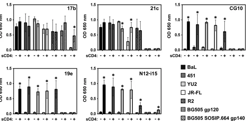
As shown in
Fig. 1
, the dependence on CD4 for gp120 binding
of the selected, representative panel of five CD4i MAbs to gp120 is
variable (the MAbs of the panel are described in
Table 1
). One of
these, MAb 17b, has been used extensively as a “gold standard”
CD4i MAb (
11
,
24
,
39
,
62
,
69–76
). The prominent CD4i status of
MAb 17b stems from the fact that cell surface trimeric envelope
requires CD4 for 17b binding (
38
), as is also the case for the
solu-ble BG505 SOSIP.664 trimer (
Fig. 1
). In contrast, MAb 17b shows
little requirement for CD4 when binding to full-length
mono-meric gp120, consistent with the conclusion of Kwon et al. that
monomeric gp120 assumes a default CD4-bound conformation
(
24
). MAb 21c also shows a similar CD4-relaxed pattern of
bind-ing to the clade B gp120s while not bindbind-ing at all to the clade A
BG505 monomer or the BG505 SOSIP trimer. In contrast, the
other three CD4i MAbs, CG10, 19e, and N12-i15, bound all clade
B gp120s in a totally CD4-stringent manner yet did not bind the
clade A BG505 monomer and the BG505 SOSIP trimer. Thus,
although some variation in binding can be seen on a
subtype-specific level, CD4i MAbs can be divided into two distinct classes:
on November 7, 2019 by guest
http://jvi.asm.org/
relaxed binders, which can bind monomeric gp120 in the absence
of CD4 (e.g., 17b and 21c), and stringent binders, whose binding
to gp120 is completely dependent on CD4 (CG10, 19e, and
N12-i15).
Mapping distinct CD4i regions on gp120.
Does the
CD4-bound state constitute a single epitope or region, or are there a
number of different epitopes in the HIV-1 gp120 outer domain
that are affected by CD4 binding? For this, we conducted a
com-petitive ELISA, thereby testing the ability of the bridging sheet
binding MAb CG10 (
44–46
) to compete against the other four
MAbs of the panel (
Fig. 2
). It should be noted that the region of
MAb 17b and 21c binding within gp120 is the bridging sheet
minidomain, as ascertained by cocrystallization of these MAbs in
complex with gp120 and CD4 (
18
,
23
), and that MAb 19e binding
has also been mapped to this region (
42
). Therefore, the
corre-sponding epitopes of four of the CD4i MAbs overlap aspects of the
bridging sheet, as also indicated by the ability of MAb CG10 to
totally obstruct binding by MAbs 17b, 21c, and 19e (
Fig. 2
).
Bind-ing of the strBind-ingent MAb N12-i15, however, shows almost no
competition with MAb CG10 and therefore defines an
indepen-dent epitope that has no immediate overlap on the bridging sheet.
Thus, we can conclude that conformational rearrangements in
gp120 due to CD4 binding include at least two outer domain
epitopes of gp120, which are spatially separated enough to
pre-clude competition by antibodies.
Stringent CD4i MAbs bind conformationally induced gp120
in the absence of CD4.
The stringent binding nature of MAbs
CG10, 19e, and N12-i15 could hypothetically be the result of the
CD4 protein directly contributing pivotal contact residues
neces-sary for MAb recognition. We previously assessed the
contribu-tion of such hypothetical critical contact residues within CD4
us-ing a molecule other than CD4. A short 14-amino-acid peptide
(designated the m2 peptide [CDRRDLPDWAIRAC]), shown to
bind to gp120 and allosterically induce the CD4i conformation,
i.e., without occluding the CD4 binding site itself, was used in
place of CD4 (
48
). We showed that the m2 peptide induces the
epitopes of the CD4i conformation recognized by the three
strin-gent MAbs CG10, 19e, and N12-i15 and allows binding to gp120
in the absence of CD4. m2 peptide induction of a CD4i
confor-mation occurs without occluding the CD4 binding site, thus
illus-trating an alternative route toward achieving a conformation
re-sembling a CD4-bound state. Together, this experiment (
48
) and
previous studies of CG10 (
62
,
77
) demonstrate that MAbs CG10,
19e, and N12-i15 can bind in the total absence of CD4, provided
that gp120 is induced to display the stringent CD4i epitopes. This
FIG 1Stringent CD4i MAbs do not bind gp120 in the absence of sCD4. An ELISA was used to analyze CD4i MAb binding to gp120s from different strains and to a gp140 trimer. MAbs 17b and 21c show binding to gp120 independent of sCD4 (relaxed binding). MAbs CG10, 19e, and N12-i15 show no binding to gp120 in the absence of sCD4 (stringent binding). Only MAb 17b was cross-reactive with the clade A BG505 SOSIP trimer and its monomer, showing relaxed binding to the monomer and stringent binding to the trimer. Statistically significant differences (P⬍0.05) between the “⫺sCD4” and “⫹sCD4” columns are marked with an asterisk. OD, optical density.
FIG 2MAb N12-i15 defines a CD4i region upon gp120 distinct from the bridging sheet. Competition ELISAs were carried out in order to discern whether all the detected CD4i epitopes cluster to one area on gp120 or not. Binding of CD4i MAbs to gp120BaL-sCD4 was measured in either the absence or the presence of prebound MAb CG10. MAbs 17b, 21c, and 19e compete strongly with MAb CG10. MAb N12-i15 defines a CD4i epitope sufficiently separated from the bridging sheet so as to show no competition with MAb CG10. Statistically significant differences (P⬍0.05) between the “⫺CG10” and “⫹CG10” columns are marked with an asterisk.
on November 7, 2019 by guest
http://jvi.asm.org/
[image:4.585.84.506.63.269.2] [image:4.585.41.287.491.638.2]illustrates that even if some residues of CD4 contact these MAbs,
they are not a prerequisite for stable binding.
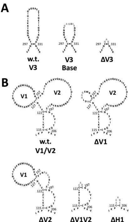
Contributions of the V1, V2, and V3 loops to the CD4i
con-formation.
Next, we analyzed the effects of the V1/V2/V3 loops
and bridging sheet hairpin 1 (the base of the V1/V2 loops) on the
CD4-bound conformation as interpreted by CD4i MAb binding.
Using BaL gp120 as the prototype, a number of mutants were
generated. For the sake of clarity,
Fig. 3
to
5
provide structural
schematics of the collection of gp120 mutations and truncations
analyzed. The V3 loop was either partially or fully deleted (the V3
base and
⌬
V3 mutants, respectively) (
Fig. 3A
), and the V1 and V2
loops were either both truncated (⌬V1V2 mutant) or truncated
separately (the
⌬
V1 and
⌬
V2 mutants) (
Fig. 3B
). Additionally, the
V2 core mutant (described in detail in the legend of
Fig. 4
) was
constructed based on the crystal structure of the V1/V2 loops
bound to a neutralizing antibody (
78
). This crystal structure
showed that the V2 loop assumes a “Greek key” fold. The V2 core
mutant was generated by truncating the V1 loop and replacing the
parts of the V2 loop that do not directly participate in the Greek
key fold (the loop designated L2) with a Gly-Ser linker, allowing us
to test the importance of the Greek key V2 loop residues for CD4i
MAb binding. Finally, the bridging sheet hairpin 1 (the base of the
V1/V2 loops) was truncated (along with the V1/V2 loops) (
Fig.
3B
) in order to test if the CD4i MAbs could bind in the absence of
a complete four-stranded bridging sheet.
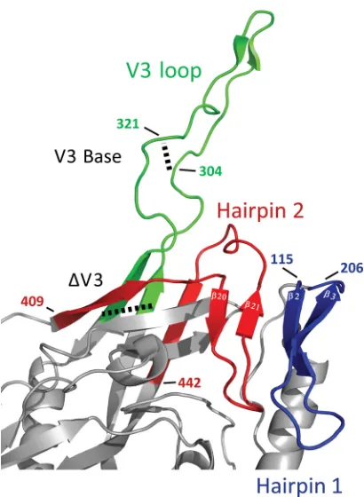
Figure 5
depicts the
spa-tial relationship between the V3 loop and the bridging sheet
minidomain comprised of hairpin 1 (the

2-

3 excursion from
the gp120 inner domain) and hairpin 2 (the
20-21 excursion
from the gp120 outer domain).
The three variations of V3 loop structures (depicted in
Fig. 3A
)
were examined with regard to their impact on the binding of the
CD4i MAb panel. As is shown in
Fig. 6
, total removal of the V3
loop, including its base (
⌬
V3), rendered MAbs 17b and 21c
abso-lutely stringent for CD4 (an effect previously demonstrated [
23
,
75
]). Relaxed binding (no requirement for CD4) could be
gained provided that the base of V3 was kept in place. Thus,
re-moval of the V3 loop in its entirety has a profound negative effect
on 17b and 21c binding that can be overcome by maintaining even
a few residues of the base of the V3 loop. MAbs CG10, 19e, and
N12-i15 continued to demonstrate stringent binding to all the V3
loop-truncated gp120 constructs.
Next, we tested the effects of systematic truncations in the
re-gion of the V1 and V2 loops and the base of these loops, hairpin 1,
on CD4i MAb binding (constructs depicted in
Fig. 3B
and
4
).
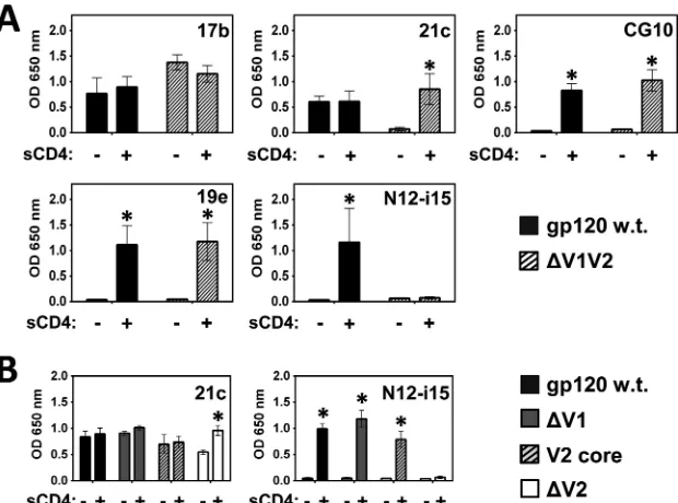
Removal of the V1 and V2 loops (
⌬
V1V2 mutant), while
main-taining an intact V3 loop, did not affect binding by MAbs 17b,
CG10, and 19e, while MAb 21c was rendered stringent for CD4,
and binding of N12-i15 was irreversibly lost (
Fig. 7A
). Hence, it is
clear that at least one of these variable loops impacts the binding of
MAbs 21c and N12-i15.
Selective removal of the V1 versus the V2 loop reveals that the
V1 loop plays no or little role in MAb 21c or N12-i15 binding, in
contrast to the requirement for a V2 loop (
Fig. 7B
) (
23
). This
conclusion can be refined further by showing that a gp120 mutant
that retains at least the V2 core residues (V2 core mutant described
in the legend of
Fig. 4
and ELISA results shown in
Fig. 7B
) binds
MAbs 21c and N12-i15 at wt levels. These core V2 loop residues
were found to adopt a Greek key structure when the V1 and V2
loops were expressed on a protein scaffold and cocrystallized with
a broadly cross-neutralizing MAb (
78
) and again when trimeric
BG505 SOSIP.664 gp140 was crystallized in the absence of sCD4
(
29–31
) and are sufficient to allow wt-level binding.
In addition, by comparing the V2 loop sequences of strains
BaL, 451, YU2, and JR-FL, which are stringently bound by MAb
N12-i15, with the V2 loop sequence of strain R2, which is barely
recognized by MAb N12-i15, we gain some indication as to which
residues within the Greek key may be critical for N12-i15 binding.
Figure 8
shows the sequence alignment of the different V2 loops,
with the four residues that differ in strain R2 within the Greek key
structure marked. These differences may explain the reduced
binding of N12-i15 to R2 compared to the other clade B isolates
(
Fig. 1
).
Finally, we asked whether the CD4i MAbs actually require an
intact four-stranded bridging sheet. For this, we removed hairpin
1 (the stem of the V1/V2 loops) altogether, thus producing the
⌬H1 mutant. The
⌬H1 mutant was combined with the
above-FIG 3Schematic of the wt and modified forms of the gp120 V1/V2 and V3 loops. The sequences of wt and modified V3 (A) and V1/V2 (B) loops are shown with disulfide bonds depicted as black lines. Amino acid numbering corresponds to BaL gp120. Landmark cysteine residues are numbered for con-venience. wt sequences are shown in black, and altered sequences are shown in gray. (A) V3 loop mutants, including (i) the wt V3 loop, (ii) V3 base (gp120 with a partially truncated V3 loop), and (iii)⌬V3 (gp120 with a truncated V3 loop). (B) V1/V2 loop mutants, including (i) wt V1/V2 loops, (ii)⌬V1 (gp120 with a truncated V1 loop), (iii)⌬V2 (gp120 with a truncated V2 loop), (iv) ⌬V1V2 (gp120 with truncated V1/V2 loops), and (v)⌬H1 (gp120 with trun-cated V1/V2 loops and stem).
on November 7, 2019 by guest
http://jvi.asm.org/
[image:5.585.60.266.63.417.2]described V3 loop base and
⌬V3 truncations (both the
⌬H1 and
⌬
V3 truncations are shown in
Fig. 3
). MAbs 21c, CG10, 19e, and
N12-i15 showed no binding to the
⌬H1 constructs, even in the
presence of CD4 (
Fig. 9
). Surprisingly, however, MAb 17b bound
the
⌬H1 construct although with a strict requirement for CD4.
FIG 4Comparison of wt V1/V2 loops and the V2 core mutant. The crystal structure of the V1/V2 loops from gp120 of strain ZM109 reveals that the V2 loop assumes a Greek key fold (PDB accession number3U2S) (78). The Greek key fold is comprised of four antiparallel-strands (designated a to d) connected by the V1 loop (dashed) and two small loops (designated L1 and L2), which are part of the V2 loop (black). Based upon this crystal structure, the V2 core mutant was constructed by using gp120 from the BaL strain (the same strain upon which all of the envelope mutants in this study are based). The V2 core mutant was constructed by removing most of the V1 loop and replacing the parts of the V2 loop that do not participate in the Greek fold (loop L2) with a Ser-Gly linker. Numbering refers to adjacent cysteine residues and is based on the BaL strain, with disulfide bonds shown as black bars. Shown are the V1 loop (dashed), the V2 loop (black), and disulfide bonds (black bars between cysteine residues). (A) Linearized schematic of the wt V1/V2 loop fold. (B) Schematic of the fold of the wt V1/V2 loops. (C) Schematic of the fold of the V2 core mutant. (D) Sequence of the V2 core mutant.
FIG 5Orientation of the bridging sheet hairpins in relation to the V3 loop. The hairpins that constitute the bridging sheet (hairpins 1 and 2, shown in blue and red, respectively) and the V3 loop (green) are shown in the CD4-liganded conformation (PDB accession number2B4C). Note that hairpin 1 (2-3) is the base for the V1/V2 loops (the V1/V2 loops are missing in the crystal struc-ture) and that the V3 loop protrudes between the proximal and distal aspects of hairpin 2 (20-21). The locations of the different V3 loop truncations (V3 base and⌬V3) are marked with dashed lines. For the sake of consistency with our mutants, the numbering is based on BaL gp120 (see Materials and Methods).
FIG 6V3 loop modifications affect binding by relaxed CD4i MAbs. A binding ELISA was carried out to ascertain the effects of V3 loop truncations on binding by the CD4i MAb panel to gp120BaL. Full truncation of the V3 loop (⌬V3) results in stringent binding by MAbs 17b and 21c, an effect not seen for the V3 base mutant. Binding by the stringent CD4i MAbs CG10, 19e, and N12-i15 was not affected by V3 loop truncations. Statistically significant differences (P⬍0.05) between the “⫺sCD4” and “⫹sCD4” columns are marked with an asterisk.
on November 7, 2019 by guest
http://jvi.asm.org/
[image:6.585.133.453.65.223.2] [image:6.585.61.266.349.629.2] [image:6.585.300.544.389.657.2]This contrasts with a previous report using a mutant very similar
to
⌬H1 in which MAb 17b failed to bind in a pulldown experiment
(
75
). The binding of MAb 17b to the
⌬
H1 mutants persisted,
provided that at least the base of the V3 loop was maintained. No
recovery of binding could be found for 17b in the
⌬
H1-
⌬
V3
con-struct, probably as a result of the lack of binding to CD4. Thus,
MAb 17b is indeed a CD4i MAb. However, its association with
gp120 does not indicate a functional bridging sheet but rather
hairpin 2 in a conformation that allows CD4 binding and MAb
recognition. A summary of the binding activities of the CD4i
MAbs tested is given in
Table 2
.
Binding activities of purified monomeric gp120 and its
mu-tations.
The formation of oligomeric forms of gp120 in
trans-fected HEK 293T cells due to aberrant disulfide bridges is well
documented (
55
,
58
,
79–81
). Oligomerization can result
specifi-cally from rearranged disulfides in the V2 loop region. Hence, we
tested the CD4 dependence of binding of MAbs 17b, CG10, and
N12-i15 that define CD4i epitopes associated with hairpin 2, the
bridging sheet, and the V2 loop, respectively, using FPLC-purified
monomeric gp120 in addition to 4 relevant mutations. As shown
in
Fig. 10
, MAb 17b binds to wt BaL purified monomeric gp120 in
the absence of CD4 as do the
⌬V2 and
⌬V1V2 truncations, with
little improvement in binding with the addition of CD4. However,
as illustrated in
Fig. 9
, the purified monomer of
⌬H1 binds 17b in
a stringent CD4-dependent manner. The bridging sheet defining
stringent MAb CG10 is totally dependent on the presence of CD4
for binding purified monomeric wt gp120 or the
⌬
V1V2 and
⌬
V2
truncations. The addition of CD4 to the
⌬H1 monomer does not
support CG10 recognition. Finally MAb N12-i15 is also
strin-gently dependent on CD4 binding for recognition of WT
mono-meric gp120; however, no binding is demonstrated for any of the
truncations missing the V2 loop, as illustrated in
Fig. 7
and
9
.
Extended core gp120s do not present stringent CD4i
epitopes.
The coree
constructs produced and characterized
previ-ously by Kwon et al. (
24
) have been proposed to represent the
CD4-bound conformation. These constructs contain most of the
N and C termini and only the V3 loop base, and hairpin 1 is
truncated just below the cysteine disulfide (C122-C197), thus
re-placing the V1 and V2 loops with a short Gly-Gly linker. We
ex-pressed three representative core
estructures, clade B YU2 core
e,
clade C C1086 coree, and clade E 93TH057 coree
(kindly provided
by Y. D. Kwon and P. D. Kwong), and tested their binding to the
five CD4i MAbs of the panel, in the presence and absence of CD4,
the logic being that if the coree
gp120s indeed represent the
ulti-mate CD4-bound conformation, CD4i MAbs CG10 and 19e
should bind them in the absence of sCD4. As shown in
Fig. 11A
,
MAb 17b binds wt gp120 and the coree
proteins equally well in the
FIG 7CD4i MAbs 21c and N12-i15 are sensitive to V1/V2 loop modifications. A binding ELISA was performed to test whether binding by the CD4i MAbs to gp120BaLis affected by the complete truncation of the V1/V2 loops (⌬V1V2) (A) or selective modification of the V1 or V2 loop (B). (A) MAb 21c becomes stringent and MAb N12-i15 loses all binding when the V1/V2 loops are fully truncated. (B) Selective modifications within the V1/V2 loops indicate that the V1 loop is not necessary for binding by MAbs 21c and N12-i15, but the core of the V2 loop (the V2 loop residues which fold into a Greek key conformation) (Fig. 4) is required. Statistically significant differences (P⬍0.05) between the “⫺sCD4” and “⫹sCD4” columns are marked with an asterisk.
FIG 8Sequence alignment of the gp120 V2 loop from different HIV strains. The gp120 V2 loop sequences from the different HIV strains tested against MAb N12-i15 for binding were aligned by using the Clustal Omega program (http://www.ebi.ac.uk/Tools/msa/clustalo/) and compared. The numbering system used is based on the BaL strain for consistency. Strand c of the Greek key fold based on the crystal structure of V1/V2 loops from the ZM109 strain (78) is shown. As MAb N12-i15 was shown to bind to elements within the Greek key fold of the V2 loop, the four residues different in the R2 V2 loop located within the Greek key fold are highlighted in dark gray. The V2 loop of the BG505 strain (both the monomer and the BG505 SOSIP trimer) is shown for comparison only, as MAb N12-i15 does not bind this strain (Fig. 1).
on November 7, 2019 by guest
http://jvi.asm.org/
[image:7.585.138.448.66.296.2] [image:7.585.39.286.558.623.2]presence and absence of CD4 (with slightly lower binding to
93TH057 core
e). CD4i MAbs 21c and 19e showed no binding,
even in the presence of CD4, to any of the core
econstructs,
possi-bly as a result of the truncations introduced into the tip of hairpin
1 of the bridging sheet in the construction of the core
eproteins
(
24
). As expected, no binding was detected for MAb N12-i15, as
the V1-V2 loops are missing. However, MAb CG10 bound to the
core
econstructs in a strictly CD4-dependent manner. This assay
was repeated with the monomeric, FPLC-purified YU2 core
epro-tein, with similar results (
Fig. 11B
).
DISCUSSION
HIV mediates target cell infection through binding of 2 to 3 spikes
to cell surface CD4 (
82
). This constitutes the first step in the
dy-namic process in which structural elements of the envelope shift
and rearrange to gain function, ultimately leading to
gp41-medi-ated membrane fusion and the introduction of the viral
nucleo-capsid into the cytoplasm of target cells (
83
). A key player in this
process is gp120, whose outer domain directly binds CD4 and
undergoes a range of conformational transitions. One way to
in-vestigate such dynamic processes is to use conditional probes,
probes that can discriminate between the various transition states
of a given target. The range of CD4i conformational transitions
within gp120 was therefore systematically interrogated by using a
panel of well-characterized conditional probes, the CD4i MAbs,
and led to a number of conclusions, as discussed below.
MAb 17b has often been taken as a gold standard indicator for
the CD4-bound conformation (
11
,
24
,
39
,
62
,
69–76
). This MAb
was originally described by Thali et al., who recognized that native
cell surface HIV-1 spike was absolutely dependent on CD4 for 17b
[image:8.585.41.287.65.393.2]FIG 9MAb 17b binds gp120 in the absence of bridging sheet hairpin 1 in a CD4-dependent manner. A binding ELISA was performed to test whether the CD4i MAbs and sCD4 bind wt gp120BaLor the following bridging sheet hair-pin 1 gp120BaLmutants (truncation sequences shown inFig. 3):⌬H1 (gp120 with truncated hairpin 1),⌬H1-V3 base (gp120 with truncated hairpin 1 and a partially truncated V3 loop), and⌬H1-⌬V3 (gp120 with truncated hairpin 1 and a fully truncated V3 loop). Only MAb 17b showed binding to the⌬H1 mutant and only in the presence of sCD4, provided that at least the base of the V3 loop was retained. Note that as sCD4 levels were detected by using a bio-tinylated anti-CD4 MAb and HRP-conjugated streptavidin (see Materials and Methods), a slightly different OD at 650 nm scale (yaxis) is used. Although ELISAs are semiquantitative, the strict requirement for CD4 binding is con-siderable and statistically significant (P⬍0.05), as indicated by an asterisk.
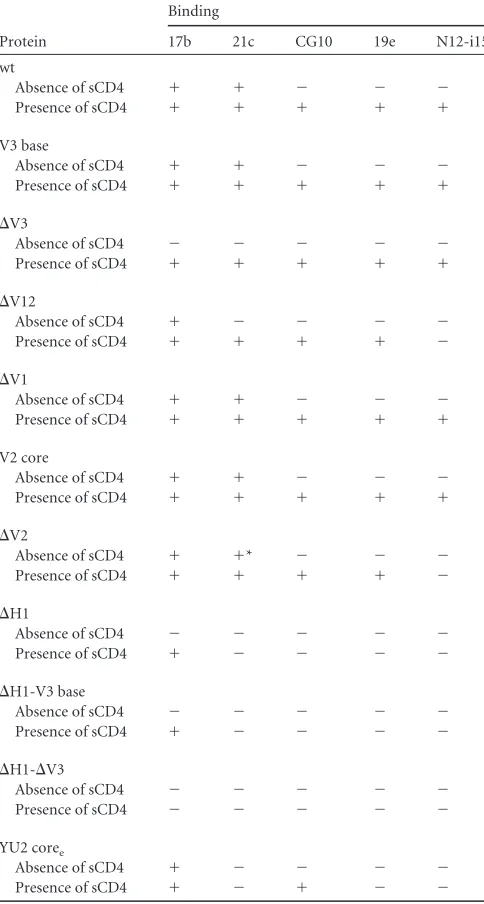
TABLE 2Summary of CD4i MAb binding to different gp120 mutantsa
Protein
Binding
17b 21c CG10 19e N12-i15
wt
Absence of sCD4 ⫹ ⫹ ⫺ ⫺ ⫺
Presence of sCD4 ⫹ ⫹ ⫹ ⫹ ⫹
V3 base
Absence of sCD4 ⫹ ⫹ ⫺ ⫺ ⫺
Presence of sCD4 ⫹ ⫹ ⫹ ⫹ ⫹
⌬V3
Absence of sCD4 ⫺ ⫺ ⫺ ⫺ ⫺
Presence of sCD4 ⫹ ⫹ ⫹ ⫹ ⫹
⌬V12
Absence of sCD4 ⫹ ⫺ ⫺ ⫺ ⫺
Presence of sCD4 ⫹ ⫹ ⫹ ⫹ ⫺
⌬V1
Absence of sCD4 ⫹ ⫹ ⫺ ⫺ ⫺
Presence of sCD4 ⫹ ⫹ ⫹ ⫹ ⫹
V2 core
Absence of sCD4 ⫹ ⫹ ⫺ ⫺ ⫺
Presence of sCD4 ⫹ ⫹ ⫹ ⫹ ⫹
⌬V2
Absence of sCD4 ⫹ ⫹* ⫺ ⫺ ⫺
Presence of sCD4 ⫹ ⫹ ⫹ ⫹ ⫺
⌬H1
Absence of sCD4 ⫺ ⫺ ⫺ ⫺ ⫺
Presence of sCD4 ⫹ ⫺ ⫺ ⫺ ⫺
⌬H1-V3 base
Absence of sCD4 ⫺ ⫺ ⫺ ⫺ ⫺
Presence of sCD4 ⫹ ⫺ ⫺ ⫺ ⫺
⌬H1-⌬V3
Absence of sCD4 ⫺ ⫺ ⫺ ⫺ ⫺
Presence of sCD4 ⫺ ⫺ ⫺ ⫺ ⫺
YU2 coree
Absence of sCD4 ⫹ ⫺ ⫺ ⫺ ⫺
Presence of sCD4 ⫹ ⫺ ⫹ ⫺ ⫺
a
Results for binding in the absence or presence of sCD4 are shown. The asterisk denotes binding at⬃50% of binding to wt gp120.
on November 7, 2019 by guest
http://jvi.asm.org/
[image:8.585.299.541.255.707.2]binding, in contrast, however, to the unrestricted ability of this
MAb to bind monomeric gp120 in the absence of CD4 (
38
).
Here we report that MAb 17b binds the
⌬
H1 mutant
com-plexed with CD4. This is surprising, as hairpin 1 constitutes half of
the bridging sheet and contributes 8 out of 18 contact residues for
17b (with the other 10 contact residues residing at the base of
hairpin 2 [
18
]). Nonetheless, gp120 devoid of hairpin 1 binds
MAb 17b robustly albeit in a strictly CD4-dependent manner.
These results point to the unique role and conformation of
hair-pin 2 (discussed below). This structural feature assumes the
re-quired conformation recognized by MAb 17b independent of
CD4 binding when the gp120 monomer dissociates from the
tri-meric spike. Furthermore, two conditions under which 17b
bind-ing to monomeric gp120 becomes strbind-ingently dependent on CD4
binding have been found: (i) truncation of the V3 loop base and
(ii) removal of hairpin 1. In gp120, the
2 strand of hairpin 1
aligns with the descending flank of hairpin 2 (

21), forming a
series of hydrogen bonds. When this stabilizing interaction with
hairpin 1 is maintained, CD4 binding can compensate for the
absence of a V3 loop base. Removal of hairpin 1 can be
compen-sated for by CD4 binding so long as at least the V3 loop base is in
place.
What makes the V3 loop base so critical for 17b binding? This
might be explained by the fact that the V3 loop naturally splays the
two strands at the base of hairpin 2. Without this splaying effect,
the overall orientation of hairpin 2 becomes distorted and
unrec-ognized by 17b. In the absence of the V3 loop base, CD4 still binds
gp120 and restores the required conformation of hairpin 2 (
18
),
thus gaining 17b recognition. This hypothesis is supported by
Zhou and collaborators, who report that tethering of the bridging
sheet hairpin 2 to the inner domain, in approximation of the CD4i
conformation, reduced the entropy of interactions with CD4 by
60% and increased the on-rate and affinity of MAb 17b in the
absence of CD4 (
84
). In conclusion, a critical aspect for a
CD4-bound conformation is a functional hairpin 2, a feature that is
measured by 17b recognition.
The physical juxtaposition of hairpins 1 and 2, via the
align-ment of

2 with

21, is the essence of a bridging sheet, a feature
that can be detected by MAb 21c. In contrast to 17b, MAb 21c
requires intimate contacts with the extended aspects of hairpin 1
in addition to residues of hairpin 2. The
⌬H1 mutation lacks these
essential elements of the 21c epitope, which obviously cannot be
restored by CD4; hence, no 21c binding is measured.
Critical evaluation of the essential elements of the V2 loop and
how they are involved in CD4i transitions of gp120 was
accom-plished by studying MAb N12-i15. The stringent dependence of
N12-i15 binding on CD4 and the V2 loop delineates a second
FIG 10Binding of MAbs 17b, CG10, and N12-i15 to FPLC-purified mo-nomeric gp120. The gp120 monomers of the truncations described in the legends ofFig. 3B,7, and9were purified by FPLC and tested by an ELISA for MAb binding in the presence and absence of CD4, as indicated. Binding to purified monomeric full-length BaL gp120 is given for comparison. Patterns of binding to monomeric gp120 were identical to the patterns of binding to mixed monomeric and oligomeric preparations. Statistically significant differences (P⬍0.05) between the “⫺sCD4” and “⫹sCD4” columns are marked with an asterisk.
FIG 11MAb CG10 retains stringent binding to coreegp120s. A binding ELISA was performed to test whether the CD4i MAbs bind the different coree gp120s. (A) MAb 17b binds all of the coreegp120s with or without sCD4, while MAbs 21c and 19e do not bind coreegp120s at all. MAb CG10 retained strin-gent binding to the coreegp120s. (B) A binding ELISA was performed to test whether monomeric, FPLC-purified YU2 coreeprotein bound the CD4i MAbs similarly to the mixed monomeric and oligomeric preparations shown in panel A. Binding to purified monomeric full-length BaL gp120 is given for comparison. Patterns of binding to the CD4i MAbs tested persisted. Statisti-cally significant differences (P⬍0.05) between the “⫺sCD4” and “⫹sCD4” columns are marked with an asterisk.
on November 7, 2019 by guest
http://jvi.asm.org/
[image:9.585.42.287.64.223.2] [image:9.585.301.539.69.477.2]measureable aspect of the CD4i conformation of gp120. The lack
of N12-i15 competition against the other MAbs of the described
panel emphasizes the uniqueness of this structural feature of the
CD4-bound conformation.
Finally, whereas it has been proposed that the core
estructures
reported by Kwon et al. (
24
) represent the default CD4-bound
conformation of monomeric gp120, our results clearly indicate
that this is not the full picture. The stringent MAb CG10 binds to
core
estructures but only in complex with CD4. This clearly
illus-trates that there are still structural features lacking in core
ethat can
be further induced upon association with CD4. This assertion is
further strengthened by a recent single-molecule fluorescence
res-onance energy transfer (smFRET) study which showed that the
viral spike can undergo transitions between three distinct
confor-mations (
14
). The authors of that study assigned the first of these
to the “closed” conformation and the second to the “CD4-bound”
conformation seen in the tripartite gp120-CD4-17b crystal
struc-tures, while the third conformation could not be assigned a
de-fined structure but was also favored after the addition of CD4.
Interestingly, the viral spikes could sample all three
conforma-tions even when unliganded and continued to fluctuate between
the two CD4-associated conformations after the addition of CD4,
with conformational dynamics being strain specific. This
multi-plicity of conformations associated with binding by CD4 clearly
shows that more work is needed to clarify their exact structural
nature. Here, our analyses of the CD4i stringent MAbs may
pro-vide specific probes to better understand and scrutinize various
gp120 transitional states.
Taking these results together, along with the crystal
struc-tures of CD4-unliganded trimeric gp140 and the CD4-bound
gp120 core, we postulate the following sequence of
conforma-tional transitions that are triggered upon CD4 binding
(illus-trated in
Fig. 12
):
1. Insertion of CD4 into its binding pocket in gp120 drives
residue Phe43 of CD4 forward, pushing the tip of hairpin 2
inwards and downwards (this transition brings the tip of
hairpin 2 closer to the base of the V3 loop by 5 Å, as shown
in
Fig. 12
).
2. The shift and reorientation of strand
21 disrupt hydrogen
bonding with strand

3.
3. This allows hairpin 1 to fully extend, flipping the V1/V2
loops outward.
4. Repositioning of the V1/V2 loops involves twisting of
hair-pin 1, thereby displacing
3 in favor of
2, which can then
form hydrogen bonds with

21, thus stabilizing a complete
bridging sheet.
5. The newly formed bridging sheet with the central

2-

21
antiparallel orientation becomes exposed and accessible for
coreceptor binding.
Obviously, more defining MAbs combined with structural
studies will further contribute to our understanding of these
events and possibly provide insights in the quest for an AIDS
vac-cine. Despite many years of intense research, we are still far from
achieving this goal. There seems to be increasing support for the
idea that immunogens that are as close as possible to the natural
trimeric conformation of HIV-1 envelope will provide a more
relevant and hopefully potent vaccine (
85
,
86
). Understanding the
atomic details of envelope structures in the bound and free states
is definitely important and extremely useful. Simpler
epitope-based vaccines, accentuating neutralizing epitopes, may prove to
be an efficient alternative for vaccine design. A better
understand-ing of the dynamics of HIV-1 envelope interactions with its
recep-tors, and in particular those of monomeric gp120, might
contrib-ute to the ultimate task of generating such an epitope-based
vaccine for AIDS (
87
).
ACKNOWLEDGMENTS
This research was supported by grants from the Israel Science Foundation (J.M.G.), the Bill and Melinda Gates Foundation (grant OPP1033109) (G.K.L.), the National Institutes of Health (grant R01 AI087181) (G.K.L), and the Frankel Foundation and a donation by Peter Kraus. J.M.G. is the incumbent of the David Furman Chair of Immunobiology in Cancer. G.K. is the recipient of the Jakov, Miriana, and Jorge Saia doctoral fellow-ship. A.R.B. received the Dan David doctoral fellowfellow-ship.
We acknowledge Yongjun Guan, James Robinson, and Ron Diskin for
FIG 12Comparison of the gp120 bridging sheet in the CD4-unliganded and CD4-bound conformations. The structures of the bridging sheet elements in the crystal of CD4-unliganded trimer (PDB accession number4NCO) (A) and the crystal of CD4-bound gp120 core (PDB accession number1G9M) (B) were compared. Hairpin 1 (2 [blue]-3 [gray]), hairpin 2 (20-21 [green]), and the orientation of the V1/V2 loops are indicated (V1/V2 loops are not shown in the unliganded crystal and are truncated in the CD4-bound crystal). Residue Phe43 of CD4 is also shown (schematically in the unliganded crystal). The distance between carbon␣of Trp422 in the tip of hairpin 2 and carbon␣of Cys297 at the base of the V3 loop is also indicated (the cysteine residues at the base of the V3 loop are shown in space-fill for orientation). (A) In the unligan-ded crystal,3 of hairpin 1 forms hydrogen bonds with21 of hairpin 2, and the V1/V2 loops are oriented toward the trimer apex. (B) After binding to CD4, hairpin 1 twists and flips. Now,2 of hairpin 1 forms hydrogen bonds with21 of hairpin 2, and the V1/V2 loops are oriented away from the trimer apex. In addition, the distance between the tip of hairpin 2 and the base of the V3 loop is decreased by 5 Å.
on November 7, 2019 by guest
http://jvi.asm.org/
[image:10.585.315.523.66.346.2]provision of antibodies; John Moore for providing the BG505 monomer and the BG505 SOSIP trimer; Young Do Kwon and Peter D. Kwong for providing the gp120 cores; and Itai Benhar and Limor Nahary for their assistance.
FUNDING INFORMATION
This work, including the efforts of George K. Lewis, was funded by HHS | National Institutes of Health (NIH) (R01 AI087181). This work, includ-ing the efforts of George K. Lewis, was funded by Bill and Melinda Gates Foundation. This work, including the efforts of Gilad Kaplan, Anna Roit-burd-Berman, and Jonathan M. Gershoni, was funded by Israel Science Foundation (ISF). This work, including the efforts of Gilad Kaplan, Anna Roitburd-Berman, and Jonathan M. Gershoni, was funded by Frankel Family Foundation.
REFERENCES
1.Klatzmann D, Champagne E, Chamaret S, Gruest J, Guetard D, Her-cend T, Gluckman JC, Montagnier L.1984. T-lymphocyte T4 molecule behaves as the receptor for human retrovirus LAV. Nature312:767–768. http://dx.doi.org/10.1038/312767a0.
2.McDougal JS, Mawle A, Cort SP, Nicholson JK, Cross GD, Scheppler-Campbell JA, Hicks D, Sligh J.1985. Cellular tropism of the human retrovirus HTLV-III/LAV. I. Role of T cell activation and expression of the T4 antigen. J Immunol135:3151–3162.
3.Dalgleish AG, Beverley PC, Clapham PR, Crawford DH, Greaves MF, Weiss RA.1984. The CD4 (T4) antigen is an essential component of the receptor for the AIDS retrovirus. Nature312:763–767.http://dx.doi.org /10.1038/312763a0.
4.Feng Y, Broder CC, Kennedy PE, Berger EA. 1996. HIV-1 entry cofactor: functional cDNA cloning of a seven-transmembrane, G pro-tein-coupled receptor. Science 272:872– 877. http://dx.doi.org/10 .1126/science.272.5263.872.
5.Alkhatib G, Combadiere C, Broder CC, Feng Y, Kennedy PE, Murphy PM, Berger EA.1996. CC CKR5: a RANTES, MIP-1alpha, MIP-1beta receptor as a fusion cofactor for macrophage-tropic HIV-1. Science272:
1955–1958.http://dx.doi.org/10.1126/science.272.5270.1955.
6.Choe H, Farzan M, Sun Y, Sullivan N, Rollins B, Ponath PD, Wu L, Mackay CR, LaRosa G, Newman W, Gerard N, Gerard C, Sodroski J.
1996. The beta-chemokine receptors CCR3 and CCR5 facilitate infection by primary HIV-1 isolates. Cell85:1135–1148.http://dx.doi.org/10.1016 /S0092-8674(00)81313-6.
7.Deng H, Liu R, Ellmeier W, Choe S, Unutmaz D, Burkhart M, Di Marzio P, Marmon S, Sutton RE, Hill CM, Davis CB, Peiper SC, Schall TJ, Littman DR, Landau NR.1996. Identification of a major co-receptor for primary isolates of HIV-1. Nature381:661– 666.http://dx.doi.org/10 .1038/381661a0.
8.Doranz BJ, Rucker J, Yi Y, Smyth RJ, Samson M, Peiper SC, Parmentier M, Collman RG, Doms RW.1996. A dual-tropic primary HIV-1 isolate that uses fusin and the beta-chemokine receptors CKR-5, CKR-3, and CKR-2b as fusion cofactors. Cell85:1149 –1158.http://dx.doi.org/10.1016 /S0092-8674(00)81314-8.
9.Dragic T, Litwin V, Allaway GP, Martin SR, Huang Y, Nagashima KA, Cayanan C, Maddon PJ, Koup RA, Moore JP, Paxton WA.1996. HIV-1 entry into CD4⫹cells is mediated by the chemokine receptor CC-CKR-5. Nature381:667– 673.http://dx.doi.org/10.1038/381667a0.
10. Wu L, Gerard NP, Wyatt R, Choe H, Parolin C, Ruffing N, Borsetti A, Cardoso AA, Desjardin E, Newman W, Gerard C, Sodroski J.1996. CD4-induced interaction of primary HIV-1 gp120 glycoproteins with the chemokine receptor CCR-5. Nature384:179 –183.http://dx.doi.org/10 .1038/384179a0.
11. Dey B, Pancera M, Svehla K, Shu Y, Xiang SH, Vainshtein J, Li Y, Sodroski J, Kwong PD, Mascola JR, Wyatt R.2007. Characterization of human immunodeficiency virus type 1 monomeric and trimeric gp120 glycoproteins stabilized in the CD4-bound state: antigenicity, biophysics, and immunogenicity. J Virol81:5579 –5593.http://dx.doi.org/10.1128 /JVI.02500-06.
12. Dey B, Svehla K, Xu L, Wycuff D, Zhou T, Voss G, Phogat A, Chakrabarti BK, Li Y, Shaw G, Kwong PD, Nabel GJ, Mascola JR, Wyatt RT.2009. Structure-based stabilization of HIV-1 gp120 enhances hu-moral immune responses to the induced co-receptor binding site. PLoS Pathog5:e1000445.http://dx.doi.org/10.1371/journal.ppat.1000445.
13. Kong L, Huang CC, Coales SJ, Molnar KS, Skinner J, Hamuro Y, Kwong PD.2010. Local conformational stability of HIV-1 gp120 in unliganded and CD4-bound states as defined by amide hydrogen/ deuterium exchange. J Virol 84:10311–10321. http://dx.doi.org/10 .1128/JVI.00688-10.
14. Munro JB, Gorman J, Ma X, Zhou Z, Arthos J, Burton DR, Koff WC, Courter JR, Smith AB, III, Kwong PD, Blanchard SC, Mothes W.2014. Conformational dynamics of single HIV-1 envelope trimers on the surface of native virions. Science346:759 –763.http://dx.doi.org/10.1126/science .1254426.
15. Munro JB, Mothes W.2015. Structure and dynamics of the native HIV-1 Env trimer. J Virol 89:5752–5755. http://dx.doi.org/10.1128 /JVI.03187-14.
16. Pancera M, Majeed S, Ban YE, Chen L, Huang CC, Kong L, Kwon YD, Stuckey J, Zhou T, Robinson JE, Schief WR, Sodroski J, Wyatt R, Kwong PD.2010. Structure of HIV-1 gp120 with gp41-interactive region reveals layered envelope architecture and basis of conformational mobil-ity. Proc Natl Acad Sci U S A107:1166 –1171.http://dx.doi.org/10.1073 /pnas.0911004107.
17. Ward AB, Wilson IA.2015. Insights into the trimeric HIV-1 envelope glycoprotein structure. Trends Biochem Sci40:101–107.http://dx.doi.org /10.1016/j.tibs.2014.12.006.
18. Kwong PD, Wyatt R, Robinson J, Sweet RW, Sodroski J, Hendrickson WA.1998. Structure of an HIV gp120 envelope glycoprotein in complex with the CD4 receptor and a neutralizing human antibody. Nature393:
648 – 659.http://dx.doi.org/10.1038/31405.
19. Chen B, Vogan EM, Gong H, Skehel JJ, Wiley DC, Harrison SC.2005. Structure of an unliganded simian immunodeficiency virus gp120 core. Nature433:834 – 841.http://dx.doi.org/10.1038/nature03327.
20. Kwong PD, Wyatt R, Majeed S, Robinson J, Sweet RW, Sodroski J, Hendrickson WA.2000. Structures of HIV-1 gp120 envelope glycopro-teins from laboratory-adapted and primary isolates. Structure8:1329 – 1339.http://dx.doi.org/10.1016/S0969-2126(00)00547-5.
21. Huang CC, Tang M, Zhang MY, Majeed S, Montabana E, Stanfield RL, Dimitrov DS, Korber B, Sodroski J, Wilson IA, Wyatt R, Kwong PD.
2005. Structure of a V3-containing HIV-1 gp120 core. Science310:1025– 1028.http://dx.doi.org/10.1126/science.1118398.
22. Pugach P, Ozorowski G, Cupo A, Ringe R, Yasmeen A, de Val N, Derking R, Kim HJ, Korzun J, Golabek M, de Los Reyes K, Ketas TJ, Julien JP, Burton DR, Wilson IA, Sanders RW, Klasse PJ, Ward AB, Moore JP.2015. A native-like SOSIP.664 trimer based on an HIV-1 sub-type B env gene. J Virol89:3380 –3395.http://dx.doi.org/10.1128/JVI .03473-14.
23. Diskin R, Marcovecchio PM, Bjorkman PJ.2010. Structure of a clade C HIV-1 gp120 bound to CD4 and CD4-induced antibody reveals anti-CD4 polyreactivity. Nat Struct Mol Biol17:608 – 613.http://dx.doi.org/10.1038 /nsmb.1796.
24. Kwon YD, Finzi A, Wu X, Dogo-Isonagie C, Lee LK, Moore LR, Schmidt SD, Stuckey J, Yang Y, Zhou T, Zhu J, Vicic DA, Debnath AK, Shapiro L, Bewley CA, Mascola JR, Sodroski JG, Kwong PD. 2012. Unliganded HIV-1 gp120 core structures assume the CD4-bound confor-mation with regulation by quaternary interactions and variable loops. Proc Natl Acad Sci U S A109:5663–5668.http://dx.doi.org/10.1073/pnas .1112391109.
25. Bartesaghi A, Merk A, Borgnia MJ, Milne JL, Subramaniam S.2013. Prefusion structure of trimeric HIV-1 envelope glycoprotein determined by cryo-electron microscopy. Nat Struct Mol Biol20:1352–1357.http://dx .doi.org/10.1038/nsmb.2711.
26. Berger EA.1997. HIV entry and tropism: the chemokine receptor con-nection. AIDS11(Suppl A):S3–S16.
27. Harris A, Borgnia MJ, Shi D, Bartesaghi A, He H, Pejchal R, Kang YK, Depetris R, Marozsan AJ, Sanders RW, Klasse PJ, Milne JL, Wilson IA, Olson WC, Moore JP, Subramaniam S.2011. Trimeric HIV-1 glycopro-tein gp140 immunogens and native HIV-1 envelope glycoproglycopro-teins display the same closed and open quaternary molecular architectures. Proc Natl Acad Sci U S A 108:11440 –11445. http://dx.doi.org/10.1073/pnas .1101414108.
28. Tran EE, Borgnia MJ, Kuybeda O, Schauder DM, Bartesaghi A, Frank GA, Sapiro G, Milne JL, Subramaniam S.2012. Structural mechanism of trimeric HIV-1 envelope glycoprotein activation. PLoS Pathog
8:e1002797.http://dx.doi.org/10.1371/journal.ppat.1002797.
29. Julien JP, Cupo A, Sok D, Stanfield RL, Lyumkis D, Deller MC, Klasse PJ, Burton DR, Sanders RW, Moore JP, Ward AB, Wilson IA.2013.
on November 7, 2019 by guest
http://jvi.asm.org/
Crystal structure of a soluble cleaved HIV-1 envelope trimer. Science342:
1477–1483.http://dx.doi.org/10.1126/science.1245625.
30. Lyumkis D, Julien JP, de Val N, Cupo A, Potter CS, Klasse PJ, Burton DR, Sanders RW, Moore JP, Carragher B, Wilson IA, Ward AB.2013. Cryo-EM structure of a fully glycosylated soluble cleaved HIV-1 envelope trimer. Science342:1484 –1490.http://dx.doi.org/10 .1126/science.1245627.
31. Pancera M, Zhou T, Druz A, Georgiev IS, Soto C, Gorman J, Huang J, Acharya P, Chuang GY, Ofek G, Stewart-Jones GB, Stuckey J, Bailer RT, Joyce MG, Louder MK, Tumba N, Yang Y, Zhang B, Cohen MS, Haynes BF, Mascola JR, Morris L, Munro JB, Blanchard SC, Mothes W, Connors M, Kwong PD.2014. Structure and immune recognition of trimeric pre-fusion HIV-1 Env. Nature514:455– 461.http://dx.doi.org/10 .1038/nature13808.
32. Julien JP, Lee JH, Ozorowski G, Hua Y, Torrents de la Pena A, de Taeye SW, Nieusma T, Cupo A, Yasmeen A, Golabek M, Pugach P, Klasse PJ, Moore JP, Sanders RW, Ward AB, Wilson IA.2015. Design and struc-ture of two HIV-1 clade C SOSIP.664 trimers that increase the arsenal of native-like Env immunogens. Proc Natl Acad Sci U S A112:11947–11952. http://dx.doi.org/10.1073/pnas.1507793112.
33. DeVico AL, Rahman R, Welch J, Crowley R, Lusso P, Sarngadharan MG, Pal R.1995. Monoclonal antibodies raised against covalently cross-linked complexes of human immunodeficiency virus type 1 gp120 and CD4 receptor identify a novel complex-dependent epitope on gp 120. Virology211:583–588.http://dx.doi.org/10.1006/viro.1995.1441. 34. Ferrari G, Pollara J, Kozink D, Harms T, Drinker M, Freel S, Moody
MA, Alam SM, Tomaras GD, Ochsenbauer C, Kappes JC, Shaw GM, Hoxie JA, Robinson JE, Haynes BF.2011. An HIV-1 gp120 envelope human monoclonal antibody that recognizes a C1 conformational epitope mediates potent antibody-dependent cellular cytotoxicity (ADCC) activ-ity and defines a common ADCC epitope in human HIV-1 serum. J Virol
85:7029 –7036.http://dx.doi.org/10.1128/JVI.00171-11.
35. Gohain N, Tolbert WD, Acharya P, Yu L, Liu T, Zhao P, Orlandi C, Visciano ML, Kamin-Lewis R, Sajadi MM, Martin L, Robinson JE, Kwong PD, DeVico AL, Ray K, Lewis GK, Pazgier M.2015. Cocrystal structures of antibody N60-i3 and antibody JR4 in complex with gp120 define more cluster A epitopes involved in effective antibody-dependent effector function against HIV-1. J Virol89:8840 – 8854.http://dx.doi.org /10.1128/JVI.01232-15.
36. Lewis GK, Finzi A, DeVico AL, Pazgier M.2015. Conformational mask-ing and receptor-dependent unmaskmask-ing of highly conserved Env epitopes recognized by non-neutralizing antibodies that mediate potent ADCC against HIV-1. Viruses7:5115–5132.http://dx.doi.org/10.3390/v7092856. 37. Richard J, Veillette M, Brassard N, Iyer SS, Roger M, Martin L, Pazgier M, Schon A, Freire E, Routy JP, Smith AB, III, Park J, Jones DM, Courter JR, Melillo BN, Kaufmann DE, Hahn BH, Permar SR, Haynes BF, Madani N, Sodroski JG, Finzi A.2015. CD4 mimetics sensitize HIV-1-infected cells to ADCC. Proc Natl Acad Sci U S A112:E2687– E2694.http://dx.doi.org/10.1073/pnas.1506755112.
38. Thali M, Moore JP, Furman C, Charles M, Ho DD, Robinson J, Sodroski J.1993. Characterization of conserved human immunodefi-ciency virus type 1 gp120 neutralization epitopes exposed upon gp120-CD4 binding. J Virol67:3978 –3988.
39. Xiang SH, Doka N, Choudhary RK, Sodroski J, Robinson JE.2002. Characterization of CD4-induced epitopes on the HIV type 1 gp120 en-velope glycoprotein recognized by neutralizing human monoclonal anti-bodies. AIDS Res Hum Retroviruses18:1207–1217.http://dx.doi.org/10 .1089/08892220260387959.
40. Zhang PF, Cham F, Dong M, Choudhary A, Bouma P, Zhang Z, Shao Y, Feng YR, Wang L, Mathy N, Voss G, Broder CC, Quinnan GV, Jr.
2007. Extensively cross-reactive anti-HIV-1 neutralizing antibodies in-duced by gp140 immunization. Proc Natl Acad Sci U S A104:10193– 10198.http://dx.doi.org/10.1073/pnas.0608635104.
41. Guan Y, Pazgier M, Sajadi MM, Kamin-Lewis R, Al-Darmarki S, Flinko R, Lovo E, Wu X, Robinson JE, Seaman MS, Fouts TR, Gallo RC, Devico AL, Lewis GK.2013. Diverse specificity and effector function among human antibodies to HIV-1 envelope glycoprotein epitopes ex-posed by CD4 binding. Proc Natl Acad Sci U S A110:E69 –E78.http://dx .doi.org/10.1073/pnas.1217609110.
42. Lewis GK, Fouts TR, Ibrahim S, Taylor BM, Salkar R, Guan Y, Kamin-Lewis R, Robinson JE, Devico AL.2011. Identification and characteriza-tion of an immunogenic hybrid epitope formed by both HIV gp120 and
human CD4 proteins. J Virol85:13097–13104.http://dx.doi.org/10.1128 /JVI.05072-11.
43. Decker JM, Bibollet-Ruche F, Wei X, Wang S, Levy DN, Wang W, Delaporte E, Peeters M, Derdeyn CA, Allen S, Hunter E, Saag MS, Hoxie JA, Hahn BH, Kwong PD, Robinson JE, Shaw GM.2005. Anti-genic conservation and immunoAnti-genicity of the HIV coreceptor binding site. J Exp Med201:1407–1419.http://dx.doi.org/10.1084/jem.20042510. 44. Denisova G, Stern B, Raviv D, Zwickel J, Smorodinsky NI, Gershoni JM.1996. Humoral immune response to immunocomplexed HIV enve-lope glycoprotein 120. AIDS Res Hum Retroviruses12:901–909.http://dx .doi.org/10.1089/aid.1996.12.901.
45. Enshell-Seijffers D, Denisov D, Groisman B, Smelyanski L, Meyuhas R, Gross G, Denisova G, Gershoni JM.2003. The mapping and reconstitu-tion of a conformareconstitu-tional discontinuous B-cell epitope of HIV-1. J Mol Biol334:87–101.http://dx.doi.org/10.1016/j.jmb.2003.09.002.
46. Gershoni JM, Denisova G, Raviv D, Smorodinsky NI, Buyaner D.1993. HIV binding to its receptor creates specific epitopes for the CD4/gp120 complex. FASEB J7:1185–1187.
47. Mazor Y, Barnea I, Keydar I, Benhar I.2007. Antibody internalization studied using a novel IgG binding toxin fusion. J Immunol Methods321:
41–59.http://dx.doi.org/10.1016/j.jim.2007.01.008.
48. Roitburd-Berman A, Dela G, Kaplan G, Lewis GK, Gershoni JM.2013. Allosteric induction of the CD4-bound conformation of HIV-1 Gp120. Retrovirology10:147.http://dx.doi.org/10.1186/1742-4690-10-147. 49. Gershoni JM, Lapidot M, Zakai N, Loyter A.1986. Protein blot analysis
of virus receptors: identification and characterization of the Sendai virus receptor. Biochim Biophys Acta 856:19 –26. http://dx.doi.org/10.1016 /0005-2736(86)90004-0.
50. Burton DR, Barbas CF, III, Persson MA, Koenig S, Chanock RM, Lerner RA.1991. A large array of human monoclonal antibodies to type 1 human immunodeficiency virus from combinatorial libraries of asymp-tomatic seropositive individuals. Proc Natl Acad Sci U S A88:10134 – 10137.http://dx.doi.org/10.1073/pnas.88.22.10134.
51. Burton DR, Pyati J, Koduri R, Sharp SJ, Thornton GB, Parren PW, Sawyer LS, Hendry RM, Dunlop N, Nara PL, Lamacchia M, Garratty E, Stiehm ES, Bryson YJ, Cao Y, Moore JP, Ho DD, Barbas CF.1994. Efficient neutralization of primary isolates of HIV-1 by a recombinant human monoclonal antibody. Science266:1024 –1027.http://dx.doi.org /10.1126/science.7973652.
52. Zhang MY, Xiao X, Sidorov IA, Choudhry V, Cham F, Zhang PF, Bouma P, Zwick M, Choudhary A, Montefiori DC, Broder CC, Burton DR, Quinnan GV, Jr, Dimitrov DS.2004. Identification and character-ization of a new cross-reactive human immunodeficiency virus type 1-neutralizing human monoclonal antibody. J Virol78:9233–9242.http: //dx.doi.org/10.1128/JVI.78.17.9233-9242.2004.
53. Roben P, Moore JP, Thali M, Sodroski J, Barbas CF, III, Burton DR.
1994. Recognition properties of a panel of human recombinant Fab frag-ments to the CD4 binding site of gp120 that show differing abilities to neutralize human immunodeficiency virus type 1. J Virol68:4821– 4828. 54. Center RJ, Earl PL, Lebowitz J, Schuck P, Moss B.2000. The human immunodeficiency virus type 1 gp120 V2 domain mediates gp41-independent intersubunit contacts. J Virol74:4448 – 4455.http://dx.doi .org/10.1128/JVI.74.10.4448-4455.2000.
55. Coutu M, Finzi A.2015. HIV-1 gp120 dimers decrease the overall affinity of gp120 preparations for CD4-induced ligands. J Virol Methods215–216:
37– 44.http://dx.doi.org/10.1016/j.jviromet.2015.02.017.
56. Doms RW, Earl PL, Moss B. 1991. The assembly of the HIV-1 env glycoprotein into dimers and tetramers. Adv Exp Med Biol300:203–219; discussion 220 –201.http://dx.doi.org/10.1007/978-1-4684-5976-0_13. 57. Earl PL, Doms RW, Moss B.1990. Oligomeric structure of the human
immunodeficiency virus type 1 envelope glycoprotein. Proc Natl Acad Sci U S A87:648 – 652.http://dx.doi.org/10.1073/pnas.87.2.648.
58. Finzi A, Pacheco B, Zeng X, Kwon YD, Kwong PD, Sodroski J.2010. Conformational characterization of aberrant disulfide-linked HIV-1 gp120 dimers secreted from overexpressing cells. J Virol Methods168:
155–161.http://dx.doi.org/10.1016/j.jviromet.2010.05.008.
59. Hallenberger S, Tucker SP, Owens RJ, Bernstein HB, Compans RW.
1993. Secretion of a truncated form of the human immuno