R E S E A R C H
Open Access
Identification of immune protective genes
of
Eimeria maxima
through cDNA
expression library screening
XinChao Yang
†, MengHui Li
†, JianHua Liu, YiHong Ji, XiangRui Li, LiXin Xu, RuoFeng Yan and XiaoKai Song
*Abstract
Background:Eimeria maximais one of the most prevalentEimeriaspecies causing avian coccidiosis, and results in huge economic loss to the global poultry industry. Current control strategies, such as anti-coccidial medication and live vaccines have been limited because of their drawbacks. The third generation anticoccidial vaccines including the recombinant vaccines as well as DNA vaccines have been suggested as a promising alternative strategy. To date, only a few protective antigens ofE. maximahave been reported. Hence, there is an urgent need to identify novel protective antigens ofE. maximafor the development of neotype anticoccidial vaccines.
Methods:With the aim of identifying novel protective genes ofE. maxima, a cDNA expression library ofE. maxima sporozoites was constructed using Gateway technology. Subsequently, the cDNA expression library was divided into 15 sub-libraries for cDNA expression library immunization (cDELI) using parasite challenged model in chickens. Protective sub-libraries were selected for the next round of screening until individual protective clones were obtained, which were further sequenced and analyzed.
Results:Adopting the Gateway technology, a high-quality entry library was constructed, containing 9.2 × 10⁶clones with an average inserted fragments length of 1.63 kb. The expression library capacity was 2.32 × 10⁷colony-forming units (cfu) with an average inserted fragments length of 1.64 Kb. The expression library was screened using parasite challenged model in chickens. The screening yielded 6 immune protective genes including four novel protective genes of EmJS-1, EmRP, EmHP-1 and EmHP-2, and two known protective genes of EmSAG and EmCKRS. EmJS-1 is the selR domain-containing protein ofE. maximawhose function is unknown.EmHP-1 and EmHP-2 are the hypothetical proteins ofE. maxima.EmRP and EmSAG are rhomboid-like protein and surface antigen glycoproteins ofE. maximarespectively, and involved in invasion of the parasite.
Conclusions:Our results provide a cDNA expression library for further screening of T cell stimulating or inhibiting antigens ofE. maxima.Moreover, our results provide six candidate protective antigens for developing new vaccines againstE. maxima.
Keywords:Eimeria maxima, cDNA expression library, Immune protective gene
* Correspondence:[email protected]
†Equal contributors
College of Veterinary Medicine, Nanjing Agricultural University, Nanjing, Jiangsu 210095, People’s Republic of China
Background
Avian coccidiosis is the disease caused by protozoan para-sites of the genusEimeria[1, 2].Eimeria maximais one of the seven Eimeria species that infects domestic chickens and results in severe lesions of the small intestine, low effi-ciency of feed utilization and weight loss [3, 4]. The global economic losses due to avian coccidiosis are more than 3 billion US dollars per year [2, 5, 6]. Control strategies against avian coccidiosis still rely heavily on anti-coccidial medication and live vaccines [1, 4]. However, the applica-tion of coccidiostats has been limited because of the rapid emergence of drug resistance and increasing consumer concerns about drug residues in food [7]. Live vaccines have inherent drawbacks such as their limited production, potential reversion to virulence and high cost of manufac-ture [2, 6, 8]. The search for new approaches for coccidiosis control turned towards third generation anticoccidial cines including recombinant vaccines as well as DNA vac-cines [4, 6, 9–13]. To date, only a few protective antigens of E. maximahave been reported. The lack of candidate pro-tective antigens represents a considerable bottleneck in de-veloping new vaccines against this parasite [14–16]. Hence, finding novel protective antigens ofE. maximais urgently required for the future development of univalent vaccines and a multivalent vaccine to protect against infections with multipleEimeriaspecies. With the aim of identifying novel protective genes of E. maxima, in the present study, a cDNA expression library of E. maxima sporozoites was constructed using Gateway technology. Subsequently, the protective genes ofE. maximawere screened using cDELI in parasite challenge model and the biological characters of protective genes were analyzed.
Methods
Vector, parasite and animals
Eukaryotic expression vector pVAX1 was purchased from Invitrogen (Carlsbad, California, USA). Sporulated oocysts ofE. maxima(Jiangsu) were collected 7 days prior to the challenge infection. The purity of E. maxima was deter-mined with ITS1-PCR as described previously [16, 17]. New-hatched Hy-Line layer chickens (commercial breed W-36) were raised in a sterilized room under coccidia-free conditions until the end of the experiment. Food and water without anti-coccidial drugs were available.
RNA isolation for library construction
Sporozoites of E. maximawere purified from sporulated oocysts on DE-52 anion exchange columns using a previ-ously described protocol [18, 19]. Total RNA was ex-tracted fromE. maximasporozoites using E.Z.N.A.® Total RNA Kit Maxi Kit (OMEGA, Norcross, Georgia, USA). Subsequently, mRNA was purified with FastTrack® MAG mRNA Isolation Kits (Invitrogen). The quality of the
isolated total RNA and mRNA were determined by de-naturing agarose gel electrophoresis.
Construction of cDNA entry library ofE. maxima
The cDNA entry library was constructed using Clone-Miner™ II cDNA Library Construction Kit (Invitrogen) following manufacturer’s protocol (see Additional file 1: Protocol for constructing entry library). To evaluate the titer of cDNA entry library, 50 μl of the 1000-fold di-luted library bacteria were cultured overnight at 37 °C on LB plates containing100μg/ml ampicillin. After that, colonies on the plate were counted. The titer was calcu-lated as follows: colony-forming units (cfu)/ml = colonies on plate × dilution factor/volume plated (ml). Total cfu of the library = titer (cfu/ml) × total volume of cDNA li-brary (ml). To determine the insert size of the lili-brary, 24 random clones were amplified by PCR with universal primers (5′-TCC CAG TCA CGA CGT TGT AAA ACG ACG GCC AGT CTT-3′/5′-AGA GCT GCC AGG AAA CAG CTA TGA CCA TGT AAT ACG ACT C-3′) targeting the pDONR222 vector. The PCR program are as following: 95 °C for 5 min with an initial denatur-ation, 35 cycles of 94 °C for 30 s, 58 °C for 30 s and 72 ° C for 2 min, after 35 cycles, 72 °C for 5 min.
Construction of cDNA expression library forE. maxima Prior to the construction of a cDNA expression library for E. maxima, we ligated the recombinant gene attR1-ccdB-attR2 into the expression vector pVAX1 to construct a Gateway system compatible vector pVAX1-DEST. Briefly, the attR1-ccdB-attR2 gene was amplified from the pDEST32 vector (Invitrogen) by PCR using the HindIII/XhoI-flanked primers (F/R 5′-GAC GAC AAG CTT CTG TAT CGT CGA GGT CGA ATC AAA CAA G-3′/5′-TCA TCC TCG AGT ACT TAC TTA GCG GCC ATC AAA CCA C-3′, restriction enzyme of Hin-dIII/XhoI are underlined). The PCR product was digested with restriction enzymes HindIII/XhoI and ligated into the pVAX1 vector to produce Gateway system compatible vector pVAX1-DEST.
Test of the cDNA expression library by PCR amplification of knownE. maximagenes
To test the representativeness of the cDNA expression library, 7 available E. maxima genes in our lab were amplified from the constructed cDNA expression library of E. maxima. Briefly, 10 μl of the cDNA expression library was inoculated into 5 ml of broth culture medium which was grown to an OD600 of 1, before the plasmid DNA was isolated from the culture. With the isolated DNA as template, the E. maxima genes of MIC3-1, MIC3-2, MIC3-3, MIC2, MIC7, MIC5 and AMA1 were amplified by PCR using specific primers (see Additional file 2: Table S1). The PCR products were analysed by 1% agarose gel electrophoresis.
The first round of cDNA expression library screening
Preparation of plasmid DNA for library screening
Ten microlitres of the cDNA expression library was divided into 15 first-level sub-libraries and plasmid DNA was isolated from each of the sub-libraries for screening. Briefly, 10 μl of cDNA expression library was diluted into 3 ml of LB broth and plated onto 15 LB agar plates (each plate representing an individual sub-library) con-taining 50μg/ml kanamycin (200μl of each plate). Next, the plates were incubated at 37 °C for 16–18 h. All resulting clones were then transferred into 150 ml broth culture medium and incubated at 30 °C for 16–18 h. Plasmid DNA was then isolated from each sub-library
using a High Pure Maxi Plasmid Kit (TIANGEN, Beijing, China) following manufacturer’s instructions. Mean-while, the plasmid DNA of empty vector pVAX1 was prepared. The concentration of the plasmid DNA was measured by NanoDrop 2000 spectrophotometer and stored at -20 °C for further use.
Immunization and challenge infection
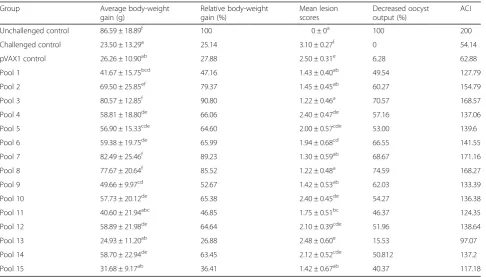
[image:3.595.57.544.447.724.2]At 14 days of age, 360 chickens were weighed individu-ally and randomly distributed into 15 experimental groups and 3 control groups of 20 chickens in each. As shown in Table 1, the experimental groups were the 15 sub-library immunized groups, the 3 control groups were pVAX1 vector control, unchallenged control and challenged control group. Experimental groups were immunized with library plasmid DNA by intramuscular injection in legs at a dose of 100μg. The vector control group was immunized with 100 μg of vector pVAX1, whereas the challenged and unchallenged controls were injected with PBS. A booster immunization was given by the same method as the primary immunization 7 days later. After 1 week of booster immunization, chickens were weighed individually and challenged orally with 1.5 × 10⁵ E. maxima oocysts except the unchallenged control group. The chickens were weighed individually and sacrificed by cervical dislocation 7 days post-chal-lenge. Average body-weight gain, survival rate, decreased oocyst output, lesion score, and anticoccidial index
Table 1Protective efficacy of the 15 sub-libraries in the first round of cDNA expression library screening
Group Average body-weight
gain (g)
Relative body-weight gain (%)
Mean lesion scores
Decreased oocyst output (%)
ACI
Unchallenged control 86.59 ± 18.89f 100 0 ± 0a 100 200
Challenged control 23.50 ± 13.29a 25.14 3.10 ± 0.27f 0 54.14
pVAX1 control 26.26 ± 10.90ab 27.88 2.50 ± 0.31e 6.28 62.88
Pool 1 41.67 ± 15.75bcd 47.16 1.43 ± 0.40ab 49.54 127.79
Pool 2 69.50 ± 25.85ef 79.37 1.45 ± 0.45ab 60.27 154.79
Pool 3 80.57 ± 12.85f 90.80 1.22 ± 0.46a 70.57 168.57
Pool 4 58.81 ± 18.80de 66.06 2.40 ± 0.47de 57.16 137.06
Pool 5 56.90 ± 15.33cde 64.60 2.00 ± 0.57cde 53.00 139.6
Pool 6 59.38 ± 19.75de 65.99 1.94 ± 0.68cd 66.55 141.55
Pool 7 82.49 ± 25.46f 89.23 1.30 ± 0.59ab 68.67 171.16
Pool 8 77.67 ± 20.64f 85.52 1.22 ± 0.48a 74.59 168.27
Pool 9 49.66 ± 9.97cd 52.67 1.42 ± 0.53ab 62.03 133.39
Pool 10 57.73 ± 20.12de 65.38 2.40 ± 0.45de 54.27 136.38
Pool 11 40.60 ± 21.94abc 46.85 1.75 ± 0.51bc 46.37 124.35
Pool 12 58.89 ± 21.98de 64.64 2.10 ± 0.39cde 51.96 138.64
Pool 13 24.93 ± 11.20ab 26.88 2.48 ± 0.60e 15.53 97.07
Pool 14 58.70 ± 22.94de 63.45 2.12 ± 0.52cde 50.812 137.2
Pool 15 31.68 ± 9.17ab 36.41 1.42 ± 0.67ab 40.37 117.18
(ACI) of each group were calculated as described in the evaluation of protection.
Evaluation of protection
Several criteria were employed for evaluating the efficacy of DNA immunization with the expression library including survival rate, lesion score, body-weight gain, decreased oocyst output and ACI. The equations for cal-culating the criteria were shown in Additional file 3: Equations of criteria for evaluating the efficacy of DNA immunization with the expression library. Any sub-li-brary with the ACI≥160 was considered protective [20, 21]. Body-weight gain and lesion scores were expressed as the mean ± standard deviation (SD) and statistical analysis was performed using the SPSS statistical pack-age (IBM SPSS Statistics 19). Differences between groups were tested with the one-way ANOVA Duncan test and were considered significant atP< 0.05.
The further rounds screening of the individual protective clone
The second round screening was performed according to the result of the first round screening. Briefly, the protect-ive first-level sub-library was divided into several second-level sub-libraries. The immunization and challenge experiment was carried out to determine the protective second-level sub-library. The experimental design and efficacy evaluation was same as the first round screening. A third, fourth and even fifth round of screening was performed until the individual protective clone was obtained, following the experimental design and efficacy evaluation described in the first round screening.
DNA sequencing and sequence analysis
The protective clones were sequenced by Invitrogen Com-pany (Shanghai, China). The open reading frame (ORF) of the protective antigen genes was determined with DNAS-TAR software and ORF Finder (https://www.ncbi.nlm.-nih.gov/orffinder/). The ORF and predicted protein sequence of the antigen gene was blasted with NCBI (the National Center for Biotechnology Information, https:// blast.ncbi.nlm.nih.gov/Blast.cgi) and ToxoDB database (www.toxodb.org). The T cell epitope motif and antigen index were predicted using DNASTAR software.
Results
Construction ofE. maximacDNA expression library
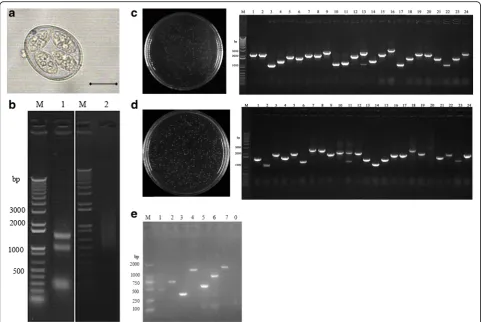
Our study adopted Gateway technology for library con-struction with the modified pVAX1 vector. Figure 1a shows sporulated oocysts of E. maxima. The integrity and purity of total RNA extracted from the sporozoites were detected by 1% denaturing agarose gel electrophor-esis (Fig. 1b, Lane 1) and nucleic acid analyzer (Thermo nanodrop) and purified mRNA were performed for the
same analysis (Fig. 1b, Lane 2). As shown in Fig. 1b (Lane 1), electrophoresis clearly revealed 3 bands of 28S, 18S, and 5S of total RNA. Furthermore, the brightness of 28 s was about 2 times of 18 s, which indicated good integrity of the total RNA (Fig. 1b, Lane 2). The quantity of the total RNA was about 600μg and the A260/A280 value at 1.92, which indicated good purity of the total RNA. The purified mRNA appeared excellent quality with an A260/A280 value of 2.32, whereas the total mRNA quantity was approximately 8.83μg.
The cDNA entry library titer was determined by serial dilution using plating assay. After growing overnight, 230 clones were counted on the plate (Fig. 1c). Accord-ing to the equations, the titer of the plate was calculated as 4.6 × 10⁶ cfu, and the total clones of the entry were 9.2 × 10⁶. Insert size of the library was detected by PCR, and the positive bands were generated from all the ran-domly picked 24 clones. Furthermore, the insert size ranged from 0.9 to 2.8 Kb with an average size of 1.63 Kb (Fig. 1c). These data indicated that the entry library was well represented and could be applied further for the construction of expression library. The cDNA expression library was evaluated in the same way. The results showed that the expression library capacity was 2.32 × 10⁷cfu. The length of insert was ranged from 1 to 3 Kb with an average size of 1.64 Kb (Fig. 1d).
Test of the cDNA expression library by PCR amplification of knownE. maximagenes
To test the representativeness of the cDNA expression library, 7 E. maxima genes with different sizes were amplified from the expression library. As showed in Fig. 1e, PCR revealed 7 bands of 450, 684, 336, 1275, 519, 888 and 1422 bp. The bands were consistent with the sizes ofE. maxima genes MIC3-1, MIC3-2, MIC3-3, MIC2, MIC7, MIC5 and AMA1, respectively.
Screening of cDNA expression library
The first round screening
be selected for the second round screening of protective clones.
The second round screening
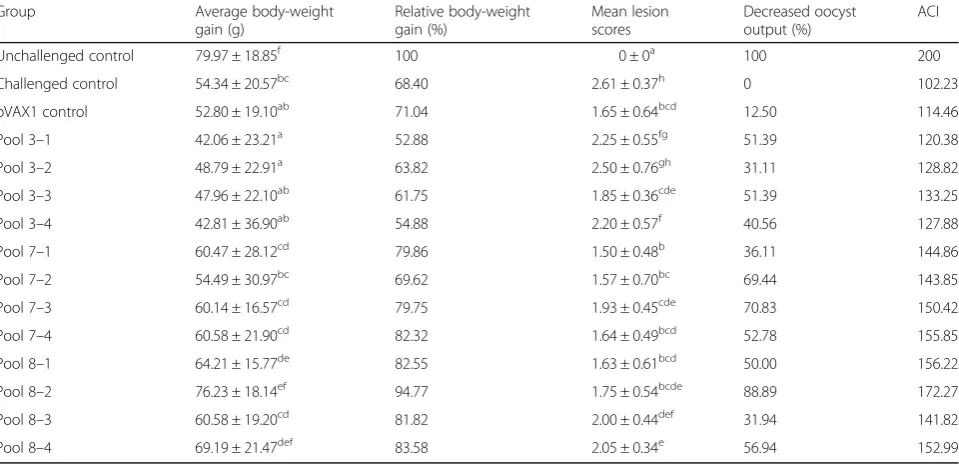
Since pool 3, 7 and 8 were the most protective, emphasis was given to identifying the protective component(s) in these pools. As shown in (Table 2), pool 3, 7 and 8 were partitioned into 4 second-level sub-libraries respectively (designated pool 3–1 to 3–4, pool 7–1 to 7–4, pool 8–1 to 8–4) with 50–75 clones per pool. Animal experiments were performed to compare the protective efficacy of the second-level sub-libraries following the experimental design described as the first round screening. The results were shown in Table 2. Pool 8–2 induced the highest de-creased oocyst output and ACI of 172.27, indicating that it could be selected for the third round screening of protective clones.
The third round screening
The protective pool 8–2 (63 clones) was partitioned into 9 third-level sub-libraries (designated pool 8–2–1 to 8– 2–9) with 7 clones per pool. The results of protective
efficacies of each clone were shown in (Table 3). Pool 8– 2–2 and 8–2–8 induced the ACIs of 164.54 and 163.43, respectively, indicating that pool 8–2–2 and 8–2–8 could be selected for the fourth round screening of pro-tective individual clones.
The fourth round screening
All the individual clones from the two positive pools (pool 8–2–2 and 8–2–8) were designated as clone 8–2– 2–1 to 8–2–2–7 and 8–2–8–1 to 8–2–8–7 respectively (Table 4). The results of protective efficacies of the 14 single clones were shown in Table 4. Six individual clones induced ACIs > 160 namely, 8–2–2–2 (162.97), 8–2–2–5 (167.21), 8–2–8–1 (162.22), 8–2–8–2 (160.02), 8–2–8–3 (160.02) and 8–2–8–6 (160.74).
DNA sequence analysis
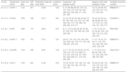
[image:5.595.57.539.87.409.2]100% identity in amino acids with SAG family member of E. maxima (GenBank: CDJ60815.1) and was named as EmSAG. The positive clone 8–2–8–1 shared 91 and 84% identity in amino acids with Eimeria tenella rhomboid-like protein (GenBank: ABC50099.1) and E. maxima rhomboid family domain-containing protein, putative (GenBank: CDJ59262.1) respectively, and was named as EmRP. The positive clone 8–2–8–2 shared 70% identity in amino acids with E. maxima hypo-thetical protein (GenBank: CDJ61108.1) and was named as EmHP-2. The positive clone 8–2–8–3 shared 100% identity in amino acids with Eimeria maxima CAMP-dependent protein kinase regulatory
subunit (GenBank: CDJ61187.1) and was named as EmCKRS. The positive clone 8–2–8–6, named as EmJS-1, shared 79% identity in amino acids with hypothetical protein of Eimeria_necatrix_Houghton (ToxoDB: ENH_00014740) and no identity with the known gene of E. maxima.
[image:6.595.59.539.100.332.2]Characterizations of these 6 protective clones were shown in (Table 5). Complete ORFs were included within the 6 antigen genes separately (Table 5, Additional file 4: Figure S1). Prediction of T cell epitope motif and antigen index revealed that the identified anti-gens are abundant in T cell epitope motifs and regions with high antigenic index (Table 5, Additional file 5: Table 2Protective efficacy of the 12 sub-libraries in the second round of cDNA expression library screening
Group Average body-weight
gain (g)
Relative body-weight gain (%)
Mean lesion scores
Decreased oocyst output (%)
ACI
Unchallenged control 79.97 ± 18.85f 100 0 ± 0a 100 200
Challenged control 54.34 ± 20.57bc 68.40 2.61 ± 0.37h 0 102.23
pVAX1 control 52.80 ± 19.10ab 71.04 1.65 ± 0.64bcd 12.50 114.46
Pool 3–1 42.06 ± 23.21a 52.88 2.25 ± 0.55fg 51.39 120.38
Pool 3–2 48.79 ± 22.91a 63.82 2.50 ± 0.76gh 31.11 128.82
Pool 3–3 47.96 ± 22.10ab 61.75 1.85 ± 0.36cde 51.39 133.25
Pool 3–4 42.81 ± 36.90ab 54.88 2.20 ± 0.57f 40.56 127.88
Pool 7–1 60.47 ± 28.12cd 79.86 1.50 ± 0.48b 36.11 144.86
Pool 7–2 54.49 ± 30.97bc 69.62 1.57 ± 0.70bc 69.44 143.85
Pool 7–3 60.14 ± 16.57cd 79.75 1.93 ± 0.45cde 70.83 150.42
Pool 7–4 60.58 ± 21.90cd 82.32 1.64 ± 0.49bcd 52.78 155.85
Pool 8–1 64.21 ± 15.77de 82.55 1.63 ± 0.61bcd 50.00 156.22
Pool 8–2 76.23 ± 18.14ef 94.77 1.75 ± 0.54bcde 88.89 172.27
Pool 8–3 60.58 ± 19.20cd 81.82 2.00 ± 0.44def 31.94 141.82
Pool 8–4 69.19 ± 21.47def 83.58 2.05 ± 0.34e 56.94 152.99
Note: Significant difference (P< 0.05) between numbers with different letters; non-significant difference (P> 0.05) between numbers with the same letter
Table 3Protective efficacy of the 9 sub-libraries in the thrid round of cDNA expression library screening
Group Average body-weight
gain (g)
Relative body-weight gain (%)
Mean lesion scores
Decreased oocyst output (%)
ACI
Unchallenged control 98.26 ± 13.09e 100 0 ± 0a 100.00 200
Challenged control 37.72 ± 41.47a 40.91 3.05 ± 0.84f 0.00 70.41
pVAX1 control 41.47 ± 27.85ab 43.81 2.60 ± 1.16e 17.95 77.81
Pool 8–2–1 41.98 ± 26.55ab 45.34 2.00 ± 1.19d 75.64 120.34
Pool 8–2–2 79.85 ± 40.00d 84.74 1.02 ± 1.25b 61.54 164.54
Pool 8–2–3 54.62 ± 42.72bc 59.28 2.12 ± 1.17d 65.38 128.08
Pool 8–2–4 44.65 ± 30.68ab 46.35 2.30 ± 1.12de 71.79 113.35
Pool 8–2–5 43.87 ± 28.02ab 47.15 1.37 ± 1.07bc 43.59 113.45
Pool 8–2–6 51.47 ± 25.97bc 55.58 2.10 ± 1.29d 62.82 124.58
Pool 8–2–7 55.07 ± 26.63c 56.80 2.32 ± 1.09de 64.10 123.6
Pool 8–2–8 73.93 ± 30.41d 82.93 1.45 ± 1.35c 84.62 163.43
Pool 8–2–9 45.56 ± 38.25abc 50.20 2.55 ± 0.95e 62.82 114.7
[image:6.595.58.540.532.724.2]Table 4Protective efficacy of the 14 single clones in the fourth round of cDNA expression library screening
Group Average body-weight
gain (g)
Relative body-weight gain (%)
Mean lesion scores
Decreased oocyst output (%)
ACI
Unchallenged control 57.61 ± 9.47f 100 0 ± 0a 100.00 200
Challenged control 36.89 ± 11.89ab 63.68 2.63 ± 0.63fg 0.00 97.30
pVAX1 control 37.42 ± 6.82abc 60.29 1.80 ± 0.38cdef 8.91 102.24
Clone 8–2–2–1 41.76 ± 18.49abcde 69.09 2.81 ± 1.50g 56.44 130.97
Clone 8–2–2–2 48.83 ± 12.79cdef 81.12 0.81 ± 0.62b 51.49 162.97
Clone 8–2–2–3 40.12 ± 13.95abcd 64.47 2.36 ± 1.24defg 13.86 100.79
Clone 8–2–2–4 43.26 ± 14.30abcde 73.50 2.84 ± 1.21g 14.85 105.08
Clone 8–2–2–5 56.63 ± 26.03f 93.89 1.66 ± 0.93bcde 56.44 167.21
Clone 8–2–2–6 38.82 ± 11.68abcd 63.52 1.92 ± 1.32cdef 23.76 104.31
Clone 8–2–2–7 39.88 ± 18.61abcd 66.43 2.78 ± 1.42g 18.81 98.54
Clone 8–2–8–1 50.04 ± 19.32def 84.86 1.26 ± 0.92bc 52.48 162.22
Clone 8–2–8–2 48.27 ± 16.57bcdef 80.03 1.50 ± 1.31bcd 82.18 160.02
Clone 8–2–8–3 49.52 ± 18.54def 85.80 1.57 ± 1.75bcd 72.28 160.02
Clone 8–2–8–4 34.84 ± 14.24a 60.28 1.52 ± 1.24bcd 15.84 105.02
Clone 8–2–8–5 46.87 ± 12.41bcdef 82.19 1.47 ± 1.19bc 66.34 157.46
Clone 8–2–8–6 52.41 ± 15.10ef 89.07 1.83 ± 1.24cdef 60.40 160.74
Clone 8–2–8–7 39.89 ± 11.89abcd 68.08 2.46 ± 1.14efg 35.64 123.40
Note: Significant difference (P< 0.05) between numbers with different letters; non-significant difference (P> 0.05) between numbers with the same letter
Table 5Characterizations of the six protective clones screened from the cDNA expression library ofE. maxima
Clones Designated name
Insert size (bp)
ORF (bp)
Predicted molecular mass (kDa)
Isoelectric point
Predicted T cell epitope motifs
Regions with high antigenic index
GenBank accession number
8–2–2–2 EmHP–1 1162 957 35.49 5.05 4–13, 84–88, 96–99, 129–132,
151–154, 175–178, 197–201, 240–243, 250–260, 271–278, 281–284
10–33, 38–48, 69– 145, 149–167, 173– 217, 229–243
KR868754.1
8–2–2–5 EmSAG 1076 708 24.73 4.81 22–25, 39–42, 45–48, 63–66, 79–
82, 106–109, 122–126, 135–138, 160–163, 172–179, 188–192, 200–203, 214–217
34–42, 45–50, 53– 66, 68–88, 94–101, 115–124, 127–136, 144–151, 156–175
CDJ60815.1
8–2–8–1 EmRP 1004 774 28.34 8.27 8–11, 19–22, 60–64, 66–70, 90–
97, 150–153, 195–198, 222–228, 245–249
1–10, 14–23, 27– 35, 54–62, 105–118, 165–175, 212–220, 226–234
KR815509
8–2–8–2 EmHP–2 1261 753 27.91 5.01 29–32, 114–118, 133–139, 149–
155, 160–163, 174–177, 192–195, 198–201, 225–232
11–27, 14–23, 32– 43, 71–124, 135– 152, 166–190, 243– 2247, 226–234
KR868755.1
8–2–8–3 EmCKRS 1150 930 32.97 4.33 2–5, 7–10, 23–27, 59–63, 76–79,
139–142, 170–174, 208–211, 219–222, 229–233, 241–244, 247–250, 268–271, 275–279, 285–289
1–17, 24–42, 55– 80, 94–109, 118– 221, 232–258, 270– 284, 292–309
CDJ61187.1
8–2–8–6 EmJS–1 1233 603 22.26 8.33 73–76, 78–81, 125–128, 132–136,
157–160, 163–166, 179–182, 192–195
54–64, 67–90, 110– 118, 123–132, 137– 145, 160–173, 180– 200, 292–309
[image:7.595.52.544.453.732.2]Figure S2). EmHP-1, EmRP, EmHP-2 and EmJS-1 are novel genes of E. maxima, their nucleotide sequences and amino acids have been submitted to GenBank with the accession numbers of KR868754.1, KR815509, KR868755.1 and KR868753.1, respectively (Table 5).
Discussion
The expression library immunization (ELI) has proven to be a useful strategy to identifying protective gene pools for novel vaccine candidates, even when little is known about the possible antigenic targets [22, 23]. The cDELI, based on a large number of cDNA clones, has additional advantages over genomic immunization approaches, because a cDNA expression library repre-sents only those genes that are being expressed and the selection of stage-specific antigens is possible. The use of cDELI could be particularly attractive for pathogens with complicated life-cycles and large genomes [24]. To date, cDELI has discovered protective genes or gene pools from a diverse set of bacterial, fungal, and parasitic pathogens [23–27]. In this research, we used cDELI to screen the protective genes of E. maxima and success-fully obtained six protective genes.
As effective protection is the key characteristic of a practical vaccine, ELI was originally designed with the intention of using actual pathogen challenge as the screening criterion [22, 23]. In this study, we screened the protective genes using actual parasite challenge model in chickens and the six screened genes did pro-vide effective protection against E. maxima. The DNA sequence analysis revealed that the six genes are abun-dant in predicted T cell epitope motifs and regions with high antigenic index. In our subsequent study, we immunized chickens with the identified genes and evalu-ated the level of cytokines, T cell subtype and IgG of the immunized chickens. The results revealed that the immunization with the genes induced significantly en-hanced T cell response and antibodies in the immunized chickens (unpublished data), compared with the control chickens, which are consistent with the DNA sequence analysis and the effective protection of these genes.
In the current study, only six protective genes were obtained through cDELI. The number of protective genes isolated through cDELI was also limited in previ-ous studies. For example, Ivey et al. [25] isolated only one protective gene of Coccidioides immitis by cDELI. Tekiel et al. [27] obtained 28 protective genes of Trypa-nosoma cruzi from a trypomastigote cDNA expression library. Huntley et al. [28] identified 26 protective genes of Mycobacterium avium paratuberculosis. There are several reasons why the number of protective genes iso-lated using cDELI is often small. The following explana-tions might answer this question. First, since the genome of Eimeria spp. is estimated to be between 55
and 60 Mbp in size, encoding 8000–9000 genes (http:// www.genedb.org/Homepage/Etenella), it is very difficult to include all genes in the cDNA library, and some protective genes might be lost during the library con-struction, such as two protective antigens AMA-1 and IMP-1 described by Blake et al. [14]. Secondly, another explanation may be the weakness of ELI approach. The simultaneous expression of many antigens could lead to antigenic competition. For example, some antigens are known to be the focus of immune responses, while others can induce immunological non-responsiveness which could mask the effective antigens in cDELI [29]. The effective antigens also could be masked by the dilu-tion effect during cDELI. For example, if one sub-library contains 100 plasmids during cDELI, each individual plasmid will be delivered in 1/100th of the maximal dose of DNA and will therefore generate a weaker response than if delivered by itself at the highest dose [23]. This could also be due to the fact that the gene length prob-ably alters the cloning/transformation efficiency. Some longer transcripts may clone less efficiently than smaller transcripts. Thirdly, we used ACI as a screening criter-ion, a synthetic criterion including weight gain, survival rate, oocysts output and lesion score. Since ACIs of some clones/sub-libraries were very close to 160, one possible way to increase the number of protective anti-gens is to pick clones with a lower ACI and then test them for the immunologic parameters.
Conclusions
This study identified six protective genes of E. maxima through cDNA expression library construction and screening. Of the six protective genes, four are new and include EmJS-1, EmRP, EmHP-1 and EmHP-2. The remaining protective genes, EmSAG and EmCKRS, are known. EmJS-1 is the selR domain-containing protein of E. maxima whose function is unknown. EmHP-1 and EmHP-2 are the hypothetical proteins ofE. maximaand EmRP and EmSAG are implicated in the invasion of the parasite. Our results provide a cDNA expression library for the further screening of T cell stimulating or inhibit-ing antigens ofE. maxima.Moreover, our data provided six candidate protective antigens for developing new vaccines againstE. maxima.
Additional files
Additional file 1:Protocol for constructing entry library. (DOCX 15 kb)
Additional file 2: Table S1.Specific primers of 7 knownE. maxima
genes used in representativeness test of the cDNA expression library. (DOCX 16 kb)
Additional file 3:Equations of criteria for evaluating the efficacy of DNA immunization with the expression library. (DOCX 17 kb)
Additional file 4: Figure S1.Open reading frame and deduced amino acid sequence of the six protective clones screened from the cDNA expression library ofE. maxima. (TIF 3156 kb)
Additional file 5: Figure S2.T cell epitope motif and antigen index of the six protective clones predicted using DNASTAR software. (TIF 1051 kb)
Abbreviations
ACI:Anticoccidial index; AMA1: Apical membrane antigen 1; cDELI: cDNA expression library immunization; cfu: Colony-forming units; ELI: Expression library; EmCKRS:Eimeria maxima:CAMP-dependent protein kinase regulatory subunit; EmHP-1:Eimeria maximahypothetical protein 1; EmHP-2:Eimeria maximahypothetical protein 2; EmRP:Eimeria maximarhomboid protein; EmSAG:Eimeria maximasurface antigen glycoprotein; ITS1-PCR: Polymerase chain reaction based on the internal transcribed spacer 1; MIC1: Microneme 1; MIC2: Microneme 2; MIC3: Microneme 3; MIC5: Microneme 5;
MIC7: Microneme 7; NCBI: The National Center for Biotechnology Information; ORF: Open reading frame; PBS: Phosphate buffer saline; PCR: Polymerase chain reaction; S.D.: Standard deviation; TM: Transmembrane
Acknowledgements
We gratefully thank LianRui Liu and Lu Tian for the sample collection. We gratefully thank ZhenChao Zhang and JingWei Huang for valuable suggestions. We gratefully thank Muhammad Ehsan for his careful polish with the English of the manuscript.
Funding
This work was supported by the National Natural Science Foundation of China (Grant No. 31672545), the Natural Science Foundation of Jiangsu Province of China (Grant No. BK20161442), the Fundamental Research Funds for the Central Universities (Grant No. KYZ201631) and the Priority Academic Program Development of Jiangsu Higher Education Institutions (PAPD).
Availability of data and materials
The nucleotide sequences and amino acids of EmHP-1, EmRP, EmHP-2 and EmJS-1 have been submitted to GenBank (National Center for Biotechnology Information, NCBI) with the accession numbers of KR868754.1, KR815509, KR868755.1 and KR868753.1, respectively.
Authors’contributions
SXK designed the study and critically revised the manuscript. LXR, YRF and XLX helped in the study design and analyzed the data. LMH performed the laboratory tests. YXC contributed to the cDNA expression library and wrote the draft. LJH and JYH contributed to the screening of the protective antigen. All authors read and approved the final manuscript.
Competing interests
The authors declare that they have no competing interests.
Consent for publication
Not applicable.
Ethics approval
The treatments of animals in our research were in conformity with the guidelines of the Animal Ethics Committee, Nanjing Agricultural University, China. All animal experiments abided by the guidelines of the Animal Welfare Council of China. All animal experiments were evaluated and approved by the Animal Ethics Committee of Nanjing Agricultural University (approval number: 2013CB100201).
Received: 13 October 2016 Accepted: 10 February 2017
References
1. Innes E, Vermeulen A. Vaccination as a control strategy against the coccidial parasitesEimeria,ToxoplasmaandNeospora. Parasitology. 2006;133(Suppl): S145–68.
2. Blake DP, Tomley FM. Securing poultry production from the ever-present
Eimeriachallenge. Trends Parasitol. 2014;30(1):12–9.
3. Song K, Lillehoj H, Choi K, Yun C, Parcells M, Huynh J, et al. A DNA vaccine encoding a conservedEimeriaprotein induces protective immunity against liveEimeria acervulinachallenge. Vaccine. 2000;19(2–3):243–52.
4. Witcombe DM, Smith NC. Strategies for anti-coccidial prophylaxis. Parasitology. 2014;141(11):1379–89.
5. Williams R. A compartmentalised model for the estimation of the cost of coccidiosis to the world’s chicken production industry. Int J Parasitol. 1999;29(8):1209–29.
6. Dalloul RA, Lillehoj HS. Poultry coccidiosis: recent advancements in control measures and vaccine development. Expert Rev Vaccines. 2006;5(1):143–63. 7. Clarke L, Fodey TL, Crooks SR, Moloney M, O’Mahony J, Delahaut P, et al. A review of coccidiostats and the analysis of their residues in meat and other food. Meat Sci. 2014;97(3):358–74.
8. Du A, Wang S. Efficacy of a DNA vaccine delivered in attenuatedSalmonella typhimuriumagainstEimeria tenellainfection in chickens. Int J Parasitol. 2005;35(7):777–85.
9. Jenkins MC. Advances and prospects for subunit vaccines against protozoa of veterinary importance. Vet Parasitol. 2001;101(3–4):291–310.
10. Chapman H. Milestones in avian coccidiosis research. A review. Poultry Sci. 2014;93(3):501–11.
11. Huang J, Zhang Z, Li M, Song X, Yan R, Xu L, et al. Immune protection of microneme 7 (EmMIC7) againstEimeria maximachallenge in chickens. Avian Pathol. 2015;44(5):392–400.
12. Huang J, Zhang Z, Li M, Song X, Yan R, Xu L, et al.Eimeria maxima
microneme protein 2 delivered as DNA vaccine and recombinant protein induces immunity against experimental homogenous challenge. Parasitol Int. 2015;64(5):408–16.
13. Meunier M, Chemaly M, Dory D. DNA vaccination of poultry: the current status in 2015. Vaccine. 2016;34(2):202–11.
14. Blake DP, Billington KJ, Copestake SL, Oakes RD, Quail MA, Wan KL, et al. Genetic mapping identifies novel highly protective antigens for an apicomplexan parasite. PLoS Pathog. 2011;7(2):e1001279.
15. Jenkins M, Fetterer R, Miska K, Tuo W, Kwok O, Dubey J. Characterization of theEimeria maximasporozoite surface protein IMP1. Vet Parasitol. 2015;211(3–4):146–52.
16. Jenkins M, Miska K, Klopp S. Application of polymerase chain reaction based on ITS1 rDNA to speciateEimeria. Avian Dis. 2006;50(1):110–4.
18. Schmatz DM, Crane MSJ, Murray PK. Purification ofEimeriasporozoites by DE-52 anion exchange chromatography. J Protozool. 1984;31(1):181–3. 19. Klotz C, Gehre F, Lucius R, Pogonka T. Identification ofEimeria tenellagenes
encoding for secretory proteins and evaluation of candidates by DNA immunisation studies in chickens. Vaccine. 2007;25(36):6625–34. 20. McManus EC, Campbell WC, Cuckler AC. Development of resistance to
quinoline coccidiostats under field and laboratory conditions. J Parasitol. 1968;54(6):1190–3.
21. Chapman H. Evaluation of the efficacy of anticoccidial drugs againstEimeria
species in the fowl. Int J Parasitol. 1998;28(7):1141–4.
22. Barry MA, Lai WC, Johnston SA. Protection against mycoplasma infection using expression-library immunization. Nature. 1995;377(6550):632–5. 23. Barry MA, Howell DP, Andersson HA, Chen JL, Singh RA. Expression library
immunization to discover and improve vaccine antigens. Immunol Rev. 2004;199:68–83.
24. Manoutcharian K, Terrazas LI, Gevorkian G, Govezensky T. Protection against murine cysticercosis using cDNA expression library immunization. Immunol Lett. 1998;62(3):131–6.
25. Ivey FD, Magee DM, Woitaske MD, Johnston SA, Cox RA. Identification of a protective antigen ofCoccidioides immitisby expression library
immunization. Vaccine. 2003;21(27–30):4359–67.
26. Melby PC, Ogden GB, Flores HA, Zhao W, Geldmacher C, Biediger NM, et al. Identification of vaccine candidates for experimental visceral leishmaniasis by immunization with sequential fractions of a cDNA expression library. Infect Immun. 2000;68(10):5595–602.
27. Tekiel V, Alba-Soto CD, González Cappa SM, Postan M, Sánchez DO. Identification of novel vaccine candidates for Chagas’disease by immunization with sequential fractions of a trypomastigote cDNA expression library. Vaccine. 2009;27(9):1323–32.
28. Huntley J, Stabel J, Paustian M, Reinhardt T, Bannantine J. Expression library immunization confers protection againstMycobacterium aviumsubsp.
paratuberculosisinfection. Infect Immun. 2005;73(10):6877–84.
29. Ulmer JB, Liu MA. ELI’s coming: expression library immunization and vaccine antigen discovery. Trends Microbiol. 1996;4(5):169–70.
30. Santos JM, Graindorge A, Soldati-Favre D. New insights into parasite rhomboid proteases. Mol Biochem Parasitol. 2012;182(1–2):27–36. 31. Zheng J, Gong P, Jia H, Li M, Zhang G, Zhang X, et al.Eimeria tenella
rhomboid 3 has a potential role in microneme protein cleavage. Vet Parasitol. 2014;201(1–2):146–9.
32. Baker RP, Wijetilaka R, Urban S. TwoPlasmodiumrhomboid proteases preferentially cleave different adhesins implicated in all invasive stages of malaria. PLoS Pathog. 2006;2(10):e113.
33. Dowse TJ, Pascall JC, Brown KD, Soldati D. Apicomplexan rhomboids have a potential role in microneme protein cleavage during host cell invasion. Int J Parasitol. 2005;35(7):747–56.
34. Buguliskis JS, Brossier F, Shuman J, Sibley LD. Rhomboid 4 (ROM4) affects the processing of surface adhesins and facilitates host cell invasion by
Toxoplasma gondii. PLoS Pathog. 2010;6(4):e1000858.
35. Ramly NZ, Rouzheinikov SN, Sedelnikova SE, Baker PJ, Chow YP, Wan KL, et al. Crystallization and preliminary crystallographic analysis of a surface antigen glycoprotein, SAG19, fromEimeria tenella. Acta Crystallogr Sect F: Struct Biol Cryst Commun. 2013;69(Pt12):1380–3.
• We accept pre-submission inquiries
• Our selector tool helps you to find the most relevant journal
• We provide round the clock customer support
• Convenient online submission
• Thorough peer review
• Inclusion in PubMed and all major indexing services
• Maximum visibility for your research
Submit your manuscript at www.biomedcentral.com/submit