0022-538X/06/$08.00⫹0 doi:10.1128/JVI.02670-05
Copyright © 2006, American Society for Microbiology. All Rights Reserved.
CD127
⫹
CCR5
⫹
CD38
⫹⫹⫹
CD4
⫹
Th1 Effector Cells Are an Early
Component of the Primary Immune Response to Vaccinia Virus and
Precede Development of Interleukin-2
⫹
Memory CD4
⫹
T Cells
John J. Zaunders,
1*† Wayne B. Dyer,
2,3† Mee Ling Munier,
1,4Susanna Ip,
1,4Jie Liu,
2,3Elisabeth Amyes,
5William Rawlinson,
6Robert De Rose,
7Stephen J. Kent,
7John S. Sullivan,
2,3David A. Cooper,
1,4and Anthony D. Kelleher
1,4Centre for Immunology, St. Vincent’s Hospital and University of NSW, Sydney, NSW, Australia1; Viral Immunology Laboratory,
Tissue Typing Department, Australian Red Cross Blood Service, Sydney, NSW 2000, Australia2; Transfusion Medicine and
Immunogenetics Research Unit, Faculty of Medicine, University of Sydney, NSW 2006, Australia3; National Centre in
HIV Epidemiology and Clinical Research, University of NSW, Sydney, NSW, Australia4; John Curtin School of
Medical Research, ANU, Canberra, ACT, Australia5; Virology Division, Microbiology Department,
Prince of Wales Hospital, Sydney, NSW, Australia6; and Department of Microbiology and
Immunology, University of Melbourne, Parkville, VIC, Australia7
Received 20 December 2005/Accepted 9 June 2006
The stages of development of human antigen-specific CD4ⴙT cells responding to viral infection and their differentiation into long-term memory cells are not well understood. The inoculation of healthy adults with vaccinia virus presents an opportunity to study these events intensively. Between days 11 and 14 postinocu-lation, there was a peak of proliferating CCR5ⴙCD38ⴙⴙⴙCD4ⴙeffector cells which contained the cytotoxic granule marker T-cell intracellular antigen 1 and included gamma interferon (IFN-␥)-producing vaccinia virus-specific CD4ⴙT cells. The majority of these initial vaccinia virus-specific CD4ⴙT cells were CD127ⴙand produced interleukin-2 (IL-2) but not CTLA-4 in response to restimulation in vitro. Between days 14 and 21, there was a switch from IFN-␥ and IL-2 coexpression to IL-2 production only, coinciding with a resting phenotype and an increased in vitro proliferation response. The early CCR5ⴙCD38ⴙⴙⴙvaccinia virus-specific CD4ⴙT cells were similar to our previous observations of human immunodeficiency virus (HIV)-specific CD4ⴙ T cells in primary HIV type 1 (HIV-1) infection, but the vaccinia virus-specific cells expressed much more CD127 and IL-2 than we previously found in their HIV-specific counterparts. The current study provides important information on the differentiation of IL-2ⴙvaccinia virus-specific memory cells, allowing further study of antiviral effector CD4ⴙT cells in healthy adults and their dysfunction in HIV-1 infection.
We recently found that human immunodeficiency virus (HIV)-specific CD4⫹T cells expressing cell surface CCR5 and CD38 were generated very early in the primary immune re-sponse to HIV type 1 (HIV-1) infection. These cells were relatively prevalent in subjects studied within 21 days of the onset of clinical symptoms of primary HIV-1 infection but then were apparently greatly reduced in subjects presenting at a slightly later stage of infection (58). One possible explanation for this rapid decline was preferential cytopathic infection due to the cell surface expression of the HIV-1 coreceptor CCR5 as well as the permissive activated phenotype.
Alternatively, these cells may play a role in normal primary immune responses as a very short-lived effector population which declines due to normal homeostatic processes, as re-ported for viral antigen-specific CD4⫹T cells in murine mod-els (8, 48, 50). High levmod-els of human virus-specific CD38⫹ CD4⫹ T cells have been reported to be transiently present during acute presentations of both primary cytomegalovirus (CMV) and Epstein-Barr virus (EBV) infections (1, 15, 37). In
acute EBV infection, this subpopulation is associated with elevated levels of both proliferation (56) and apoptosis (57). Therefore, we asked whether a transitory appearance of CCR5⫹CD38⫹⫹⫹CD4⫹T cells is a typical component of the normal primary antiviral immune response in healthy adult subjects. We chose to use primary vaccinia virus inoculation as a model system to study the generation of virus-specific CD4⫹ T cells in detail and to use these events as a comparator for the events we have described during primary HIV-1 infection.
An inoculation with vaccinia virus represents an attractive model for the study of human antiviral CD4⫹T-cell responses since, unlike primary HIV-1, EBV, and CMV infections, the exact date of infection is known and it is possible to undertake a scheduled study of the very early stages in the immune response. Long-term memory, vaccinia virus-specific CD4⫹T cells are well described (11), but the earliest they have been studied is about 4 weeks postinoculation (41). Therefore, the process of differentiation of human vaccinia virus-specific memory CD4⫹ T cells is largely unknown. We recruited a group of healthy adult subjects undergoing routine vaccinia virus inoculation (indicated for occupational reasons). This approach allowed us to study longitudinal samples from base-line to 3 weeks postinoculation and enabled the accurate mea-surement of early changes in small antigen-specific subsets of CD4⫹T cells.
* Corresponding author. Mailing address: Centre for Immunology, St. Vincent’s Hospital, Victoria St., Darlinghurst, NSW 2010, Austra-lia. Phone: 61-2-8382-3700. Fax: 61-2-8382-2391. E-mail: j.zaunders @cfi.unsw.edu.au.
† J.J.Z and W.B.D. contributed equally to this paper.
10151
on November 8, 2019 by guest
http://jvi.asm.org/
MATERIALS AND METHODS
Subjects.A total of seven healthy adults were recruited from university staff undergoing routine inoculation with vaccinia virus as required by local occupa-tional health and safety regulations. The vaccinia virus was administered into the left or right deltoid region using established techniques (43). Vaccine recipients were excluded if they had any condition recognized as an exclusion criterion consistent with established risk groups (7). Dryvax vaccine (Wyeth Laboratories, Marietta, PA) was obtained directly from the Centers for Disease Control and Prevention in the United States and administered under the Australian Thera-peutic Goods Administration special access scheme.
In three subjects studied intensively, peripheral blood samples were scheduled in the protocol for visits at baseline and days 7, 10, 14, and 21 postinoculation. However, actual visit days differed as shown in Fig. 1A. Full blood counts were done at each visit. The subject demographics were as follows: subject VS001 was male, aged 24 years; VS002, male, aged 25 years; and VS003, female, aged 36 years. The inoculation of subject VS002 was rescheduled at short notice, so
baseline blood was actually from day⫺14 but is shown throughout as day 0 for
consistency.
A second group of four subjects, VS004, -5, -6, and -7, was also studied prospectively, with peripheral blood samples collected at baseline and day 13
postinoculation. The study was approved by the ARCBS Human Research Ethics Committee, and all subjects gave written informed consent.
Reagents.The monoclonal antibodies (MAb) used were CD3-PerCP-Cy5.5, CD4-phycoerythrin (PE)-Cy7, CD8-allophycocyanin (APC)-Cy7, CD11a-fluo-rescein isothiocyanate (FITC), CD27-FITC, CD28-PE, CD38-APC, -PE, and -FITC, CD56-APC, HLA-DR-FITC and -PerCP, CD57-FITC, CD62L-FITC,
CD123-PE, CD154 (CD40L)-PE, gamma interferon (IFN-␥)-APC, interleukin-2
(IL-2)-FITC, CD40L (CD154)-PE, tumor necrosis factor alpha (TNF-␣)-FITC,
and lineage cocktail-FITC (from Becton Dickinson, San Jose, CA); CCR5-APC, -PE, and -FITC, CCR7 (purified immunoglobulin M [IgM]), CD45RA-APC, CD45RO-FITC, TNFR2 (CD120b)-PE, inducible costimulator (ICOS)-PE,
Ki-67-FITC, Bcl-2-PE and -FITC, IL-12R1-PE, perforin-FITC, granzyme A-FITC,
OX40 (CD134)-FITC, CD11c-APC, and CTLA-4-PE (Pharmingen); T-cell
in-tracellular antigen 1 (TIA-1) (GMP-17)-PE, IL-2R(CD122)-PE, and IL-7R
(CD127)-PE (Beckman Coulter, Hialeah, FL); IL-18R-PE (R&D Systems, Min-neapolis, MN); and granzyme B-APC (Caltag, Burlingame, CA). Anti-mouse
IgM (chain specific)-PE was obtained from Jackson Laboratories (West Grove,
PA). All antibodies were used according to the manufacturers’ directions. Vaccinia virus antigen for in vitro assays was prepared following the propa-gation of NYCBH strain in HeLa cells. Whole viral lysate of infectious
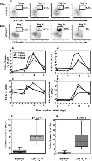
intracel-FIG. 1. Clinical course following inoculation. (A) The timelines of clinical features, following inoculation at day 0, are shown above the line for each individual. For each subject, the roman numerals and arrows indicate the sequence of clinical features, while the numbers under the timelines show when peripheral blood samples were collected. Changes in (B) CD4 T-cell counts and (C) CD8 T-cell counts, following inoculation with vaccinia virus, are also shown.
on November 8, 2019 by guest
http://jvi.asm.org/
[image:2.585.134.453.67.468.2]lular mature virus was prepared by multiple freeze-thawing of infected cells,
followed by the removal of cell debris by centrifugation at 700⫻gfor 10 min.
Viral antigen was further treated by heat inactivation at 56°C for 30 min, which
reduced the infectious titer by a factor of 106
(data not shown). This vaccinia virus lysate was used at a final concentration of 1/250 in the assays of antigen-specific T cells, as described below. A control lysate of uninfected HeLa cells was also prepared in parallel and used at the same concentration.
T-lymphocyte phenotyping of fresh whole blood.The measurement of CD4⫹ T-cell subsets in fresh peripheral blood was performed by six-color flow cyto-metric analysis on a dual-laser LSR II flow cytometer (Becton Dickinson), as previously described (55). Subset analysis was performed on whole blood within 1 h of venipuncture to minimize spontaneous loss of CCR5 expression, as previously described (55, 58). For CCR7 analysis, whole blood was first incubated with purified anti-CCR7 IgM MAb simultaneously with other directly conjugated IgG MAbs, and cells were washed once and then further incubated with
anti-mouse IgM (chain specific)-PE. Intracellular staining was performed using
FACS lyse and FACS permeabilizing reagents (Becton Dickinson) according to the manufacturer’s directions and analyzed as previously described (55, 56).
More detailed analysis of CCR5⫹CD38⫹⫹⫹CD4⫹T cells at day 14 was
performed for subjects VS002 and VS003. The binding of various PE-conjugated
MAbs to CCR5⫹CD38⫹⫹⫹CD4⫹T cells was studied by first gating on CCR5-,
FITC-, and CD38-APC-stained CD4⫹T lymphocytes, while FITC-conjugated
MAbs were studied on CCR5-PE- and CD38-APC-stained CD4⫹T lymphocytes.
NK cells were identified in a forward scatter/side scatter lymphocyte gate as either CD3-negativeCD56dim or CD3-negativeCD56bright.
Assays of antigen-specific CD4ⴙT cells.Vaccinia virus-specific T-cell
prolif-eration was determined by a [3
H]thymidine incorporation assay, as previously
described (55). A total of 1⫻105peripheral blood mononuclear cells (PBMCs)
was cultured in triplicate with live or heat-inactivated vaccinia virus (NYCBH
strain) diluted to an equivalent antigen dose of 5⫻105
PFU. An equivalent dilution of the mock-infected cell lysate was used as the control for the vaccinia virus antigens. Tetanus toxoid (2 lipofloculation units/ml; CSL, Melbourne, Aus-tralia) was also used to determine the kinetics of responses to a recall antigen. Results were expressed as a stimulation index.
A standard whole-blood flow cytometric intracellular cytokine assay of
anti-gen-specific CD4⫹T cells (32, 55, 58) was used to quantify and phenotype
vaccinia virus-specific CD4⫹T cells. Cultures of 0.5 ml sodium
heparin-antico-agulated whole blood were incubated for 6 h at 37°C in 5% CO2with anti-CD28
and anti-CD49d MAb (1g/ml each) as well as brefeldin A (10g/ml) for the
last 4 h. Cultures were performed in the absence or presence of vaccinia virus lysate (see above).
The expression of CD134 (OX40) was studied in cultures of 0.25 ml sodium heparin-anticoagulated whole blood plus 0.25 ml Iscove’s modified Dulbecco’s medium, in the presence or absence of vaccinia lysate at a 1/250 final
concen-tration. After 40 to 44 h of culturing at 37°C in 5% CO2, 100-l aliquots of the
cultures were stained and analyzed as for fresh whole blood.
Neutralizing antibody titer.A plaque reduction assay was used to measure neutralizing activity in serum samples from the study subjects. Sera were heat inactivated for 30 min at 56°C. An initial 1:5 dilution of serum was performed in RPMI with 0.2% bovine serum albumin, followed by five to eight further twofold
serial dilutions. A total of 600l of serum dilutions (or medium alone to control
for maximum virus plaques) was added to 600l of vaccinia virus (NYCBH
strain) at 800 PFU/ml in RPMI with 0.2% bovine serum albumin. Mixtures of
serum and virus were incubated at 37°C for 60 min, and 500l was added to each
of duplicate wells (six-well plates) of confluent BSC-1 cell monolayers. After incubation for 60 min at 37°C (with regular mixing of the virus mixture over the monolayer surface), 3 ml of RPMI with 2% fetal calf serum was added to each well and the plates were incubated for 48 h. The medium was then removed, monolayers were stained with 1 ml/well of crystal violet stain (0.5% in methanol) for 10 min and washed with water, and virus plaques were counted manually. Results for each serum dilution were expressed as percent neutralization:
[1⫺(mean plaques/control plaques)]⫻100%.
The neutralization titer (50% infective dose [ID50]) was calculated from the
percent neutralization data from each serum sample by using a variable slope sigmoidal dose-response nonlinear regression curve-fitting program, which gave the exact serum dilution that reduced plaques by 50% (Prism; GraphPad Soft-ware, San Diego, CA).
Statistics.The Wilcoxon signed-rank test was performed to compare paired
percentages of CCR5⫹CD38⫹⫹⫹CD4⫹T cells for each subject at baseline
versus days 13 and 14 using StatView 5.0 for Macintosh (Abacus Concepts,
Berkeley, CA). A two-sidedPvalue of⬍0.05 was considered statistically
signif-icant.
RESULTS
Clinical features.All three of the subjects who were studied intensively, VS001, -2, and -3, each developed a typical single large pustule at the inoculation site by day 10 and reported a fairly uniform sequence of events: mild local lymph node ten-derness at days 7 to 8, which typically reduced over 1 to 3 days, was quickly followed by inflammation and induration at the inoculation site by days 9 and 10, which in turn was followed 1 to 2 days later by a rapid reduction in induration. These events are depicted for each individual in Fig. 1A. Also, all three subjects reported mild to moderate malaise around days 9 to 11 and had at least 1 day away from work but were well by day 14, with a drying, resolving pustule.
Longitudinal CD4 T-cell counts for these subjects are shown in Fig. 1B. All three subjects exhibited a variable decline in CD4 T-cell counts, coinciding with the period of inoculation site induration, followed by a rebound to baseline levels after the resolution of induration (see above). Similarly, all subjects exhibited a decline in CD8 T-cell counts at the time of inocu-lation site induration, followed by a large rebound by day 14 (Fig. 1C). The CD8 T-cell count for subject VS001 rebounded above the baseline cell count, such that the CD4:CD8 ratio was inverted at day 14 but returned to the baseline CD4:CD8 ratio by day 21.
We also investigated whether there were changes in NK cells following inoculation. There were no significant changes in NK cell numbers in whole blood, but there was an approximately fivefold increase in the intracellular expression of Ki-67 in CD3-CD56dim NK cells, from an average baseline value of 2.2% to an average of 9.8% at day 14 (data not shown).
Kinetics of CD4ⴙT-cell subsets.A sharp increase in circu-lating activated CCR5⫹CD38⫹⫹⫹CD4⫹T cells was observed at days 11 to 14 (Fig. 2A and B), which peaked at an average of 5.5% of CD4⫹T cells for subjects VS001, -2, and -3 by day 14 and then declined by day 21 (Fig. 2A and B). A similar pattern was also observed for the absolute cell count of CCR5⫹CD38⫹⫹⫹ CD4⫹ T cells in whole blood (data not shown). The rapidity of this increase was most apparent in subject VS001, where a blood sample was obtained at day 11, which was the day after the decrease in inoculation site indu-ration (Fig. 1A and 2A and B).
In these subjects, at day 14 there was a peak in proliferating Ki-67⫹CD4⫹T cells (Fig. 2C). More than half of the Ki-67⫹ CD4⫹T cells were CCR5⫹(data not shown). Similarly, there was a rise in TIA-1⫹CD4⫹T cells (Fig. 2D). The antibody TIA-1 identifies granules in cytotoxic T lymphocytes, and these cells were also mostly CCR5⫹ (data not shown). However, there was no apparent increase in the expression of cytotoxic effector molecules, granzyme A or B, or perforin in these TIA-1⫹CD4⫹T cells (data not shown).
There was also a rise in CD4⫹ T cells which underwent apoptosis in vitro (Fig. 2E), also peaking at day 14, measured as intracellular activated caspase-3 at the end of the 6-h cul-tures used to identify vaccinia virus-specific CD4⫹T cells (see below).
The marked increase in circulating CCR5⫹CD38⫹⫹⫹ CD4⫹ T cells was confirmed in a second group of four subjects studied at baseline and day 13. For all seven sub-jects, the CCR5⫹CD38⫹⫹⫹ subset of CD4⫹ T cells rose
on November 8, 2019 by guest
http://jvi.asm.org/
FIG. 2. Kinetics of changes in CD4⫹ T-cell subsets following inoculation with vaccinia virus. (A) Flow cytometric histograms of CCR5⫹CD38⫹⫹⫹CD4⫹(upper histograms) and CCR5⫹CD38⫹⫹⫹CD8⫹T cells (lower histograms) for subject VS001. Also shown are kinetics of (B) CCR5⫹CD38⫹⫹⫹ CD4⫹ T cells for individual subjects, (C) proliferating (Ki-67⫹) CD4⫹ T cells, (D) TIA-1⫹ CD4⫹ T cells, and (E) spontaneously apoptotic (activated caspase-3⫹) CD4⫹T cells. For the symbol keys for panels C, D, and E, see panel B. The increases in CCR5⫹ CD38⫹⫹⫹cells in CD4⫹and CD8⫹T cells from baseline to day 13 or 14 for all seven subjects studied are summarized in panels F and G, respectively. Box plots represent 10th, 25th, 50th, 75th, and 90th percentiles. ThePvalues shown were determined by the paired nonparametric Wilcoxon signed-rank test.
on November 8, 2019 by guest
http://jvi.asm.org/
from a median of 0.7% at baseline to 6.0% at days 13 to 14 (P⫽ 0.018) (Fig. 2F).
In CD8⫹ T cells, parallel changes in the corresponding CCR5⫹CD38⫹⫹⫹ subset were also observed, again with a peak at day 14 for subjects VS001, -2, and -3 (Fig. 2A). Overall
[image:5.585.302.543.64.513.2]for the seven subjects, the CCR5⫹CD38⫹⫹⫹subset of CD8⫹T cells rose from a median of 0.9% at baseline to a median of 12.8% at days 13 to 14 (P⫽0.018) (Fig. 2G). Similarly, CD8⫹ T cells that were Ki-67⫹and apoptotic also peaked at day 14, with an average of 16.1 and 6.2%, respectively, for subjects VS001, -2, and -3 (data not shown).
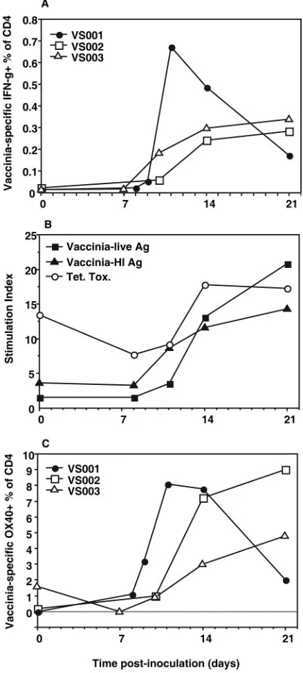
[image:5.585.53.272.71.557.2]FIG. 3. Kinetics of vaccinia virus-specific CD4⫹ T cells. Appear-ance of vaccinia virus-specific CD4⫹T cells following inoculation with vaccinia virus, including (A) total IFN-␥⫹CD4⫹T cells in response to vaccinia virus heat-inactivated antigen (HI Ag) by intracellular cyto-kine assay (average of six replicates) for each subject, (B) average PBMC lymphoproliferation stimulation index (for all three subjects) in response to vaccinia virus antigen preparations (live Ag and HI Ag) and tetanus toxoid recall antigens, and (C) appearance of OX40⫹ CD4⫹ T cells in response to vaccinia virus HI Ag in whole-blood cultures for each individual.
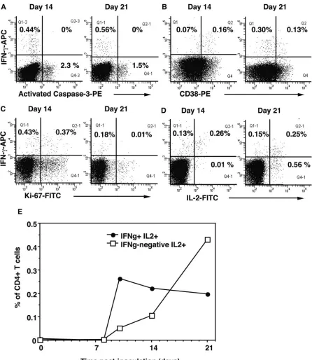
FIG. 4. Characterization of vaccinia virus-specific CD4⫹T cells at day 14. Phenotype of vaccinia virus-specific CD4⫹ T cells at day 14, including (A) CD38⫹⫹IFN-␥⫹CD4⫹T cells, (B) Bcl-2 low IFN-␥⫹ CD4⫹T cells, (C) TIA-1⫹IFN-␥⫹CD4⫹T cells, (D) Ki-67⫹IFN-␥⫹ CD4⫹T cells, (E) CD127⫹IFN-␥⫹CD4⫹T cells, and (F) CTLA-4⫹ IFN-␥⫹ CD4⫹ T cells. Representative histograms are shown, with similar results obtained for subjects VS001, -2, and -3; the percentages of CD3⫹CD4⫹cells in relevant quadrants are shown. Cursor settings for panel A are based on results from Fig. 2, and those for panel B are based on results from previous studies (55, 56). The overall phenotype of IFN-␥⫹vaccinia virus-specific CD4⫹T cells for subjects VS001, -2, and -3 is shown in panel G, representing mean⫾standard error (error bars) for each marker for all three subjects.
on November 8, 2019 by guest
http://jvi.asm.org/
Kinetics of appearance of vaccinia virus-specific CD4ⴙT cells. IFN-␥-producing vaccinia virus-specific CD4⫹ T cells appeared in the circulation of subjects VS001, -2, and -3 from day 11 onward (Fig. 3A), coinciding with the peak of activated proliferating CD4⫹T cells. The average for the three subjects was 0.34% of CD4⫹T cells at day 14, whereas in all control cultures with uninfected HeLa cell lysate, IFN-␥⫹cells were less than 0.03% of CD4⫹T cells (data not shown).
However, it must be noted that the level of IFN-␥vaccinia virus-specific CD4⫹T cells was approximately 10-fold lower at day 14 than that of the subpopulations of activated proliferat-ing effector CD4⫹ T cells, as identified in the phenotyping measurements described above.
Vaccinia virus-specific proliferative responses of PBMCs were initially detected in subjects VS001, -2, and -3 between days 11 and 14, with a further increase by day 21 (Fig. 3B). In the same individuals, proliferative responses to the recall an-tigen tetanus toxoid appeared to decrease slightly at days 8 to 11, rebounded to baseline levels at day 14, and remained con-stant from days 14 to 21 (Fig. 3B).
We also investigated the effect of 40 to 44 h of incubation with antigen on the expression of the costimulatory molecule OX40 (CD134) on CD4⫹T cells. The results show that there was a large subpopulation, averaging 6% of CD4⫹T cells at day 14, that responded to vaccinia virus antigen with up-regu-lation of OX40 (Fig. 3C), corresponding more closely with the levels of activated proliferating cells than to the subpopulation which produced IFN-␥.
Phenotype of vaccinia virus-specific CD4ⴙT cells.IFN-␥⫹ vaccinia virus-specific CD4⫹T cells at day 14 were predomi-nantly CD38⫹⫹ (Fig. 4A), Bcl-2low (Fig. 4B), TIA-1⫹ (Fig. 4C), CD127⫹(Fig. 4E), CD40L⫹(Fig. 4G), and CD57 nega-tive (Fig. 4G). Heterogeneity of IFN-␥⫹vaccinia virus-specific CD4⫹T cells was also observed at day 14, with just over half containing Ki-67⫹(Fig. 4D) and expressing IL-2 (see below), but with just under half expressing CTLA-4 (Fig. 4F). The overall phenotype results for the IFN-␥⫹vaccinia virus-specific CD4⫹T cells at day 14 from subjects VS001, -2, and -3 for all markers studied are summarized in Fig. 4G.
Even though CCR5 expression cannot be reliably measured on in vitro-restimulated antigen-specific CD4⫹T cells (55), we can reasonably infer its presence on these cells by the ex vivo CCR5⫹phenotype of CD38⫹⫹⫹CD4⫹T cells (Fig. 2A), as we have recently also done for HIV-specific CD4⫹T cells (58).
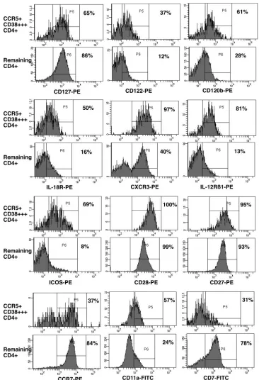
Phenotype of CCR5ⴙCD38ⴙⴙⴙCD4ⴙT cells at day 14.The detailed phenotype of CCR5⫹CD38⫹⫹⫹CD4⫹T cells at day 14 is shown in Fig. 5 relative to that of other CD4⫹T cells. These activated cells expressed increased levels of markers characteristic of Th1 effector cells, including IL-12R1, CXCR3, and IL-18R. They also expressed increased levels of CD120b (TNFR2) as well as thechain of IL-2R (CD122). However, these activated cells predominantly retained the ex-pression of the IL-7R␣chain (CD127) and also expressed the costimulatory molecules ICOS, CD27, and CD28 but at the same time there was clear down-regulation of CD7 expression. The potential trafficking of these cells to sites of inflamma-tion was indicated by relative down-regulainflamma-tion of CCR7, in addition to the up-regulation of CXCR3 and CCR5 as well as increased expression of the integrins CD49d (not shown) and CD11a. However, there was no up-regulation of integrin7
(not shown) nor was there any apparent change in the expres-sion of CD62L (not shown).
Survival of vaccinia virus-specific CD4ⴙT cells between day 14 and day 21. Since there was a rapid disappearance of CCR5⫹CD38⫹⫹⫹CD4⫹T cells between day 14 and day 21, we investigated a possible role for apoptosis in limiting the peak of vaccinia virus-specific CD4⫹T cells. Although we saw a clear overall increase in spontaneous apoptosis within the CD4⫹ T-cell subset at day 14 (Fig. 2E), we were unable to demonstrate directly that IFN-␥⫹ antigen-specific CD4⫹ T cells exhibited activated caspase-3 during short-term cultures with antigen (Fig. 6A). In fact, there was a very clear distinc-tion between IFN-␥⫹antigen-specific CD4⫹T cells and apop-totic cells. We also examined whether CD38⫹⫹⫹CD4⫹T cells had activated caspase-3 after short-term culture in vitro, but apoptotic CD4⫹ T cells generally had only intermediate ex-pression of CD38, whereas apoptotic CD8⫹T cells had higher CD38 expression (data not shown).
There was a change in the activated proliferative phenotype of IFN-␥⫹vaccinia virus-specific CD4⫹T cells between day 14 and day 21, as indicated by the decrease in CD38 and Ki-67 expression (Fig. 6B and C). This change towards a resting phenotype coincided with the appearance of a relatively large population of vaccinia virus-specific CD4⫹T cells which pro-duced IL-2 but not IFN-␥at day 21 (Fig. 6D). There were also similar populations of vaccinia virus-specific CD4⫹T cells at day 21 which expressed CD40L and TNF-␣ but not IFN-␥ (data not shown). The overall results show that the initial burst of antigen-specific CD4⫹ T cells predominantly coexpressed IFN-␥and IL-2, but between day 14 and day 21, there was a switch to cells which predominantly produced IL-2 but not IFN-␥(Fig. 6E).
However, Bcl-2 expression in vaccinia virus-specific CD4⫹T cells remained low at day 21 (data not shown), similar to the low levels of Bcl-2 in the corresponding cells at day 14 (see above).
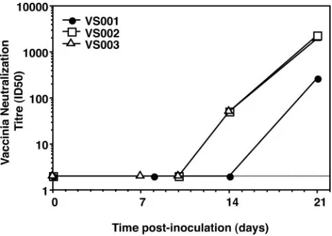
Kinetics of appearance of vaccinia virus-specific neutraliz-ing antibodies.The three subjects, VS001, -2, and -3, studied in detail longitudinally were negative for neutralizing antibodies at baseline (neutralization titer [ID50] wasⱕ2), with low-titer antibodies first appearing at day 14 in two out of three subjects and continuing to rise to high titers in all three subjects by day 21 (Fig. 7).
DISCUSSION
The combination of cell surface phenotyping and intracellu-lar cytokine staining of longitudinal samples of fresh whole blood has allowed the characterization of a unique population of CCR5⫹CD38⫹⫹⫹ antigen-specific effector CD4⫹ T cells during the primary antiviral response to vaccinia virus inocu-lation in healthy adults. The current study confirms the ephem-eral nature of the CCR5⫹CD38⫹⫹⫹antigen-specific CD4⫹T cells in the circulation, as was previously suggested for primary HIV-1 infection (58). Interestingly, these cells appeared in peripheral blood only after the resolution of, first, the local lymph node involvement and, second, the swelling at the inoc-ulation site, suggesting that the vaccinia virus-specific cells proliferated and were sequestered at those sites until viral replication was controlled. A similar effect has recently been
on November 8, 2019 by guest
http://jvi.asm.org/
reported for tuberculin-specific memory CD4⫹T cells in the induration associated with a positive Mantoux test (40).
We have also now shown that there is a second phase of resting CD38-negative IL-2-producing CD4⫹T cells which fol-lows the effector cells within days. These cells were associated
[image:7.585.107.473.67.603.2]with lymphoproliferative responses in vitro and did not neces-sarily produce IFN-␥ but did produce CD40L and TNF-␣. Therefore, as the cells with the CCR5⫹CD38⫹⫹⫹effector cell phenotype disappeared, they were rapidly replaced with cells with the phenotype of long-term memory cells.
FIG. 5. Detailed phenotype of CCR5⫹CD38⫹⫹⫹CD4⫹T cells at day 14. Analysis of various markers on CCR5⫹CD38⫹⫹⫹CD4⫹T cells at day 14. For each marker, a pair of histograms is shown: the upper histogram is gated on CCR5⫹CD38⫹⫹⫹CD4⫹T cells and the lower histogram is gated on remaining CD4⫹T cells (Fig. 2A). The histograms shown are for subject VS003; similar results were obtained for subject VS002. The percentage of positive cells is shown for each histogram.
on November 8, 2019 by guest
http://jvi.asm.org/
The CCR5⫹CD38⫹⫹⫹antigen-specific CD4⫹T cells had a phenotype and cytokine profile of Th1 effector cells. In murine models employing either gene knockouts or vaccinia virus con-structs containing cytokine genes, IFN-␥has been shown to be very important in the control of vaccinia virus replication in vivo (39). These results imply a likely role for IFN-␥⫹CD4⫹T
[image:8.585.68.514.67.581.2]cells in the initial control of poxvirus primary infections of humans since the effector CD4⫹and CD8⫹T cells as well as increased turnover of NK cells were much better correlated with the timing of the resolution of the vaccinia virus pustule than were either the IL-2⫹memory CD4⫹T cells or the hu-moral response. This result is consistent with the observations
FIG. 6. Comparison of vaccinia virus-specific CD4⫹T cells at days 14 and 21. Attributes of vaccinia virus-specific IFN-␥⫹CD4⫹T cells at day 14 and day 21 are shown, including the coexpression of (A) activated caspase-3, (B) CD38, (C) Ki-67, and (D) IL-2. The results shown are representative of all three subjects, and the percentages of CD3⫹CD4⫹ cells in relevant quadrants are shown in the histograms. (E) The coexpression of IFN-␥by vaccinia virus-specific IL-2⫹CD4⫹T cells over time is also shown. The results shown represent the average for all three subjects.
on November 8, 2019 by guest
http://jvi.asm.org/
of progressive vaccinia virus infection in subjects with cell-mediated immune defects, whereas subjects with humoral im-munity abnormalities control vaccinia virus infection normally (14). In fact, in mouse models of vaccinia virus infection in the setting of T-cell deficiency, a combination of IL-2 and NK cells appears to be sufficient to clear virus (23). These direct cell-mediated effects contrast with results showing that neutralizing antibodies are the most important components of long-term immune protection against reinfection with smallpox (10, 13) as well as the greater effectiveness of proliferative central memory CD8⫹ T cells in mouse models of reinfection with lymphocytic choriomeningitis virus (53).
The CD4⫹effector T cells observed in individuals undergo-ing a primary response to vaccinia virus also expressed a marker of cytotoxic granules, TIA-1. Cytotoxic clones of vac-cinia virus-specific CD4⫹ T cells have been previously de-scribed in vitro (26). However, we did not observe high-level expression of the effector molecule granzyme A or B or per-forin ex vivo, unlike our previous observations of CD4⫹ cyto-toxic T lymphocyte in both chronic and primary HIV-1 infec-tion (2, 58). It is possible, though, that these TIA-1⫹cells may rapidly up-regulate the effector molecules if restimulated in vitro, as did similar cells from a HIV⫹long-term nonprogres-sor (55). Alternatively, the contents of TIA-1⫹ granules in vaccinia virus-specific CD4⫹T cells may differ from cytotoxic effector molecules (5, 44).
Our results suggest that results from the intracellular cyto-kine assay of IFN-␥⫹antigen-specific CD4⫹T cells may be as much as 10-fold lower than other indicators of antigen-specific CD4⫹T cells, such as the CCR5⫹CD38⫹⫹⫹effector pheno-type, expression of Ki-67⫹and ICOS ex vivo, or induction of OX40 expression in vitro. We had previously observed this discrepancy with primary HIV-1 infection (58), but it now appears to be a more general phenomenon. It does not seem likely that bystander proliferation (49) could account for a difference of this magnitude, and we previously found no evi-dence of bystander proliferation of CMV-specific CD4⫹T cells in primary HIV-1 infection (58). Furthermore, the healthy adults studied here did not have significant CD4⫹T-cell
defi-ciency or concurrent opportunistic infections, often cited as a cause of the higher level of T-cell activation in HIV-1 infection (12). Also, the levels of activated cells in the current study are comparable to those reported in vaccinia virus infection in mouse models (18). Purification and in vitro studies of putative antigen-specific activated CD4⫹T cells may clarify their spec-ificity.
An increased rate of apoptosis of CD4⫹T cells was observed at the peak of the response of activated proliferating CD4⫹T cells immediately prior to their decline. Also, the low level of expression of Bcl-2 in the antigen-specific CD4⫹T cells in the current study would suggest that they are short-lived cells (46). However, when we investigated whether antigen-specific CD4⫹T cells also contained activated caspase-3 as a marker of apoptosis, we could not demonstrate a direct link between the two processes. It is possible that the detection of activated caspase-3 represented a relatively late event in apoptosis, when cells were no longer able to secrete cytokines and had already down-regulated CD38.
Surprisingly, we found that Bcl-2 levels were still low in resting antigen-specific CD4⫹T cells that expressed IL-7R and produced IL-2 at day 21. According to a current model of memory cell differentiation, IL-7R⫹effector cells are precur-sors for memory cells which re-express high levels of Bcl-2 (22). Our results suggest that such a process of differentiation must still be in process at day 21. This will be further investigated in future studies. Alternatively, another anti-apoptotic Bcl-2 fam-ily member may have been protecting these nascent memory cells from apoptosis, possibly Mcl-1 (34) or survivin (45), while they are completing differentiation to Bcl-2high memory cells. Another probable mechanism constraining the extent of an-tigen-specific CD4 T-cell expansion may be the expression of CTLA-4, based on gene knockout models in mice (47, 52). We observed that just under half of the IFN-␥⫹ vaccinia virus-specific CD4⫹T cells coexpressed CTLA-4.
Balanced against the negative effects of apoptosis and CTLA-4 expression are the known positive effects of costimu-lation via the Ig superfamily member ICOS (3, 33) and the TNF superfamily member OX40 (9), which were found in this study to be expressed by vaccinia virus-specific CD4⫹T cells in the early stages of the response. Gene knockout studies in murine models have demonstrated an important role for OX40 expression in CD4⫹memory T-cell differentiation (3, 9). The role of ICOS is less clear but has been seen to be important in the absence of CD28 (4). The stimulation with antigen in vitro was necessary to see OX40 expression, but ICOS appeared to be constitutively expressed on the activated effector cells, sug-gesting differing roles for these molecules. In particular, OX40 appears to exert its prosurvival effect (42, 45) toward the end of a primary response (9). Interestingly, in vivo stimulation of OX40 using the administration of an agonistic MAb led to enhanced CD4⫹T-cell responses in a mouse model of vaccinia virus infection (30).
[image:9.585.43.284.68.239.2]Another reported positive influence on CD4⫹ memory T-cell differentiation is the maintenance of the expression of the IL-7 receptor (24, 25). A high proportion of IFN-␥⫹vaccinia virus-specific CD4⫹T cells retained the expression of CD127, the␣chain of the IL-7R, whereas we had previously found that a much lower proportion of early IFN-␥⫹HIV-specific CD4⫹ T cells expressed CD127 (58). These results suggest that a
FIG. 7. Appearance of neutralizing antibodies. The titers of vac-cinia virus-specific neutralizing antibodies, following inoculation, are shown for each vaccinia virus-naı¨ve subject.
on November 8, 2019 by guest
http://jvi.asm.org/
major difference in memory cell generation between these two infections occurs very early, possibly at the level of initial activation of naı¨ve CD4⫹T cells by dendritic cells. Little is known of the regulation of CD127 expression, but the reduced expression of CD127 in CD8⫹ T cells is characteristic of chronic (51) and primary (57) HIV-1 infection and in CD4⫹T cells (58) during primary HIV-1 infection.
This study provides important information on which cyto-kines may be important in the primary anti-vaccinia virus re-sponse, including not only IL-7 but also IL-15, IL-12, IL-18, and TNF-␣, since receptors for all these factors could be seen to be up-regulated on antigen-specific CD4⫹T cells. An im-proved knowledge of the effects of these cytokines may allow their rational use as adjuvants, in addition to the potential use of agonists directed towards ICOS or OX40.
Our results show that the antigen-specific CD4⫹T-cell re-sponse to vaccinia virus consisted of two phases within a week of each other. The acute Th1 effector phase involving highly activated proliferating effector cells was closely followed by a second phase during which resting cells that produced more IL-2 than IFN-␥appeared. Previous studies of viral antigen-specific T cells in murine models provided evidence that effector cells differentiate to become resting memory cells. In one study, the marking of CD8⫹memory cells which had previously expressed perforin demonstrated a lineage rela-tionship (35) and it is interesting that the antigen-specific CD4⫹T effector cells in the current study also prominently expressed a marker of cytotoxic T lymphocyte, namely TIA-1. In other studies, CD4⫹effector cells were purified and became resting central memory cells following adoptive transfer (17, 19), while a corresponding study of CD8⫹ T cells yielded similar results (53).
Therefore, our results provide some circumstantial support for the concept of a lineage relationship between effector cells and resting memory cells in human viral infection. An impor-tant implication of this model is that in the case of primary HIV-1 infection, the expression of CCR5 as well as the state of activation would suggest that such precursors of memory cells are highly susceptible to cytopathic infection during the differ-entiation in the environment of the lymph node. This may leave patients with the characteristic lack of proliferative IL-2⫹ HIV-specific CD4⫹memory T cells (16, 20, 29, 36, 54). An alternative temporal model may be that effector cells gener-ated during the primary response simply disappear by apopto-sis (6, 31) or trafficking (27, 28), while the production of a separate population of resting memory cells is delayed until active viral replication is greatly reduced (21). Careful study of T-cell receptor clonotypes (38) of effector and memory cells following vaccinia virus inoculation may provide an opportu-nity to directly observe the relationship between the different subsets.
In summary, the results from the current study indicate that the early development of CCR5⫹CD38⫹⫹⫹ antigen-specific CD4⫹T cells is a normal feature of primary viral infection. These markers will now enable the purification and more de-tailed study of antigen-specific CD4⫹T cells at this important early stage of antiviral response. Such studies may also suggest reasons for the observed differences between vaccinia virus-specific CD4⫹T cells and HIV-specific CD4⫹T cells.
ACKNOWLEDGMENTS
We thank the study participants for their cooperation in this anal-ysis.
This study was supported by the ACH2 and ARCBS K and I Grants Scheme (W.B.D.) and by program grants from the Australian National Health and Medical Research Council (NHMRC) to J.J.Z., E.A., S.J.K., R.D.R., A.D.K., and D.A.C.), and AIEDRP, NIH Division of AIDS to J.J.Z., A.D.K., and D.A.C. M.L.M. is the recipient of a Dora Lush postgraduate scholarship from the NHMRC. The National Cen-tre in HIV Epidemiology and Clinical Research is supported by the Commonwealth Department of Health and Ageing.
REFERENCES
1.Amyes, E., C. Hatton, D. Montamat-Sicotte, N. Gudgeon, A. B. Rickinson, A. J. McMichael, and M. F. Callan.2003. Characterization of the CD4⫹T cell response to Epstein-Barr virus during primary and persistent infection.
J. Exp. Med.198:903–911.
2.Appay, V., J. J. Zaunders, L. Papagno, J. Sutton, A. Jaramillo, A. Waters, P. Easterbrook, P. Grey, D. Smith, A. J. McMichael, D. A. Cooper, S. L. Rowland-Jones, and A. D. Kelleher.2002. Characterization of CD4⫹CTLs
ex vivo. J. Immunol.168:5954–5958.
3.Bertram, E. M., W. Dawicki, and T. H. Watts.2004. Role of T cell
costimu-lation in anti-viral immunity. Semin. Immunol.16:185–196.
4.Bertram, E. M., A. Tafuri, A. Shahinian, V. S. Chan, L. Hunziker, M. Recher, P. S. Ohashi, T. W. Mak, and T. H. Watts.2002. Role of ICOS
versus CD28 in antiviral immunity. Eur. J. Immunol.32:3376–3385.
5.Bossi, G., and G. M. Griffiths.2005. CTL secretory lysosomes: biogenesis
and secretion of a harmful organelle. Semin. Immunol.17:87–94.
6.Callan, M. F., C. Fazou, H. Yang, T. Rostron, K. Poon, C. Hatton, and A. J. McMichael.2000. CD8⫹T-cell selection, function, and death in the primary
immune response in vivo. J. Clin. Investig.106:1251–1261.
7.Centers for Disease Control and Prevention.2003. Smallpox vaccination and adverse reactions: guidance for clinicians. Morb. Mortal. Wkly. Rep.
52:1–30.
8.Christensen, J. P., and P. C. Doherty.1999. Quantitative analysis of the
acute and long-term CD4⫹T-cell response to a persistent
gammaherpesvi-rus. J. Virol.73:4279–4283.
9.Croft, M.2003. Co-stimulatory members of the TNFR family: keys to
effec-tive T-cell immunity? Nat. Rev. Immunol.3:609–620.
10.Crotty, S., P. Felgner, H. Davies, J. Glidewell, L. Villarreal, and R. Ahmed.
2003. Cutting edge: long-term B cell memory in humans after smallpox
vaccination. J. Immunol.171:4969–4973.
11.Demkowicz, W. E., Jr., R. A. Littaua, J. Wang, and F. A. Ennis.1996. Human cytotoxic T-cell memory: long-lived responses to vaccinia virus. J. Virol.
70:2627–2631.
12.Douek, D. C., L. J. Picker, and R. A. Koup.2003. T cell dynamics in HIV-1
infection. Annu. Rev. Immunol.21:265–304.
13.Edghill-Smith, Y., H. Golding, J. Manischewitz, L. R. King, D. Scott, M. Bray, A. Nalca, J. W. Hooper, C. A. Whitehouse, J. E. Schmitz, K. A. Reimann, and G. Franchini.2005. Smallpox vaccine-induced antibodies are necessary and sufficient for protection against monkeypox virus. Nat. Med.
11:740–747.
14.Fulginiti, V. A., A. Papier, J. M. Lane, J. M. Neff, and D. A. Henderson.2003. Smallpox vaccination: a review, part II. Adverse events. Clin. Infect. Dis.
37:251–271.
15.Gamadia, L. E., E. B. Remmerswaal, J. F. Weel, F. Bemelman, R. A. van Lier, and I. J. Ten Berge.2003. Primary immune responses to human CMV: a
critical role for IFN-gamma-producing CD4⫹T cells in protection against
CMV disease. Blood101:2686–2692.
16.Harari, A., F. Vallelian, P. Meylan, and G. Pantaleo.2005. Functional het-erogeneity of memory CD4 T cell responses in different conditions of antigen
exposure and persistence. J. Immunol.174:1037–1045.
17.Harbertson, J., E. Biederman, K. E. Bennett, R. M. Kondrack, and L. M. Bradley.2002. Withdrawal of stimulation may initiate the transition of
ef-fector to memory CD4 cells. J. Immunol.168:1095–1102.
18.Harrington, L. E., R. van der Most, J. L. Whitton, and R. Ahmed.2002. Recombinant vaccinia virus-induced T-cell immunity: quantitation of the
response to the virus vector and the foreign epitope. J. Virol.76:3329–3337.
19.Hu, H., G. Huston, D. Duso, N. Lepak, E. Roman, and S. L. Swain.2001.
CD4⫹T cell effectors can become memory cells with high efficiency and
without further division. Nat. Immunol.2:705–710.
20.Iyasere, C., J. C. Tilton, A. J. Johnson, S. Younes, B. Yassine-Diab, R. P. Sekaly, W. W. Kwok, S. A. Migueles, A. C. Laborico, W. L. Shupert, C. W. Hallahan, R. T. Davey, Jr., M. Dybul, S. Vogel, J. Metcalf, and M. Connors.
2003. Diminished proliferation of human immunodeficiency virus-specific
CD4⫹T cells is associated with diminished interleukin-2 (IL-2) production
and is recovered by exogenous IL-2. J. Virol.77:10900–10909.
21.Jelley-Gibbs, D. M., D. M. Brown, J. P. Dibble, L. Haynes, S. M. Eaton, and S. L. Swain.2005. Unexpected prolonged presentation of influenza antigens
promotes CD4 T cell memory generation. J. Exp. Med.202:697–706.
on November 8, 2019 by guest
http://jvi.asm.org/
22.Kaech, S. M., J. T. Tan, E. J. Wherry, B. T. Konieczny, C. D. Surh, and R. Ahmed.2003. Selective expression of the interleukin 7 receptor identifies effector CD8 T cells that give rise to long-lived memory cells. Nat. Immunol.
4:1191–1198.
23.Karupiah, G., B. E. Coupar, M. E. Andrew, D. B. Boyle, S. M. Phillips, A. Mullbacher, R. V. Blanden, and I. A. Ramshaw.1990. Elevated natural killer cell responses in mice infected with recombinant vaccinia virus encoding
murine IL-2. J. Immunol.144:290–298.
24.Kondrack, R. M., J. Harbertson, J. T. Tan, M. E. McBreen, C. D. Surh, and L. M. Bradley.2003. Interleukin 7 regulates the survival and generation of
memory CD4 cells. J. Exp. Med.198:1797–1806.
25.Li, J., G. Huston, and S. L. Swain.2003. IL-7 promotes the transition of CD4
effectors to persistent memory cells. J. Exp. Med.198:1807–1815.
26.Littaua, R. A., A. Takeda, J. Cruz, and F. A. Ennis.1992. Vaccinia
virus-specific human CD4⫹cytotoxic T-lymphocyte clones. J. Virol.66:2274–2280.
27.Marshall, D. R., S. J. Turner, G. T. Belz, S. Wingo, S. Andreansky, M. Y. Sangster, J. M. Riberdy, T. Liu, M. Tan, and P. C. Doherty.2001. Measuring
the diaspora for virus-specific CD8⫹T cells. Proc. Natl. Acad. Sci. USA
98:6313–6318.
28.Masopust, D., V. Vezys, A. L. Marzo, and L. Lefrancois.2001. Preferential
localization of effector memory cells in nonlymphoid tissue. Science291:
2413–2417.
29.McNeil, A. C., W. L. Shupert, C. A. Iyasere, C. W. Hallahan, J. A. Mican, R. T. Davey, Jr., and M. Connors.2001. High-level HIV-1 viremia suppresses
viral antigen-specific CD4⫹T cell proliferation. Proc. Natl. Acad. Sci. USA
98:13878–13883.
30.Munks, M. W., D. V. Mourich, R. S. Mittler, A. D. Weinberg, and A. B. Hill.
2004. 4-1BB and OX40 stimulation enhance CD8 and CD4 T-cell responses
to a DNA prime, poxvirus boost vaccine. Immunology112:559–566.
31.Murali-Krishna, K., J. D. Altman, M. Suresh, D. J. Sourdive, A. J. Zajac, J. D. Miller, J. Slansky, and R. Ahmed.1998. Counting antigen-specific CD8 T cells: a reevaluation of bystander activation during viral infection.
Immu-nity8:177–187.
32.Nomura, L. E., J. M. Walker, and H. T. Maecker.2000. Optimization of
whole blood antigen-specific cytokine assays for CD4⫹T cells. Cytometry
40:60–68.
33.Nurieva, R. I.2005. Regulation of immune and autoimmune responses by
ICOS-B7h interaction. Clin. Immunol.115:19–25.
34.Opferman, J. T., A. Letai, C. Beard, M. D. Sorcinelli, C. C. Ong, and S. J. Korsmeyer.2003. Development and maintenance of B and T lymphocytes
requires antiapoptotic MCL-1. Nature426:671–676.
35.Opferman, J. T., B. T. Ober, and P. G. Ashton-Rickardt. 1999. Linear differentiation of cytotoxic effectors into memory T lymphocytes. Science
283:1745–1748.
36.Picker, L. J., and V. C. Maino.2000. The CD4⫹T cell response to HIV-1.
Curr. Opin. Immunol.12:381–386.
37.Precopio, M. L., J. L. Sullivan, C. Willard, M. Somasundaran, and K. Luzuriaga.2003. Differential kinetics and specificity of EBV-specific CD4⫹
and CD8⫹T cells during primary infection. J. Immunol.170:2590–2598.
38.Price, D. A., S. M. West, M. R. Betts, L. E. Ruff, J. M. Brenchley, D. R. Ambrozak, Y. Edghill-Smith, M. J. Kuroda, D. Bogdan, K. Kunstman, N. L. Letvin, G. Franchini, S. M. Wolinsky, R. A. Koup, and D. C. Douek.2004. T cell receptor recognition motifs govern immune escape patterns in acute SIV
infection. Immunity21:793–803.
39.Ramshaw, I. A., A. J. Ramsay, G. Karupiah, M. S. Rolph, S. Mahalingam, and J. C. Ruby.1997. Cytokines and immunity to viral infections. Immunol.
Rev.159:119–135.
40.Reed, J. R., M. Vukmanovic-Stejic, J. M. Fletcher, M. V. Soares, J. E. Cook, C. H. Orteu, S. E. Jackson, K. E. Birch, G. R. Foster, M. Salmon, P. C. Beverley, M. H. Rustin, and A. N. Akbar.2004. Telomere erosion in memory T cells induced by telomerase inhibition at the site of antigenic challenge in
vivo. J. Exp. Med.199:1433–1443.
41.Rock, M. T., S. M. Yoder, P. F. Wright, T. R. Talbot, K. M. Edwards, and J. E. Crowe, Jr.2005. Differential regulation of granzyme and perforin in
effector and memory T cells following smallpox immunization. J. Immunol.
174:3757–3764.
42.Rogers, P. R., J. Song, I. Gramaglia, N. Killeen, and M. Croft.2001. OX40 promotes Bcl-xL and Bcl-2 expression and is essential for long-term survival
of CD4 T cells. Immunity15:445–455.
43.Rotz, L. D., D. A. Dotson, I. K. Damon, and J. A. Becher.2001. Vaccinia (smallpox) vaccine: recommendations of the Advisory Committee on Immu-nization Practices (ACIP), 2001. Morb. Mortal. Wkly. Rep. Recomm. Rep.
50:1–25.
44.Smyth, M. J., J. M. Kelly, V. R. Sutton, J. E. Davis, K. A. Browne, T. J. Sayers, and J. A. Trapani.2001. Unlocking the secrets of cytotoxic granule
proteins. J. Leukoc. Biol.70:18–29.
45.Song, J., T. So, M. Cheng, X. Tang, and M. Croft.2005. Sustained survivin expression from OX40 costimulatory signals drives T cell clonal expansion.
Immunity22:621–631.
46.Strasser, A., and M. Pellegrini.2004. T-lymphocyte death during shutdown
of an immune response. Trends Immunol.25:610–615.
47.Tivol, E. A., F. Borriello, A. N. Schweitzer, W. P. Lynch, J. A. Bluestone, and A. H. Sharpe.1995. Loss of CTLA-4 leads to massive lymphoproliferation and fatal multiorgan tissue destruction, revealing a critical negative
regula-tory role of CTLA-4. Immunity3:541–547.
48.Topham, D. J., and P. C. Doherty.1998. Longitudinal analysis of the acute
Sendai virus-specific CD4⫹T cell response and memory. J. Immunol.161:
4530–4535.
49.Tough, D. F., P. Borrow, and J. Sprent.1996. Induction of bystander T cell
proliferation by viruses and type 1 interferon in vivo. Science272:1947–1950.
50.Varga, S. M., and R. M. Welsh.2000. High frequency of virus-specific
interleukin-2-producing CD4⫹T cells and Th1 dominance during
lympho-cytic choriomeningitis virus infection. J. Virol.74:4429–4432.
51.Vingerhoets, J., E. Bisalinkumi, G. Penne, R. Colebunders, E. Bosmans, L. Kestens, and G. Vanham.1998. Altered receptor expression and decreased sensitivity of T cells to the stimulatory cytokines IL-2, IL-7 and IL-12 in HIV
infection. Immunol. Lett.61:53–61.
52.Waterhouse, P., J. M. Penninger, E. Timms, A. Wakeham, A. Shahinian, K. P. Lee, C. B. Thompson, H. Griesser, and T. W. Mak.1995. Lymphopro-liferative disorders with early lethality in mice deficient in Ctla-4. Science
270:985–988.
53.Wherry, E. J., V. Teichgraber, T. C. Becker, D. Masopust, S. M. Kaech, R. Antia, U. H. von Andrian, and R. Ahmed.2003. Lineage relationship and
protective immunity of memory CD8 T cell subsets. Nat. Immunol.4:225–
234.
54.Younes, S. A., B. Yassine-Diab, A. R. Dumont, M. R. Boulassel, Z. Gross-man, J. P. Routy, and R. P. Sekaly.2003. HIV-1 viremia prevents the
establishment of interleukin 2-producing HIV-specific memory CD4⫹T cells
endowed with proliferative capacity. J. Exp. Med.198:1909–1922.
55.Zaunders, J. J., W. B. Dyer, B. Wang, M. L. Munier, M. Miranda-Saksena, R. Newton, J. Moore, C. R. Mackay, D. A. Cooper, N. K. Saksena, and A. D. Kelleher.2004. Identification of circulating antigen-specific CD4⫹T
lym-phocytes with a CCR5⫹, cytotoxic phenotype in an HIV-1 long-term
non-progressor and in CMV infection. Blood103:2238–2247.
56.Zaunders, J. J., G. R. Kaufmann, P. H. Cunningham, D. Smith, P. Grey, K. Suzuki, A. Carr, L. E. Goh, and D. A. Cooper.2001. Increased turnover of
CCR5⫹and redistribution of CCR5-CD4 T lymphocytes during primary
human immunodeficiency virus type 1 infection. J. Infect. Dis.183:736–743.
57.Zaunders, J. J., L. Moutouh-de Parseval, S. Kitada, J. C. Reed, S. Rought, D. Genini, L. Leoni, A. Kelleher, D. A. Cooper, D. E. Smith, P. Grey, J. Estaquier, S. Little, D. D. Richman, and J. Corbeil.2003. Polyclonal
prolif-eration and apoptosis of CCR5⫹ T lymphocytes during primary human
immunodeficiency virus type 1 infection: regulation by interleukin (IL)-2,
IL-15, and Bcl-2. J. Infect. Dis.187:1735–1747.
58.Zaunders, J. J., M. L. Munier, D. E. Kaufmann, S. Ip, P. Grey, D. Smith, T. Ramacciotti, D. Quan, R. Finlayson, J. Kaldor, E. S. Rosenberg, B. D. Walker, D. A. Cooper, and A. D. Kelleher.2005. Early proliferation of
CCR5⫹CD38⫹⫹⫹antigen-specific CD4⫹Th1 effector cells during primary
HIV-1 infection. Blood106:1660–1667.