Selective Sorption
DOI: 10.1002/anie.201307656Shape Selectivity by Guest-Driven Restructuring of a Porous
Material**
J. E. Warren, C. G. Perkins, K. E. Jelfs, P. Boldrin, P. A. Chater, G. J. Miller, T. D. Manning,
M. E. Briggs, K. C. Stylianou, J. B. Claridge, and M. J. Rosseinsky*
Abstract:A flexible metal-organic framework selectively sorbs para- (pX) over meta-xylene (mX) by synergic restructuring around pX coupled with generation of unused void space upon mX loading. The nature of the structural change suggests more generally that flexible structures which are initially mismatched in terms of fit and capacity to the preferred guest are strong candidates for effective molecular separations.
P
orous materials are widely used in shape-selective sorp-tion.[1] Metal-organic frameworks (MOFs) have chemicallytailorable internal surfaces[2]bearing a wide range of
func-tional groups and can respond flexibly[3]to guest uptake.[4, 5]
Shape selectivity is achieved in rigid porous hosts by matching their fixed channel geometries to the target molecule.[6]MOFs
are attractive for a range of separation applications[1, 7]
because of electronic[8] and geometrical features that are
hard to access in other classes of porous crystalline materi-als.[9] Flexible MOFs display excellent figures of merit for
CO2/CH4[10]and N2/CO separations.[11]The separation of the
xylene and ethylbenzene C8isomers has been demonstrated
by zeolites[12]and by rigid MOFs.[13]Flexible MOFs can also
perform this separation in vapor[14]and liquid phases,[15]and
undergo “breathing”-type structural changes when sorbing C8
isomers,[16] but currently show lower selectivities than rigid
hosts. We present a flexible MOF that differentially restruc-tures aroundpara- (pX) and meta-xylene (mX) to achieve high selectivity, and demonstrate how the restructuring distinguishes between the two isomers at the atomic level.
[Ce(HTCPB)·(EtOH)0.28(H2O)2.75] (1; Figure 1; see also
the Supporting Information, Figures S1–6),[17]was synthesized
by solvothermal reaction of Ce(NO3)3with the tetradentate
carboxylic acid H4TCPB[18] in EtOH/H2O, with incomplete
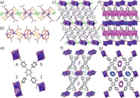
deprotonation of the linker. The rectangular linker affords a large channel 1 containing the ethanol guests and a smaller channel 2 containing water (Figure 1 c,f). [Ce(HTCPB)] (3) is accessed by desolvation of 1 (Figures 1 and S7–9), is permanently porous[19]to CO
2and N2, and displays a
rever-sible water sorption isotherm (Figures S10 and S11). The defining structural units of1are Ce2dimers, formed by four
bridging carboxylates (from carboxyphenyl rings 4 and 5 of the linker), and coordinated terminally by non-bridging protonated COOH (from carboxyphenyl ring 1) and carbox-ylate (from carboxyphenyl ring 2), plus EtOH and H2O
ligands (Figure 1 a). These dimers are connected into sheets through the HTCPB molecule, but isolated from dimers in adjacent sheets by the capping H2O and EtOH ligands.
[image:1.595.304.545.175.344.2]Compound3is formed in a stepwise desolvation process (via an unusual intermediate,2), in which both of these ligands are substituted by the carboxylate on carboxyphenyl ring 2, which now bridges two neighboring dimers (Figure 1 b) to link the sheets in three dimensions (Figure 1 d). This requires reor-ientation of the HTCPB linker with the rotation of carboxy-phenyl ring 2 (Table S2), and reorients the dimers in3to align their Ce–Ce vector more closely to the channel direction (Table S3), thus opening up channel 2 (Figure 1 f,g). Hydro-gen atoms at the 3 and 6 positions of the central benzene ring project into channel 1, with channel 2 decorated by hydrogen Figure 1. Structures of1and3. a) Coordination environment of Ce in 1: coordinated EtOH and H2O shown in green and cyan, respectively; the purple Ce centers form carboxylate-bridged dimers; the non-bridging carboxylate on carboxyphenyl ring 2 orange; C gray, O red, H white. b) Coordination environment of Ce in3; the carboxylates on carboxyphenyl ring 2 connect the dimers. c) View along [010] of1, Ce coordination environment shown as purple polyhedra. d) View along [010] of3: the carboxyphenyl ring 2 carboxylate connects the Ce dimers; Channel 1 magenta, channel 2 blue. e) Connectivity of the HTCPB ligand viewed normal to the central benzene ring plane in3 with pendant ring numbering (Figure S8 is the parallel view; Figure S6 shows equivalent views of1). f)1viewed along [100], with channel EtOH magenta, H2O dark blue. g) View of3along [100].
[*] Dr. J. E. Warren, C. G. Perkins, Dr. K. E. Jelfs, Dr. P. Boldrin, Dr. P. A. Chater, Dr. G. J. Miller, Dr. T. D. Manning, Dr. M. E. Briggs, Dr. K. C. Stylianou, Dr. J. B. Claridge, Prof. M. J. Rosseinsky Department of Chemistry, University of Liverpool Liverpool, L69 7ZD (UK)
E-mail: [email protected]
Homepage: http://www.liv.ac.uk/chemistry/research/rosseinsky-group/
[**] We thank the EPSRC for support under EP/H000925. We thank A. McLennan for experimental assistance.
atoms from the four pendant benzoates. Carboxyphenyl ring 1 is unique, as the COOH group coordinates solely to one metal center, with the OH moiety lining channel 2. Compound3thus displays a hierarchy of structure-forming bonds, with pore shapes defined by ligand torsions and displacements from1, and has channel surfaces comprising a range of functionalities.
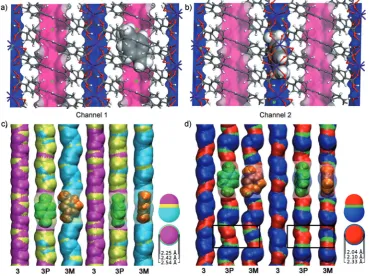
To investigate the potential for selective xylene isomer uptake, docking calculations were performed with rigid3as a host. These indicated that the topography of the pores permits occupation by pX, while excluding uptake of the similarly sized mX (Figure 2 a,b). In the larger channel 1,
several positions are available for the guest, with the preferred location defined by the formation of two sym-metrical C H···pinteractions[20]from the central benzene ring
of the HTCPB ligand of the framework to the pX benzene ring (Figure 2 a). The Ce2dimer defines a pocket in channel 2
the length of which matches that of pX, with the constriction beyond this pocket defined by the terminal COOH on carboxyphenyl ring 1 preventing occupation of other loca-tions in this channel (Figure 2 b). The pX orientation is fixed by the smaller channel 2 dimensions, which from GCMC
simulations produce a lower computed occupancy of 13 % than the fully occupied channel 1, thus showing that3does not have optimized capacity for pX.
Batch experiments on bulk powder samples in liquid C8
isomer mixtures established that3is selective for the uptake of pX over the other xylene isomers and ethylbenzene (EB) (Figure S16): the selectivities are determined by GC measure-ments. An apXmX selectivity of 4.5 (Tables S8–10: kinetic
diameters pX 5.8 , mX 6.4 ) was measured. The selectiv-ities for pX overortho-xylene (6.5 ) and EB (5.8 ) of 5.6 and 2.4, respectively, are also high.[21] Current pX/mX
separation processes use (K, Ba)-exchanged and K-exchanged zeolite Y, for which the selectivity param-eter apXmX=4 and 4.5,
respectively.[22] Rigid MOF
materials can produce equiv-alent performance (e.g. MIL-125(Ti)-NH2 with
apXmX=4.4)[23]based on the
same intrapore xylene pack-ing separation mechanism.
The observation that 3 sorbs mX is contrary to the rigid lattice docking predic-tions, and indicates a flexible structural response to guests. The reasons for this combi-nation of selectivity and flex-ibility were then identified in single-crystal structure determinations of the [Ce-(HTCPB)·(xylene)] phases, denoted 3 P and 3 M, formed by loading pX and mX, respectively, into 3 (Figures S18–S24 and Table S12). Both xylene iso-mers are adsorbed individu-ally to the same extent, with the observed capacity for pX almost double that com-puted for rigid3because of the guest-driven restructur-ing of the host. The HTCPB linker pivots about its cen-tral benzene ring upon xylene loading to exchange the dimer-forming and -bridging roles of rings 5 and 2 in 3 by correlated motion along the dimer chain, while leaving the Ce coordination intact (Figure 3 and S21).
The channel geometries in3and the guest-loaded3 Pand 3 Mare shown in Figure 2 c,d. In3, the larger channel 1 is also more cylindrical than channel 2, which is straight, but pocketed at the dimer positions. Channel 1 largely retains cylindrical character when pX is sorbed to form 3 P, but becomes less regular in diameter with the formation of pockets at the pX guest locations. This is achieved by an Figure 2. Computed guest locations in rigid3, and measured channel relaxation upon guest loading.
[image:2.595.53.423.215.491.2]expansion (of ca. 0.2 ) around the larger benzene ring section of the pX guest coupled with narrowing (magenta in Figure 2 c) around the smaller methyl groups. This distortion of the host to match the shape of the guest results in crystallographic order of pX in these well-defined locations. In contrast, mX loading expands channel 1 in3 Mto a greater extent (ca. 0.3 ; cyan in Figure 2 c) and imposes a zig-zag geometry on the channel. This more pronounced structural change is required by the angular disposition of the methyl groups in mX, but does not however produce a unique guest position:3 Mhas extensive translational positional disorder of mX along the channel.
The long axis of pX aligns with the channel 1 direction in 3 P (Figure 4 a), in contrast to the modeled orientation in unrelaxed3where this channel is sufficiently wide at the guest location for pX to tilt across it (Figure 2 a). This is consistent with relaxation of the channel to optimize the fit between
guest and host van der Waals surfaces. The pX position is defined by two symmetrical C H···p interactions on both faces of the guest with the hydrogen atoms of the HTCPB central benzene ring (Figures S24–26, and Tables S13 and S14).[24]mX in channel 1 of3 Mis rotated away from the pX
orientation by 168 about the channel direction (Figure 4 c), forming only one C H···p interaction with the host—two symmetrical contacts are not possible for mX, which has a poor shape fit at long distances from the framework and unfavorable steric H···H clashes at short distances.
The reconstruction of the narrower, pocketed channel 2 is less homogeneous than that of channel 1 because more guest-specific relaxation is needed to enable xylene sorption (Figure 2 d). The guest occupies the pockets defined by the good match between the length of the Ce2 dimer and pX
(Figure 4 f). These pockets become more pronounced in3 P through shape-driven relaxation of the host around the guest. The narrow region now occupied by the pX methyls extends further along the channel direction in3 Pthan in3, whereas the wide region occupied by the guests benzene ring is reduced in extent. Channel 2 in 3 M has similar dog-leg geometry and pocket dimensions to3 P, but the imperfect mX fit is signaled by a guest ring hydrogen clashing with the channel surface (Figure 5 c) and the guest positional disorder over two sites, in contrast to the single well-matched site occupied by pX (Figure S20).
[image:3.595.299.544.60.225.2]The relaxation of channel 2 affords full xylene occupancy of the pockets for both3 Pand3 M. In3 P (Figure 4 b) this expansion is spatially modulated to optimize the match between the channel surface chemistry and the two structural components of pX (Figure 4 e,f). The guest methyl groups form two C H···O bonds to the terminal COOH units of the Figure 3. The structural roles of rings 2 and 5 in bridging and forming
[image:3.595.50.289.61.360.2]the Ce2dimers are exchanged when3 Pforms from3upon xylene loading (the same change occurs upon formation of3 M); carboxy-phenyl ring 1 purple, ring 2 green, ring 4 yellow, ring 5 blue. a)3viewed along [010]; the 1,2 edge of the central benzene ring of the ligand is located between the dimers, with carboxyphenyl ring 2 bridging them, and with the 4,5 edge carboxylates forming the dimers. b)3 Pviewed along [010]; each edge of the central benzene now has one group (2 or 4) involved in dimer formation, and one (1 or 5) between the dimers, with 5 bridging. c)3viewed along [001]. d)3 Pviewed along [001]. Rotation of the central benzene ring and displacement of successive dimer chains pivots the HTCPB linker about the fixed rings (1 and 4), allowing rings 2 and 5 to change function in3 P. Figure S21 shows the changes of the carboxylate positions along the dimer chain associated with this transformation.
Ce2dimer: these units can rotate to optimize this 3.442(6)
interaction as carboxyphenyl ring 1 only forms a single Ce O bond. The aromatic ring surface of the guest forms two symmetrical C H···p interactions with hydrogen atoms on carboxyphenyl ring 4 of the linker. The mX in channel 2 of 3 M(Figure 4 d) undergoes rotation and disordered positional displacement away from the pX location to minimize unfavorable close contacts to the framework (Figures S27– 30). The angular disposition of the mX methyl groups permits only one weak (4.03(2) ) C H···O interaction and the mX C H···p interactions are also asymmetric (Figure 4 g,h). Reconstruction of channel 2 creates one optimized guest site in3 P, but two close poorly-matched sites in 3 M, each half-occupied in the average structure.
The distortion required to accommodate mX forces the unfavorable creation of a large diameter region within channel 2 of3 M, which isnotoccupied by any part of the mX molecule (Figure 2 d), and is defined by three H atoms from rings 1 and 5 of HTCPB (Figure 5 a,b). Rotation of ring 5 is driven by its close contact with hydrogen at position 5 of the mX aromatic ring (Figure 5 c,e). This does not occur in 3 P, where the resulting free space is visible in Figure 5 d. The carboxyphenyl ring 5 torsion angle (O58-C56-C53-C52) thus differs by 4.168 between 3 P and 3 M (Figure S32 and Table S15). Enhanced ring 1 rotation over3 Pis required to allow the COOH unit to form the single C H···O bond in3 M. The guest-induced rotation of rings 5 and 1 relocates their hydrogen atoms to create the guest-free expanded region.
Structural analysis reveals that3is not well-matched to pX or mX, and relaxes on loading to optimize capacity and fit for each guest—the superior fit to pX in3 Pis achieved with less distortion than required for the inferior match to mX in 3 M. GCMC calculations (Table S4) on the3 Pstructure show that the structural relaxation from3doubles the capacity for
pX as found experimentally, demonstrating that flexibility is essential for the observed uptake. The linker rotation observed cooperatively modifies the interactions highlighted in channels 1 (e.g. C H···p interaction from the central benzene ring) and 2 (e.g. C H···O interaction from carboxy-phenyl ring 1). Competitive 2-component calculations give a computed thermodynamic selectivity ofapXmX=6.25. As the
initial structural match to, and the subsequent differential host relaxation around, the pX and mX guests are both involved in selection between them, variation in selectivity with lanthanide size across the family of [Ln(HTCPB)] phases (accessible for Ln=La–Sm) might be expected. The exper-imentally measuredapXmXselectivity reaches a maximum of
[image:4.595.61.538.57.256.2]6.33 at Nd(HTCPB) (Table S16). Rigid host GCMC calcu-lations (Table S17) indicate [Ce(HTCPB)]3should bemore selective than the Nd analogue, suggesting that the dynamic structural relaxation of the host around the competing guests, rather than their match to the initially rigid lattice structure, controls the extent of selectivity. Consistent with this, determination of the crystal structures of the Nd analogues 3-Nd, 3 P-Nd and 3 M-Nd (Tables S18 and S19) reveal structural relaxation of the host on xylene loading, producing guest molecules located in very similar positions to their Ce analogues, but with smaller cell dimensions in each case. This is consistent with the reduced contact distances in the Nd materials3 P-Nd and3 M-Nd cooperatively amplifying both unfavorable and favorable interactions that are present in3 P and3 M, and thus enhancing the selectivity over that found in the Ce system because of the differential relaxation around the two guests, in contrast to calculations based on a rigid structural response of the Ce and Nd hosts. Detailed studies of desorption kinetics and cyclability will be needed to evaluate the suitability of3and its analogues for practical separations based on this selective sorption.
In conclusion,3responds flexibly to two similarly-shaped guests—it expands to enhance its capacity for pX and to admit mX, which cannot enter rigid3at all due to shape mismatch, to the same loading level—and yet distinguishes strongly between them despite adapting its shape to both of them because the flexible response differentiates between the two molecules. The restructuring around the preferred pX is synergic, with positive feedback between distortion of the host and enhanced fit to the guest through localized expansion and contraction. The flexibility needed to accom-modate mX involves negative feedback between rearrange-ment to generate a compromise guest location, signaled by mX positional disorder, and the creation of unused void space remote from the guest. Nature frequently exploits conforma-tional change of an initially mismatched biomolecule host during molecular recognition to enhance specificity.[25] This
differential relaxation around similar guests is a route to high selectivity for synthetic porous solids, when larger host restructuring is needed to accommodate molecules other than the preferred target, but gives a poorer fit. This suggests that when flexible hosts are used, identification of “off-target” as well as “perfect match” structures, defined in terms of capacity for and structural fit to the preferred guest, is a valuable approach when selecting between molecules with complex shapes.
Received: August 30, 2013 Revised: December 23, 2013 Published online: March 26, 2014
.
Keywords: host-guest systems · metal-organic frameworks · structural rearrangement · xylenes[1] J. R. Li, J. Sculley, H. C. Zhou,Chem. Rev.2012,112, 869 – 932. [2] H.-C. Zhou, J. R. Long, O. M. Yaghi,Chem. Rev.2012,112, 673 –
674.
[3] C. Serre, C. Mellot-Draznieks, S. Surble, N. Audebrand, Y. Filinchuk, G. Ferey,Science2007,315, 1828 – 1831.
[4] S. Horike, S. Shimomura, S. Kitagawa,Nat. Chem.2009,1, 695 – 704.
[5] a) T. K. Maji, R. Matsuda, S. Kitagawa,Nat. Mater.2007,6, 142 – 148; b) S. B. Choi, H. Furukawa, H. J. Nam, D. Y. Jung, Y. H. Jhon, A. Walton, D. Book, M. OKeeffe, O. M. Yaghi, J. Kim, Angew. Chem.2012,124, 8921 – 8925;Angew. Chem. Int. Ed. 2012,51, 8791 – 8795.
[6] A. Corma,Chem. Rev.1995,95, 559 – 614.
[7] D. Peralta, G. Chaplais, A. Simon-Masseron, K. Barthelet, G. D. Pirngruber,Ind. Eng. Chem. Res.2012,51, 4692 – 4702. [8] a) Y.-S. Bae, C. Y. Lee, K. C. Kim, O. K. Farha, P. Nickias, J. T.
Hupp, S. T. Nguyen, R. Q. Snurr,Angew. Chem.2012,124, 1893 – 1896; Angew. Chem. Int. Ed. 2012, 51, 1857 – 1860; b) E. D.
Bloch, W. L. Queen, R. Krishna, J. M. Zadrozny, C. M. Brown, J. R. Long,Science2012,335, 1606 – 1610.
[9] Z. R. Herm, B. M. Wiers, J. A. Mason, J. M. van Baten, M. R. Hudson, P. Zajdel, C. M. Brown, N. Masciocchi, R. Krishna, J. R. Long,Science2013,340, 960 – 964.
[10] a) H. Hayashi, A. P. Cote, H. Furukawa, M. OKeeffe, O. M. Yaghi,Nat. Mater.2007,6, 501 – 506; b) P. Serra-Crespo, M. A. van der Veen, E. Gobechiya, K. Houthoofd, Y. Filinchuk, C. E. A. Kirschhock, J. A. Martens, B. F. Sels, D. E. De Vos, F. Kapteijn, J. Gascon,J. Am. Chem. Soc.2012,134, 8314 – 8317. [11] H. Sato, W. Kosaka, R. Matsuda, A. Hori, Y. Hijikata, R. V. Belosludov, S. Sakaki, M. Takata, S. Kitagawa,Science2013,343, 167 – 170.
[12] J.-P. Bellat, M.-H. Simonot-Grange, S. Jullian,Zeolites1995,15, 124 – 130.
[13] a) P. S. Brcia, D. Guimaraes, P. A. P. Mendes, J. A. C. Silva, V. Guillerm, H. Chevreau, C. Serre, A. E. Rodrigues,Microporous Mesoporous Mater.2011,139, 67 – 73; b) Z. Y. Gu, X. P. Yan, Angew. Chem.2010, 122, 1519 – 1522;Angew. Chem. Int. Ed. 2010,49, 1477 – 1480.
[14] L. Alaerts, M. Maes, L. Giebeler, P. A. Jacobs, J. A. Martens, J. F. M. Denayer, C. E. A. Kirschhock, D. E. De Vos, J. Am. Chem. Soc.2008,130, 14170 – 14178.
[15] R. El Osta, A. Carlin-Sinclair, N. Guillou, R. I. Walton, F. Vermoortele, M. Maes, D. de Vos, F. Millange,Chem. Mater. 2012,24, 2781 – 2791.
[16] V. Finsy, C. E. A. Kirschhock, G. Vedts, M. Maes, L. Alaerts, D. E. De Vos, G. V. Baron, J. F. M. Denayer,Chem. Eur. J.2009, 15, 7724 – 7731.
[17] a) G. Sheldrick, Acta Crystallogr. Sect. A 2008, 64, 112 – 122; b) O. V. Dolomanov, L. J. Bourhis, R. J. Gildea, J. A. K. Howard, H. Puschmann,J. Appl. Crystallogr.2009,42, 339 – 341. [18] O. K. Farha, K. L. Mulfort, J. T. Hupp,Inorg. Chem.2008,47,
10223 – 10225.
[19] J. Rouquerol, F. Rouquerol, K. S. W. Sing, Adsorption by Powders and Porous Solids: Principles, Academic Press, London,1999.
[20] a) G. R. Desiraju, T. Steiner, The Weak Hydrogen Bond: In Structural Chemistry and Biology, Oxford University Press, Oxford,2001; b) S. K. Burley, G. A. Petsko,Science1985,229, 23 – 28; c) S. Tsuzuki,Struct. Bonding (Berlin)2005,115, 149 – 193.
[21] L. Alaerts, C. E. A. Kirschhock, M. Maes, M. A. van der Veen, V. Finsy, A. Depla, J. A. Martens, G. V. Baron, P. A. Jacobs, J. E. M. Denayer, D. E. De Vos,Angew. Chem.2007,119, 4371 – 4375;Angew. Chem. Int. Ed.2007,46, 4293 – 4297.
[22] J.-P. Bellat, M.-H. Simonot-Grange,Zeolites1995,15, 219 – 227. [23] F. Vermoortele, M. Maes, P. Z. Moghadam, M. J. Lennox, F. Ragon, M. Boulhout, S. Biswas, K. G. M. Laurier, I. Beurroies, R. Denoyel, M. Roeffaers, N. Stock, T. Duren, C. Serre, D. E. De Vos,J. Am. Chem. Soc.2011,133, 18526 – 18529.
[24] S. K. Wolff, D. J. Grimwood, J. J. McKinnon, M. J. Turner, D. Jayatilaka, M. A. Spackman, CrystalExplorer, Version 3.0,2012. [25] Y. Savir, T. Tlusty,Plos One2007,2, e468.
![Figure 4. Experimentally determined environments of the xylene guestsxylene plane with xylene aligned flat for pX inxylene shown in purple, these are removed for clarity in (f) and (h).e) View down [100] for pX inof pocket surrounding the xylenes in channe](https://thumb-us.123doks.com/thumbv2/123dok_us/8056623.224957/3.595.50.289.61.360/figure-experimentally-determined-environments-guestsxylene-inxylene-surrounding-xylenes.webp)
![Figure 5. The guest-free expanded region in channel 2 of 3M (highlighted in Figure 2d) shown in dark blue viewed down a) [001] and b) [100];H12, H51, and H52 from rings 1 and 5 are represented as spheres of vdW radius](https://thumb-us.123doks.com/thumbv2/123dok_us/8056623.224957/4.595.61.538.57.256/figure-expanded-channel-highlighted-figure-viewed-represented-spheres.webp)
