JOURNAL OFVIROLOGY, Feb. 2003, p. 2157–2164 Vol. 77, No. 3
0022-538X/03/$08.00⫹0 DOI: 10.1128/JVI.77.3.2157–2164.2003
Copyright © 2003, American Society for Microbiology. All Rights Reserved.
Nuclear Localization of the Duck Hepatitis B Virus Capsid Protein:
Detection and Functional Implications of Distinct Subnuclear
Bodies in a Compartment Associated with RNA
Synthesis and Maturation
He´le`ne Mabit,† Andreas Knaust, Klaus M. Breiner,‡ and Heinz Schaller*
ZMBH, University of Heidelberg, 69120 Heidelberg, Germany
Received 12 August 2002/Accepted 24 October 2002
We have reinvestigated the subcellular distribution of the duck hepatitis B virus (DHBV) core protein in infected duck hepatocytes and in transfected cells. By using indirect immunofluorescence, the protein was found to be localized not only in the cytoplasm, as described previously, but also within the cell nucleus, being concentrated in distinct, brightly staining nuclear core bodies (NCBs). In colocalization studies using confocal microscopy, the NCBs were found exclusively in the periphery of nuclear subdomains characterized as splicing factor compartments and distal to other subnuclear domains. Also relevant for their functional significance is that the NCBs formed during the establishment of virus infection, i.e., at very low overall concentrations of newly synthesized core protein, and persisted throughout all stages of infection. Moreover, a subset of NCBs colocalized with foci of pregenomic DHBV RNA present at concentrations detectable by fluorescence in situ hy-bridization. Taken together, these findings indicate that a minor fraction of the DHBV core protein molecules escapes the major cytoplasmic assembly pathway to accumulate in specific subnuclear domains, and they fur-thermore suggest that these NCBs serve a role in the synthesis and/or maturation of the DHBV RNA pregenome.
The hepadnaviruses (hepatitis B viruses [HBVs]) are a fam-ily of small, enveloped DNA viruses comprising the medically important human HBV and other members infecting rodents and birds (9). The HBVs are characterized by containing a small, circular, noncovalently closed DNA genome and having a compact genome organization and also a unique replication strategy that involves reverse transcription of an RNA prege-nome. Early in infection, the incoming DNA genome is deliv-ered to the host cell nucleus, where it is converted from an open circle to a covalently closed circular form, which in turn serves as the transcriptional template for pregenomic and sub-genomic RNA synthesis (9, 31). Out of a small set of gene products, the polymerase and the core protein assemble with the pregenomic RNA to form an immature, RNA-containing nucleocapsid. Genome maturation progresses in a multistep process involving reverse transcription and finally leading to mature nucleocapsids containing the open circular DNA ge-nome as present in the virion (35). Early in infection, these nucleocapsids deliver the genome back to the nucleus, ampli-fying the pool of intranuclear DNA templates (36). At later stages with abundant envelope protein expression, mature nu-cleocapsids attach to intracellular membranes containing bud-ding envelope structures, initiating virion formation and secre-tion (22).
According to the basic replication cycle outlined above, the
core protein serves primarily as the structural, nucleic acid binding component of the nucleocapsid and is additionally a cofactor in viral DNA synthesis, including reverse transcrip-tion. These functions require, as essential elements, several clusters of arginine residues which are located in the carboxy-terminal segment of the amino acid sequence following the capsid assembly domain (13, 24, 28). These sequence elements are additionally part of a nuclear localization signal, which is involved in targeting the nucleocapsid to the nuclear pore for genome import and which is multipartite in mammalian hep-adnaviruses (6, 17, 39) and monopartite in the avian duck HBV (DHBV) (21). The nuclear localization signal function seems to be counterbalanced, probably by phosphorylation (17, 19; A. K. Weigand, A. Knaust, and H. Schaller, unpublished data), thus precluding nuclear accumulation of core protein and sup-porting the cytoplasmic localization and capsid assembly ob-served for all hepadnaviruses in productively infected or trans-fected cells. However, a predominant nuclear localization of HBV core protein has been reported to occur in various ex-perimental systems, such as in the livers of chronic HBV car-riers (3) or of HBV-transgenic mice (11, 12), as well as in a particular HBV-transfected cell line (40). Electron microscopy of liver sections and further biochemical analysis revealed that this phenotype correlated with the presence of capsids free of viral nucleic acid, a result interpreted as arising from unbal-anced import of core protein subunits (11; H. W. Zentgraf, C. Kuhn, and H. Schaller, unpublished results). No functional significance has so far been attributed to these observations, and presently the assumption prevails that intranuclear core protein assembly may be a functionally irrelevant by-product of disregulated intracellular core protein traffic.
Further support for this notion come from studies with DHBV, an animal model that has been extensively employed
* Corresponding author. Mailing address: ZMBH, University of Heidelberg, Im Neuenheimer Feld 282, 69120 Heidelberg, Germany. Phone: 49 6221 54 68 85. Fax: 49 6221 54 58 93. E-mail: hshd@zmbh .uni-heidelberg.de.
† Present address: Europa¨isches Patentamt, 80331 Munich, Ger-many.
‡ Present address: Global LifeScience Ventures, 80539 Munich, Germany.
2157
on November 8, 2019 by guest
http://jvi.asm.org/
to elucidate the molecular biology of hepadnavirus replication in vivo and in vitro. In contrast to the results with mammalian hepadnaviruses, for DHBV only cytoplasmic and no intranu-clear core protein accumulation has been reported (see e.g., references 8 and 41), except for a recent report describing unidirectional nuclear import and retention of assembly-in-competent, green fluorescent protein (GFP)-fused DHBV core polypeptides (21). In a follow-up to these observations, we have now used the DHBV system to readdress the question of the general nature of nuclear core protein import and of its potential functional significance. Modifying previous reports, we demonstrate that a minor fraction of the assembly-compe-tent, full-length protein escapes the major assembly pathway to accumulate in the cell nuclei of DHBV-infected duck hepato-cytes. There it forms distinct nuclear core bodies (NCBs) that map very close to two subnuclear splicing factor compartments and that accumulate pregenomic DHBV RNA to concentra-tions occasionally detectable by fluorescence in situ hybridiza-tion (FISH). Taken together, these findings suggest that the hepadnavirus core protein has a so-far-unrecognized, non-structural function in the posttranscriptional viral RNA me-tabolism.
MATERIALS AND METHODS
Virus, plasmids, and antibodies.DHBV subtype 16 was used throughout the study. pCD16, a plasmid starting at position 2520 under control of the cytomeg-alovirus immediate-early promoter-enhancer, has been described previously (26), as have pCDcore, a plasmid expressing the DHBV core protein as the only
gene product (21), and pHBV-P7, a plasmid containing the full-size DHBV genome bearing a TTG3TAG mutation at DHBV nucleotide 183 that intro-duces a stop codon at amino acid 5 of the polymerase open reading frame (29). The core protein was detected by immunofluorescence with a polyclonal anticore antiserum (D087) as described previously (21). We performed colocalization studies of the NCBs with several focus-forming nuclear factors by using the following antibodies: a mouse anti-P80-coilin monoclonal antibody (immuno-globulin G2a) (provided by A. Lamond, University of Dundee, Dundee, Scotland United Kingdom); an anti-splicing cofactor SC35 monoclonal antibody (immu-noglobulin G1) (Sigma); a guinea pig antinucleolin antibody (30) (provided by M. Schmidt-Zachmann, DKFZ, Heidelberg, Germany); and a mouse anti-Sm protein (systemic lupus erythematosus marker) monoclonal antibody (clone Y12; Neomarkers). Anti-nuclear pore complex monoclonal antibody 414 was provided by D. Go¨rlich, ZMBH, Heidelberg, Germany.
Cell culture, infection, and transfection.DHBV-positive livers were obtained from 4- to 6-week-old ducks infected congenitally or experimentally. Primary duck hepatocytes (PDHs) were prepared and cultivated as described previously (14). For infection of negative PDHs, DHBV-containing duck serum was applied to one well of a 12-well culture dish containing 8⫻105cells at a multiplicity of
infection of 100. After 14 h of incubation, the cells were washed and further cultivated. After 3 days, the cells were fixed and immunostained for DHBV core protein as described below. The human hepatoma cell line HuH7 was cultivated on coverglass chamber slides (Nunc) and transfected with the plasmids by using a standard calcium phosphate protocol. For immunofluorescence analysis, the cells were fixed 3 to 4 days after transfection as described below.
Immunofluorescence and Western blotting.At 3 days postinfection or post-transfection, the cells, PDHs, or transfected HuH7 cells were washed with phos-phate-buffered saline (PBS) and fixed with 3% formalin for 30 min. The fixed cells were permeabilized with 0.25% Triton X-100 in PBS and immunostained with a rabbit polyclonal antibody against the DHBV core protein (D087) as a primary antibody and an fluorescein isothiocyanate (FITC)-conjugated anti-rabbit immunoglobulin as a secondary antibody. Cell nuclei were isolated by using a standard protocol based on the method of Widnell and Tata (38).
FIG. 1. DHBV core protein forms nuclear foci in DHBV-infected hepatocytes. Primary duck hepatocytes (A and C) or isolated nuclei (B) were fixed and immunostained for DHBV core protein. After counterstaining for chromatin (4⬘,6⬘-diamidino-2-phenylindole [DAPI]) (A) or the nuclear pore complex (NPC) (B and C), the cells or nuclei were analyzed by conventional fluorescence microscopy (A) or confocal laser scanning microscopy (B and C). The NCBs remained stable in the isolated nuclei after removal of the outer nuclear membrane and chromatin extraction
(B) (for details, see Materials and Methods).
on November 8, 2019 by guest
http://jvi.asm.org/
Isolated nuclei were washed twice in 1% Triton X-100 in 10 mM Tris–5 mM MgCl2–0.25 M sucrose. Chromatin was extracted by digestion with 400 U of
DNase I per ml in cytoskeletal buffer {10 mM PIPES [piperazine-N,N ⬘-bis(2-ethanesulfonic acid)] [pH 6.8], 300 mM sucrose, 50 mM NaCl, 3 mM MgCl2,
1 mM EGTA, 20 mM vanadyl riboside complex, 1 mM 4-(2-aminoethyl)benzene sulfonyl fluoride} for 30 min at 37°C followed by washing twice with 1 M NaCl buffer (cytoskeltal buffer adjusted to 1 M NaCl) (25).
For costaining, the samples were incubated at the same time with D087 and one of the different primary mouse antibodies. FITC-conjugated anti-rabbit or tetramethyl rhodamine isocyanate-conjugated anti-mouse immunoglobulin was used as a secondary antibody. Fluorescence was analyzed either with a conven-tional microscope or with a Leica TCS NT confocal laser scanning microscope (63⫻/1.23 objective). For monitoring core expression biochemically (see Fig. 2), cells were lysed in sample buffer and equivalents of 8⫻104cells per time sample
were loaded on a sodium dodecyl sulfate gel. Western blotting analysis was performed as described previously (22).
In situ hybridization.To generate an antisense probe specifically detecting the pregenomic RNA (from restriction siteAvrII toEcoRI, nucleotide [nt] 403 to 3018 of DHBV16), we used the plasmid pCD16, displaying a T7 promoter after theNheI restriction site in antisense direction. After deleting theAvrII-NheI fragment from pCD16 (nt 911 to 3335) and linearizing the obtained plasmid with EcoRI, we generated a digoxigenin-labeled RNA probe by in vitro transcription with T7 polymerase as detailed by the manufacturer (Boehringer Mannheim). To generate a sense probe detecting genomic DHBV DNA (from nt 3018 to 391 of DHBV16), we subcloned theEcoRI-BamHI fragment of pCD16 (nt 504 to 2166) downstream of an SP6 promoter sequence of plasmid pSPT19 (Boehringer Mannheim). The resulting plasmid was cut withBglII, serving as starting product for in vitro transcription.
To examine colocalization of DHBV RNA relative to DHBV core protein, we used the protocol used by Seguin et al. (32). PDHs prepared from a noninfected duck liver were cultivated on glass coverslips and infected as described above. At 3 days postinfection, coverslips were washed with PBS and fixed at room tem-perature for 10 min in 4% paraformaldehyde–PBS buffered to pH 7.4. Following fixation, coverslips were washed twice with PBS and stored in 70% ethanol at 4°C. For prehybridization, each coverslip was inverted onto 100l of prehybrid-ization solution (50% deionized formamide, 2⫻SSPE [1⫻SSPE is 0.18 M NaCl, 10 mM NaH2PO4, and 1 mM EDTA {pH 7.7}], 5⫻Denhardt’s reagent, 1 mg of
herring sperm DNA per ml), and the chamber was sealed with Parafilm and incubated for 1 h at 37C. The appropriate RNA probe was resuspended at 1g/
l in fresh hybridization solution and heated at 80°C for 10 min. The coverslips were then placed on 30l of fresh hybridization solution, and the chambers were sealed with Parafilm and incubated overnight at 37°C. Unbound probe was removed by four 15-min washes in 50% deionized formamide–2⫻SSPE at 37°C. Samples were then incubated with 1% blocking reagent (Boehringer Mannheim) containing the rabbit polyclonal anti-DHBV core antibody. Samples were then washed and incubated with both FITC-conjugated sheep digoxigenin anti-body (1/40 dilution) and Texas Red-conjugated goat anti-rabbit immunoglobulin antibody (1/80 dilution). Coverslips were mounted in antibleach medium (Boehr-inger Mannheim), and the fluorescence was analyzed with a Leica TCS NT confocal laser scanning microscope (63⫻/1.23 objective). To test for colocaliza-tion of DHBV DNA, we followed the same protocol except that (i) the fixed cells were heated at 70°C for 2 min in 70% deionized formamide–2⫻SSPE prior to hybridization with the sense DHBV RNA probe and (ii) the cells were incubated with the sense DHBV RNA probe at 42°C overnight.
RESULTS
[image:3.603.49.277.68.293.2]DHBV core protein localizes to distinct nuclear foci in DHBV-infected cultured hepatocytes. PDHs that had been prepared from a DHBV-infected animal were analyzed, after fixation with paraformaldehyde, for the subcellular distribution of the core protein by using indirect immunofluorescence with a DHBV core-specific antiserum. As shown in Fig. 1A, micro-scopic analysis of such samples revealed that all DHBV core-positive cells contained, in addition to the diffuse cytoplasmic staining, DHBV core protein in one, or occasionally several,
FIG. 2. Kinetics of NCB formation after DHBV infection. PDHs were infected in vitro with a multiplicity of 100 DNA-containing DHBV particles per cell. After 1, 2, 3, or 4 days, cells were fixed, immunostained for DHBV core protein, and analyzed with a confocal microscope (main panels). In parallel, cell lysates were analyzed by Western blotting for DHBV core protein content (inset).
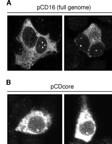
FIG. 3. Formation of nuclear core protein foci does not depend on additional viral components. HuH7 cells were transfected with pCD16, encoding the full DHBV genome (A), or pCDcore, a plasmid express-ing the core protein only (B). At 2 days posttransfection, the cells were fixed, immunostained for DHBV core protein, and analyzed with a confocal microscope. For each transfection, two examples of DHBV core-positive cells with NCBs were selected.
VOL. 77, 2003 SUBNUCLEAR COMPARTMENTS ACCUMULATING DHBV CORE PROTEIN 2159
on November 8, 2019 by guest
http://jvi.asm.org/
[image:3.603.304.537.74.372.2]on November 8, 2019 by guest
http://jvi.asm.org/
brightly staining, spherical foci which apparently resided within the cell nucleus. No such NCBs were detected in noninfected control cells (not shown here; see Fig. 2, day 1). The NCBs were also detected, although with somewhat reduced intensity and frequency, after fixation with methanol, a result arguing against artifacts due to the fixation procedure. Moreover, their nuclear localization was confirmed by confocal laser scanning microscopy sectioning through fixed PDHs, or cell nuclei that had been prepared from DHBV-infected duck livers, and ad-ditionally immunostained for the nuclear pore complex, a marker delineating the nuclear membrane (Fig. 1B and C). Interestingly, the NCB pattern was affected neither by destruc-tion of the nuclear membrane with 1% Triton X-100 nor by subsequent chromatin extraction (Fig. 1B), suggesting a rather stable structure which is not attached to chromatin.
NCBs arise during the establishment of viral infection.To characterize the kinetics of NCB formation after experimental DHBV infection of PDHs, cells were fixed at daily intervals, stained with an anti-DHBV core antiserum, and analyzed by confocal microscopy. A parallel set of infected cells was lysed and analyzed by Western blotting for total core protein con-tent. As shown in Fig. 2, nuclear foci of core protein appeared as early as day 2 postinfection, a time point when cytoplasmic core protein was not yet detected by immunostaining and when the protein was barely detectable by Western blotting (Fig. 2, inset). Thereafter, the NCBs remained essentially unchanged in size and numbers and persisted in the infected cells through-out prolonged cell culture up to 4 weeks (data not shown). For the functional relevance of these observations, it is important to note that the accumulation of core protein in NCBs, and in the cell nucleus in general, occurred well before the high steady-state levels of cytoplasmic core protein were reached, thus making an artifactual aggregation in the nuclear compart-ment unlikely. In contrast, NCBs were formed less frequently in transfected cells producing core protein more rapidly and to high cytoplasmic concentrations (see below).
NBC formation does not require any further viral compo-nent.Next, we wanted to assess whether NCB formation would depend on viral gene products other than the core protein or on the presence of the intact DHBV replication machinery. To this end, hepatoma cells of human or avian origin (Huh7 and LMH, respectively) were transfected with a set of plasmids containing different genomic and subgenomic DHBV DNA segments (for details, see Materials and Methods). The human cell line was preferentially chosen for these and further trans-fection experiments to facilitate subsequent immunofluores-cence studies determining colocalization with cellular marker proteins (see below). At 2 days posttransfection, the cells were fixed, immunostained, and analyzed by confocal microscopy for the distribution of the DHBV core protein. Expression from pCD16, a plasmid expressing a replication-competent
DHBV RNA pregenome under the control of the cytomega-lovirus immediate-early promoter, resulted only in about 5 to 10% of core-positive cells in an NCB pattern similar to the one observed in infected duck hepatocytes (Fig. 3A). A comparable intracellular DHBV core distribution was observed in cells transfected with a related genomic plasmid displaying a stop codon at the beginning of the polymerase open reading frame (data not shown) or with a plasmid in which all DHBV coding sequences following the core gene, as well as the upstream encapsidation signal, had been deleted (pCDcore) (Fig. 3B). These results demonstrate that the accumulation of core pro-tein in nuclear bodies is an intrinsic property of the propro-tein itself, predicting an interaction with specific cellular partners. They further indicate that structures relevant for such interac-tions are conserved between avian and human cells.
NCBs are located in the immediate vicinity of interchroma-tin granule speckles containing the spliceosome assembly co-factor SC35.The cell nucleus contains distinct classes of sub-nuclear bodies, including nucleoli, splicing speckles, and coiled bodies (Cajal bodies), etc., which are either attached to chro-matin or located in the interchrochro-matin space (33, 18). To define whether NCB domains were associated with known subnuclear compartments, we performed colocalization stud-ies by confocal fluorescence microscopy, using antibodstud-ies de-tecting various focus-forming nuclear proteins. Analysis of im-munostained NCBs in pCD16-transfected HuH7 cells revealed that these occupied quite characteristic positions relative to reference structures identified by antibodies specific for nucle-oli (N038), coiled bodies (p80/coilin), Sm proteins, and pre-mRNA splicing cofactor SC35 (Fig. 4): NCBs localized occa-sionally at the border of nucleoli (in about 1 out of 10 cases [Fig. 4A]), always distal to coiled bodies (in no case out of more than 20 [Fig. 4B]), and preferentially in the immediate vicinity of Sm speckles (in about 5 out of 10 cases [Fig. 4C]) but without exact overlap between domains stained for the marker protein or the core protein (visualized at a higher magnifica-tion in Fig. 4C, second column).
Even more significantly, NCBs always mapped at the periph-ery of SC35-positive substructures. This is exemplified in the two panels of Fig. 4D showing eight strong NCBs (in two focal planes through three nuclei), seven of which are also covered in the threefold-enlarged inserts. Although SC35 speckles rep-resent rather extended intranuclear substructures, NCBs were never found to map within these domains, nor were they de-tected in the much more extended space in between. Even weak NCBs that were occasionally observed very close to but slightly detached from SC35 speckles, like the one detectable at the higher magnification in the upper right corner of the left inset in Fig. 4D, were found to be directly attached to the nearby SC35-positive substructure when viewed in successive focal planes (data not shown). Taken together, these
observa-FIG. 4. NCBs map at distinct nuclear subdomains. HuH7 cells were transfected with a plasmid encoding the full DHBV genome (pCD16). At 2 days posttransfection, the cells were fixed and coimmunostained for DHBV core protein (green fluorescence) and for either nucleolus (N038 antibody) (A), coiled bodies (anti-p80/coilin antibody) (B), Sm protein (Y12 antibody) (C), or spliceosome (anti-splicing factor SC35 antibody) (D) (red fluorescence). They were then analyzed with a confocal microscope. Sequential excitation and scanning of the two fluorescent channels (separate excitations at 488 and 568 nm) were used to avoid cross bleeding of the fluorochromes between channels. For each antibody, two representative cells are shown. Bars, 10m. The insets in panels D represent threefold enlargements of the area marked by the dotted lines.
VOL. 77, 2003 SUBNUCLEAR COMPARTMENTS ACCUMULATING DHBV CORE PROTEIN 2161
on November 8, 2019 by guest
http://jvi.asm.org/
tions strongly argue that the association of NCB structures with SC35 speckles is specific, suggesting a functional correla-tion.
Sm proteins have functions in the biogenesis and are com-ponents of a subset of the small ribonucleoprotein particles that catalyze pre-mRNA splicing (27). SC35 speckles are as-sumed to be nuclear compartments for storage or recycling of splicing factors or to be more directly involved in the splicing mechanism (7, 10, 15, 16). Thus, our results indicate that the NCBs are part of distinct nuclear subdomains, which are lo-cated in the close vicinity of cellular RNA splicing compart-ments.
Foci of pregenomic DHBV RNA colocalize with core protein in NCBs.The results described above suggest that NCB for-mation reflects a specific association of core protein with a nuclear compartment involved in the control of alternative RNA splicing of the DHBV RNA pregenome (26). In a first attempt to assess this possibility, we used FISH to test whether NCB also contained viral nucleic acids. DHBV-infected PDHs were fixed and hybridized under the appropriate annealing conditions (see Materials and Methods) with an RNA probe complementary to a sequence unique for the RNA pregenome and absent from all subgenomic DHBV RNAs (map positions 1 to 415). As a control, we used a probe of opposite polarity assaying for antisense or double-stranded viral nucleotide se-quences. Within the sensitivity reached, no signal was observed with the latter probe or with a non-DHBV-specific control RNA (not shown), which is not surprising in view of the
ap-proximately 20 copies of DHBV DNA template per cell (4, 36). In contrast to the case for these controls, we saw a distinct spot-like concentration of the genomic RNA probe in about 1% of the infected hepatocytes. As exemplified in Fig. 5 by two representative RNA-positive cells, these pregenomic RNA dots consistently colocated with an NCB. Conversely, most NCBs were without RNA dots, even in cells where other NCBs overlapped with DHBV RNA foci, and there was also no clear correlation of the intensities of core staining and the prege-nome signal between individual NCB structures (Fig. 5, lower panel). While this absence of RNA dots in most NCBs is probably due to the insensitivity of our RNA analysis, the strict colocalization of all of the DHBV RNA foci detected with NCBs nevertheless confirms that both structures occupy the same nuclear compartment. Furthermore, this colocalization strongly suggests that these domains play a discrete role in the viral RNA metabolism at the level of hepadnaviral RNA syn-thesis and/or maturation in the cell nucleus.
DISCUSSION
[image:6.603.100.484.68.338.2]While the hepadnavirus core proteins were up to now be-lieved to be destined solely to become a structural and func-tional component of the cytoplasmic nucleocapsid and the secreted virion, we now demonstrate that a fraction of the assembly-competent DHBV core protein subunits accumulate in specific nuclear subdomains, termed NCBs, for which we
FIG. 5. Foci of DHBV pregenomic RNA colocalize with NCBs in a subset of infected hepatocytes. DHBV-infected PDHs were stained for pregenomic DHBV RNA by FISH (green fluorescence) (for details, see Materials and Methods) and immunostained for DHBV core protein (red fluorescence). Analysis was performed with a confocal microscope (sequential excitation and scanning of the two fluorochromes). The upper and lower panels show two representative examples of RNA-positive cells. Note that in the lower panel, the RNA colocalize with a weak NCB and not with a nearby strong NCB in the same nucleus.
on November 8, 2019 by guest
http://jvi.asm.org/
propose a role in the biosynthesis of the unspliced DHBV RNA pregenome.
The formation of these NCBs probably depends on the availability of yet-unassembled core protein subunits, which are capable of rapid nucleocytoplasmic shuttling: NCBs formed preferentially at the low overall DHBV core concentrations as present during establishment of DHBV infection, whereas they were only rarely observed in DHBV-transfected cells with high DHBV core concentrations leading to cytoplasmic capsid as-sembly. In keeping with this interpretation, an NCB-like nu-clear retention pattern was also observed in cells transfected with an assembly-incompetent GFP fusion with the C-terminal half of DHBV core (GFP/C125-262) but not in those trans-fected with a fusion to the assembly-competent full-length DHBV core (21). Together, our data indicate that the DHBV core protein subunit carries the potential for selective, high-affinity retention at yet-undefined subnuclear structures. The structures targeted seem to be limited in size and numbers, as they were already largely saturated during establishment of infection, so that the NCBs represented only a minor fraction of the total cellular core protein once the steady state of pro-ductive infection had been reached. Our findings further show that this process did not require the presence of other virus-encoded interaction partners, at least not for formation of the basic NCB structure. However, as demonstrated by the colo-calization of pregenomic DHBV RNA, our data do not ex-clude a nonessential participation of additional viral gene products such as the viral P protein, which is known to bind to the genomic RNA packaging signal as well as to contain inter-action domains for core protein sequences (20).
At present, our data allow only speculation as to the poten-tial NCB functions. The first clues come from colocalization studies showing (i) an overlap with foci accumulating the DHBV RNA pregenome and (ii) a preferential location adja-cent to nuclear speckles associated with the SR protein SC35, which is prototypic of essential, multifunctional splicing factors required at different steps of spliceosome assembly and mod-ulating constitutive and alternative pre-mRNA splicing (7, 10, 37, 42). Actively transcribed cellular genes have been shown to locate at the periphery of these subcellular domains, support-ing the existence of compartments couplsupport-ing synthesis and mat-uration of primary transcripts (15, 16, 18, 33, 34). In analogy, the NCBs may be part of factories involved in the synthesis and maturation of nascent DHBV transcripts, acting either by binding to the DHBV minichromosome as reported for HBV by Bock et al. (1) or as a more general nucleic acid interaction partner. Additionally, and not mutually exclusive, the DHBV core protein, being a nucleocytoplasmic shuttling RNA binding protein (21), may also participate in the nuclear export of the unspliced pregenomic DHBV RNA, as exemplified by the role of structural virus proteins in the ribonucleoprotein complex incorporating the unspliced RNA genomes of influenza A virus (2).
In this context it is important to recall that the DHBV RNA pregenome is subject to alternative splicing (26) and is ex-pected to pass through a cellular splicing compartment. Fur-thermore, the hepadnavirus core proteins, containing an RNA binding motif and an SR-rich protein interaction domain, ap-pear to be related to the SR protein splicing cofactor family, an assumption supported by the recent observations that the HBV
core protein is the target of SR-specific protein kinases (5). The formation of DHBV NCBs at the periphery of nuclear splicing speckles thus suggests the existence of a particular speckle subcompartment (23) which is used by DHBV to fa-cilitate the nuclear interaction of its unspliced RNA prege-nome with the core protein as a putative viral RNA export adaptor. In further speculation, the DHBV nuclear core pro-tein might also be envisaged to be directly involved in the regulation of alternative splicing of the DHBV RNA pre-genome. In initial attempts to prove this concept by RNA analysis of core gene knockout mutants, we have observed enhanced ratios of spliced RNA relative to the pregenome but have not obtained conclusive answers due to overlapping
effects from cis-acting elements also influencing pregenome
splicing (B. Zachmann-Brand and H. Schaller, unpublished data).
Finally, we may ask whether NCB formation is a feature generally associated with hepadnavirus replication. To our knowledge, an NCB-like pattern has so far not been detected in hepatocytes infected by mammalian hepadnaviruses, and even on close inspection, we have observed nuclear foci of HBV core protein in only a very minor fraction of HBV-transfected cells. This difference from the pronounced NCB formation in DHBV parallels the absence of any significant or functionally relevant RNA splicing in the replication of the mammalian hepadnaviruses and is therefore consistent with the hypothesis that the NCBs play an SR protein-like role in the maturation of the DHBV RNA pregenome.
ACKNOWLEDGMENTS
We thank Ba¨rbel Glass for the preparation of PDHs and for pro-viding duck liver tissue and sera. We are also grateful to Angus Lam-ond, Marion Schmidt-Zachmann, Dirk Go¨rlich, and Christa Kuhn for antibodies and to Karin Coutinho for expert editorial assistance.
This work was supported by a ZMBH fellowship to H.M., a grant (Scha 134/11-1) from the Deutsche Forschungsgemeinschaft to H.S., and the Fonds der Chemischen Industrie.
REFERENCES
1. Bock, C. T., S. Schwinn, S. Locarnini, J. Fyfe, M. P. Manns, C. Trautwein, and H. Zentgraf. 2001. Structural organization of the hepatitis B virus minichromosome. J. Mol. Biol.307:183–196.
2. Bui, M., E. G. Wills, A. Helenius, and G. R. Whittaker.2000. Role of the influenza virus M1 protein in nuclear export of viral ribonucleoproteins. J. Virol.74:1781–1786.
3. Chu, C. M., and Y. F. Liaw.1987. Intrahepatic distribution of hepatitis B surface and core antigens in chronic hepatitis B virus infection. Hepatocyte with cytoplasmic/membranous hepatitis B core antigen as a possible target for immune hepatocytolysis. Gastroenterology92:220–225.
4. Civitico, G. M., and S. A. Locarnini.1994. The half-life of duck hepatitis B virus supercoiled DNA in congenitally infected primary hepatocyte cultures. Virology203:81–89.
5. Daub, H., S. Blencke, P. Habenberger, A. Kurtenbach, J. Dennenmoser, J. Wissing, A. Ullrich, and M. Cotten.2002. Identification of SRPK1 and SRPK2 as the major cellular protein kinases phosphorylating hepatitis B virus core protein. J. Virol.76:8124–8137.
6. Eckhardt, S. G., D. R. Milich, and A. McLachlan.1991. Hepatitis B virus core antigen has two nuclear localization sequences in the arginine-rich carboxyl terminus. J. Virol.65:575–582.
7. Fu, X. D., and T. Maniatis.1992. Isolation of a complementary DNA that encodes the mammalian splicing factor SC35. Science256:535–538. 8. Galle, P. R., H. J. Schlicht, M. Fischer, and H. Schaller.1988. Production of
infectious duck hepatitis B virus in a human hepatoma cell line. J. Virol. 62:1736–1740.
9. Ganem, D., and R. Schneider.2001. Hepadnaviridae: the viruses and their replication, 4th ed. Lippincott Williams & Wilkins, Philadelphia, Pa. 10. Graveley, B. R.2000. Sorting out the complexity of SR protein functions.
RNA6:1197–1211.
11. Guidotti, L. G., V. Martinez, Y. T. Loh, C. E. Rogler, and F. V. Chisari.1994.
VOL. 77, 2003 SUBNUCLEAR COMPARTMENTS ACCUMULATING DHBV CORE PROTEIN 2163
on November 8, 2019 by guest
http://jvi.asm.org/
Hepatitis B virus nucleocapsid particles do not cross the hepatocyte nuclear membrane in transgenic mice. J. Virol.68:5469–5475.
12. Guidotti, L. G., B. Matzke, H. Schaller, and F. V. Chisari.1995. High-level hepatitis B virus replication in transgenic mice. J. Virol.69:6158–6169. 13. Hatton, T., S. Zhou, and D. N. Standring.1992. RNA- and DNA-binding
activities in hepatitis B virus capsid protein: a model for their roles in viral replication. J. Virol.66:5232–5241.
14. Hild, M., O. Weber, and H. Schaller.1998. Glucagon treatment interferes with an early step of duck hepatitis B virus infection. J. Virol.72:2600–2606. 15. Huang, S., and D. L. Spector.1996. Intron-dependent recruitment of pre-mRNA splicing factors to sites of transcription. J. Cell Biol.133:719–732. 16. Jimenez-Garcia, L. F., and D. L. Spector.1993. In vivo evidence that
tran-scription and splicing are coordinated by a recruiting mechanism. Cell73: 47–59.
17. Kann, M., B. Sodeik, A. Vlachou, W. H. Gerlich, and A. Helenius.1999. Phosphorylation-dependent binding of hepatitis B virus core particles to the nuclear pore complex. J. Cell Biol.145:45–55.
18. Lamond, A. I., and W. C. Earnshaw.1998. Structure and function in the nucleus. Science280:547–553.
19. Liao, W., and J. H. Ou.1995. Phosphorylation and nuclear localization of the hepatitis B virus core protein: significance of serine in the three repeated SPRRR motifs. J. Virol.69:1025–1029.
20. Lott, L., B. Beames, L. Notvall, and R. E. Lanford.2000. Interaction between hepatitis B virus core protein and reverse transcriptase. J. Virol.74:11479– 11489.
21. Mabit, H., K. M. Breiner, A. Knaust, B. Zachmann-Brand, and H. Schaller. 2001. Signals for bidirectional nucleocytoplasmic transport in the duck hep-atitis B virus capsid protein. J. Virol.75:1968–1977.
22. Mabit, H., and H. Schaller.2000. Intracellular hepadnavirus nucleocapsids are selected for secretion by envelope protein-independent membrane bind-ing. J. Virol.74:11472–11478.
23. Mintz, P. J., and D. L. Spector.2000. Compartmentalization of RNA pro-cessing factors within nuclear speckles. J. Struct. Biol.129:241–251. 24. Nassal, M.1992. The arginine-rich domain of the hepatitis B virus core
protein is required for pregenome encapsidation and productive viral posi-tive-strand DNA synthesis but not for virus assembly. J. Virol.66:4107–4116. 25. Nickerson, J. A., G. Krockmalnic, K. M. Wan, and S. Penman.1997. The nuclear matrix revealed by eluting chromatin from a cross-linked nucleus. Proc. Natl. Acad. Sci. USA94:4446–4450.
26. Obert, S., B. Zachmann-Brand, E. Deindl, W. Tucker, R. Bartenschlager, and H. Schaller.1996. A splice hepadnavirus RNA that is essential for virus replication. EMBO J.15:2565–2574.
27. Pannone, B. K., and S. L. Wolin.2000. Sm-like proteins wRING the neck of mRNA. Curr. Biol.10:R478–R481.
28. Schlicht, H. J., R. Bartenschlager, and H. Schaller.1989. The duck hepatitis B virus core protein contains a highly phosphorylated C terminus that is essential for replication but not for RNA packaging. J. Virol.63:2995–3000. 29. Schlicht, H. J., G. Radziwill, and H. Schaller.1989. Synthesis and encapsi-dation of duck hepatitis B virus reverse transcriptase do not require forma-tion of core-polymerase fusion proteins. Cell56:85–92.
30. Schmidt-Zachmann, M. S., B. Hugle-Dorr, and W. W. Franke.1987. A constitutive nucleolar protein identified as a member of the nucleoplasmin family. EMBO J.6:1881–1890.
31. Seeger, C., and W. S. Mason.2000. Hepatitis B virus biology. Microbiol. Mol. Biol. Rev.64:51–68.
32. Seguin, B., A. Staffa, and A. Cochrane.1998. Control of human immunode-ficiency virus type 1 RNA metabolism: role of splice sites and intron se-quences in unspliced viral RNA subcellular distribution. J. Virol.72:9503– 9513.
33. Spector, D. L.1993. Macromolecular domains within the cell nucleus. Annu. Rev. Cell Biol.9:265–315.
34. Spector, D. L., X. D. Fu, and T. Maniatis.1991. Associations between distinct pre-mRNA splicing components and the cell nucleus. EMBO J. 10:3467–3481.
35. Summers, J., and W. S. Mason.1982. Replication of the genome of a hepatitis B-like virus by reverse transcription of an RNA intermediate. Cell 29:403–415.
36. Tuttleman, J. S., C. Pourcel, and J. Summers.1986. Formation of the pool of covalently closed circular viral DNA in hepadnavirus-infected cells. Cell 47:451–460.
37. Wang, H. Y., X. Xu, J. H. Ding, J. R. Bermingham, Jr., and X. D. Fu.2001. SC35 plays a role in T cell development and alternative splicing of CD45. Mol. Cell7:331–342.
38. Widnell, C. C., and J. R. Tata.1964. A procedure for the isolation of enzymically active rat-liver nuclei. Biochem. J.92:313–317.
39. Yeh, C. T., Y. F. Liaw, and J. H. Ou.1990. The arginine-rich domain of hepatitis B virus precore and core proteins contains a signal for nuclear transport. J. Virol.64:6141–6147.
40. Yeh, C. T., S. W. Wong, Y. K. Fung, and J. H. Ou.1993. Cell cycle regulation of nuclear localization of hepatitis B virus core protein. Proc. Natl. Acad. Sci. USA90:6459–6463.
41. Yu, M., and J. Summers.1994. Phosphorylation of the duck hepatitis B virus capsid protein associated with conformational changes in the C terminus. J. Virol.68:2965–2969.
42. Zhu, J., A. Mayeda, and A. R. Krainer.2001. Exon identity established through differential antagonism between exonic splicing silencer-bound hnRNP A1 and enhancer-bound SR proteins. Mol. Cell8:1351–1361.