Copyright © 2002, American Society for Microbiology. All Rights Reserved.
Search for Hepatitis C Virus Negative-Strand RNA Sequences and
Analysis of Viral Sequences in the Central Nervous System:
Evidence of Replication
Marek Radkowski,
1,2Jeffrey Wilkinson,
1Marek Nowicki,
3Debra Adair,
1Hugo Vargas,
1Craig Ingui,
1Jorge Rakela,
1and Tomasz Laskus
1*
Division of Transplantation Medicine, Mayo Clinic Scottsdale, Scottsdale, Arizona 852591; Institute of Infectious
Diseases, Medical Academy, Warsaw, Poland2; and Maternal-Child Virology Research Laboratory, University of
Southern California, Los Angeles, California 900333
Received 8 August 2001/Accepted 4 October 2001
Patients with chronic hepatitis C are more likely to have significant changes in their physical and mental well-being than patients with liver disease of other etiology, and hepatitis C virus (HCV) has been occasionally implicated in diseases of the central nervous system. We analyzed the presence of the HCV negative-strand RNA sequence, which is the viral replicative intermediary, in autopsy brain tissue samples from six
HCV-infected patients. Negative-strand HCV RNA was searched for by a strand-specificTth-based reverse
tran-scriptase PCR, and viral sequences amplified from brain tissue and serum were compared by single-strand conformational polymorphism analysis and direct sequencing. HCV RNA negative strands were detected in brain tissue in three patients. In two of these patients, serum- and brain-derived viral sequences were different and classified as belonging to different genotypes. In one of the latter patients, HCV RNA negative strands were detected in lymph node and, while being different from serum-derived sequences, were identical to those present in the brain. The results of the present study suggest that HCV can replicate in the central nervous system, probably in cells of the macrophage/monocyte lineage.
Hepatitis C virus (HCV) is a common etiologic agent of chronic hepatitis, cirrhosis, and hepatocellular carcinoma (1). Although HCV is a primary hepatotropic virus, there is mount-ing evidence that it can also replicate at extrahepatic sites, particularly under conditions of immunodeficiency associated with human immunodeficiency type 1 (HIV-1) infection (16, 17, 22).
Whether HCV can infect the central nervous system (CNS) remains unclear. HCV belongs to theFlaviviridaefamily, which includes several well-known neurotropic viruses (e.g., yellow fever, dengue, and tick-borne encephalitis viruses), and several reports have implicated HCV as an occasional cause of various CNS and peripheral nervous system pathologies (3, 6, 11, 13, 27). Moreover, HCV RNA has been detected in cerebrospinal fluid from both HIV-positive and HIV-negative patients (23, 25), and viral sequences have been amplified directly from brain tissue from a patient diagnosed with progressive enceph-alomyelitis (3). However, the presence of viral sequences in any particular compartment cannot be regarded as evidence for replication, and to prove the latter, the presence of repli-cative intermediates must be established. In the case of posi-tive-strand viruses such as HCV, cells supporting replication should contain viral negative-strand RNA sequences.
In the present study we analyzed HCV RNA in autopsy brain tissue samples from six subjects, three of whom were HIV-1 positive. In addition to strand-specific detection of
HCV RNA negative strands, we compared viral sequences amplified from various CNS structures and serum, assuming that in the presence of independent viral compartments they could be different, much like what has been described for HIV-1 (10). To our knowledge, this is the first attempt to detect HCV replicative intermediaries in brain tissue and to analyze viral sequences derived from various parts of the brain.
MATERIALS AND METHODS
Biological samples.Serum and brain tissue samples were collected from six HCV-positive patients, five of whom had liver cirrhosis (Table 1). Brain tissue samples were obtained during routine autopsies conducted within 36 h after death and stored at⫺80°C until analysis. The following samples were collected: subcortical white matter and cerebral cortex from the frontal region, nucleus lentiformis, cerebellum, and medulla oblongata. However, in patient 1 only the last of these tissues was available for analysis. In addition, mediastinal lymph nodes were collected from four patients. After tissue homogenization, RNA was extracted by means of a modified guanidinium thiocyanate-phenol-chloroform technique using a commercially available kit (RNAzol; Gibco/BRL). Total RNA (1 and 5g as determined by spectrophotometry) was routinely used for reverse transcriptase PCR (RT-PCR). We found the latter amount of RNA to be the upper limit of the template, beyond which the amplification reactions would be commonly inhibited. In the case of serum, the amount of extracted RNA loaded into the reaction mixture corresponded to 100l. This study was approved by the respective ethical committees of the involved institutions.
Strand-specific RT-PCR.Strand specificity of our RT-PCR for the detection of HCV negative-strand RNA was ascertained by conducting cDNA synthesis at high temperature using the thermostable enzymeTth. The sensitivity and strand specificity of this reaction were established using synthetic RNAs as templates. A detailed description of our strand-specific assay and sequence of employed prim-ers was published previously (17, 20). In brief, the cDNA was generated in 20l of a reaction mixture containing 50 pM sense primer, 1⫻RT buffer (Perkin-Elmer), 1 mM MnCl2, 200M concentrations of each deoxynucleoside triphos-phate, and 5 U ofTth(Perkin-Elmer). After 20 min at 65°C, Mn2⫹was chelated with 8l of 10⫻EGTA chelating buffer (Perkin-Elmer), 50 pM antisense primer was added, the volume was adjusted to 100l, and the MgCl2concentration was
* Corresponding author. Mailing address: Division of Transplanta-tion Medicine, SC Johnson Bldg Sj3, Mayo Clinic Scottsdale, Scotts-dale, AZ 85259. Phone: (480) 301-6370. Fax: (480) 301-3384. E-mail: [email protected].
600
on November 8, 2019 by guest
http://jvi.asm.org/
adjusted to 2.2 mM. The amplification was performed in a Perkin-Elmer GenAmp PCR System 9600 thermocycler as follows: initial denaturing for 1 min at 94°C; 50 cycles of 94°C for 15 s, 58°C for 30 s, and 72°C for 30 s; followed by a final extension at 72°C for 7 min. Twenty microliters of the final product was analyzed by agarose gel electrophoresis and Southern hybridization with a32 P-labeled internal oligoprobe. For the detection of positive-strand RNA, the prim-ers were added in revprim-erse order.
The strand-specific assay was capable of detecting approximately 100 genomic eq molecules of the correct strand while unspecifically detectingⱖ108genomic eq of the incorrect strand. The addition of 1 to 5g of total cellular RNA extracted from human tissues would lower the sensitivity of the reaction by no more than 1 log, while the specificity of the assay was not affected. Thus, the strand-specific assay was capable of detecting between 102and 103viral genomic eq in 1g of RNA. In serum the approximate detection limit was 103eq/ml. The sensitivity and specificity of our assay for the detection of the positive strand were identical to those for the detection of the negative strand.
Standard RT-PCR.Moloney murine leukemia virus RT-based detection of HCV has been described in detail previously (17). This assay was capable of detecting approximately 10 genomic eq of the correct synthetic template but was not strand specific. Similarly toTth-based assay, the addition of cellular RNA would slightly lower the sensitivity by up to 1 log. The established detection limit was approximately 10 to 100 genomic eq per 1g of total RNA. In serum the approximate detection limit was 100 genomic eq per 1 ml.
The NS5 region was amplified by RT-PCR using primers described previously (21). Appropriate measures, described elsewhere (17, 20), were employed to prevent and detect contamination. Nested protocols, which are prone to car-ryover contamination, were not used for detection purposes. All RT-PCR runs included positive controls consisting of end point dilutions of respective RNA strands, and negative controls included brain tissue samples from uninfected subjects and normal sera.
Analysis of HCV quasispecies.The analysis was conducted on the stable 5⬘
untranslated region (5⬘UTR) because a small number of expected viral variants within quasispecies allows for reliable comparison and we have previously found that variations in this region may correlate with extrahepatic replication (16, 17). In addition, comparison of highly variable E2 regions may be unreliable due to selective adsorption by human cells of viral quasispecies differing in the E2 region (18). For the purpose of sequence comparison, nested protocols were used to maximize the yield of PCR product. Amplification of the 5⬘UTR was conducted by using the RT-PCR assay as previously described (17).
HCV quasispecies were compared by the single-strand conformation polymor-phism (SSCP) assay as described elsewhere (17), with minor modifications. In brief, PCR products were purified with a DNA binding resin system (Wizard PCR; Promega, Madison, Wis.) and resuspended in 50l of water. Next, 2 to 4
l of the purified product was diluted in15l of low-ionic-strength solution (10% saccharose, 0.5% bromophenol blue, 0.5% xylene cyanol), denatured by heating at 97°C for 3 min, immediately cooled on ice, and subjected to nondenaturing 8% polyacrylamide gel electrophoresis in 1⫻Tris-borate-EDTA buffer with 400 V applied for 5 to 6 h at a constant temperature of 25°C. The bands were visualized with silver staining (Silver Stain; Promega). This assay enables detection of minor variants representingⱖ3% of the whole population (17).
All analyzed products were sequenced directly in both directions using a Perkin-Elmer ABI 377 automatic sequencer. To rule out incorporation errors by Taqpolymerase, direct sequencing was repeated from a new amplification reac-tion. HCV genotypes were determined by direct sequencing of the NS5 region (33).
The presence of CD2 (T cells), CD19 (B cells), and CD14 (monocyte/macro-phage) phenotypes was determined by RT-PCR as described by others (34). The following primers were used: for CD2, 5⬘-AGACCGATGATCAGGATAT-3⬘
and 5⬘-TGGGAAGTTGCTGGATTCTG-3⬘(expected product size, 547 bp); for CD14, 5⬘-ATGCATGTGGTCCAGCGCCC-3⬘and 5⬘-CCACCGACAGGGTC GAACG-3⬘(expected product size, 266 bp); for CD19, 5⬘-GACCTCACCATG GCCCCTGG-3⬘and 5⬘-CTGGCCGAGCAGTGATCTCC-3⬘(expected product size, 277 bp). To prevent contamination with genomic DNA, extracted RNA was digested with DNase I (1 U/g of RNA), extracted with phenol-chloroform, and ethanol precipitated. In addition, to check the integrity of isolated RNA and to detect contaminating genomic DNA,-actin primers specific for two different exons separated by an intron were used (5⬘-TCATGTTTGAGACCTTCAA-3⬘
and 5⬘-GTCTTTGCGGATGTCCACG-3⬘). Amplification of genomic DNA would result in a 607-bp product, while amplification of cDNA would result in a 513-bp product.
RESULTS
Tests for the presence of HCV RNA were positive in serum and in every sample of brain tissue analyzed from all six pa-tients, the only exception being patient 3, from whom viral sequences could not be amplified from medulla oblongata. HCV RNA titers, determined by analyzing 10-fold serial dilu-tions of the RNA template, ranged from 105to 107genomic
eq/ml and 101 to 103 genomic eq/g of RNA in serum and
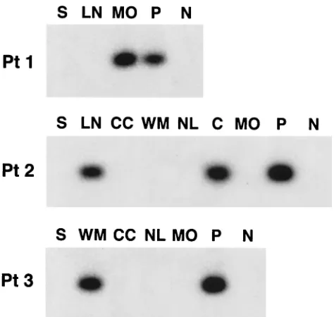
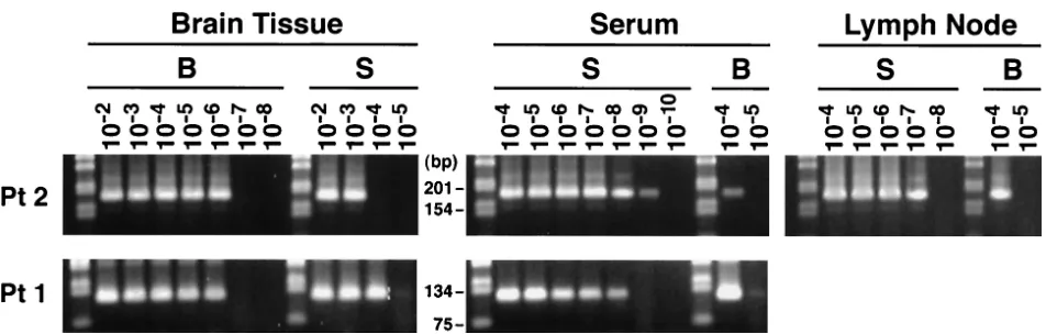
[image:2.587.43.543.93.231.2]brain tissue, respectively (Table 2). In patients 4 to 6, viral negative strand was not detected in any of the analyzed brain tissue samples in two independent sets of experiments using RNA from two separate extractions. However, medulla oblon-gata from patient 1 and cerebellum and subcortical white mat-ter from patients 2 and 3, respectively, were repeatedly positive for the presence of strand HCV RNA. HCV negative-strand RNA was also detected in one out of the four analyzed lymph nodes (patient 2), while none of the six analyzed sera tested positive. These results are summarized in Tables 1 and 2, and Fig. 1 illustrates detection of HCV negative-strand RNA in patients 1 to 3. These reactions were unlikely to represent false-positive results, because nonspecific detection of the in-correct strand might be expected when the latter is present at
TABLE 1. Clinical and virologic data on six HCV-infected patients whose autopsy CNS tissue samples were analyzed for the presence of HCV replicationa
Patient
no. Age(yr) Gender Diagnosis Cause ofdeath
HIV-1 status (CD4⫹cell
count)
Presence of HCV
negative-strand RNA in: HCV genotype
Serum CNS Lymphnode Serum CNS Lymphnode
1 28 M i.v. drug abuse; decompensated cirrhosis;
bacterial endocarditis Sepsis Pos (418) Neg Medulla oblon-gata Neg 1b 3a 1b
2 70 M Decompensated cirrhosis; HCC Liver failure Neg Neg Cerebellum Pos 1b 1a 1ab
3 45 M Alcoholism; decompensated cirrhosis;
miliary tuberculosis Acute pancreatitis Neg Neg Subcorticalwhite matter NA 1b 1b NA 4 57 M Alcoholism, decompensated liver
cirrhosis, HCC Liver failure,hepatic coma Neg Neg Neg Neg 1b 1b 1b
5 34 M i.v. drug abuse Drug overdose Pos (440) Neg Neg Neg 1b 1b 1b
6 29 F i.v. drug abuse; AIDS; alcoholism;
cirrhosis Drug overdose Pos (120) Neg Neg NA 1b 1b NA
aAbbreviations: M, male; F, female; i.v., intravenous; HCC, hepatocellular carcinoma; Neg, negative; Pos, positive; NA, not available. bThe positive HCV RNA strand was type 1b, while the negative strand was type 1a.
on November 8, 2019 by guest
http://jvi.asm.org/
high number, at least 108genomic eq/reaction. However, the
approximate concentration of HCV RNA in brain samples containing the viral negative strand was only 103 genomic
eq/g of total RNA while the concentration of HCV RNA in
lymph node tissue in patient 2 was 104genomic eq/g of total
RNA (Table 2).
To determine the proportion of positive-strand HCV RNA to negative-strand HCV RNA at the sites of putative replica-tion, serial dilutions of extracted RNA were tested for the presence of positive- and negative-strand HCV RNA using Tth-based strand-specific RT-PCR. As can be seen in Fig. 2, negative-strand HCV RNA titers in brain tissue were 1 log lower than titers of the positive strand. In lymph node from patient 2, this difference was 2 logs.
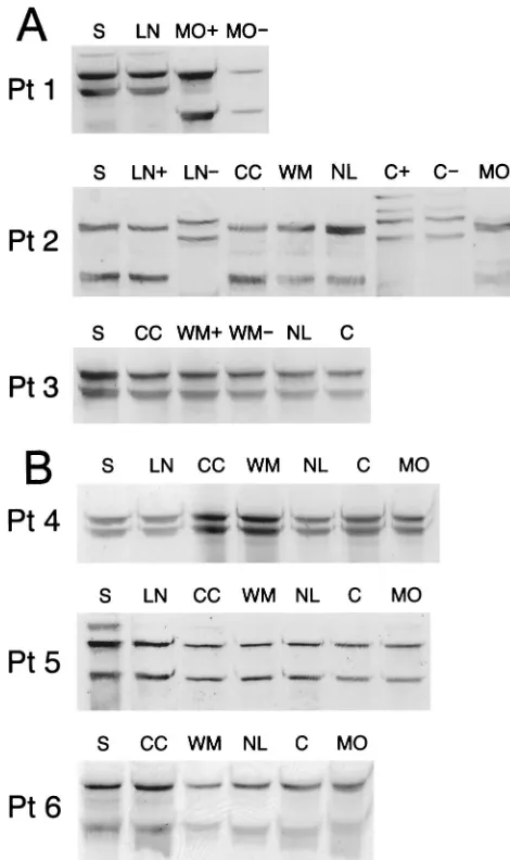
In the next step, viral sequences amplified from different parts of brain were compared by the SSCP assay with one another and with respective viral sequences amplified from serum. In patients 4 to 6, in whom viral negative strands were not detected in brain tissue, all the band patterns were iden-tical. Similarly, no differences were observed for patient 3. However, in patients 1 and 2, negative- and positive-strand viral sequences recovered from medulla oblongata and cere-bellum, respectively, were different from those derived from serum (Fig. 3). Importantly, in the latter patient
[image:3.587.42.544.93.206.2]cerebellum-FIG. 1. Detection of negative-strand HCV RNA in various brain tissue samples and lymph nodes (LN) in patients 1 to 3 (Pt 1 to Pt 3). The presence of viral negative strands was determined using strand-specificTth-based RT-PCR. Twenty microliters (20%) of the reaction mixture was fractionated on agarose, transferred to a nylon membrane by Southern blotting, and subsequently hybridized to a32P-labeled probe. The amount of RNA loaded into each reaction mixture was 5 g; in the case of serum (S), it corresponded to 100l. The examined brain tissue samples included cerebral cortex (CC), subcortical white matter (WM), nucleus lentiformis (NL), cerebellum (C), and medulla oblongata (MO). In patient 1 only medulla oblongata was available for study. Positive sensitivity controls (lanes P) consisted of 103genomic eq of the correct synthetic strand mixed with 5g of RNA extracted from brain tissue from an HCV-negative subject. Negative controls (lanes N) consisted of 5g of RNA extracted from brain tissue from uninfected patients.
[image:3.587.45.282.354.579.2]FIG. 2. The detection of HCV negative-strand (⫺str) and positive-strand (⫹str) RNA in brain tissue samples from patients 1 to 3 (Pt 1 to Pt 3). The analyzed tissues were medulla oblongata, cerebellum, and subcortical white matter in patients 1, 2, and 3, respectively. In addi-tion, lymph node (LN) from patient 2 was studied. Tenfold serial dilutions of extracted RNA were tested for the presence of positive-and negative-strpositive-and HCV RNA byTth-based RT-PCR. The amount of RNA loaded into the reaction mixture at dilution 100corresponds to 5 g. Negative controls (lanes N) consisted of RNA extracted from brain tissue from uninfected subjects, and positive/sensitivity controls (lanes P) consisted of 103genomic eq of the correct synthetic strand mixed with 5g of RNA from an uninfected subject.
TABLE 2. The detection and titers of positive and negative strands of HCV RNA in serum, lymph nodes, and different parts of brain in six subjectsa
Patient no.
Serum titer (eq/ml) in
serum
Titer (eq/g of RNA) in:
Cerebral cortex white matterSubcortical oblongataMedulla lentiformisNucleus Cerebellum Lymph node
⫹str ⫺str ⫹str ⫺str ⫹st ⫺str ⫹str ⫺str ⫹str ⫺str ⫹str ⫺str ⫹str ⫺str
1 107 Nc NDd ND ND ND 103 102 ND ND ND ND 104 N
2 106 N 101b N 102 N 102 N 102 N 103 102 104 102
3 106 N 102 N 103 102 N N 101 N 101 N ND ND
4 107 N 102 N 102 N 102 N 103 N 101 N 103 N
5 105 N 102 N 102 N 101 N 102 N 102 N 103 N
6 106 N 101 N 102 N 102 N 101 N 101 N ND ND
aThe titers of the negative strand (⫺str) were determined byTth-based strand-specific assays, while the titers of the positive strand (⫹str) were determined by a Tth-based assay or, when negative, by the more sensitive MMLV-based assay.
bA titer of 101represents a sample which was negative by theTth-based assay but was positive by the MMLV-based assay in 5g of RNA template only.
cN, negative. dND, not done.
on November 8, 2019 by guest
http://jvi.asm.org/
[image:3.587.302.537.526.617.2]derived sequences were identical to the viral negative strand amplified from the lymph node. The discrepancy between lymph node-derived positive and negative HCV RNA strands can be explained by the fact that the former represents in large part serum-derived contamination and the latter represents indigenous replicating virus. To lower the risk that these
re-sults are artifactual, all SSCP analysis was duplicated in an independent experiment using new RNA template.
Direct sequencing confirmed the presence of identical mas-ter sequences in serum and brain tissue in patients 3 to 6. However, both positive- and negative-strand “master” se-quences recovered from medulla and cerebellum of patients 1 and 2, respectively, differed by several nucleotide substitutions from the serum consensus sequences (Fig. 4). Thus, in two out of three patients in whom HCV negative-strand RNA was found in the CNS, sequences amplified from brain tissue were different from those circulating in serum.
To determine the genotype of infecting strains, the NS5 region of HCV was amplified from all analyzed samples. When serum samples were analyzed, all patients were found to be infected with genotype 1b strains. In patients 4 to 6, the geno-types of strains amplified from the CNS were concordant with those of strains circulating in serum. However, in patient 1 the viral strain found in medulla oblongata was classified as type 3a, while in patient 2 the strain found in cerebellum was clas-sified as type 1a.
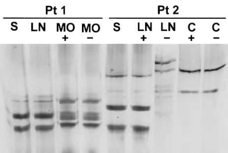
To further clarify the issue of different HCV genotypes in the CNS and circulation in patients 1 and 2, we conducted SSCP analysis of NS5 region sequences amplified from serum, lymph nodes, and pertinent brain structures. Positive and neg-ative strands were amplified using theTth-based assay. As can be seen in Fig. 5, similar to the results of 5⬘UTR analysis, positive and negative strands amplified from medulla oblon-gata and cerebellum in patients 1 and 2, respectively, were different than those amplified from serum. In addition, in pa-tient 2 the pattern for the positive strand from the lymph node was identical to the serum pattern, while the negative strand’s pattern resembled the pattern found in the brain (some addi-tional bands present in neither the serum- nor brain-derived viral sequences were also present). Direct sequencing of the viral negative strand amplified from this lymph node allowed its classification as type 1a, while the positive strand was clas-sified as type 1b.
In patients 1 and 2, HCV strains replicating in the CNS and those circulating in serum were different and classified as be-longing to different genotypes. However, the other strain could have been present below the sensitivity level of direct sequenc-ing (20 to 25%) and even that of SSCP (⬃3%). A commonly used alternative, sequencing of cloned PCR products, would require a large number of clones to be processed and se-quenced, making it laborious and thereby impractical.
We decided to use sequence-specific primers that would allow specific amplification of one sequence from the back-ground of other sequences. This strategy takes advantage of the observation that mismatches localized at the 3⬘terminus of the primer can dramatically decrease amplification efficiency (14, 26). However, as relatively few differences were present between the brain-derived and serum-derived strains in the 5⬘UTR, the analysis was conducted on the NS5 region. We previously used this approach to analyze virological outcome in cases of infection with multiple HCV strains (21).
In both cases, strain-specific primers were designed to match either the serum- or brain-derived sequence but to provide a 3⬘
end mismatch with respect to the other strain (Fig. 6). Thus, these primers should preferentially amplify only one of the strains present. To provide the control template necessary to
FIG. 3. (A) Analysis by SSCP of 5⬘UTR HCV sequences amplified from serum (S) and various autopsy brain tissue samples from patients 1 to 3 (Pt 1 to Pt 3). The following brain tissue samples were examined: cerebral cortex (CC), subcortical white matter (WM), nucleus lenti-formis (NL), cerebellum (C), and medulla oblongata (MO). In patient 1 only medulla oblongata was available for study, while in patient 3 HCV RNA from medulla oblongata could not be amplified. In patients 1 and 2, mediastinal lymph nodes (LN) were also available for analysis. HCV RNA negative strands were detected in medulla oblongata in patient 1, in lymph node and cerebellum in patient 2, and in subcortical white matter in patient 3. The presence of identical and dissimilar viral sequences in the analyzed samples was verified by direct sequencing. Symbols:⫹, positive strand;⫺, negative strand. (B) Analysis by SSCP of 5⬘UTR HCV sequences amplified from serum (S) and various autopsy brain tissue samples from patients 4 to 6. Abbreviations de-noting various analyzed tissues are the same as those for panel A. HCV RNA negative strands were not detected in any of the samples. The presence of identical viral sequences in the analyzed samples was verified by direct sequencing.
on November 8, 2019 by guest
http://jvi.asm.org/
[image:4.587.42.277.71.467.2]check the specificity of the reactions, the first-round PCR product representing either the serum- or brain-derived viral sequence was end point diluted so that no more than one in five reactions was positive when amplified with the second round of primers. In this case, the second-round PCR product would be for the most part derived from single template cop-ies, thus ensuring that it is homogenous (32). The sequence of these control templates was ascertained by direct sequencing. As seen in Fig. 7, in both patients serum-specific sequences were detected in the CNS and brain-specific sequences were detected in serum. However, the latter detection was not uni-form from experiment to experiment, most likely due to sto-chastic phenomena related to low-copy template number. All positive reactions were sequenced directly, and sequence anal-ysis confirmed that indeed the strain-specific PCR detected the proper strain in each case. By analysis of serial dilutions of the PCR products, it was determined that the titers of the minor strains were several logs lower than the titers of the major strains (Fig. 8).
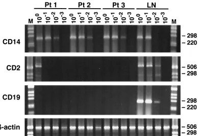
There remains a theoretical possibility that the detectable HCV RNA negative strands were originating in peripheral blood mononuclear cells (PBMC) present in contaminating blood. To exclude the PBMC as the significant source of HCV negative-strand RNA, we conducted mRNA phenotyping by RT-PCR of cells present in the implicated brain tissue from patients 1 to 3. Tested phenotypes were CD2 (T cells), CD19 (B cells), and CD14 (monocytes/macrophages). As can be seen in Fig. 9, expression of CD14 was found in all three brain
[image:5.587.47.539.238.364.2]samples, CD19 was not detected, and CD2 expression was found only in the brain tissue sample from patient 1, but its titer was low. Importantly, in the latter patient viral negative strands were not detected in the lymph node, which makes it even less likely that lymphoid cells in blood were the direct source of viral replicative forms detectable in his brain.
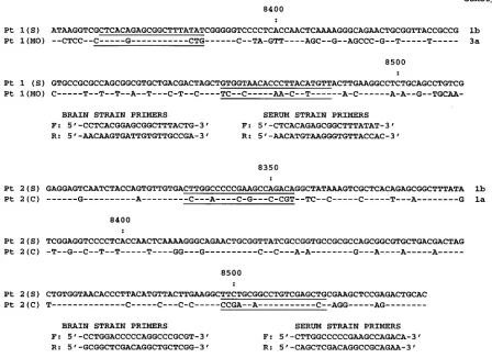
[image:5.587.306.537.533.688.2]FIG. 4. Nucleotide sequence alignment of 5⬘UTR fragments of HCV recovered from serum (S) and brain tissue samples from patients 1 to 3 (Pt1 to Pt3). Sequences are compared with the prototype sequence published by Choo et al. (7) shown on the top line. Symbols and abbreviations: -, sequence identity;⫹, positive strand;⫺, negative strand; MO, medulla oblongata; WM, subcortical white matter; LN, lymph node; C, cerebellum.
FIG. 5. Analysis by SSCP of NS5 region sequences amplified from serum (S), lymph node (LN), and brain tissue samples from patients 1 and 2. Symbols and abbreviations: MO, medulla oblongata; C, cere-bellum;⫹, positive strand;⫺, negative strand.
on November 8, 2019 by guest
http://jvi.asm.org/
DISCUSSION
The findings of a recently published study demonstrated the common presence of HCV replication in blood macrophages/ monocytes in HIV-infected subjects (16). As brain microglia cells are essentially tissue-resident macrophages of blood monocytic origin (8), we hypothesized that they could support HCV replication as well. The present study supports this con-jecture, as HCV replicative forms were detected in the CNS of three out of six studied patients, and in two of these subjects viral sequences in serum and the CNS differed enough to be classified as belonging to different genotypes. These two inde-pendent lines of evidence, viral sequence differences and the presence of negative-strand RNA, argue for the genuine pres-ence of HCV replication in the CNS, most likely in cells of the macrophage/monocyte lineage. Moreover, negative-strand HCV RNA titers were 1 log lower than titers of the positive strand, which is the same proportion as that found commonly in the liver (15, 19). The assumption about lymphoid origin of infected cells is supported by the observation that in patient 2 the strain replicating in the CNS, while being different from that circulating in serum, was the same genotype as the one replicating in the lymph node.
An important question is that of how the HCV got into the CNS. In theory, HCV could gain access to the brain by way of cerebrospinal fluid as occurs in visna virus infection (12). Al-ternatively, and more likely, neuroinvasion is related to traf-ficking of infected cells of monocyte/macrophage lineage through the blood-brain barrier, in a process similar to that postulated for HIV-1 infection (28, 35). Subsequently, there could be a secondary spread of HCV to permissive resident microglial cells within the brain. Replication in the CNS could be facilitated by immunosuppression, some degree of which was likely to be present in all of our patients. This possibility is supported by the observations that while HCV negative-strand RNA is rarely detected in PBMC from normal subjects (15, 24), it is commonly found in HIV-coinfected patients or liver transplant recipients (16, 30). Moreover, HCV replication was demonstrated in hematopoietic cells inoculated into severe combined immunodeficiency mice (5). However, HCV repli-cation in bone marrow was also found in some obviously im-munocompetent subjects (29, 31).
The observed concomitant infection of the same host by two different HCV strains, each replicating in a different compart-ment, is probably the consequence of coinfection or
superin-FIG. 6. Nucleotide sequence alignment of the NS5 region fragments of HCV recovered from patients 1 and 2 (Pt 1 and Pt 2) from serum (S) and autopsy brain tissue. In patient 1 viral fragments recovered from medulla oblongata (MO) and in patient 2 sequences recovered from cerebellum (C) were different from those found in respective serum. Sequence differences between the serum- and brain-derived strains were exploited to design strain-specific primers which would allow specific amplification of one strain from the background of the other strain. The underlined sequence segments show the location of the strain-specific primers. The nucleotide numbering system follows that of the type 1a wild-type strain described by Choo et al. (8).
on November 8, 2019 by guest
http://jvi.asm.org/
[image:6.587.60.508.79.405.2]fection with strains manifesting different tropisms for different cells. For example, it has been demonstrated for lymphocytic choriomeningitis virus that strains differing by a single amino acid substitution, when inoculated together into a mouse, are competitively selected either by the liver and spleen or by neurons (9). We have recently reported that infection with multiple HCV strains results in rapid predominance of a single strain that presumably replicates in the liver and that all other strains are either eliminated or constitute a tiny fraction of circulating virions (21). It is thus possible that HCV adap-tation to an extrahepatic niche may be a strategy to elude competitive exclusion by the dominant strain that replicates in the liver.
In the present study the HCV negative-strand RNA was detected in one site of the brain in each of the three patients. However, as the titer of the replicating virus seemed to be low, replication may have been present at other brain sites but below the level of detection. This could be compounded by the fact that the studied biological material constituted autopsy tissues which were obtained within 36 h after death and there-fore some RNA might have been degraded. Interestingly, HIV-1, which undoubtedly infects brain microglia cells, is also not uniformly detected throughout the brain, even in the same subjects (2, 4).
[image:7.587.126.457.72.219.2]In summary, we found evidence of HCV replication in CNS
FIG. 7. Specific detection of serum-derived HCV sequences in brain tissue (A) and of brain-derived sequences in serum (B) in two patients with evidence of viral replication in the CNS. One microgram of RNA extracted from medulla oblongata (patient 1) (Pt1) and cerebellum (patient 2) (Pt2) or RNA corresponding to 100l of serum was subjected to 35 cycles of RT-PCR, after which the product was diluted 1:10 and 1l was amplified for another 35 cycles with strain-specific primers. Each reaction was repeated in 4 independent experiments (lanes 1 to 4). As can be seen, serum-derived sequences were detected from the background of derived sequences in all four independent experiments, while the brain-derived sequences were detected from the background of serum-brain-derived sequences less uniformly. The RT-PCR products were sequenced and it was determined that they matched the sequences they were designed to amplify. The positive controls (P) contain approximately 104to105 template copies (as determined by optical density readings) of the correct template, while negative controls (N) contain approximately 1010 template copies of the incorrect template. Samples were analyzed by agarose gel electrophoresis (3% NuSieve). Lane m, 1-kb molecular ladder (Gibco/BRL).
FIG. 8. The determination of the proportion of brain- and serum-derived sequences in serum and brain tissue from patient 1 and 2 (Pt 1 and Pt 2). In addition, in patient 2 lymph node was also studied. One microgram of RNA extracted from medulla oblongata (patient 1) and cerebellum (patient 2) or RNA corresponding to 100l of serum was subjected to 20 cycles of RT-PCR, after which the product was serially diluted 1:10 and 1l was amplified for another 35 cycles with primers specific for the serum (S)- or brain (B)-derived strain. As seen, in brain tissue the approximate ratio of serum- to brain-derived strains was 1:102to 1:103, while in serum the brain-derived virus was present at a level 3 to 5 logs lower than that of the major serum-derived virus. In the lymph node in patient 2 the ratio between the two strands was approximately 3 logs. Leftmost lane on each gel, 1-kb molecular ladder (Gibco/BRL).
on November 8, 2019 by guest
http://jvi.asm.org/
[image:7.587.55.531.508.660.2]autopsy samples from three out of six studied patients. How-ever, the consequences of this are presently unclear.
ACKNOWLEDGMENT
This work was supported in part by National Institutes of Health grant DA13760.
REFERENCES
1.Alter, M. J., H. S. Margolis, K. Krawczynski, F. N. Judson, A. Mares, W. J. Alexander, P. Y. Hu, J. K. Miller, M. A. Gerber, R. E. Sampliner, E. L. Meeks, and M. J. Beach.1992. The natural history of community-acquired hepatitis C in the United States. The Sentinel Counties Chronic non-A, non-B Hepatitis Study Team. N. Engl. J. Med.327:1899–1905.
2.Bagasra, O., E. Lavi, L. Bobroski, K. Khalili, J. P. Pestaner, R. Tawadros, and R. J. Pomerantz.1996. Cellular reservoirs of HIV-1 in the central nervous system of infected individuals: identification by the combination of in situ polymerase chain reaction and immunohistochemistry. AIDS10:573– 585.
3.Bolay, H., F. Soylemezoglu, G. Nurlu, S. Tuncer, and K. Varli.1996. PCR detected hepatitis C virus genome in the brain of a case with progressive encephalomyelitis with rigidity. Clin. Neurol. Neurosurg.98:305–308. 4.Brew, B. J., M. Rosenblum, K. Cronin, and R. W. Price.1995. AIDS
de-mentia complex and HIV-1 brain infection: clinical-virological correlations. Ann. Neurol.38:563–570.
5.Bronowicki, J. P., M. A. Loriot, V. Thiers, Y. Grignon, A. L. Zignego, and C. Brechot.1998. Hepatitis C virus persistence in human hematopoietic cells injected into SCID mice. Hepatology28:211–218.
6.Caudai, C., D. Maimone, P. Almi, P. Annunziata, I. Bastianoni, C. A. Bog-giano, G. C. Guazzi, M. Padula, and P. E. Valensin.1997. The potential role of hepatitis C virus in the pathogenesis of the neurological syndrome in chronic hepatitis C. Gut41:411–412.
7.Choo, Q. L., K. H. Richman, J. H. Han, K. Berger, C. Lee, C. Dong, C. Gallegos, D. Coit, R. Medina-Selby, P. J. Barr, A. J. Weiner, D. W. Bradley, G. Kuo, and M. Houghton.1991. Genetic organization and diversity of the
hepatitis C virus. Proc. Natl. Acad. Sci. USA88:2451–2455.
8.Davis, E. J., T. D. Foster, and W. E. Thomas.1994. Cellular forms and functions of brain microglia. Brain Res. Bull.34:73–78.
9.Dockter, J., C. F. Evans, A. Tishon, and M. B. Oldstone.1996. Competitive selection in vivo by a cell for one variant over another: implications for RNA virus quasispecies in vivo. J. Virol.70:1799–1803.
10.Epstein, L. G., C. Kuiken, B. M. Blumberg, S. Hartman, L. R. Sharer, M. Clement, and J. Goudsmit.1991. HIV-1 V3 domain variation in brain and spleen of children with AIDS: tissue-specific evolution within host-deter-mined quasispecies. Virology180:583–590.
11.Fujita, H., Y. Chuganji, M. Yaginuma, M. Momoi, and T. Tanaka.1999. Case report: acute encephalitis immediately prior to acute onset of hepatitis C virus infection. J. Gastroenterol. Hepatol.14:1129–1131.
12.Haase, A. T.1986. Pathogenesis of lentivirus infections. Nature322:130–136. 13.Heckmann, J. G., C. Kayser, D. Heuss, B. Manger, H. E. Blum, and B. Neundorfer.1999. Neurological manifestations of chronic hepatitis C. J. Neurol.246:486–491.
14.Kwok, S., D. E. Kellogg, N. McKinney, D. Spasic, L. Goda, C. Levenson, and J. J. Sninsky.1990. Effects of primer-template mismatches on the polymer-ase chain reaction: human immunodeficiency virus type 1 model studies. Nucleic Acids Res.18:999–1005.
15.Lanford, R. E., D. Chavez, F. V. Chisari, and C. Sureau.1995. Lack of detection of negative-strand hepatitis C virus RNA in peripheral blood mononuclear cells and other extrahepatic tissues by the highly strand-specific rTthreverse transcriptase PCR. J. Virol.69:8079–8083.
16.Laskus, T., M. Radkowski, A. Piasek, M. Nowicki, A. Horban, J. Cianciara, and J. Rakela.2000. Hepatitis C virus in lymphoid cells of patients coin-fected with human immunodeficiency virus type 1: evidence of active repli-cation in monocytes/macrophages and lymphocytes. J. Infect. Dis.181:442– 448.
17.Laskus, T., M. Radkowski, L. F. Wang, S. J. Jang, H. Vargas, and J. Rakela. 1998. Hepatitis C virus quasispecies in patients infected with HIV-1: corre-lation with extrahepatic viral replication. Virology248:164–171.
[image:8.587.101.489.70.338.2]18.Laskus, T., M. Radkowski, L. F. Wang, M. Nowicki, and J. Rakela.2000. Uneven distribution of hepatitis C virus quasispecies in tissues from subjects with end-stage liver disease: confounding effect of viral adsorption and FIG. 9. mRNA phenotyping by RT-PCR of cells present in brain tissue from patients 1 to 3 (Pt 1 to Pt 3). Tested phenotypes were CD2 (T cells), CD14 (moncytes/macrophages), and CD19 (B cells). The studied tissues were medulla oblongata, cerebellum, and subcortical white matter in patients 1, 2, and 3, respectively. In addition, as a positive control, lymph node (LN) from patient 2 was analyzed. Tenfold serial dilutions of extracted RNA were tested; the amount of RNA loaded into the reaction mixture at dilution 100corresponds to 1g. The expected product sizes were 266 bp for CD14, 547 bp for CD2, and 277 bp for CD19. As can be seen, expression of CD14 was found in all three brain samples, CD19 was not detected, and CD2 expression was found in the brain tissue sample from patient 1 (but the titer was low). To check the integrity of isolated RNA and to detect contaminating genomic DNA,-actin primers specific for two different exons separated by an intron were used. Amplification of genomic DNA results in a 607-bp product (not seen), while amplification of cDNA results in a 513-bp product.
on November 8, 2019 by guest
http://jvi.asm.org/
mounting evidence for the presence of low-level extrahepatic replication. J. Virol.74:1014–1017.
19.Laskus, T., M. Radkowski, L. F. Wang, H. Vargas, and J. Rakela.1998. Detection of hepatitis G virus replication sites by using highly strand-specific Tth-based reverse transcriptase PCR. J. Virol.72:3072–3075.
20.Laskus, T., M. Radkowski, L. F. Wang, H. Vargas, and J. Rakela.1997. Lack of evidence for hepatitis G virus replication in the livers of patients coin-fected with hepatitis C and G viruses. J. Virol.71:7804–7806.
21.Laskus, T., L. F. Wang, M. Radkowski, H. Vargas, M. Nowicki, J. Wilkinson, and J. Rakela.2001. Exposure of hepatitis C virus (HCV) RNA-positive recipients to HCV RNA-positive blood donors results in rapid predomi-nance of a single donor strain and exclusion and/or suppression of the recipient strain. J. Virol.75:2059–2066.
22.Lerat, H., S. Rumin, F. Habersetzer, F. Berby, M. A. Trabaud, C. Trepo, and G. Inchauspe.1998. In vivo tropism of hepatitis C virus genomic sequences in hematopoietic cells: influence of viral load, viral genotype, and cell phe-notype. Blood91:3841–3849.
23.Maggi, F., M. Giorgi, C. Fornai, A. Morrica, M. L. Vatteroni, M. Pistello, G. Siciliano, A. Nuccorini, and M. Bendinelli.1999. Detection and quasispecies analysis of hepatitis C virus in the cerebrospinal fluid of infected patients. J. Neurovirol.5:319–323.
24.Mellor, J., G. Haydon, C. Blair, W. Livingstone, and P. Simmonds.1998. Low level or absent in vivo replication of hepatitis C virus and hepatitis G virus/GB virus C in peripheral blood mononuclear cells. J. Gen. Virol. 79:705–714.
25.Morsica, G., M. T. Bernardi, R. Novati, C. Uberti Foppa, A. Castagna, and A. Lazzarin.1997. Detection of hepatitis C virus genomic sequences in the cerebrospinal fluid of HIV-infected patients. J. Med. Virol.53:252–254. 26.Nassal, M., and A. Rieger.1990. PCR-based site-directed mutagenesis using
primers with mismatched 3⬘-ends. Nucleic Acids Res.18:3077–3078. 27.Origgi, L., M. Vanoli, A. Carbone, M. Grasso, and R. Scorza.1998. Central
nervous system involvement in patients with HCV-related cryoglobulinemia. Am. J. Med. Sci.315:208–210.
28.Price, R. W., J. Sidtis, and M. Rosenblum.1988. The AIDS dementia complex: some current questions. Ann. Neurol.23:S27–S33.
29.Radkowski, M., J. Kubicka, E. Kisiel, J. Cianciara, M. Nowicki, J. Rakela, and T. Laskus.2000. Detection of active hepatitis C virus and hepatitis G virus/GB virus C replication in bone marrow in human subjects. Blood 95:3986–3989.
30.Radkowski, M., L. F. Wang, H. E. Vargas, J. Rakela, and T. Laskus.1998. Detection of hepatitis C virus replication in peripheral blood mononuclear cells after orthotopic liver transplantation. Transplantation66:664–666. 31.Sansonno, D., C. Lotesoriere, V. Cornacchiulo, M. Fanelli, P. Gatti, G.
Iodice, V. Racanelli, and F. Dammacco.1998. Hepatitis C virus infection involves CD34⫹hematopoietic progenitor cells in hepatitis C virus chronic carriers. Blood92:3328–3337.
32.Simmonds, P., P. Balfe, J. F. Peutherer, C. A. Ludlam, J. O. Bishop, and A. J. Brown.1990. Human immunodeficiency virus-infected individuals contain provirus in small numbers of peripheral mononuclear cells and at low copy numbers. J. Virol.64:864–872.
33.Simmonds, P., E. C. Holmes, T. A. Cha, S. W. Chan, F. McOmish, B. Irvine, E. Beall, P. L. Yap, J. Kolberg, and M. S. Urdea.1993. Classification of hepatitis C virus into six major genotypes and a series of subtypes by phy-logenetic analysis of the NS-5 region. J. Gen. Virol.74:2391–2399. 34.Thoma, S. J., C. P. Lamping, and B. L. Ziegler.1994. Phenotype analysis of
hematopoietic CD34⫹cell populations derived from human umbilical cord blood using flow cytometry and cDNA-polymerase chain reaction. Blood 83:2103–2114.
35.Zheng, J., and H. E. Gendelman.1997. The HIV-1 associated dementia complex: a metabolic encephalopathy fueled by viral replication in mono-nuclear phagocytes. Curr. Opin. Neurol.10:319–325.