0022-538X/95/$04.0010
Copyrightq1995, American Society for Microbiology
Human T-Cell Leukemia Virus Type II Nucleotide Sequences
between env and the Last Exon of tax/rex Are Not Required
for Viral Replication or Cellular Transformation
PATRICK L. GREEN,
1* TED M. ROSS,
1IRVIN S. Y. CHEN,
2AND
SHERRIE PETTIFORD
1Department of Microbiology and Immunology, Vanderbilt University School of Medicine, Nashville, Tennessee 37232-2363,
1and Department of Medicine and Department of Microbiology and Immunology, University of California, Los Angeles,
School of Medicine and Jonsson Comprehensive Cancer Center, Los Angeles, California 90024-1736
2Received 1 August 1994/Accepted 14 October 1994
Human T-cell leukemia virus types I (HTLV-I) and II (HTLV-II) and bovine leukemia virus contain a region
of approximately 600 nucleotides located 3
*
to the
env
gene and 5
*
to the last exon of the
tax
and
rex
regulatory
genes. This region was originally termed nontranslated or untranslated (UT) since it did not appear to be
expressed. Several studies have identified novel mRNAs in HTLV-I-, HTLV-II-, and bovine leukemia
virus-infected cells that splice into open reading frames (ORFs) contained in the UT region and, thus, have the
potential to produce proteins that might contribute to the biological properties of these viruses. The HTLV-II
infectious molecular clone pH6neo has several ORFs in the UT region (nucleotides 6641 to 7213) and a large
ORF which overlaps the third exon of
tax
/
rex
. To investigate the importance of these ORF-containing sequences
on viral replication and transformation in cell culture, proviral clones containing deletions in UT (pH6neo
D
UT) or a stop codon insertion mutation (pH6neoST) were constructed. Lymphoid cells were transfected with
mutant proviral constructs, and stable cell clones, designated 729pH6neo
D
UT and 729pH6neoST, were
characterized. Viral protein production, reverse transcriptase activity, and the capacity to induce syncytia were
indistinguishable from cells transfected with the wild-type clone. Finally, 729pH6neo
D
UT- and
729pH6neoST-producer cells cocultured with primary blood T lymphocytes resulted in cellular transformation characteristic
of HTLV. These results indicate that putative protein-coding sequences between
env
and the last exon of
tax
/
rex
are not required for viral replication or transformation in cell culture.
Human T-cell leukemia virus types I (HTLV-I) and II
(HTLV-II) are members of a class of retroviruses that includes
simian T-cell leukemia virus and bovine leukemia virus (BLV).
The genome organization as well as some of the biological
properties of these viruses are similar. As with all retroviruses,
they contain the gag, pol, and env genes. In addition, they
encode the trans-regulatory gene products Tax and Rex, both
of which are essential for viral replication and cellular
trans-formation (7, 29). Tax localizes to the nucleus of infected cells
(13, 19, 33) and acts to increase the rate of transcription of all
viral genes from the viral long terminal repeat (LTR) (6, 7, 12,
31). Rex effects posttranscriptional regulatory steps and
in-duces the cytoplasmic expression of incompletely spliced viral
mRNA species that encode the Gag, Pol, and Env proteins (18,
19, 24, 30).
HTLV-I, HTLV-II, and BLV contain a region of
approxi-mately 600 nucleotides (nt) located 3
9
to the env gene and 5
9
to
the last exon of the tax/rex genes. This region has been termed
nontranslated or untranslated (UT) because expression of
open reading frames (ORFs) contained within UT has not
been documented to date in virus-infected cells. However, this
region of the genome in the HTLV family of viruses is highly
conserved, which suggests that UT sequences play an
impor-tant role in the biological properties of the virus. Reverse
transcriptase-PCR analyses have identified viral RNAs in
HTLV-I-, HTLV-II-, and BLV-infected cells that splice into
ORFs contained in the UT region (2–4, 10, 22, 26).
Charac-terization of four novel alternatively spliced viral mRNAs
identified in HTLV-I-infected individuals have the potential to
encode proteins from sequences termed ORF I and ORF II of
the UT region. Putative proteins encoded by ORF I have been
designated p27
I(Rof) and p12
I, and those encoded by OFR II
have been termed p30
II(Tof) and p13
II. These proteins can be
detected following overexpression of ORF-containing plasmid
constructs in cell culture, but there is no direct evidence that
they are expressed in cells of persons with adult T-cell
leuke-mia or HTLV-associated myelopathy or HTLV-I-infected cells
in culture.
The UT region of the HTLV-II proviral clone pH6neo
extends from nt 6641 to 7213. This region contains several
ORFs, one of which begins in the UT region and extends into
and overlaps the last exon of tax/rex. Alternatively spliced
mRNAs have been identified in HTLV-II-infected cells that
have the potential to encode proteins from ORFs contained
within the UT region (10). The availability of an infectious
proviral clone of HTLV-II allowed us to directly address the
role of these ORFs in viral replication and cellular
transfor-mation in cell culture. Mutant viruses were generated in which
the coding potential of UT region containing ORFs was
removed by either large deletion or stop codon insertional
mutations. This mutational strategy eliminated the potential to
generate novel mRNAs or proteins by deleting identified 3
9
splice sites and ORF sequences or by termination of ORFs.
Our study indicates that the region between env and the last
exon of tax/rex, which contains putative protein-coding
se-quences, is dispensable and is not required for the replicative
or transforming properties of HTLV-II in cell culture.
MATERIALS AND METHODS
Cells.Human 729-6 cells, an Epstein-Barr virus-transformed B-cell line, were maintained in Iscove’s medium supplemented to contain 5% fetal calf serum
* Corresponding author.
387
on November 9, 2019 by guest
http://jvi.asm.org/
(FCS), 100 U of penicillin per ml, 100mg of streptomycin per ml, and 2 mM glutamine. BJAB cells, an Ebstein-Barr virus-negative Burkitt’s lymphoma cell line, were maintained in RPMI 1640 medium supplemented to contain 10% FCS, 100 U of penicillin per ml, 100mg of streptomycin per ml, and 2 mM glutamine. Peripheral blood was obtained from normal donors by venipuncture. Peripheral blood lymphocytes (PBL) were isolated by centrifugation over Ficoll-Paque (Pharmacia) and depleted of macrophages by adherence to plastic for 4 h. These cells were cultured in RPMI 1640 medium supplemented to contain 20% FCS, 100 U of penicillin per ml, 100mg of streptomycin per ml, and 2 mM glutamine. Plasmids.An infectious HTLV-II full-length proviral clone, pH6neo, LTR-II-CAT, and SV2neo have been described previously (8, 32). All clones with the prefix pH6neo contain mutations within the full-length clone. pH6neoCla produces neither Tax nor Rex because of a frameshift mutation at a unique ClaI restriction site at nt 7386, and pH6neoSph does not produce Rex because of removal of the rex initiator methionine codon contained within an SphI restric-tion site at nt 5123 (17, 29). A 324-nt delerestric-tion in the 59part of the UT region removing nt 6661 to 6984 was constructed by deleting a PstI fragment (nt 6660 to 6984) from a subclone containing the XhoI-ClaI fragment of pH6neo (nt 6209 to 7385). The deleted fragment was reinserted into the pH6neo backbone, resulting in the proviral clone pH6neoDUT6661-6984. A 509-nt deletion removing all of the UT region with the exception of the 3960 nt was constructed by digesting the XhoI-ClaI subclone with PstI and BglII endonucleases and then digesting it with exonuclease III for different lengths of time, resulting in a series of unidirectional 39deletions extending from the BglII site at nt 6994. The resulting single strand was removed with mung bean nuclease. Samples were further digested with NheI (nt 6648) and treated with mung bean nuclease, and truncated clones were religated with T4 DNA ligase. The precise size of the deletion was determined by dideoxy sequencing. One deleted clone (nt 6645 to 7153) was reinserted into the pH6neo backbone, resulting in the proviral clone pH6neoDUT6645-7153. pH6neoST-7333 contains a C-to-A point mutation at nt 7333 introduced by oligonucleotide-directed site-specific mutagenesis (23). This mutation introduces a stop codon into the large ORF which begins in the UT region and extends into tax/rex; this mutation does not alter the Tax or Rex amino acid sequence.
Transfections and chloramphenicol acetyltransferase (CAT) assay.Plasmid DNAs were introduced into cells by electroporation as previously described (5). Briefly, cells were washed with phosphate-buffered saline (PBS) and resuspended at a concentration of 23107
cells per ml in RPMI 1640 medium supplemented to contain 20% FCS, 100 U of penicillin per ml, 100mg of streptomycin per ml, and 2 mM glutamine. A total of 53106
cells and 25mg of plasmid DNA were exposed to a 960-mF charge with a 250-V potential. Cells were transferred to 10 ml of medium and grown at 378C for 48 h. Stable transfectants containing pH6neo-derived DNA were isolated following incubation in 24-well culture dishes (53105
cells per well) in medium supplemented to contain 10% FCS, 100 U of penicillin per ml, 100mg of streptomycin per ml, 2 mM glutamine, and 1.5 mg of geneticin per ml. Following a 3-week selection period, cells that grew were pooled, and these mass cultures were maintained for further analysis. Stable transfectants are designated by the prefix ‘‘729’’ followed by the clone with which they were transfected (e.g., 729pH6neo is a cell line stably transfected with the wild-type clone pH6neo).
Cell extracts for CAT assays were processed 48 h posttransfection as described previously (14). Three independent CAT assays were performed, and the mean and standard deviation for each sample was calculated. All CAT reactions were standardized for equivalent levels of protein, and incubation for a period of 1 h resulted in an enzymatic activity that was in the linear range. Percentages of
14[C]chloramphenicol acetylation were quantified by scintillation counting of
excised spots.
Metabolic labeling and immunoprecipitation.Stable 729-6-transfected cell lines were metabolically labeled at a concentration of 106cells per ml with
[35S]methionone-cysteine (Trans35S-label, 100mCi/ml; ICN Biochemicals, Inc.)
in methionine-cysteine-free RPMI 1640 medium supplemented to contain 10% dialyzed FCS. Cells were lysed in RIPA buffer (0.05 M Tris-HCl [pH 8.0], 0.1% sodium dodecyl sulfate [SDS], 1.0% Triton X-100, 0.15 M NaCl, 2.0 mM phenylmethylsulfonyl fluoride) and the lysates were clarified by centrifugation at 100,0003g for 1 h at 48C. Lysates were immunoprecipitated with HTLV-II-specific human antisera. Immune complexes were collected with protein A-Sepharose (Pharmacia) and subjected to SDS-polyacrylamide gel electrophoresis (SDS-PAGE). Gels were treated with En3Hance (NEN Research Products) for
fluorography and subjected to autoradiography.
DNA transfer and hybridization.High-molecular-weight DNA was extracted from stable 729-6 transfectants or HTLV-II-infected BJAB cells and subjected to Southern blot analysis (34), as described elsewhere (16). The probe consisted of an HTLV-II-specific32
P-oligo-labeled fragment (401-bp PstI-ClaI fragment, nt 6984 to 7384).
Syncytia and transformation assays.Stably transfected 729 cells were irradi-ated with 10,000 rads, and 53105
cells were cocultivated with either 105
BJAB cells or 106
PBL in 24-well culture plates. Cells were fed twice a week with RPMI 1640 supplemented to contain 10% FCS, 100 U of penicillin per ml, 100mg of streptomycin per ml, and 2 mM glutamine. Syncytia were scored in BJAB cocultures microscopically 3 to 5 days postplating. Long-term BJAB cocultures resulted in a chronically infected BJAB producer cell lines, which were syncytium free. Transformed T-cells defined as continuous growth in the absence of interleukin-2 grew out from PBL cocultures 3 to 4 weeks postplating. In both
cases expression of p19 Gag was detected by indirect immunofluorescence as described previously (9). Briefly, cells were washed in PBS, spotted onto slides, and allowed to air dry. Cells were fixed for 10 min in acetone-methanol (1:1) and allowed to air dry. Cells were incubated with anti-p19 Gag antibody (Cellular Products Inc.) for 30 min at room temperature under a humidified chamber. Cells were washed with PBS and incubated with fluorescein-conjugated F(ab9)2fragment rabbit anti-mouse immunoglobulin G (Organon Teknika
Corp.) for 30 min at room temperature in a humidified chamber. Cells were washed three times with PBS and examined immediately under a fluorescence microscope.
RESULTS
Construction of HTLV-II UT region mutants.
Mutations
were constructed in the HTLV-II proviral genome (Fig. 1A) to
determine the contribution of UT region sequences to the
biological properties of the virus. The HTLV-II molecular
clone pH6neo was used throughout these studies. Upon stable
introduction into mammalian cells, pH6neo directs the
synthe-sis of virions, which by coculture assay are capable of infecting
and transforming human PBL. Two deletion mutants and one
point mutant were constructed in a subclone containing
HTLV-II UT region sequences and introduced into the
full-length proviral clone pH6neo. pH6neo
D
UT6661-6984 contains
a deletion of 324 nt of the 5
9
portion of UT sequences, whereas
pH6neo
D
UT6645-7153 removes all UT sequences (509 nt)
with the exception of 60 nt (Fig. 1B). The 3
9
60 bp of UT must
be present to maintain efficient splicing of the tax/rex mRNA
(17). The mutant proviral clone pH6neoST-7333 contains a
point mutation at nucleotide 7333 that introduces a stop codon
in the large ORF that begins in UT and overlaps the last exon
of tax/rex (Fig. 1B). This mutation was designed to preserve the
amino acid sequence of Tax or Rex. All mutations were
confirmed by determination of their nucleotide sequences
(data not shown).
Effects of UT region mutations on HTLV-II LTR-linked
gene expression.
The mutant proviral clones described above
were tested for their capacity to trans activate LTR-linked gene
expression by using transient CAT assays. The pH6neo
provi-ral clone contains the complete HTLV-II provirus, and as
previously reported, transcription from a native LTR responds
to regulation by the trans-regulatory gene products, Tax and
Rex (6, 17). Thus, CAT linked to an LTR can be used as an
assay for tax and rex expression from wild-type and mutant
HTLV-II proviruses.
Wild-type and mutant HTLV-II proviral clones were
co-transfected with LTR-II-CAT into 729 B cells, and functional
levels of Tax and Rex were assessed by measuring CAT activity
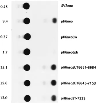
(Fig. 2). The wild-type proviral clone (pH6neo) resulted in a
33-fold increase in CAT activity above that with the vector
control (SV2neo). The full-length proviral clone pH6neoCla,
which does not produce functional Tax or Rex, resulted in no
trans activation above baseline levels, as previously reported
(17, 29). The full-length proviral clone pH6neoSph, which does
not produce functional Rex, resulted in significantly reduced
CAT activity compared with that of the wild-type, also
consis-tent with previous results (17). The mutant clones pH6neo
D
UT6661-6984, pH6neo
D
UT6645-7153, and pH6neoST-7333
resulted in wild-type levels of CAT activity, indicating that both
Tax and Rex produced by these mutants are functional in this
assay. These results indicate that the ORFs in the UT region as
well as the large ORF that overlaps tax/rex do not contribute to
trans activation and these sequences are dispensable for tax
and rex gene expression and function.
Isolation of stable transfectants.
To determine the effect of
mutations in the UT region on synthesis of viral proteins, viral
genome replication, and cellular transformation, stable 729 cell
transfectants containing pH6neo, pH6neo
D
UT6661-6984, pH6
388 GREEN ET AL. J. VIROL.
on November 9, 2019 by guest
http://jvi.asm.org/
neo
D
UT6645-7153, and pH6neoST-7333 were isolated. To
confirm the presence of HTLV-II proviral DNA in the stable
transfectants, cellular DNA was analyzed by nucleic acid
hybridization following digestion with diagnostic restriction
enzymes. Each of the stable transfectants analyzed contained
complete copies of the HTLV-II provirus (data not shown).
Comparison of the hybridization intensities of the predicted
size fragments in the different transfectants indicated that the
HTLV-II DNA was present in relatively similar copy numbers
(Fig. 3). The smaller sizes of the hybridizing bands in both
pH6neo
D
UT6661-6984 and pH6neo
D
UT6645-7153 confirmed
the respective deletions of 324 and 509 nt in the UT region.
Cell lines containing the pH6neoST-7333 DNA, which cannot
be distinguished from wild-type DNA, were confirmed by
determining the sequences of the PCR products of stable
transfectant cell DNA (data not shown). These results indicate
that the stable transfectants have integrated HTLV-II proviral
DNA and the constructed mutations are present following
transfection and selection.
Gag protein expression by UT region mutants.
To
deter-mine whether viral proteins were being produced by the stable
transfectants, cells were metabolically labeled with [
35S]methi-onine-cysteine, and immunoprecipitations were performed on
cell lysates. Immunoprecipitation of cell lysates with human
anti-HTLV-II antisera followed by SDS-PAGE and
autora-diography indicated that the stable transfectants containing
pH6neo
D
UT6661-6984, pH6neo
D
UT6645-7153, and pH6neo
ST-7333 produced significant levels of p24 Gag capsid protein
(Fig. 4). These results indicate that UT region mutants are
indistinguishable from the wild type in the synthesis of p24 Gag
capsid protein.
Production of infectious virus and cellular transformation.
We next determined the effect of UT deletions and the large
ORF region stop codon mutation on virus replication and
transformation. Supernatants of the stable transfectants were
assayed for the presence of HTLV-II by determining reverse
transcriptase activity. Reverse transcriptase activity was
de-tected in the supernatant of all stable transfectants, and the
[image:3.612.94.263.457.635.2]FIG. 1. Organization of HTLV-II genome and HTLV-II-coding region. (A) The complete proviral genome is shown schematically. LTRs are depicted with their U3, R, and U5 regions. The location of the gag, pro, pol, env, tax, and rex genes and their corresponding reading frames are indicated. Sequences (nt 6641 to 7213) located between the env gene and the last exon of tax/rex have been termed the UT region. The locations of the BamHI restriction sites are denoted by arrows. Numbers below the genome denote kilobases. (B) HTLV-II genome 39to the env gene and the locations of ORFs based on the nucleotide sequence of the proviral clone pH6neo are shown. ORFs are denoted by open boxes, and the last coding exons for Tax and Rex are indicated. Vertical arrows denote splice acceptor sites, as reported previously (10); these mRNAs have the potential to encode proteins (see Fig. 7). The location of two deletions in the UT region and a mutation introducing a stop codon (p) in the large ORF in frame 3 are indicated.
FIG. 2. Representative CAT assay testing Tax and Rex functional activity. LTR-II-CAT and various HTLV-II full-length proviral clones were electropo-rated into 729 B cells, and the percent chloramphenicol acetylation was determined. The percent acetylation values shown (on the left) are a represen-tative of three experiments. The mean and standard deviation for each sample were 0.2360.05 for SV2neo, vector control; 11.661.9 for pH6neo, wild-type full-length HTLV-II; 0.2460.03 for pH6neoCla, Tax2and Rex2; 1.8360.15 for pH6neoSph, Rex2; 12.260.95 for pH6neoDUT6661-6984, 324-bp deletion in the UT region; 14.361.2 for pH6neoDUT6645-7153, 509-bp deletion in the UT region; and 12.660.46 for pH6neoST-7333, stop codon in reading frame 3 which overlaps the tax and rex genes.
on November 9, 2019 by guest
http://jvi.asm.org/
activity did not differ by more than twofold (data not shown).
These results indicate that reverse transcriptase levels in
supernatants are equivalent and suggest that the progeny
virions produced are identical as well.
To demonstrate that the two UT deletion mutant viruses
and the ORF stop codon mutant virus are capable of
produc-tive infection, the stable transfectants were cocultivated with
the BJAB cell line. Productive infection of BJAB cells by
HTLV-II results in a rapid induction of syncytia and some
cytopathicity (1). Efficiency of syncytium formation was
deter-mined by microscopic enumeration of syncytia following
cocul-tivation of irradiated stable transfectants with BJAB cells.
Co-cultivation of irradiated 729pH6neo
D
UT6661-6984, 729pH6
neo
D
UT6645-7153, 729pH6neoST-7333, and the wild-type
HTLV-II producer 729pH6neo with BJAB cells resulted in
syncytium formation (Fig. 5 and Table 1). Syncytium formation
is dependent on the efficient expression of viral Env, and the
presence of infectious virus capable of spreading throughout
the culture dramatically reduces the time required for
syncy-tium induction as well as cytopathology. In an effort to address
the efficiency at which mutant viruses replicate and induce
syncytia, 10-fold serial dilutions of irradiated producer cells
were cocultured with BJAB cells. Syncytia could be induced
with as few as 10 irradiated producer cells, and there was no
apparent difference in the time course of syncytium
induc-tion by mutant viruses and wild-type virus. We also noted
that long-term growth of each of these HTLV-infected
cultures resulted in the establishment of syncytium-free
BJAB cell lines. These cell lines were found to be infected
with HTLV-II and express virus as documented by new
syncytium formation upon the addition of fresh uninfected
BJAB cells.
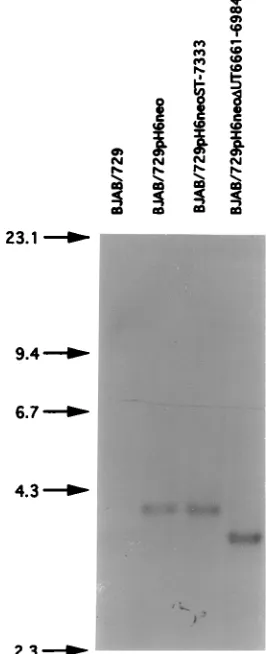
To determine whether HTLV-II proviral DNA was present
in the BJAB cells cocultured with the 729 transfectants, cell
DNA was analyzed by nucleic acid hybridization following
digestion with diagnostic restriction enzymes. It was important
to determine the stability of these mutations in the infected
BJAB cultures and to ascertain whether these mutations
reverted in culture. All long-term BJAB cocultures contained
HTLV-II proviral DNA (Fig. 6). The deletion in the UT region
was confirmed in the infected BJAB cell population (Fig. 6).
The point mutation introduced in pH6neoST-7333 was still
present in BJAB cells as determined by nucleotide sequence
analysis of PCR products of DNA obtained from infected
BJAB cells (data not shown). Therefore, we conclude that the
UT region mutant viruses are stable and infectious for BJAB
cells.
[image:4.612.351.526.68.347.2]To determine whether the UT region mutations have altered
the capacity of HTLV-II to transform primary T cells,
irradi-ated 729 producer cells were cocultured with primary blood
lymphocytes. HTLV transformation of primary T lymphocytes
is apparent 3 to 4 weeks following coculture. HTLV-II UT
region mutant viruses and wild-type HTLV-II had the capacity
to transform primary T lymphocytes with similar efficiencies
under these experimental conditions. These results confirm
that the region between env and the last exon of tax/rex,
containing putative protein-coding sequences, are not
neces-sary for the replication of the virus and that the deletion or
FIG. 3. Southern hybridization analysis of stable transfectant 729 cell DNA. Ten micrograms of high-molecular-weight cellular DNA was digested with BamHI, electrophoresed through a 0.7% agarose gel, blotted onto nitrocellulose paper, and hybridized with a32
[image:4.612.115.238.70.331.2]P-labeled HTLV-II-specific 401-nt probe (PstI [nt 6984]-ClaI [nt 7384]). A 3.46-kb fragment is expected to be detected in 729pH 6neo- and 729pH6neoST-7333-digested cell DNA, whereas 3.14- and 2.95-kb fragments are expected in 729pH6neoDUT6661-6984 and 729pH6neoD UT6645-7153, respectively (Fig. 1). The sizes (in kilobases) indicated on the left were determined by comparison with a HindIII digest of lambda DNA.
FIG. 4. Immunoprecipitation of [35S]methionone-cysteine-labeled stable
transfectant cell lines. Cells were metabolically labeled, and cell lysates were prepared. Stable transfectant cell lysates, normalized by scintillation counting of trichloroacetic acid precipitates, were immunoprecipitated with human anti-HTLV-II antisera (primary viral products detected were the p24 Gag capsid protein and Gag precursor). The sizes (in kilodaltons [K] indicated on the right) were determined by comparison to protein markers (Amersham) (lane M).
390 GREEN ET AL. J. VIROL.
on November 9, 2019 by guest
http://jvi.asm.org/
disruption of these coding sequences does not affect the
capacity of HTLV-II to infect or transform primary T cells in
cell culture.
DISCUSSION
Using an infectious molecular clone of HTLV-II, we
ad-dressed the importance of sequences between env and the last
exon of tax/rex in viral replication and cellular transformation
in cultured cells. Viral mutants containing deletions in this
region or a stop codon in the large ORF overlapping tax/rex
were constructed in the context of a full-length proviral clone.
These mutations had no adverse effects on trans activation by
Tax or Rex as well as viral replication. Most importantly, the
mutant viruses were stable and had the capacity to transform
primary blood lymphocytes in culture, a hallmark of the HTLV
family of retroviruses. Thus, the region between env and the
last exon of tax/rex (UT region) containing putative
protein-coding sequences is dispensable for viral replication and
transformation in cell culture.
The genome complexity of HTLV-I, HTLV-II, and BLV
appears to be increased by alternative splicing within the UT
region (2–4, 10, 22, 25). Alternatively spliced viral mRNAs
identified in HTLV-I-infected individuals have the potential to
encode proteins from two ORFs, termed ORF I and ORF II,
contained within the UT region. Putative proteins encoded by
ORF I have been designated p27
Ior Rof and p12
I. p12
Icorresponds to the C-terminal 99 amino acids of p27
Iand
localizes to endomembranes of cells transfected with an ORF
I cDNA expression vector, whereas p27
Idoes not appear to be
expressed in this system (21). The p12
Iprotein exhibits amino
acid sequence similarity to the E5 oncoprotein of bovine
papillomavirus type 1 (11). p12
Ialso potentiates the
[image:5.612.64.553.71.487.2]transform-ing activity of E5, suggesttransform-ing a potential role in the HTLV
transformation process (11). However, one study found that a
naturally occurring HTLV-I variant containing a 11-nt deletion
FIG. 5. HTLV-II syncytium induction in BJAB cells. 729 stable transfectants were irradiated with 10,000 rads and cocultured with BJAB cells. Syncytia were scored in BJAB cell cocultures microscopically 3 to 5 days postplating. Cells were photographed at 72 h postplating. (A) 729 mock-transfected control; (B) 729pH6neo, wild-type HTLV-II; (C) 729pH6neoDUT6645-7153; (D) 729pH6neoST-7333.
on November 9, 2019 by guest
http://jvi.asm.org/
removing the p12
Iinitiation start codon was competent to
transform normal human umbilical cord blood cells in cell
culture (27). The putative ORF II product, p30
IIor Tof
protein, expressed from a cDNA-containing vector localizes to
the nucleoli and has been reported to have distant homology to
several transcription activators, such as oct-1, oct-2, pit-1, and
POU-M1 (10, 21). p30
IIdoes not appear to bind DNA or to
have transcriptional trans-activating activity for the viral LTR
(28). Alternative splicing of ORF II results in production of
p13
II, which localizes to nuclei of cells transfected with the
relevant ORF II cDNA expression vector (21). It is important
to note that although these novel mRNAs corresponding to
ORF I and ORF II have been identified, the detection of their
protein products in either HTLV-I-infected patients or tissue
culture cells infected in vitro has eluded investigators.
There-fore, it has not yet been possible to assign functions to these
putative proteins or determine their contribution to the virus
life cycle.
[image:6.612.108.241.75.402.2]The UT region of the HTLV-II proviral clone pH6neo
contains five ORFs of substantial size as well as an additional
ORF that begins in the UT region and overlaps the last exon
of tax/rex (Fig. 1B). Alternatively spliced mRNAs containing
several of these ORFs have been identified in
HTLV-II-infected cells (10). On the basis of the sequence of the
HTLV-II pH6neo isolate, these mRNAs have the potential to
encode six proteins of various sizes depending on splice
acceptor and translation start codon utilization (Fig. 7).
HTLV-II and HTLV-I UT-containing ORFs have been
aligned to determine their relatedness or similarity. Amino
acid sequence alignment of HTLV-I and HTLV-II Rof
puta-tive proteins reveals 62% similarity, with 46% identity (10).
Much of this relatedness is attributed to the amino-terminal
Rex amino acids, since protein alignment without those
se-quences results in a 35% similarity and 26% identity (Fig. 7
FIG. 6. Southern hybridization analysis of DNA prepared from BJAB cells infected with HTLV-II. Ten micrograms of high-molecular-weight cellular DNA was digested with BamHI, electrophoresed through a 0.7% agarose gel, blotted onto nitrocellulose paper, and hybridized with a32P-labeled HTLV-II-specific
401-bp probe (PstI [nt 6984]-ClaI [nt 7384]). Expected fragment sizes (on the left) are as described in the legend to Fig. 3.
FIG. 7. Amino acid sequence of putative protein products. Putative proteins shown (labeled 1 to 6) were based on the previously identified splice acceptor sites in UT region ORFs (10) and the nucleotide sequence of the HTLV-II molecular clone pH6neo (Fig. 1B). Splicing to nt 6807 generates a 31-amino-acid protein starting at the Rex AUG and continuing into frame 3 (putative protein 1) or a 63-amino-acid protein starting at the Tax AUG continuing into frame 2 (putative protein 4). Splicing to nt 6827 generates a 76-amino-acid protein starting at the Rex AUG and continuing into frame 2 (putative protein 2) or a 76-amino-acid protein starting at the Tax AUG and continuing into frame 1 (putative protein 5). Splicing to nt 6944 generates a 37-amino-acid protein starting at the Rex AUG and continuing into frame 2 (putative protein 3) or a 37-amino-acid protein starting at the Tax AUG and continuing into frame 1 (putative protein 6). Underlined amino acids in putative proteins 1 to 3 denote the contribution of Rex, and the underlined amino acid in putative proteins 4 to 6 denote the Tax initiator methionine. The asterisks in proteins 2 and 4 denote potential internally initiated proteins (31 amino acids) and, by alignment, correspond to p12I
of HTLV-I (21). Protein 2 has been termed Rof-2 (10, 21).
TABLE 1. Infection and transformation of cellsa
Stable transfectant Syncytium formationb
Primary T-cell
trans-formationc
Immuno-fluorescence (BJAB/primary)d
729 2 2 2/2
729pH6neo 1(10) 1 1/1
729pH6neoDUT6661-6984 1(10) 1 1/1
729pH6neoDUT6645-7153 1(10) 1 1/1
729pH6neoST-7333 1(10) 1 1/1
aStably transfected 729 cells were irradiated with 10,000 rads, and 53105
cells were cocultivated with 53105stimulated PBL or serial 10-fold dilutions of
irradiated cells were incubated with 53105BJAB cells in 24-well culture plates.
Cells were fed twice a week with RPMI 1640 supplemented to contain 10% FCS and antibiotics.
bSyncytium formation was scored as positive or negative and photographed at
72 h postplating. The numbers in parentheses indicate the minimum number of 729 producer cells required for syncytium induction following coculture with BJAB cells.
cTransformation was scored as positive or negative 3 to 4 weeks following
coculture of 729 producer cells with PBL.
dThe presence of HTLV was confirmed in both BJAB and primary cells by
detection of HTLV p19 Gag by immunofluorescence.
392 GREEN ET AL. J. VIROL.
on November 9, 2019 by guest
http://jvi.asm.org/
[image:6.612.317.555.84.165.2]and data not shown). Sequence alignment of HTLV-I p12
I(21,
22) and the corresponding region in HTLV-II (Fig. 7) reveal
56% similarity, with 37% identity (data not shown). The same
alignment of HTLV-I and HTLV-II Tax or Rex shows 77 and
63% identity, respectively. Therefore, on the basis of sequence
alignment and our current knowledge, if proteins are
ex-pressed from ORFs contained within the conserved region
between env and the last exon of tax/rex in both HTLV-I- and
HTLV-II-infected cells, it is possible that they would serve
similar functions.
The conservation of genome organization and the
expres-sion of alternatively spliced mRNAs in the HTLV family of
retroviruses suggest a function in the virus life cycle for the
sequences between env and the last exon of tax/rex. Our results
suggest that current assays using cultured cells may not allow
us to determine the importance or function of these sequences
or their putative proteins. These findings may be similar to
those obtained in studies of the Nef protein of the
immuno-deficiency viruses. Nef appears to be largely dispensable in cell
culture but plays a critical role in pathogenicity and the
development of AIDS when assayed in monkeys (20). It is
conceivable that sequences of the UT region do not encode
functional proteins but function in some other capacity. By
genome comparison to the human immunodeficiency virus
type 1, one possibility is that the sequences between HTLV
env and the last exon of tax/rex might contain a Rex response
element. However, immunobinding and gel shift assays failed
to detect any binding of Rex to RNAs containing these
sequences (15). One opportunity of gaining insight into the
importance of the HTLV UT region in the biology of the
virus will most likely come from comparative in vivo studies
with BLV since there currently exists no animal model
sys-tem for HTLV pathogenesis. One report has provided
evi-dence that two of the alternatively spliced mRNAs, which
have the potential to encode putative BLV proteins, are
ex-pressed at particular stages of BLV infection and may play
a role in BLV-induced lymphocytosis (2). Further in vivo
analysis will be required to determine the contribution that
putative proteins encoded by sequences between env and the
last exon of tax/rex play in the pathogenesis of this family of
retroviruses.
ACKNOWLEDGMENTS
We thank Terry Dermody for editorial comments.
This work was supported by Public Health Service grant CA59581 from the National Institutes of Health, by National Cancer Institute Training grant CA09385, and by Leukemia Society of America grant H910404. P.L.G. is a scholar of the Leukemia Society of America.
REFERENCES
1. Agadjanyan, M. G., K. E. Ugen, B. Wang, W. V. Williams, and D. B. Weiner. 1994. Identification of an 80-kilodalton membrane glycoprotein important for human T-cell leukemia virus type I and type II syncytium formation and infection. J. Virol. 68:485–493.
2. Alexandersen, S., S. Carpenter, J. Christensen, T. Storgaard, B. Viuff, Y. Wannemuehler, J. Belousov, and J. A. Roth.1993. Identification of alterna-tively spliced mRNAs encoding potential new regulatory proteins in cattle infected with bovine leukemia virus. J. Virol. 67:39–52.
3. Berneman, Z. N., R. B. Gartenhaus, M. J. Reitz, M. E. Klotman, and R. C. Gallo.1992. cDNA sequencing confirms HTLV-I expression in adult T-cell leukemia/lymphoma and different sequence variations in vivo and in vitro. Leukemia 3:67–71.
4. Berneman, Z. N., R. B. Gartenhaus, M. S. Reitz, W. A. Blattner, A. Manns, B. Hanchard, O. Ikehara, R. C. Gallo, and M. E. Klotman.1992. Expression of alternately spliced human T-lymphotropic virus type I pX mRNA in infected cell lines and in primary uncultured cells from patients with adult T-cell leukemia/lymphoma and healthy carriers. Proc. Natl. Acad. Sci. USA 89:3005–3009.
5. Cann, A. J., Y. Koyanagi, and I. S. Y. Chen. 1988. High efficiency transfection of primary human lymphocytes and studies of gene expression. Oncogene 3: 123–128.
6. Cann, A. J., J. D. Rosenblatt, W. Wachsman, N. P. Shah, and I. S. Y. Chen. 1985. Identification of the gene responsible for human T-cell leukemia virus transcriptional regulation. Nature (London) 318:571–574.
7. Chen, I. S. Y., A. J. Cann, N. P. Shah, and R. B. Gaynor. 1985. Functional relationship of HTLV-II x and adenovirus E1A proteins in transcriptional activation. Science 230:570–573.
8. Chen, I. Y., J. McLaughlin, J. C. Gasson, S. C. Clark, and D. W. Golde. 1983. Molecular characterization of genome of a novel human T-cell leukaemia virus. Nature (London) 305:502–505.
9. Chen, I. S. Y., S. G. Quan, and D. W. Golde. 1983. Human T-cell leukemia virus type II transforms normal human lymphocytes. Proc. Natl. Acad. Sci. USA 80:7006–7009.
10. Ciminale, V., G. N. Pavlakis, D. Derse, C. P. Cunningham, and B. K. Felber. 1992. Complex splicing in the human T-cell leukemia virus (HTLV) family of retroviruses: novel mRNAs and proteins produced by HTLV type I. J. Virol. 66:1737–1745.
11. Franchini, G., J. C. Mulloy, I. J. Koralnik, M. A. Lo, J. J. Sparkowski, T. Andresson, D. J. Goldstein, and R. Schlegel.1993. The human T-cell leukemia/ lymphotropic virus type I p12I protein cooperates with the E5 oncoprotein of bovine papillomavirus in cell transformation and binds the 16-kilodalton subunit of the vacuolar H1ATPase. J. Virol. 67:7701–7704. 12. Fujisawa, J., M. Seiki, T. Kiyokawa, and M. Yoshida. 1985. Functional activation of the long terminal repeat of human T-cell leukemia virus type I by a trans-acting factor. Proc. Natl. Acad. Sci. USA 82:2277–2281. 13. Goh, W. C., J. Sodroski, C. Rosen, M. Essex, and W. A. Haseltine. 1985.
Subcellular localization of the product of the long open reading frame of human T-cell leukemia virus type I. Science 227:1227–1228.
14. Gorman, C. M., L. F. Moffat, and B. H. Howard. 1982. Recombinant genomes which express chloramphenicol acetyltransferase in mammalian cells. Mol. Cell. Biol. 2:1044–1051.
15. Green, P. L. Unpublished results.
16. Green, P. L., D. A. Kaehler, L. M. Bennett, and R. Risser. 1989. Multiple steps are required for the induction of tumors by Abelson murine leukemia virus. J. Virol. 63:1989–1994.
17. Green, P. L., Y. Xie, and I. S. Y. Chen. 1990. The internal methionine codons of human T-cell leukemia virus type II rex gene are not required for p24rex
production or virus replication and transformation. J. Virol. 64:4914– 4921.
18. Hidaka, M., J. Inoue, M. Yoshida, and M. Seiki. 1988. Post transcrip-tional regulator (rex) of HTLV-I initiates expression of viral structural proteins but suppresses expression of regulatory proteins. EMBO J. 7:519–523.
19. Inoue, J. I., M. Yoshida, and M. Seiki. 1987. Transcriptional (p40x) and
posttranscriptional (p27xIII) regulators are required for the expression and
replication of human T-cell leukemia virus type I genes. Proc. Natl. Acad. Sci. USA 84:3653–3657.
20. Kestler, H. W., III, D. J. Ringler, K. Mori, D. L. Panicali, P. K. Sehgal, M. D. Daniel, and R. C. Desrosiers. 1991. Importance of the nef gene for maintenance of high virus loads and for development of AIDS. Cell 65:651–662.
21. Koralnik, I. J., J. Fullen, and G. Franchini. 1993. The p12I, p13II, and p30II proteins encoded by human T-cell leukemia/lymphotropic virus type I open reading frames I and II are localized in three different cellular compart-ments. J. Virol. 67:2360–2366.
22. Koralnik, I. J., A. Gessain, M. E. Klotman, M. A. Lo, Z. N. Berneman, and G. Franchini.1992. Protein isoforms encoded by the pX region of human T-cell leukemia/lymphotropic virus type I. Proc. Natl. Acad. Sci. USA 89: 8813–8817.
23. Kunkel, T. A. 1985. Rapid and efficient site-specific mutagenesis without phenotypic selection. Proc. Natl. Acad. Sci. USA 82:488–492.
24. Ohta, M., H. Nyunoya, H. Tanako, T. Okamoto, T. Akagi, and K. Shimo-tohno.1988. Identification of a cis-regulatory element involved in accumu-lation of human T-cell leukemia virus type II genomic mRNA. J. Virol. 62: 4445–4449.
25. Orita, S., H. Kobayashi, Y. Aono, A. Saiga, M. Maeda, and H. Igarashi. 1993. p21X mRNA is expressed as a singly spliced pX transcript from defective provirus genomes having a partial deletion of the pol-env region in human T-cell leukemia virus type 1-infected cells. Nucleic Acids Res. 21:3799– 3807.
26. Orita, S., A. Saiga, S. Takagi, T. Tanaka, K. Okumura, Y. Aono, Y. Hinum, and H. Igarashi.1991. A novel alternately spliced viral mRNA transcribed in cells infected with human T-cell leukemia virus type I is mainly responsible for expressing p21X protein. FEBS Lett. 295:127–134.
27. Ratner, L., S. F. Josephs, B. Starcich, B. Hahn, G. M. Shaw, R. C. Gallo, and F. Wong-Staal.1985. Nucleotide sequence analysis of a variant human T-cell leukemia virus (HTLV-1b) provirus with a deletion in pX-I. J. Virol. 54:781– 790.
28. Roithmann, S., C. Pique, A. Le Cesne, L. Delamarre, D. Pham, T. Tursz, and M.-C. Dokhe´lar.1994. The open reading frame I (ORFI)/ORF II part of the
on November 9, 2019 by guest
http://jvi.asm.org/
human T-cell leukemia virus type I X region is dispensable for p40tax, p27rex,
or envelope expression. J. Virol. 68:3448–3451.
29. Rosenblatt, J. D., A. J. Cann, D. J. Slamon, I. S. Smalberg, N. P. Shah, J. Fujii, W. Wachsman, and I. S. Y. Chen. 1988. HTLV-II trans-activa-tion is regulated by two overlapping nonstructural genes. Science 240:916– 919.
30. Seiki, M., J. Inoue, M. Hikada, and M. Yoshida. 1988. Two cis-acting elements responsible for posttranscriptional trans-regulation of gene expres-sion of human T-cell leukemia virus type I. Proc. Natl. Acad. Sci. USA 85: 7124–7128.
31. Seiki, M., J. Inoue, T. Takeda, and M. Yoshida. 1986. Direct evidence that
p40xI of human T-cell leukemia virus type I is a trans-acting transcriptional activator. EMBO J. 5:561–565.
32. Shimotohno, K., Y. Takahashi, N. Shimizu, T. Gojobori, I. S. Y. Chen, D. W. Golde, M. Miwa, and T. Sugimura.1985. Complete nucleotide sequence of an infectious clone of human T-cell leukemia virus type II: a new open reading frame for the protease gene. Proc. Natl. Acad. Sci. USA 82:3101–3105. 33. Slamon, D. J., W. J. Boyle, D. E. Keith, M. F. Press, D. W. Golde, and L. M.
Souza.1988. Subnuclear localization of the trans-acting protein of human T-cell leukemia virus type I. J. Virol. 62:680–686.
34. Southern, E. M. 1975. Detection of specific sequences among DNA frag-ments separated by gel electrophoresis. J. Mol. Biol. 98:503–517.
394 GREEN ET AL. J. VIROL.