0022-538X/97/$04.0010
Copyrightq1997, American Society for Microbiology
Identification of a Replication-Competent Pathogenic Human
Immunodeficiency Virus Type 1 with a Duplication in the
TCF-1
a
Region but Lacking NF-
k
B Binding Sites
LINQI ZHANG,1YAOXING HUANG,1HANNAH YUAN,1BENJAMIN K. CHEN,2
JAMES IP,1ANDDAVID D. HO1*
The Aaron Diamond AIDS Research Center, The Rockefeller University, New York, New York 10016,1and
Department of Biology, Massachusetts Institute of Technology, Cambridge, Massachusetts 021392
Received 1 August 1996/Accepted 24 October 1996
Multiple human immunodeficiency virus type 1 (HIV-1) sequences with deletions of NF-kB binding sites at
both the 5*and 3*long terminal repeats (LTRs) were identified in serial samples collected from an infected
individual. The effect of this deletion on the level of transcription was studied by transient transfection of an LTR-driven luciferase reporter gene and by infection with a full-length recombinant HIV-1 containing a
luciferase reporter (HIVHXBluc). Detectable levels of gene expression were found in both systems, in the
presence or absence of the viral transactivator Tat. Interestingly, a duplication of a putative TCF-1amotif was
found in place of the NF-kB elements in these viruses. Higher transcriptional activity was observed with
HXBLTR (NF-kB intact) than with the patient’s LTR (NF-kB deleted), suggesting that the NF-kB binding sites
may promote optimal levels of viral gene transcription. The ability of these viruses with NF-kB deleted to
replicate and cause substantial decline in CD4 cell counts demonstrates that the NF-kB binding sites are not
absolutely required for viral replication or pathogenicity in vivo. These results are consistent with the notion that the HIV-1 LTR possesses functional redundancy which allows it to interact with multiple transcription factors, thereby ensuring viral replication in a variety of cell types.
The human immunodeficiency virus type 1 (HIV-1) provi-rus, like all retroviruses, contains two identical long terminal repeats (LTRs) flanking the three structural genes gag, pol, and
env that are essential for viral replication. Each LTR consists
of three regions, namely, U3, R, and U5 (12, 14). HIV-1 transcription is regulated by multiple viral and cellular tran-scription factors that bind to distinct sequence elements in the
59 LTR (12–14, 17, 22, 26, 35). Moreover, the initiation of
HIV-1 transcription is under the control of cellular factors; the subsequent activation of HIV-1 transcription, however, is largely mediated by virus-encoded transactivators such as Tat (12–14, 26, 33, 35). Previous reports have identified several sequence elements within the LTR that are important in reg-ulating the level of viral transcription. For example, efficient transcription of HIV-1 in T lymphocytes or monocytes/macro-phages requires the presence of the TATA box motif, the
binding sites for NF-kB and Sp1, and the Tat-response region
(TAR) (12–14, 16, 22, 26, 33, 34). Mutagenesis of these se-quence elements either individually or summarily inhibits or inactivates HIV-1 replication (12, 14, 16, 22, 25, 31, 33, 34, 36).
The HIV-1 59LTR contains two NF-kB binding sites located
upstream of the Sp1 binding sites and the TATA promoter (12, 14). Conservation of these two binding sites among most HIV-1 isolates suggests their importance in viral replication (9,
12, 14, 24, 26, 29, 30). NF-kB binding sites interact with and
respond to the rel/NF-kB family of transcription factors which
can be induced by a variety of mitogens, cytokines, viruses, and
other agents (3, 12–14). Members of the human rel/NF-kB
family include p50, p52, p65, c-rel, and relB (3, 14, 35, 38).
NF-kB exists typically as a heterodimer composed of p50 and
p65 subunits (3, 38). While p50 functions as a strong DNA
binding subunit, p65 is responsible for the transcriptional
ac-tivity of the NF-kB complex (38). Originally, NF-kB was
be-lieved to be present only in B lymphocytes, but it was later found in a variety of cell types, including T lymphocytes and monocytes/macrophages upon activation (3).
Regulation of HIV-1 LTR by NF-kB and other cellular and
viral factors is quite complex. Duplication of NF-kB binding
sites can enhance both LTR-driven reporter gene activity and viral replication (11, 25, 31, 36), whereas its deletion can impair viral replicative capacity in a variety of cell types such as pe-ripheral blood mononuclear cells (PBMCs), CD4 lymphocytes, monocytic cell line U937, and lymphoblastoid T-cell lines CEM and 11.8 (1, 12, 14, 21, 23, 35, 42). In contrast, other reports have demonstrated that mutated HIV-1 missing one or
both NF-kB binding sites is still capable of efficient replication
in stimulated PBMCs, although a 2- to 4-day delay in peak virus production was observed in CEM and MT-4 cells (11, 25,
36, 40). Moreover, a prerequisite for HIV-1 with an NF-kB
binding site deleted to replicate is the presence of three Sp1 binding sites (25). Elimination of the Sp1 binding sites can reduce HIV-1 replication to below detectable levels in all cell types tested (25, 36). The conflicting observations on the role
of NF-kB binding site in HIV-1 replication could be partially
explained by differences in HIV-1 constructs, cell types, and exogenous stimulating factors used in the assay systems (1, 11, 23, 25, 31, 36, 42), as was reported by Antoni et al. (2). Initi-ation and completion of HIV-1 replicIniti-ation therefore are not merely dependent on the presence of specific sequence ele-ments in the LTR, but are also dependent on the availability and the relative abundance of transcriptional factors present in the target cells. The high degree of functional redundancy observed in the HIV-1 LTR may enable viral replication to occur in various cell types (12, 14, 36). The observation that a great variety of cell types are productively infected by HIV-1 in vitro and in vivo further supports this notion (7, 26).
* Corresponding author. Mailing address: Aaron Diamond AIDS Research Center, 455 First Ave., 7th floor, New York, NY 10016. Phone: (212) 725-0018. Fax: (212) 725-1126. E-mail: [email protected].
1651
on November 9, 2019 by guest
http://jvi.asm.org/
To date, much of our knowledge about HIV-1 LTR regula-tion comes from in vitro studies with LTR-driven reporter genes or full-length viruses with mutations in certain elements (1, 12, 14, 16, 23, 25, 34, 36). Although these studies provide substantial insight into the regulatory mechanism of HIV-1 LTR, less is known about the effect of these mutations on the degree of HIV-1 replication or pathogenicity in vivo. Efforts
have been made to evaluate the relative importance of NF-kB
and Sp1 binding sites in the replication of HIV-2 and simian immunodeficiency virus SIVmac. Results of a study of patho-genicity in pig-tailed macaques suggest that duplication of the
NF-kB binding site within the LTR is not responsible for the
rapid, severe syndrome associated with SIVsmmPBj6.6 infec-tion (10, 32). However, a chimera in which the SIVsmmPBj6.6 LTR was replaced with an SIVsmH4 LTR caused disease in only 50% of inoculated macaques (32). Further studies are needed to determine whether this chimera may be somewhat
attenuated because of the presence of only a single NF-kB in
the LTR (10, 32). While Bellas et al. (4) found that the NF-kB
binding site has a substantial effect on SIVmac replication in primary macrophages, Ilyinskii and Desrosiers showed that
even complete elimination of the NF-kB and the Sp1 binding
sites has only a modest effect on SIVmac replication in rhesus
PBMCs (20). Markovitz et al. suggested that the NF-kB
bind-ing site plays a significant role in the response of the HIV-2 LTR to extracellular stimulation (28). Ideally, the replicative capacity of these mutated SIVmac and HIV-2 should be stud-ied with appropriate animal models, since in vivo results would have a greater relevance for understanding the replicative and pathogenic consequences of these LTR mutations. However, even if mutated SIVmac and HIV-2 were studied with animal models, the findings may not be relevant for the HIV-1 LTR, which differs considerably in its organization and function from SIVmac and HIV-2 (10, 20, 28, 32, 37).
During the course of studying HIV-1 LTR sequence varia-tion in HIV-1-infected individuals, we came upon one subject
whose HIV-1 LTR sequences lack both NF-kB binding sites,
while retaining the Sp1 binding sites, TATA box motif, and TAR (see below). Taking advantage of this unique situation in
which a natural deletion of both NF-kB binding sites was
found, systematic studies of the HIV-1 species present in this subject were performed to elucidate some of the in vivo regu-latory mechanisms of the HIV-1 LTR that would otherwise be difficult to determine by in vitro studies. Therefore, serial sam-ples collected from this individual were subjected to virologic and immunologic characterizations. Quantitative viral culture, viral phenotypic analysis, quantitative proviral DNA measure-ment, PCR amplification of LTRs and direct sequencing, and
in vitro functional analysis of the 59LTRs were performed with
samples collected serially. Our results suggested the following
conclusions. First, NF-kB binding sites within the LTR do not
appear to be absolutely required for HIV-1 replication or pathogenicity in this patient. Second, compared to the wild
type, the LTR with NF-kB deleted demonstrated less
tran-scriptional activity in the context of the luciferase reporter
gene system and the whole HIV-1 molecular clone HIVHXB.
Thus, NF-kB binding sites appear to play a role in efficiently
transcribing HIV-1. Detailed descriptions and the implications of these studies are presented and discussed below.
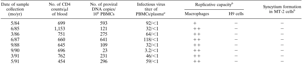
The study subject, patient D from our previous study (8), is a homosexual male who became seropositive for HIV-1 be-tween February and May of 1980. Eight sequential PBMC samples were collected between May of 1984 and May of 1991. During this period, he did not receive any antiviral therapy for HIV-1 and remained clinically well, with the exception of re-current genital herpes simplex infections. His CD4 cell counts remained relatively stable within the normal range until 1991,
when they dropped to 454 cells perml. The cell count declined
to below 200 perml by 1994 (18), thus meeting the Centers for
Disease Control definition of AIDS (5). The general clinical and virologic characteristics of this individual are summarized in Table 1. HIV-1 was successfully isolated from all of the PBMC samples collected. The infectious titer of HIV-1 ranged from 3.2 to 118 50% tissue culture infective doses per million cells, as quantified by a standard limiting dilution assay (19). The quantity of HIV-1 proviral DNA copies in PBMCs
corre-lates significantly (P,0.001) with the infectious titer.
Further-more, the amount of HIV-1 measured by these two techniques lies at the low end compared with that found in patients with moderate rates of disease progression (19, 39). In addition, sequential isolates from this individual were able to replicate in both normal donor PBMCs and macrophages, but failed to infect H9 or MT-2 cells or to induce syncytium formation (8). Plasma cultures were uniformly negative for infectious virus
(,1 50% tissue culture infective dose per milliliter) in all of the
samples tested. Unfortunately, the patient has been lost to follow up, and no samples after 1991 are available.
In order to study the LTR sequences in this individual, genomic DNA was first extracted by a standard proteinase K-phenol protocol (39) from serial PBMC samples pre- and
post viral culture. For each sample, approximately 1 mg of
genomic DNA was subjected to limiting dilution (39) before proceeding to two rounds of PCR. Nested primers were used
for both the 59and 39LTRs to increase the sensitivity and the
specificity of the amplification. Both rounds of PCR consisted
of 25 cycles, with each cycle involving three steps: 1 min at 948C
for denaturation, 1 min at 558C for annealing, and 1.5 min at
[image:2.612.57.562.82.191.2]728C for elongation. A final extension of 7 min at 728C was
TABLE 1. General clinical and virologic characterization of serial samples from the study subject
Date of sample collection
(mo/yr)
No. of CD4 counts/ml
of blood
No. of proviral DNA copies/
106PBMCs
Infectious virus titer of PBMCs/plasmaa
Replicative capacityb
Syncytium formation in MT-2 cellsb
Macrophages H9 cells
5/84 699 593 92/,1 1 2 2
6/85 1,153 121 32/,1 11 2 2
3/86 751 275 64/,1 11 2 2
6/87 660 641 118/,1 11 2 2
9/88 645 109 32/,1 11 2 2
9/90 696 23 3.2/,1 11 2 2
3/91 762 231 46/,1 11 2 2
5/91 454 296 59/,1 11 2 2
a
Fifty percent tissue culture infective dose measured per 106
PBMCs or per milliliter of plasma.
b
Results have been published elsewhere (8) and are included here solely for clarity.
on November 9, 2019 by guest
http://jvi.asm.org/
performed at the end of the 25 cycles. A Pfu polymerase (Stratagene) with proofreading activity was used in conjunc-tion with Taq (Boehringer Mannheim) polymerase to improve the accuracy of the amplification while maintaining the high processivity of Taq. The nucleotide sequences of the outer and
inner primer pairs for both the 59and 39LTRs are listed below;
the coordinates shown in parentheses denote their positions in
the HIVNL43 sequences (1, sense; 2, antisense): 59 LTR
outer, 59-CACACACAAGGCTACTTCCCTGATTGGC
AGA (157); 59-TCTGATAATGCTGAAAACATGGG
(21319); 59 LTR inner, 59-CAAGGCTACTTCCCTGATTG
GCAGAACTACACACCAGG (163); 59-AATGATCTAAG
TTCTTCTGATCCTGT (21022); 39LTR outer, 59-ATGGG
TGGCAAGTGGTCAA (18787); 59-TGCTAGAGATTTTC
CAC (29709); 39LTR inner, 59-TTTCCAGTCAGACCTCA
GGTACC (18988); 59-GTCTGAGGGATCTCTAGTTACC
AGAGTC (29680). Direct sequencing of the PCR products
was performed by the method of Winship et al. (41). Viral RNA sequences in the culture supernatant of the last sample were also determined as previously described (43). Briefly, viral RNA was extracted by guanidinium thiocyanate-phenol
solution. cDNA was then synthesized at 428C for 30 min with
[image:3.612.66.554.72.201.2]avian myeloblastosis virus reverse transcriptase (Promega). The nested PCR amplification and sequencing of cDNA prod-ucts were then carried out as stated above. Appropriate neg-ative controls were included throughout the entire course of study to monitor the possibility of cross-contamination.
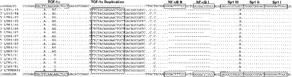
Figure 1 shows the consensus sequence of the 59 and 39
LTRs obtained from each of the serial samples. Both NF-kB
binding sites had apparently been deleted in all of the samples
tested from both the 59and 39LTRs, suggesting that the loss of
the NF-kB binding sites in this individual is a genuine
phe-nomenon. Three Sp1 binding sites, the TATA box motif, and TAR regions are well conserved among the sequences
ob-tained. Interestingly, compared with the HIVHXBLTR, a
27-nucleotide insertion was found upstream from the deleted
NF-kB binding sites in all of the sequences (Fig. 1). Some of
the 27 nucleotides are, in fact, a duplication of the upstream
region, previously designated as TCF-1asites (12, 14).
Dupli-cation of TCF-1ain HIV-1 LTR is not an uncommon finding,
because similar observations have been made in the studies of naturally occurring LTR variants in both asymptomatic as well as symptomatic patients (24, 30). In addition, enhanced activity of the LTR in directing both viral and heterologous gene
expression has been observed with the duplication of TCF-1a
(15). It is therefore plausible to suspect that the duplication of
TCF-1ain this individual may compensate for some of the lost
enhancer activity due to the deletion of the NF-kB binding
sites.
The degree of sequence diversity of the 59LTR varies from
year to year. The intrayear diversities were 1.7% in 1985, 2.3% in 1988, 1.3% in 1990, and 0.5% in 1991; thus, no clear pattern of change can be observed. However, by comparing the inter-year diversities, it is quite clear that the viral population had been diverging away from the early viral species. The genetic distance between 1985 and 1988 is 2.3%, that between 1985 and 1989 is 2.8%, and that between 1985 and 1991 is 3.3%. The consistent increase in genetic distance over time towards the early viral species suggests that viral evolution continues de-spite the asymptomatic stage of HIV-1 infection, and there is no truly latent period for viral variation in vivo.
The impact of the NF-kB deletion on the level of
transcrip-tional activity was evaluated in the context of a luciferase reporter gene system (Fig. 2) (6). The LTR sequences obtained from the last sample were cloned into the upstream region of the luciferase reporter gene in plasmid pGL-2 (Promega), and the resultant constructs are named DLTRluc. The LTR
se-quence from HIVHXBwas also cloned into the same reporter
vector for comparison and named HXBLTRluc. Five, 1, 0.2,
and 0.04mg of each construct were used in the transfection of
293 cells, a human embryonic kidney cell line, in the presence
(0.1mg) or absence of a Tat expression vector (pCMVTAT).
The cells were incubated at 378C and harvested 48 h
posttrans-FIG. 1. The 59and 39LTR consensus sequences derived from the study subject over time, aligned against HIVHXBLTR. Dots indicate identical sequences, and dashes indicate gaps introduced to preserve alignment. The locations of binding sites for TCF-1a, NF-kB, and Sp1 are shown in boxes, according to the findings of Gaynor (14).
FIG. 2. Schematic illustration of the constructs used in evaluating the LTR transcriptional activities, both in the context of a luciferase (Luc) reporter vector (upper panel) and the whole infectious virus (lower panel). The 39LTR that has been replaced with the LTR with NF-kB deleted is highlighted.
on November 9, 2019 by guest
http://jvi.asm.org/
[image:3.612.318.556.545.693.2]fection for luciferase assays. Harvested cells were lysed with
200ml of 13luciferase lysis buffer (Promega). Twenty
micro-liters of the lysates was then assayed for photon emission with a luminometer (Packard). The final readings of luciferase ac-tivity were corrected for both the transfection efficiency and the protein concentration in the cell lysates. Transfection
effi-ciencies were controlled by cotransfecting a b-galactosidase
expression vector, pSV-b-galactosidase (Promega). The
pro-tein content in the cell lysates was determined by the Bio-Rad protein assay system (Bio-Rad). All transfections were per-formed at least in duplicate.
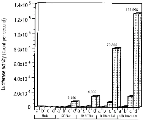
Figure 3 shows the average luciferase activity driven by the
LTR with NF-kB deleted (DLTRluc) and the intact HXBLTR
(HXBLTRluc) in the presence and absence of Tat. Detectable levels of luciferase activity were found in cells transfected with either DLTRluc alone or with DLTRluc and Tat (Fig. 3), although the latter demonstrated a substantially higher activity. In addition, the transcriptional activity of HXBLTRluc is
higher than that of DLTRluc (P ,0.05), in the presence or
absence of Tat. This observation is in agreement with previous reports in which higher transcriptional activity was found to
correlate with more NF-kB binding sites on the LTR (11, 36).
The patient’s LTR sequences were also introduced into the
infectious provirus HIVHXBlucto study the effect of the NF-kB
deletion on the level of viral transcription (Fig. 2). Provirus HIVHXBlucwas constructed by insertion of the Photinus pyralis
luciferase gene in nef (6). It is known that the U3 region of the
viral RNA is derived from the 39LTR of the provirus. This U3
region is then duplicated upon formation of the new provirus
(26, 27). We therefore replaced the 39LTR of HIVHXBlucwith
the LTR with NF-kB deleted and named the provirus
con-struct HIVDluc(Fig. 2). The new provirus produced after one
round of HIVDlucreplication will contain the NF-kB deletions
in its 59and 39LTR. The level of luciferase activity measured
in the infected target cells will directly correlate with the
tran-scriptional capacity of the corresponding 59LTR, thus enabling
us to study the effect of the NF-kB deletion in the context of
the whole virus. In brief, the LTR with NF-kB deleted was
cloned between the XhoI and AscI sites of HIVHXBluc. The
XhoI site is a naturally occurring site, whereas the AscI site was
introduced at the end of the 39LTR by site-directed
mutagen-esis. Twenty micrograms of provirus constructs was used to transfect 293 cells. Forty-eight hours posttransfection, culture supernatant was harvested and filtered. The virus concentra-tion in the culture supernatant was standardized by measuring
the p24gagconcentration (Abbott Laboratories) before use in
the subsequent infection assay. Five million phytohemaggluti-nin (PHA)-stimulated donor PBMCs or Jurkat T cells were
inoculated with 5 ng of p24gag of the virus. After a 2-h
incu-bation at 378C, the cells were washed extensively and
resus-pended in growth medium supplemented with 10% fetal calf serum and 10 U of interleukin 2 per ml. Infected PBMCs and Jurkat T cells were harvested for the luciferase assay on days 0, 3, 5, 7, and 12. Cell lysis and measurement of luciferase activity were carried out as described above.
Figure 4 shows the level of luciferase activity detected in HIVHXBluc- or HIVDluc-infected PBMCs and Jurkat T cells at
days 0, 3, 5, 7, and 12, respectively. In HIVDluc-infected
PB-MCs and Jurkat T cells, viral transcription continues
indepen-dent of the NF-kB binding sites. Peak levels of transcription
were recorded approximately 7 days postinfection, with a mod-erate decline in the ensuing period, although a generally higher level of transcription was found in PHA-stimulated PBMCs than in Jurkat T cells (Fig. 4). In addition, luciferase activity in
both HIVHXBluc-infected PBMCs and Jurkat T cells registers
almost twice that in HIVDluc-infected PBMCs and Jurkat T
cells in all samples detected (Fig. 4). This finding suggests that HXBLTR has a higher transcriptional capacity than DLTR in
both PBMCs and T cells (P , 0.05). These results clearly
demonstrate that NF-kB binding sites are not indispensable in
the course of viral replication during the infection of primary
cells. The higher transcriptional activity found in HIVHXBluc
with respect to HIVDlucalso suggests that NF-kB binding sites
are required for optimal levels of viral gene transcription. This observation is in agreement with those of previous reports in
which increased numbers of NF-kB binding sites were shown
to correlate with higher levels of transcriptional activity by the LTR and increased viral replication in almost all cell types studied (11, 36).
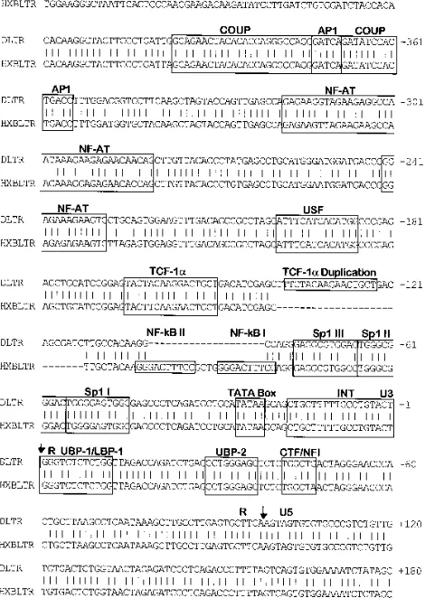
In order to confirm that the observed differences in
tran-scriptional activity are indeed due to mutations in the TCF-1a
and NF-kB regions, PCR amplification and sequencing of the
entire 59LTR were carried out after one round of infection of
both PHA-stimulated PBMCs and Jurkat T cells. Figure 5
shows the actual 59DLTR nucleotide sequence compared with
the 59HXBLTR nucleotide sequence. It is clear that the
[image:4.612.59.298.70.269.2]du-FIG. 3. Comparison of transcriptional activities of the LTR with NF-kB deleted (DLTRluc) and the LTR with NF-kB intact (HXBLTRluc) in the pres-ence and abspres-ence of transactivator Tat. Luciferase activities were measured 48 h posttransfection of 293 cells. A total of 0.04, 0.2, 1, and 5mg of each construct was used in the transfection, indicated by the letters a, b, c, and d, respectively.
FIG. 4. Comparison of transcriptional activities of the LTR with NF-kB deleted (HIVDluc) and the LTR with NF-kB intact (HIVHXBluc) in the context of the whole infectious virus. Luciferase activities were measured after infection of 53106
PHA-stimulated donor PMBCs (a) and Jurkat T cells (b) on days 0, 3, 5, 7, and 12.
on November 9, 2019 by guest
http://jvi.asm.org/
[image:4.612.318.557.71.186.2]plication of TCF-1a region and the deleted NF-kB binding
sites are present in the U3 region of 59DLTR. The sequence
variation in other regions of 59DLTR, however, is quite
min-imal, which is reflected by the high degree of conservation in almost all of the elements that are functionally important (Fig. 5).
In summary, a naturally occurring HIV-1 strain with NF-kB
binding sites deleted was identified in all available samples collected from an HIV-1-infected individual. The ability of this
virus to replicate and force a decline in CD41 lymphocyte
counts clearly indicated that a virus lacking NF-kB binding
sites can cause disease in humans. While many possible expla-nations may exist, functional redundancy of the LTR is likely to be the most plausible one (36). Multiple sequence elements present in the HIV-1 LTR permit their interactions with mul-tiple transcription factors individually or in particular combi-nations, depending on the availability and relative abundance of the factors present in the infected target cells. “Safety in numbers” would not be a far-fetched description of the strat-egy that the HIV-1 LTR uses to ensure its replication in a great variety of cell types. Furthermore, although dispensable,
NF-kB binding sites are found to enhance the capacity of the
HIV-1 LTR in directing transcription of both viral and
heter-ologous genes, suggesting that the presence of NF-kB binding
sites in the vast majority of viral isolates may function to promote optimal levels of viral gene transcription. Finally, a
duplication of a putative TCF-1amotif was also found in place
of the NF-kB elements in these viruses, but the biological
impact of this duplication warrants further investigation.
Nucleotide sequence accession number.The sequences
ob-tained were given GenBank accession numbers U80224 and U80225.
REFERENCES
1. Alcami, J., T. L. de Lera, L. Folgueira, M. Pedraza, J. Jacque, F. Bachelerie,
A. R. Noriega, R. T. Hay, D. Harrich, R. B. Gaynor, J. Virelizier, and F. Arenzana-Seisdedos.1995. Absolute dependence onkB responsive elements for initiation and Tat-mediated amplification of HIV transcription in blood CD4 T lymphocytes. EMBO J. 14:1552–1560.
2. Antoni, B. A., A. B. Rabson, A. Kinter, M. Bodkin, and G. Poli. 1994. NF-kB-dependent and -independent pathways of HIV activation in a chron-ically infected T cell line. Virology 202:684–694.
3. Baeuerle, P. A. 1991. The inducible transcription activator NF-kB: regulation by distinct protein subunits. Biochim. Biophys. Acta 1072:63–80. 4. Bellas, R. E., N. Hopkins, and Y. Li. 1993. The NF-kB binding site is
necessary for efficient replication of simian immunodeficiency virus of ma-caques in primary macrophages but not in T cells in vitro. J. Virol. 67:2908– 2913.
5. Castro, K. G., J. W. Ward, L. Slutsker, J. W. Buehler, H. W. Jaffe, and R. L.
Berkelman.1992. 1993 revised classification system for HIV infection and expanded surveillance case definition for AIDS among adolescents and adults, RR-17. Morbid. Mortal. Weekly Rep. 41:1–19.
6. Chen, B. K., K. Saksela, R. Andino, and D. Baltimore. 1994. Distinct modes of human immunodeficiency virus type 1 proviral latency revealed by super-infection of nonproductively infected cell lines with recombinant luciferase-encoding viruses. J. Virol. 68:654–660.
7. Cheng-Mayer, C. 1990. Biological and molecular features of HIV-1 related to tissue tropism. AIDS 4(Suppl. 1):s49–s56.
8. Connor, R. I., H. Mohri, Y. Cao, and D. D. Ho. 1993. Increased viral burden and cytopathicity correlate temporally with CD41T-lymphocyte decline and clinical progression in human immunodeficiency virus type 1-infected indi-viduals. J. Virol. 67:1772–1777.
9. Delassus, S., R. Cheynier, and S. Wain-Hobson. 1991. Evolution of human immunodeficiency virus type 1 nef and long terminal repeat sequences over 4 years in vivo and in vitro. J. Virol. 65:225–231.
10. Dewhurst, S., E. J. Embretson, D. C. Anderson, J. I. Mullins, and P. N. Fultz. 1990. Sequence analysis and acute pathogenicity of molecularly cloned SIVsmm-PBj14. Nature 345:636–640.
11. Englund, G., D. Hoggan, T. S. Theodore, and M. A. Martin. 1991. A novel HIV-1 isolate containing alterations affecting the NF-kB element. Virology
181:150–157.
12. Garcia, J. A., and R. B. Gaynor. 1994. Regulatory mechanisms involved in the control of HIV-1 gene expression. AIDS 8(Suppl. 1):s3–s17. 13. Garcia, J. A., F. K. Wu, R. Mitsuyasu, and R. B. Gaynor. 1987. Interactions
of cellular proteins involved in the transcriptional regulation of the human immunodeficiency virus. EMBO J. 6:3761–3770.
14. Gaynor, R. 1992. Cellular transcription factors involved in the regulation of HIV-1 gene expression. AIDS 6:347–363.
15. Golub, E., G. Li, and D. J. Volsky. 1990. Differences in the basal activity of the long terminal repeat determine different replicative capacities of two closely related human immunodeficiency virus type 1 isolates. J. Virol. 64: 3654–3660.
16. Harrich, D., J. Garcia, F. Wu, R. Mitsuyasu, J. Gonzalez, and R. Gaynor. 1989. Role of SP1-binding domains in in vivo transcriptional regulation of the human immunodeficiency virus type 1 long terminal repeat. J. Virol.
63:2585–2591.
17. Henderson, A. J., X. Zou, and K. L. Calame. 1995. C/EBP proteins activate transcription from the human immunodeficiency virus type 1 long terminal repeat in macrophages/monocytes. J. Virol. 69:5337–5344.
18. Ho, D. D., and Y. Cao. 1995. Long-term survivors of human immunodefi-ciency virus type 1 infection. N. Engl. J. Med. 332:1647–1648.
19. Ho, D. D., T. Moudgil, and M. Alam. 1989. Quantitation of human immu-nodeficiency virus type 1 in the blood of infected persons. N. Engl. J. Med.
321:1621–1625.
20. Ilyinskii, P. O., and R. C. Desrosiers. 1996. Efficient transcription and rep-lication of simian immunodeficiency virus in the absence of NF-kB and Sp1 binding elements. J. Virol. 70:3118–3126.
21. Jacque´, J. M., B. Ferna´ndez, F. Arenzana-Seisdedos, D. Thomas, F. Baleux, J.-L. Virelizier, and F. Bachelerie.1996. Permanent occupancy of the human immunodeficiency virus type 1 enhancer by NF-kB is needed for persistent viral replication in monocytes. J. Virol. 70:2930–2938.
[image:5.612.62.297.85.416.2]22. Jones, K. A., J. T. Kadonaga, P. A. Luciw, and R. Tjian. 1986. Activation of FIG. 5. Comparison of the nucleotide sequence of the LTR with NF-kB
deleted (DLTR) with that of the LTR with NF-kB intact (HXBLTR) after one round of infection. Regions that are bound by the cellular transcription factors are shown in boxes, and their locations are indicated according to the findings of Gaynor (14). Vertical lines indicate identical sequences, and dashes indicate gaps introduced to preserve alignment.
on November 9, 2019 by guest
http://jvi.asm.org/
the AIDS retrovirus promoter by the cellular transcription factor, Sp1. Sci-ence 232:755–759.
23. Kim, J. Y. H., F. Gonzalez-Scarano, S. L. Zeichner, and J. C. Alwine. 1993. Replication of type 1 human immunodeficiency viruses containing linker substitution mutations in the2201 to2130 region of the long terminal repeat. J. Virol. 67:1658–1662.
24. Koken, S. E. C., J. L. B. van Wamel, J. Goudsmit, B. Berkhout, and
J. L. M. C. Geelen.1992. Natural variants of the HIV-1 long terminal repeat: analysis of promoters with duplicated DNA regulatory motifs. Virology 191: 968–972.
25. Leonard, J., C. Parrott, A. J. Buckler-White, W. Turner, E. K. Ross, M. A.
Martin, and A. B. Rabson.1989. The NF-kB binding sites in the human
immunodeficiency virus type 1 long terminal repeat are not required for virus infectivity. J. Virol. 63:4919–4924.
26. Levy, J. A. 1993. Pathogenesis of human immunodeficiency virus infection. Microbiol. Rev. 57:183–289.
27. Lu, Y., M. Stenzel, J. G. Sodroski, and W. A. Haseltine. 1989. Effects of long terminal repeat mutations on human immunodeficiency virus type 1 repli-cation. J. Virol. 63:4115–4119.
28. Markovitz, D. M., M. J. Smith, J. Hilfinger, M. C. Hannibal, B. Petryniak,
and G. J. Nabel.1992. Activation of the human immunodeficiency virus type 2 enhancer is dependent on purine box andkB regulatory elements. J. Virol.
66:5479–5484.
29. McNearney, T., Z. Hornickova, A. Templeton, A. Birdwell, M. Arens, R.
Markham, A. Saah, and L. Ratner.1995. Nef and LTR sequences variation from sequentially derived human immunodeficiency virus type 1 isolates. Virology 208:388–398.
30. Michael, N. L., L. D’arcy, P. K. Ehrenberg, and R. R. Redfield. 1994. Nat-urally occurring genotypes of the human immunodeficiency virus type 1 long terminal repeat display a wide range of basal and Tat-induced transcriptional activities. J. Virol. 68:3163–3174.
31. Moses, A. V., C. Ibanez, R. Gaynor, P. Ghazal, and J. A. Nelson. 1994. Differential role of long terminal repeat control elements for the regulation of basal and Tat-mediated transcription of the human immunodeficiency virus in stimulated and unstimulated primary human macrophages. J. Virol.
68:298–307.
32. Novembre, F. J., P. R. Johnson, M. G. Lewis, D. C. Anderson, S. Klumpp,
H. M. McClure, and V. M. Hirsch.1993. Multiple viral determinants con-tribute to pathogenicity of the acutely lethal simian immunodeficiency virus
SIVsmmPBj variant. J. Virol. 67:2466–2474.
33. Olsen, H. S., and C. A. Rosen. 1992. Contribution of the TATA motif to Tat-mediated transcriptional activation of human immunodeficiency virus gene expression. J. Virol. 66:5594–5597.
34. Parrott, C., T. Seidner, E. Duh, J. Leonard, T. S. Theodore, A.
Buckler-White, M. A. Martin, and A. B. Rabson.1991. Variable role of the long terminal repeat Sp1-binding sites in human immunodeficiency virus replica-tion in T lymphocytes. J. Virol. 65:1414–1419.
35. Perkins, N. D., N. L. Edwards, C. S. Duckett, A. B. Agranoff, R. M. Schmin,
and G. J. Nabel.1993. A cooperative interaction between NF-kB and Sp1 is required for HIV-1 enhancer activation. EMBO J. 12:3551–3558. 36. Ross, E. K., A. J. Buckler-White, A. B. Rabson, G. Englund, and M. A.
Martin.1991. Contribution of NF-kB and Sp1 binding motifs to the repli-cative capacity of human immunodeficiency virus type 1: distinct patterns of viral growth are determined by T-cell types. J. Virol. 65:4350–4358. 37. Sakuragi, J., M. Fukasawa, R. Shibata, H. Sakai, M. Kawamura, H. Akari,
T. Kiyomasu, A. Ishimoto, M. Hayami, and A. Adachi.1991. Functional analysis of long terminal repeats derived from four strains of simian immu-nodeficiency virus SIVagm in relation to other primate lentiviruses. Virology
185:455–459.
38. Schmitz, M. L., and P. A. Baeuerle. 1991. The p65 subunit is responsible for the strong transcription activating potential of NF-kB. EMBO J. 10:3805– 3817.
39. Simmonds, P., P. Balfe, J. F. Peutherer, C. A. Ludlam, J. O. Bishop, and A. J.
Leigh Brown. 1990. Human immunodeficiency virus-infected individuals contain provirus in small numbers of peripheral mononuclear cells and at low copy numbers. J. Virol. 64:864–872.
40. Vlach, J., A. Garcia, J. Jacque, M. S. Rodriguez, S. Michelson, and J.
Virelizier.1995. Induction of Sp1 phosphorylation and NF-kB-independent HIV promoter domain activity in T lymphocytes stimulated by okadaic acid. Virology 208:753–761.
41. Winship, P. R. 1989. An improved method for directly sequencing PCR amplified material using dimethyl sulphoxide. Nucleic Acids Res. 17:1266. 42. Zeichner, S. L., J. Y. H. Kim, and J. C. Alwine. 1991. Linker-scanning
mutational analysis of the transcriptional activity of the human immunode-ficiency virus type 1 long terminal repeat. J. Virol. 65:2436–2444. 43. Zhang, L., P. Simmonds, C. A. Ludlam, and A. J. Leigh Brown. 1991.
Detection, quantification and sequencing of HIV-1 from the plasma of seropositive individuals and from factor VIII concentrates. AIDS 5:675–681.