Enhancement of limepiride dissolution
profile by solid dispersion technique
Dissertation work submitted to
THE TAMIL NADU DR. M.G.R. MEDICAL UNIVERSITY, CHENNAI
In partial fulfillment of the award of degree of
MASTER OF PHARMACY (PHARMACEUTICS)
Submitted by
MR. S.THANGAMUTHU
Under the guidance of
Asst.Prof.K.MUTHUSAMY, M.Pharm.,(Ph.D.),
Department of Pharmaceutics
March 2009
COLLEGE OF PHARMACY
Certificate
This is to certify that the dissertation entitled “ENHANCEMENT OF GLIMEPIRIDE DISSOLUTION PROFILE BY SOLID DISPERSION TECHNIQUE” was carried out by Mr.S.THANGAMUTHU., in the Department of Pharmaceutics, College of Pharmacy, Sri Ramakrishna Institute of Paramedical
Sciences, Coimbatore, which is affiliated to the Tamilnadu Dr.M.G.R. Medical
University, Chennai, under my direct supervision and complete satisfaction.
K.Muthusamy, M.Pharm., (Ph.D.), Assistant Professor, Department of Pharmaceutics, College of Pharmacy, S.R.I.P.M.S., Coimbatore – 641 044. Place : Coimbatore
Certificate
This is to certify that the dissertation entitled “ENHANCEMENT OF GLIMEPIRIDE DISSOLUTION PROFILE BY SOLID DISPERSION TECHNIQUE” was carried out by Mr.S.THANGAMUTHU., in the Department of Pharmaceutics, College of Pharmacy, Sri Ramakrishna Institute of Paramedical
Sciences, Coimbatore, which is affiliated to the Tamilnadu Dr.M.G.R. Medical
University, Chennai, under the direct supervision and guidance of
Asst.Prof.K.Muthusamy, M.Pharm., (Ph.D.), Department of Pharmaceutics, College of Pharmacy, SRIPMS, Coimbatore.
Prof.M.Gopal Rao, M.Pharm., Ph.D., Head – Department of Pharmaceutics, College of Pharmacy, S.R.I.P.M.S., Coimbatore – 641 044. Place : Coimbatore
Certificate
This is to certify that the dissertation entitled “ENHANCEMENT OF GLIMEPIRIDE DISSOLUTION PROFILE BY SOLID DISPERSION TECHNIQUE” was carried out by Mr.S.THANGAMUTHU., in the Department of Pharmaceutics, College of Pharmacy, Sri Ramakrishna Institute of Paramedical
Sciences, Coimbatore, which is affiliated to the Tamilnadu Dr.M.G.R. Medical
University, Chennai, under the direct supervision and guidance of
Asst.Prof.K.Muthusamy, M.Pharm., (Ph.D.), Department of Pharmaceutics, College of Pharmacy, SRIPMS, Coimbatore.
Dr.T.K.Ravi, M.Pharm., Ph.D., FAGE, Principal, College of Pharmacy, S.R.I.P.M.S., Coimbatore – 641 044. Place : Coimbatore
ACKNOWLEDGEMENT
The task of preparing the dissertation has been fascinating experience and it is really a moment of great pleasure for me to express my hearty gratitude to those who have helped me in the successful completion of this dissertation.
First of all I thank almighty for giving me the opportunity to carry myself forward in the path of my dream and for blessing me with the best also for making me who I am. I would take pride in tendering my deep sense of gratitude and indebtedness to my esteemed teacher and guide to Asst.Prof.K. Muthusamy, M.Pharm., (Ph.D.), Department of pharmaceutics, College of pharmacy, SRIPMS, Coimbatore, for his excellent suggestions, invaluable guidance, constant inspiration, sustained interest and good nature willingness throughout my work.
I am elated to place on record my profound sense of gratitude to Prof. M.Gopal Rao, M.Pharm., (Ph.D.), Vice Principal, Head, Department of pharmaceutics, college of pharmacy, SRIPMS, coimbatore, for his valuable support during my studies.
My sincere thanks goes to Dr. T.K. Ravi, M.Pharm., Ph.D., FAGE., Principal, College of Pharmacy, SRIPMS, Coimbatore, for providing the necessary facilities to carry out the study.
I extent my hearty thanks to Dr C. Vijayaraghavan, M.Pharm., Ph.D., and Prof. S. Kuppusamy, M.Pharm., (Ph.D.,) faculty, Department of Pharmaceutics, College of pharmacy, SRIPMS, for their support & help in making this project as a successful one.
I wish to extend my special thanks to Prof.S.Krishnan,M.Pharm., (Ph.D.,) Head, Department of pharmaceutical Bio- Technology, college of pharmacy, SRIPMS, for valuable help and permitting me to use the facilities of Department of Pharmaceutical Bio- technology.
I owe heartfelt thanks to S.Venkatesh, for his generous help during my project works.
My special thanks goes to Mr. J. Yuvaraju M.Pharm, K.Balakumar. M.Pharm., My dearest senior for their valuable guidance suggestion during my project.
My sincere thanks to Mr. A. Ramakrishnana, M.S.C., B.Ed., (Ph.D.,); Mr.S. Muruganandham; Mrs.Geetha and Mrs. Kalaivani for their help kind cooperation during the study.
I would like to thanks to the Librarian & Other staffs who have played a vital role in my project.
I owe Special thanks to Aristro Pharmaceutical Pvt.Ltd., Mumbai, & Franco- Indian Pharmaceutical Pvt. Ltd., Mumbai, for their courtesy extended to me by providing the required gift sample.
I wish to extent my thanks to Sophisticated Test & Instrumentation Centre, Cochin; Materials Research Centre.
I express my thanks to of M/s. Netsoft Computers Centre especially for designing the operation tools & completing thesis in a presentation form.
My sincere thanks & respect goes to the Managing Trustee Sevaratna Dr. R. Venkatasalu Naidu for all the facilities provided in the institution.
I submit my awesome thanks to my Seniors & my Batch mates for their support & co- operation during the course of my work.
whose help, support & encouragement had always been a source of inspiration through my project work.
I bow to my affectionate parents & grand father for their blessing and guidance in all my step of my life.
Above all, I humbly submit my dissertation work, into the hands of ALMIGHTY, who is the source of all wisdom & knowledge to find out WHO AM I.
CONTENTS
TITLE PAGE
NO CHAPTER – I
INTRODUCTION
1. SOLID DISPERSION SYSTEM AND ITS HISTORICAL BACKGROUND
1
2. CARRIERS USED FOR SOLID DISPERSIONS 3
3. CHARACTERIZATION OF SOLID DISPERSIONS 5 4. DEFINITION AND METHODS OF PREPARATION OF SOLID
DISPERSIONS
8
5. FEATURES OF SOLID DISPERSION 12
6. CLASSIFICATION AND FAST RELEASE MECHANISMS 17
SCHEME OF WORK
1. AIM 21
2. EXPERIMENTAL DESIGN 22
3. CHARACTERISATION OF SAMPLES BY DIFFERENT METHODS 22 4. FORMULATION STUDIES ON SELECTED SOLID DISPERSION 22
CHAPTER – II
DRUG PROFILE 24
CHAPTER – III
POLYMER PROFILE 29
PEG4000 29
HPMC 33
CHAPTER – IV
CHAPTER - V
ANALYTICAL METHOD 58
CHAPTER - VI
EXPERIMENTAL SECTION 61
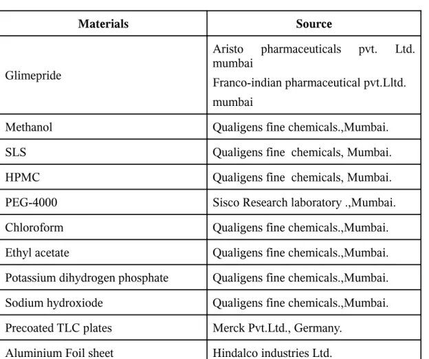
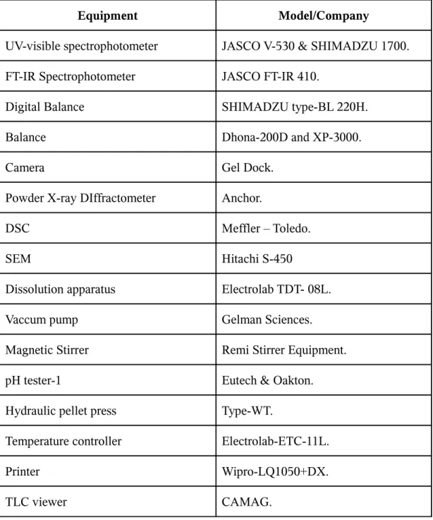
1. Materials and equipment used 61
2. Preparation and evaluation of glimepiride solid dispersion 63 3. Characterization of glimepiride solid dispersion 64
4. Evaluation of solid dispersion 81
CHAPTER – VII
RESULTS AND DISCUSSION
1. Preparation of solid dispersion 103
2. Compatibilities studies 103
3. Chacterization 104
4. In vitro analysis 105
Chapter – VIII
Summary and conclusion 106
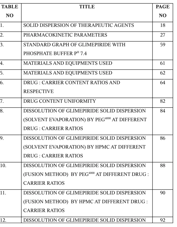
LIST OF TABLES
TABLE
NO
TITLE
PAGE
NO
1.
SOLID DISPERSION OF THERAPEUTIC AGENTS
18
2.
PHARMACOKINETIC PARAMETERS
27
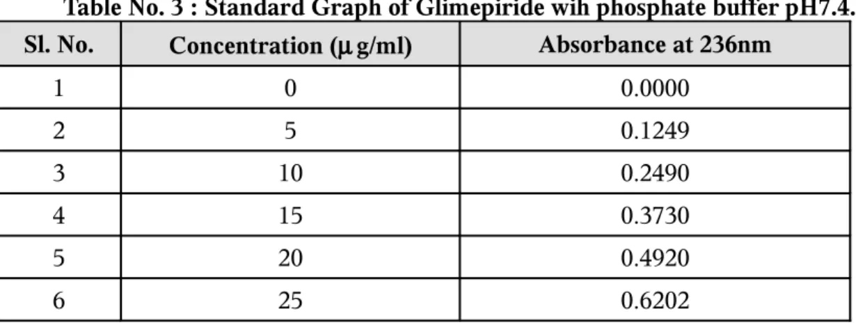
3.
STANDARD GRAPH OF GLIMEPIRIDE WITH
PHOSPHATE BUFFER P
H7.4
59
4.
MATERIALS AND EQUIPMENTS USED
61
5.
MATERIALS AND EQUIPMENTS USED
62
6.
DRUG : CARRIER CONTENT RATIOS AND
RESPECTIVE
64
7.
DRUG CONTENT UNIFORMITY
82
8.
DISSOLUTION OF GLIMEPIRIDE SOLID DISPERSION
(SOLVENT EVAPORATION) BY PEG
4000AT DIFFERENT
DRUG : CARRIER RATIOS
84
9.
DISSOLUTION OF GLIMEPIRIDE SOLID DISPERSION
(SOLVENT EVAPORATION) BY HPMC AT DIFFERENT
DRUG : CARRIER RATIOS
86
10.
DISSOLUTION OF GLIMEPIRIDE SOLID DISPERSION
(FUSION METHOD) BY PEG
4000AT DIFFERENT DRUG :
CARRIER RATIOS
88
11.
DISSOLUTION OF GLIMEPIRIDE SOLID DISPERSION
(FUSION METHOD) BY HPMC AT DIFFERENT DRUG :
CARRIER RATIOS
90
(PHYSICAL MIXTURE) WITH VARIOUS CARRIERS
13.
DISSOLUTION OF GLIMEPIRIDE IN PURE FORM AND
FROM SOLID DISPERSION S.E WITH VARIOUS
CARRIERS AT DRUG : CARRIER RATIO OF 1:2
94
14.
DISSOLUTION OF GLIMEPIRIDE IN PURE FORM AND
FROM SOLID DISPERSION (FUSION METHOD ) WITH
VARIOUS CARRIERS AT DRUG : CARRIER RATIO OF
1:2
96
15.
DISSOLUTION OF GLIMEPIRIDE IN PURE FROM,
PHYSICAL MIXTURE AND SOLID EVAPORATION
WITH PEG
4000AT DRUG : CARRIER RATIO OF 1 :2
98
16.
LOG PERCENTAGE GLIMEPIRIDE UNDISSOLVED
FROM PURE FORM AND FROM PEG
4000SOLID
DISPERSIONS (SOLVENT EVAPORATION METHOD)
1: 2 RATIO
100
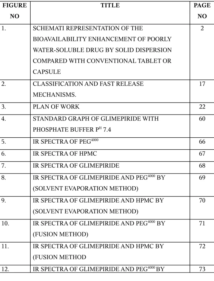
LIST OF FIGURES
FIGURE
NO
TITLE
PAGE
NO
1.
SCHEMATI REPRESENTATION OF THE
BIOAVAILABILITY ENHANCEMENT OF POORLY
WATER-SOLUBLE DRUG BY SOLID DISPERSION
COMPARED WITH CONVENTIONAL TABLET OR
CAPSULE
2
2.
CLASSIFICATION AND FAST RELEASE
MECHANISMS.
17
3.
PLAN OF WORK
22
4.
STANDARD GRAPH OF GLIMEPIRIDE WITH
PHOSPHATE BUFFER P
H7.4
60
5.
IR SPECTRA OF PEG
400066
6.
IR SPECTRA OF HPMC
67
7.
IR SPECTRA OF GLIMEPIRIDE
68
8.
IR SPECTRA OF GLIMEPIRIDE AND PEG
4000BY
(SOLVENT EVAPORATION METHOD)
69
9.
IR SPECTRA OF GLIMEPIRIDE AND HPMC BY
(SOLVENT EVAPORATION METHOD)
70
10.
IR SPECTRA OF GLIMEPIRIDE AND PEG
4000BY
(FUSION METHOD)
71
11.
IR SPECTRA OF GLIMEPIRIDE AND HPMC BY
(FUSION METHOD
72
(PHYSICAL MIXTURE)
13.
IR SPECTRA OF GLIMEPIRIDE AND HPMC BY
(PHYSICAL MIXTURE )
74
14.
X-RAY DIFFRACTION STUDIES OF GLIMEPIRIDE
SOLID DISPERSION WITH PEG
400075
15.
X-RAY DIFFRACTION STUDIES OF GLIMEPIRIDE
SOLID DISPERSION WITH HPMC
76
16.
X-RAY DIFFRACTION STUDIES OF GLIMEPIRIDE
77
17.
DSC STUDIES OF GLIMEPIRIDE SOLID DISPERSION
WITH PEG
4000BY (SOLVENT EVAPORATION METHOD)
78
18.
DSC STUDIES OF GLIMEPIRIDE SOLID DISPERSION
WITH HPMC BY (SOLVENT EVAPORATION METHOD
79
19.
DSC STUDIES OF GLIMEPIRIDE
80
20.
DISSOLUTION OF GLIMEPIRIDE SOLID DISPERSION
(SOLVENT EVAPORATION) BY PEG
4000AT DIFFERENT
DRUG : CARRIER RATIOS
85
21.
DISSOLUTION OF GLIMEPIRIDE SOLID DISPERSION
(SOLVENT EVAPORATION) BY HPMC AT DIFFERENT
DRUG : CARRIER RATIOS
87
22.
DISSOLUTION OF GLIMEPIRIDE SOLID DISPERSION
(FUSION METHOD) BY PEG
4000AT DIFFERENT DRUG:
CARRIER RATIOS
89
23.
DISSOLUTION OF GLIMEPIRIDE SOLID DISPERSION
(FUSION METHOD) BY HPMC
AT DIFFERENT DRUG:
24.
DISSOLUTION OF GLIMEPIRIDE SOLID DISPERSION
(PHYSICAL MIXTURE ) WITH VARIOUS CARRIERS
93
25.
DISSOLUTION OF GLIMEPIRIDE IN PURE FORM AND
FROM SOLID DISPERSION (SOLVENT
EVAPORATION) WITH VARIOUS CARRIERS AT
DRUG : CARRIER RATIO OF 1:2
95
26.
DISSOLUTION OF GLIMEPIRIDE IN PURE FORM AND
FROM SOLID DISPERSION (FUSION METHOD) WITH
VARIOUS CARRIERS AT DRUG : CARRIER RATIO OF
1:2
97
27.
DISSOLUTION OF GLIMEPIRIDE IN PURE FORM,
PHYSICAL MIXTURE AND SOLVENT EVAPORATION
WITH PEG
4000AT DRUG : CARRIER RATIO OF 1:2
99
28.
LOG PERCENTAGE GLIMEPIRIDE IN DISSOLVED
FROM PURE FORM AND FROM PEG
4000SOLID
DISPERSIONS (SOLVENT EVAPORATION METHOD)
OF 1:2 RATIO
CHAPTER -1
INTRODUCTION
1-91 . SOLID DISPERSION SYSTEM AND ITS
HISTORICALBACKGROUND
Fincher1 reviewed in 1968 the effect of the particle size of drugs on their dissolution
rates and biological availability comprehensively. The enhancement of oral
bioavailability of poorly water-soluble drugs remains one of the most challenging aspects
of drug development. Bioavailability can be defined as the rate and extent at which the
drug is delivered to the systemic circulation from dosage form and reaches the site of
action to produce the desired effect. Any new drug whose aqueous solubility is less than
0.01 µg/ml will definitely create a bioavailability problem and thereby affecting the therapeutic efficiency of a new drug. Once if we are able to increase the aqueous
solubility of a drug, the disintegration and dissolution properties can be easily altered, as
a result, an increase in bioavailability can be easily achieved. Methods to increase
aqueous solubility of a drug are Salt formation, solubilization, particle size reduction,
complexation, solvent evaporation, solid solution and solvent formation. They have been
commonly used to increase dissolution rate and thereby oral absorption and
bioavailability of such drugs2. There are practical limitations to these techniques. In 1961,
Sekiguchi and Obi2 developed a practical method whereby many of the limitations with
the bioavailability enhancement of poorly water-soluble drugs just mentioned can be
formation of eutectic mixtures of drugs with water-soluble carriers by the melting of their
physical mixtures.
Poorly water soluble drugs Tablet/ Capsule
Dosage form olid particles
Fig. 1: Schematic representation of the bioavailability enhancement of poorly water-soluble drug by solid dispersion compared with conventional tablet or capsule.
Sekiguchi and Obi2 in 1961 suggested that the drug was present in a eutectic
mixture in a microcrystalline state. Later, Goldberg et al 3 in 1966 demonstrated that the
entire drug in solid dispersions might not be necessarily present in a microcrystalline
state; a certain fraction of the drug might be molecularly dispersed in the matrix, thereby
forming a solid solution. In either case, once the solid dispersion was exposed to aqueous
media and carrier dissolved, the drug was released as very fine and colloidal particles.
Because of greatly enhanced surface area obtained in this way, the dissolution rate and
the bioavailability of poorly water-soluble drugs were expected to be high.
2. CARRIERS USED FOR SOLID DISPERSIONS
2.1. Poly Ethylene Glycols
Poly ethylene glycols are polymers of ethylene oxide a molecular weight usually
falling in the range 200 - 300,000. For solid dispersions PEGs with a molecular weight of
1500 - 20,000 are usually used. As the molecular weight increases, so does the viscosity
of PEG. Their solubility in water is generally good but decreases with molecular weight.
A particular advantage of PEGs for the formation solid dispersions is that, they have good
2.2. Poly Vinyl Pyrrolidine (PVP)
Polymerization of Vinyl Pyrrolidine leads to Poly Vinyl Pyrrolidine (PVP) of
molecular weight from 2500 - 3000,000. Due to their good solubility in a wide variety of
organic solvents, they are particularly suitable for solvent method. Similarly to the PEGs,
the PVPs have good water solubility and can improve the wettability of the dispersed
compound in many cases. The aqueous solubility of PVPs becomes poorer with
increasing chain length and further much higher viscosity at a given concentration.
E.g. Poly Vinyl Alcohol, Poly Vinyl Pyrrolidine Acetate co-polymer.
2.3 HYDROXY PROPYL METHYL CELLULOSE (HPMC)
Hydroxy propyl methyl cellulose (HPMC) also known as Hypromellose of
molecular weight from 10,000 – 1500000 due to their good solubility in water and in
mixtures of organic solvents like Ethan and chloroform , Mixtures of methanol and
dichloromethane like PEGS HPMC also have good water solubility and improving the
waltability of the compounds, they are particularly suitable for solvent method.
3. CHARACTERIZATION OF SOLID DISPERSIONS
The different methods that have been used to characterize solid dispersion are;
Thermo analytical methods, differential thermo analysis and hot stage microscopy.
Powder X-Ray diffraction.
Spectroscopic methods, especially IR spectroscopy.
Colorimetric analysis of the solution or melting enthalpy for calculation of entropy change.
Dissolution testing.
3.1. Therrmo Analytical Methods
Thermo analytical methods include all that examine a characteristic of the systems
as a function of temperature. Of this, Differential scanning calorimetry is the most highly
regarded method. DSC enables the quantitative detection of all process in which energy is
required or produced, i.e., endothermic and exothermic phase transition. The usual
method of measurement is to heat the reference and two test samples in such a way that
the temperature of two is kept identical.
If an energy requirement requiring transition occurs in the test samples, extra heat
is applied to this sample so that its temperature climbs at the same rate as in the reference.
The additional heat required is recorded and used to quantitate the energy or the phase
transition.
Exothermic transitions, such as conversion of one polymorph to a more stable
polymorph, can be also detected. Lack of a melting peak in DSC of solid dispersion
indicates that the drug is present in amorphous than the crystalline form. Since the
method is quantitative in nature, the degree of crystallinity can also be calculated for
systems in which the drug is partly amorphous and partly crystalline. However
crystallinities of fewer than 2% cannot be generally detected with DSC.
3.2. X-ray Diffraction
The principle behind X-RD is that when an x-ray beam is applied to sample,
can be detected depends on the wavelength applied and the geometry of the sample with
respect to periodicities in the structure. Crystalline sample is reflected by a characteristic
finger point region in the diffraction pattern. Owing to the specificity of the finger print,
crystallinity in the drug can be separately identified from crystallinity in the carrier.
Therefore, it is possible with
X-Ray Diffraction to differentiate between solid dispersions, in which it is partly present
in crystalline form, regardless of whether the carrier is amorphous or crystalline.
However, crystallinities of under 5-10% cannot generally be detected with X-RD.
3.3. Infra Red Spectroscopy
Structural changes and lack of a crystal structure can lead to changes in bonding
between functional groups which can be detected by Infra Red Spectroscopy. Since not
all the peaks in the IR spectrum are sensitive to crystalline changes, it is possible to
differentiate between those that are sensitive to changes in crystallinity and those that are
not.
3.4. Dissolution Testing
Release rate cannot be used on a stand alone basis to determine whether a solid
dispersion has been on a basis to determine whether a solid dispersion has been formed or
not. However in conjunction with other physiochemical data, they provide strong
evidence for the formation of a molecularly dispersed or nearly molecularly dispersed
system. When the goal of preparing a solid dispersion is to improve dissolution
characteristics of the drug, the results of the release rate experiments are obviously of
enhanced, and also whether the resulting supersaturated solution is stable or tends to
precipitate quickly. Comparison of results with those for pure drug powder and physical
mixture can help to indicate the dissolution via solubilization and wetting which could be
affected.
4. DEFINITION AND METHODS OF PREPARATION OF SOLID DISPERSIONS
4.1 Definition
The term refers to the dispersion of one or more active ingredients in an inert
carrier or matrix at solid state prepared by the melting (fusion), solvent evaporation
method and melting solvent method.
4.2. Methods Of Preparation
Basically there are three methods;
Melting method.
Solvent evaporation method.
Melting-solvent method.
4.2.1. Melting Method
Sekiguchi and Obi3 in 1961 first proposed the melting or fusion method, to
prepare fast release solid dispersion dosage forms. In this method, the physical mixture of drug and water-soluble carrier is heated directly until it is melted. The melted mixture is then cooled and solidified in an ice bath under vigorous stirring. The final mass is
ferrite plate or stainless steel plate and cooled by flowing air or water onto the opposite side of the plate. The solidified masses were stored in the dessicator at ambient
temperature.
Advantages
Simplicity and economy.
Less time consuming.
This method is also advantageous for compounds, which do not
undergo significant thermal degradation.
Disadvantages
The main disadvantage of the melt method includes thermal degradation,
sublimation, and polymeric transformation, which can affect the physicochemical properties of the drug including its rate of dissolution.
The temperature, at which the dispersion solidifies, affects crystallization rates and may alter both the size of the crystals and the hardness of the dispersion. This may result in tacky or glossy and unmanageable dispersions, which will require storage at elevated temperature, to facilitate hardening.
Examples:
Solid dispersions of Sulphamethoxazole, Acetaminophen, Griseofulvin,
Primidone, Chloropropamide, Chloramphenicol, Tolazamide, Steroids, Ketoprofen,
Nimesulide.
4.2.2. Solvent Evaporation Method
This method involves dissolving the drug and carrier in a suitable organic solvent,
followed by evaporation of the solvent to form solid dispersion. The mass was then
stored in a dessicator, pulverized and sieved.
Solvent removal is accomplished by various means. The most common approach
is the application of reduced pressure at a fixed temperature to evaporate the organic
fastest way of removing solvent. The freeze-drying technique is also employed to prepare
solid dispersions by removal of aqueous solutions.
Advantages
The procedure is suitable for drugs that are thermolabile.
The thermal decomposition of drugs or carriers can be prevented because of the low temperature required for the evaporation of the organic solvents.
For aqueous systems, frozen temperature can be used to evaporate the solvent, which can enhance the integrity of the drug.
Disadvantages
Difficulty in complete removal of the solvent.
Finding a suitable solvent that will dissolve both the drug and carrier is very difficult.
Plasticization of some polymers such as polyvinyl pyrrolidone has occurred with the use of some solvents.
It is important that the rate of evaporation of a solvent is controlled so as to control the particle size of the drug, which in turn will affect the rate of dissolution of the drug in the solid dispersion.
Examples:
β-Carotene–PVP, Griseofulvin–PVP, Sulfathiazole–PVP, Steroids–PVP, Reserpine–deoxycholic acid.
4.2.3. Melting-Solvent Method
The drug is first dissolved in a suitable liquid solvent and solution is then
incorporated directly into a melt of PEG obtained below 70°C without removing the
liquid solvent. It was shown that 5 -10%w/w of liquid-components would be incorporated
into PEG 4000 without significant loss of its solid property. Advantages
Possess the advantage of both melting and solvent methods.
Disadvantages
Limited to drugs having therapeutic index below 50 mg.
Selected solvents or dissolved solution may not be miscible with melt of PEG.
Examples:
Solid dispersion of Clofibrate, Methyl Salicylate, Benzyl Benzoate.
5. FEATURES OF SOLID DISPERSION
Includes recent developments in theory and practice.
An aid in new drug formulation and improvement of existing drugs.
Techniques for improved dissolution rate, sustained release, altered solid-state properties and improved solubility and stability.
6. CLASSIFICATION AND FAST RELEASE MECHANISMS
6.1. Simple Eutectic Mixtures
The simple eutectic mixture is usually prepared from the rapid solidification of the fused liquid of two components, which show solid solubility. When the preparation is dissolved in aqueous medium, the carrier will dissolve rapidly, releasing very fine crystals of drug, which offers large surface area, thereby improvement in dissolution is effected. Thermodynamically, such a system is regarded as an intimately blended physical mixture of its two crystalline components.
6.2. Solid Solutions
A solid solution is made-up of a solute dissolved in a solid solvent. It is often called a mixed crystal because the two components crystallize together in a homogenous one-phase system. A solid solution achieves faster dissolution rate than eutectic mixture because, the particle size of the drug in the solid solution is reduced to a minimum state i.e. molecular size.
Solid solution can be classified according to the extent of miscibility between the two components or the crystalline structure of the solid solution. Based on this, they can be divided into four groups: continuous (or isomorphous, unlimited, complete) solid solutions, discontinuous (or limited, restricted, partial, incomplete) solid solutions, substantial solid solutions and interstitial solid solutions.
6.2.1. Continuous Solid Solutions
In this system, the two components are miscible or solid state in all proportions. The bonding strength between two components is stronger than that between the molecules of each component.
6.2.2. Discontinuous solid solution
6.2.3 Substitutional crystalline solid solution
In this, the solute molecules substitute for the solvent molecules in the crystal lattice of the solid solvent. It can form a continuous solid solution.
6.2.4. Interstitial Crystalline Solid Solution
The solute (guest) molecule occupies the interstitial space of the solvent (host) in the lattice; it usually forms only a discontinuous (limited) solid solution.
6.3. Glass Solutions
A Glass solution is a homogeneous, glassy system in which a solute dissolves in a glassy solvent. It is another potential modification of dosage forms in increasing drug dissolution and absorption. The familiar term “glass,” however can be used to describe either a pure chemical or a mixture of chemicals in a glossy or vitreous state.
6.4. Amorphous Preparations
The amorphous form is the highest energy form of a pure drug. It should under
almost all conditions, produce faster dissolution and high absorption rates than the
crystalline form. In amorphous solid solution, the solute molecules are dispersed
molecularly but irregularly within the amorphous solvent. Novobiocine has been reported
to have a ten-fold higher solubility than its crystalline form.
6.5. Complex Formulations
The availability of a drug depends on the intrinsic absorption rate of the complex.
The water-soluble polymers have been considered as ideal carriers for the solid
dispersion of poorly soluble drugs.
Advantages Of Solid Dispersions
Solid dispersion of drugs in solid state is helpful in stabilizing unstable drugs. Many of the advantages claimed for SD are derived from their rapid
dissolution rates. The increased rate of nitrazepam from the citric acid dispersion produces increase in the rate and extent of absorption.
Various fast release solid dispersions can be prepared by solid dispersion technique. For example: fast release solid dispersions of Lorazepam can be prepared by using urea, PEG 6000 or Mannitol as carriers.
Solid dispersions may be a thermodynamically more active form of drug and directly influence the diffusion and release rate.
An increased diffusion of steroid from the ointment was obtained, example: solid dispersion of prednisolone urea dispersion.
Solid dispersion technology can be used to solidify liquid drugs, example: clofibrate and benzyl benzoate.
Disadvantages Of Solid Dispersions
Tackiness and decommission during preparation and formulation.
The oral administration of solid dispersions without concomitant reduction in dose may result in higher incidence of adverse effects.
Ex: ulceration of Indomethacin–PEG 6000 dispersion.
The physical and chemical stability of drug and vehicle.
Reproducibility of its physicochemical properties.
The scale up of manufacturing process.
Its formulation into dosage forms.
Difficulty in pulverization.
Drug carrier incompatibility.
Poor flow and mixing properties.
Sifting of the dispersions, which are usually soft and tacky.
The surface modification technique can significantly improve the dissolution of hydrophobic drug, by the adsorption of very small amounts of urea at the drug particle surface. Techniques have been commonly used to improve dissolution and bioavailability of poorly water-soluble drugs, which includes micronization, the use of surfactants, and the formation of solid dispersions.
Chiou and Riegelmann4 in 1971 outlined six types of drug-carrier interactions in
solid-state dispersions. Simple eutectic mixtures, solid solutions, glass solutions, glass suspensions, amorphous precipitates in a crystalline carrier and compound or complex formation. Other factors such as increased wettability, solubilization of the drug by the carrier at the diffusion layer and the reduction or absence of aggregation and
agglomeration may also contribute to increased dissolution.
Fig 2: Classification And Fast Release Mechanisms
FAST RELEASE MECHANISMS
DISCONTINUOUS SOLID SOLUTIOUBSTANTIAL SOLID SOL
INTEL STable 1 : Solid Dispersions Of Therapeutic Agents
Drug Carrier Method
Type of solid Dispersion Effect of Dissolution Rate Triamterene Β-cyclodextrin
S, K Not Studied Increased
Flurbiprofen PVP S Not Studied Increased
Caffeine Nicotinamide M Peritechc Increased
Chloramphenicol Urea M
Solid
Solution Increased
Clofibrate PEG 6000 M, S Not Studied Increased
Corticosteroids Sugars M Not Studied Increased
Diazepam PEG 4000 M
Eutectic with
solid
Solution
Not Studied
Griseofulvin Succinic acid M
Solid
Solution
Increased
PVP S Not Studied Increased
PVP-30 Spray
Embedding
Solid
Solution
Increased
PEG -4000 M, S Not Studied Increased
PEG -6000 M, S Not Studied Increased
PEG-2000 M, S Not Studied Increased
Anhycitric
acid M
Glass
suspension Increased
Indomethacin PEG 6000 M Not Studied Increased
Methyl salicylate PEG 6000 M,S Not Studied Increased
Paracetamol Urea M
Solid
solution Increased
Mannitol M Eutectic Increased
Reserpine PVP S Not Studied Increased
Cholanic acid S Not Studied Increased
Deoxychoilic
acid S Not Studied Increased
Sulfathiazole Urea M Simple
eutectic Not Studied
Tolubutamide PEG-4000 S Not Studied Increased
PEG-6000 S Not Studied Increased
PEG
4000+6000
M, S Not Studied Increased
PEG-2000 M, S Monoacetic Not Studied
PVP S Not Studied Increased
Polyoxyl 40
stearate M, S Not Studied Increased
PEG-8000 M, S Not Studied Increased
PVP S Not Studied Increased
PEG-6000 S Not Studied Increased
PEG-4000 M Not Studied Increased
Allopurinol PVP S Not Studied Increased
Benzybenzoate PEG 6000 M, S Not Studied Increased
*M-Melting Method, S-Solvent Method, MS- Melting Solvent Method, K-Kneading
SCHEME OF WORK
The main perspective of the present study aims at overcoming these problems with solid
dispersion technology by using carriers like HPMC and peg-4000 in a view to develop fast
release formulation of glimepiride and hence improve its dissolution characteristic. Glimepiride is
an effective anti-diabetic, which is practically insoluble in water, hence dissolution is rate
limiting.
The research work envisaged was,
Literature survey on solid dispersion, method and carriers for solid dispersion.
Preparation, characterisation and evaluation of solid dispersion of Glimepiride
with HPMC, and peg-4000.
Formulation studies on solid dispersion of Glimepiride.
1. AIM
To prepare solid dispersions of glimepiride using HPMC and peg-4000 as the inert
carriers.
To assay the solid dispersions.
To evaluate the solid dispersions by in-vitro dissolution studies in phosphate buffer pH
7.4. The in-vitro release profiles of prepared solid dispersions were compared with pure drug.
The samples were also evaluated by using various instrumental techniques.
2. EXPERIMENTAL DESIGN
Preparation of glimepiride-peg4000 and glimepiride - HPMC solid dispersions
by solvent evaporation method and fusion method.
Assay of solid dispersions.
Release studies on solid dispersions in phosphate buffer (pH 7.4).
3. CHARACTERISATION OF SAMPLES BY DIFFERENT METHODS
DSC-Analysis.
IR spectroscopic analysis.
To study the effect of concentration and type of carrier on the release of Glimepiride
from its solid dispersions.
4. FORMULATION STUDIES ON SELECTED SOLID DISPERSION
Release studies on selected solid dispersion with pure drug.
Fig 3: Plan of Work
Plan of work
Glimepiride Method validation by Standard graph of glimepiride
Preformulation studies
Preparation Polymer used HPMC, Peg-4000 Drug:Carrier 1;1
1;2 1;3 1;4 Glimepiride-HPMC SD
Glimepiride-Peg-4000 SD Dispersion
Development Of Dissolution System Of Glimepiride Evaluation
DSCC XRD
IR Comparative Study
CHAPTER - 2
DRUG PROFILE
10,13,14GLIMEPIRIDE, a new generation sulphonyl urea [Endocrine Journal, 2007] has several
benefits: rapid and complete absorption after oral administration, a lower dose,
long duration action, and possible insulin sensitizing effect. Glimepiride is an oral
blood glucose lowering drug of sulfonyl urea class.
Chemical Name
Glimepiride (1–[[p-[2-(3-ethyl-4-methyl-2-oxo-3-pyrroline-1-carboxamido)ethyl]
phenyl] sulfonyl]-3-(trans-4-methylcyclohexyl) urea.
The CAS registry number is 93479-97-1.
Molecular formula: C24H34N4O5S
Molecular Weight: 490.62
Structural formula:
Figure 5: Structure of Glimepiride
Physical properties: Glimepiride is a white to yellowish white crystalline odorless
powder.
Solubility: Glimepiride is insoluble in water, acid, base, borate and phosphate
buffers but partially soluble in methanol, ethanol, acetone, and completely
Chemical properties: Methanolic solution of Glimepiride gives UV absorption at
229nm and aqueous solution of Glimepiride gives maximum absorption between
229 and 236nm [Indian Journal of Pharmaceutical Sciences].
Log P value: 2.5
C Log P value: 3.96 [www.tsrlinc.com]
BCS: Class 2 drug (low solubility high permeability)
Available strength: 1mg, 2mg, and 4mg
Mechanism of Action
The primary mechanism of Glimepiride in lowering blood glucose appears to be
dependent on stimulating the release of insulin from functioning pancreatic beta
cells.
In addition extra pancreatic effect may also play a role in the activity of sulfonyl
urea such as Glimepiride. This is supported by both clinical and pre-clinical
studies demonstrating the Glimepiride administration can lead to increased
Pharmacokinetics
Absorption: After oral administration, Glimepiride is completely (100%) absorbed from the GI tract. Studies with single oral doses in normal subjects and
with multiple oral doses in patients with Type2 has shown significant absorption
of Glimepiride with 1 hour after administration and peak drug levels at 2 to
3hours.
Distribution: After intravenous (IV) dosing in normal subjects, the volume of distribution (Vd) was 8.8L (113ml/kg), and the total body clearance (Cl) was 47.8
ml/min. Protein binding was greater than 99.5%.
Metabolism: Glimepiride is completely metabolized by oxidative biotransformation after either an IV or oral dose. The major metabolites are the
cyclohexyl hydroxyl methyl derivative (M1) and the carboxyl derivative (M2).
Cytochrome P450 2C9 has been shown to be involved in the biotransformation of
Glimepiride to M1. M1 is further metabolized to M2 by one or several cytosolic
enzymes. M1, but not M2, possesses about 1/3 of the pharmacological activity as
compared to its parent in an animal model.
Excretion: When 14C-Glimepiride was given orally, approximately 60% of the
total radioactivity was recovered in the urine in 7 days and M1 (predominant) and
M2 accounted for 80 – 90% of that recovered in the urine. Approximately 40% of
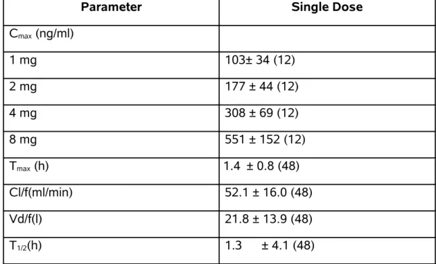
Pharmacokinetic Parameters
Table 2: Pharmacokinetic Parameters
Parameter Single Dose
Cmax (ng/ml)
1 mg 103± 34 (12)
2 mg 177 ± 44 (12)
4 mg 308 ± 69 (12)
8 mg 551 ± 152 (12)
Tmax (h) 1.4 ± 0.8 (48)
Cl/f(ml/min) 52.1 ± 16.0 (48)
Vd/f(l) 21.8 ± 13.9 (48)
T1/2(h) 1.3 ± 4.1 (48)
CL/f=Total body clearance after oral dosing
Vd/f=Volume of distribution calculated after oral dosing
Adverse reactions
GASTRO INTESTINAL REACTIONS
Vomiting, gastro intestinal pain and diarrhoea have been reported.
DERMATOLOGIC REACTIONS
Allergic skin reactions, e.g. Pruritus, Erythema, Urticaria occurs.
HEAMATOLOGIC REACTIONS
Leukopenia, Agranulocytosis, Thrombocytopenia, Hemolytic Anemia, Aplastic
Indications and Usage
Glimepiride is indicated as an adjunct to diet and exercise to lower the blood
glucose in patient with Type 2 diabetes mellitus. Hypoglycemia cannot be
controlled by diet and exercise alone. Glimepiride may be used concomitantly
with Metformin when diet, exercise, Glimepiride or Metformin alone do not
adequate glycemic control.
Dosage and Administration [Package insert of Amaryl tablets 1, 2 and 4 mg.]
Usual starting dose of Glimepiride initial therapy is 1mg to 2mg once daily,
administered with breakfast or first main meal.
Patient who may be more sensitive to Hypoglycemic drugs should be started at
1mg once daily, and should be titrated carefully.
The maximum starting dose of Glimepiride should not be more than 2mg. The
usual maintenance dose is 1 to 4mg once daily. The maximum recommended
CHAPTER - 3
POLY ETHYLENE GLYCOL PROFILE
15-241. NONPROPRIETARY NAMES
BP: Macrogols, JP: Macrogol 400, Macrogol 1500, Macrogol
4000, Macrogol 6000, Macrogol 20000, PhEur: Macrogola,
USPNF: Polyethylene glycol
2. SYNONYMS
Carbowax; Carbowax Sentry; Lipoxol; Lutrol E; PEG; Pluriol E; polyoxyethylene glycol.
3. CHEMICAL NAME
α-Hydro-ω-hydroxypoly(oxy-1,2-ethanediyl)
4. FUNCTIONAL CATEGORY
Ointment base; plasticizer; solvent; suppository base; tablet and capsule lubricant.
5. APPLICATIONS IN PHARMACEUTICAL TECHNOLOGY
Polyetylene glycols (PEGs) are widely used in a variety of pharmaceutical formulations
including parenteral, topical, ophthalmic, oral, and rectal preparations. It has been used in
controlled-release systems.
Polyethylene glycols are water-soluble and are easily removed from the skin by washing,
making them useful as ointment bases.2 Aqueous polyethylene glycol solutions can be used
either as suspending agents or to adjust the viscosity and consistency of other suspending
vehicles. When used in conjunction with other emulsifiers, polyethylene glycols can act as
stabilizers.
enhance the aqueous solubility or dissolution characteristics of poorly soluble compounds by
making solid dispersions with an appropriate polyethylene glycol.
In film coatings, solid grades of polyethylene glycol can be used alone for the film-coating
of tablets. Solid grades are also widely used as plasticizers in conjunction with film-forming
polymers.The presence of polyethylene glycols in film coats, especially of liquid grades, tends to
increase their water permeability and may reduce protection against low pH in enteric-coating
films.
6. PHARMACOPEIAL SPECIFICATIONS
6.1. Typical Properties
Density
: 1.11–1.14 g/cm
3at 25°C for liquid PEGs;
15–1.21 g/cm
3at 25°C for solid PEGs.
Flash point
: 238°C for PEG 4000;
Freezing point
: 4–8°C for PEG 4000
Melting point : 50–58°C for PEG 4000.
Moisture content : Liquid polyethylene glycols are very hygroscopic,
hygroscopicity decreases with increasing
molecular weight. Solid grades.
7. SOLUBILITY
Soluble in water, liquid PEG are soluble in acetone, alcohols, benzene,
glycerin, and glycols. Solid polyethylene glycols are soluble in acetone,
dichloromethane, ethanol (95%), and methanol; they are slightly soluble in
8. SURFACE TENSION
Approximately 44 mN/m (44 dynes/cm) for liquid polyethylene glycols; approximately
55 mN/m (55 dynes/cm) for 10% w/v aqueous solution of solid polyethylene glycol.
9. STABILITY AND STORAGE CONDITIONS
Polyethylene glycols are chemically stable in air and in solution, although grades with a
molecular weight less than 2000 are hygroscopic. Polyethylene glycols do not support microbial
growth, and they do not become rancid.
Polyethylene glycols should be stored in well-closed containers in a cool, dry place.
Stainless steel, aluminum, glass, or lined steel containers are preferred for the storage of liquid
grades.
10. INCOMPATIBILITIES
All grades can exhibit some oxidizing activity owing to the presence of peroxide impurities
and secondary products formed by autoxidation. Liquid and solid polyethylene glycol grades may
be incompatible with some coloring agents.The antibacterial activity of certain antibiotics is
reduced in polyethylene glycol bases. The preservative efficacy of the parabens may also be
impaired owing to binding with polyethylene glycols.
11. HANDLING PRECAUTIONS
Observe normal precautions appropriate to the circumstances and quantity of material
handled. Eye protection is recommended.
12. REGULATORY STATUS
Included in the FDA Inactive Ingredients Guide (dental preparations; IM and IV injections;
ophthalmic preparations; oral capsules, solutions, syrups, and tablets; rectal, topical, and vaginal
preparations). Included in nonparenteral medicines licensed in the UK. Included in the Canadian
List of Acceptable Non-medicinal Ingredients.
JP: Hydroxypropylmethylcellulose
PhEur: Hypromellosum
USP: Hypromellose
Synonyms
Benecel MHPC; hydroxypropyl methyl ether; E464; hydroxypropyl
methylcellulose; HPMC; Methocel; methylcellulose propylene glycol ether;
merthyl hydroxypropylcellulose; Metolose ; Pharmacoat ; Spectracel 6 ;
Spectracel 15; Tylopur.
Chemical Name and CAS registry number
Cellulose, 2-hydroxypropyl-methyl ether [9004-65-3]
Empirical formula molecular weight
The PhEur 2002 describes hypromellose as a partly methylated and
O-(2-hydroxypropylated) cellulose. It is available in several grades that vary in
viscosity and extent of substitution. Grades may be distinguished by appending a
number indicative of the apparent viscosity, in mPa s, of a 2% w/w aqueous
solution at 20°C. Hypromellose defined in the USP 25 specifies the substitution type by appending a four-digit number to the nonproprietary name: e.g.,
hypromellose 1828. The first two digits refer to the approximate percentage
content of the methoxy group (OCH3). The second two digits refer to the
approximate percentage content of the methoxy group (OCH3). The second two
digits refer to the approximate percentage content of the hydroxypropoxy group
approximately 10 000-1 500 000. The Jp 2001 includes three separate
monographs for hypromellose: hydroxypropylmethylcellulose 2208, 2906, and
2910, respectively.
Functional category
Coating agent; film-former; rate – controlling polymer for sustained
release; stabilizing agent; suspending agent; tablet binder; viscosity – increasing
agent.
Applications in pharmaceutical formulation or technology
Hypromellose is widely used in oral and topical pharmaceutions,
Particularly ophthalmic preparations. Compared with methylcellulose,
hypromellose produces solutions of greater clarity, with fewer undispersed fibers
present, and is therefore preferred in formulations for ophthalmic use.
Hypromellose at concentrations between 0.45-1.0% w/w may be added as a
thickening agent to vehicles for eye drops and artificial tear solutions.
Hypromellose is also used as an emulsifier, suspending agent, and
stabilizing agent in topical gels and ointments. As a protective colloid, it can
prevent droplets and particles from coalescing or agglomerating, thus inhibiting
the formation of sediments.
In addition, hypromellose is used in the manufacture of capsules, as an
Description
Hypromellose is an odorless and tasteless, white or creamy white fibrous
or granular powder.
Pharmacopeial specifications Typical Properties
Acidity / alkalinity : pH = 5.5 – 8.0 for a 1% w/w aqueous solution
Ash : 1.5-3.0% depending upon the grade
Autoignition temperature: 360°C Density (tapped) : 0.557 g/cm3
Density (tapped) : 1.326 g/cm3
Melting point : browns at 190-200°C; chars at 225-230°C. Glass transition temperature is 170-180°C.
Moisture content: hypromellose absorbs moisture from the atmosphere, the amount of water absorbed depending and relative humidity of the surrounding air.
Solubility: soluble in cold water, forming a viscous colloidal solution; practically insoluble in chloroform, ethanol (95%), and ether, but soluble in mixtures of
ethanol and dichloromethane, mixtures of methanol and dichloromethane, and
mixtures of water and alcohol. Certain grades of hypromellose are soluble in
aqueous acetone solutions, mixtures of dichloromethane and propan-2-ol, and
other organic solvents.
Viscosity (dynamic): Wide ranges of viscosity types are commercially available. Aqueous solutions are most commonly prepared, although hypromellose may
also be dissolved in aqueous alcohols such as ethanol and propan-2-ol provided
the alcohol content is less than 50% w/w. Dichloromethane and ethanol mixtures
may also be used to prepare viscous hypromellose solutions. Solutions prepared
using organic solvents tend to be more viscous; increasing concentration also
produces more viscous solutions;
Methocel grade Nominal Viscosity (MPa s)
K 100LVP 100 80-120
K4M 4000 3000-5600
K15MP 15000 12000-2100
K100MP 100 000 80 000-120 000
To prepare an aqueous solution, it is recommended that hypromellose is
dispersed and thoroughly hydrated in about 20-30% of the required amount of
water. The water should be vigorously stirred and heated to 80-90°C, then the remaining hypromellose added. Cold water should then be added to produce the
required volume.
When a water – miscible organic solvent such as ethanol, glycol, or
mixtures of ethanol and dichloromethane is used, the hypromellose should first
be dispersed into the organic solvent, at a ratio of 5-8 parts of solvent to part of
hypromellose. Cold water is then added to produce the required volume.
Stability and storage conditions
Solutions are stable at pH 3-11. Increasing temperature reduces the
viscosity of solutions. Hypromellose undergoes a reversible sol-gel
transformation upon heating and cooling, respectively. The gel point is 80-90°C, depending upon the grade and concentration of material.
Aqueous solutions are comparatively enzyme-resistant, providing good
viscosity stability during long-term storage.However, aqueous solutions are liable
to microbial spoilage and should be preserved with an antimicrobial preservative:
when hypromellose is used as a viscosity – increasing agent in ophthalmic
solutions, benzalkonium chloride is commonly used as the preservative. Aqueous
solutions may also be sterilized by autoclaving; the coagulated polymer must be
redispersed on cooling by shaking.
Hypromellose powder should be stored in a well-closed container, in a
cool, dry place.
Incompatibilities
Hypromellose is incompatible with some oxidizing agents. Since it is
nonionic, hypromellose will not complex with metallic salts or ionic organics to
form insoluble precipitates.
safety
Hypromellose is widely used as an excipient in oral and topical
pharmaceutical formulations. It is also used extensively in cosmetics and food
Hypromellose is generally regarded as a nontoxic and nonirritant material,
although excessive oral consumption may have a laxative effect. The WHO has
not specified an acceptable daily intake fro hypromellose since the levels
consumed were not considered to represent a hazard to health.
LD 50 (mouse, IP): 5 g/kg (16)
LD 50 (rat, IP): 5.2 g/kg
Handling precautions
Observe normal precautions appropriate to the circumstances and
quantity of material handled. Hypromellose dust may be irritant to the eyes and
eye protection is recommended. Excessive dust generation should be avoided to
CHAPTER - 4
LITERATURE REVIEW
34-60Sethia and Squillante (2004)58 were prepared solid dispersion of
carbamazepine in PVP K30 by conventional solvent evaporation and supercritical
methods. They have suggested that the best intrinsic dissolution rate (IDR) was
obtained for super critically processed CBZ/PVP K30 that was four-fold higher
than pure CBZ and the supercritical-based process produced augmented with
amphiphilic carriers.
Eun-Jung et al., (2006)59 has studied the dissolution rates of felodipine
musing PVP and HPMC carriers by solvent wetting method. It could be shown
that the dissolution rates of felodipine in PVP and HPMC solid dispersion were
much faster than those for they corresponding physical mixtures. However
dissolution profiles were found to depend on the carrier used; the dissolution rate
of felodipine increased slowly for solid dispersions prepared using HPMC,
whereas rapid initial dissolution rate were observed for solid dispersions
prepared using PVP or poloxamer. Increases in dissolution rate were partly
dependent on the ratios of felodipine to carrier. No significant changes in crystal
form were observed by X-ray diffraction or thermal analysis, and no significant
changes in dissolution rate were observed when sorbitol and mannitol were used
Naveen et al., (2007)60 have studied enhancement of dissolution and
mathematical modeling of drug release of a poorly water-soluble drug (rofecoxib)
using water-soluble carriers viz. polyethylene glycols (PEG 4000 and 6000),
polyglycolized fatty acid ester (Gelucire 44/14), polyvinylpyrollidone K25 (PVP),
poloxamers (Lutrol F127 and F68), polyols (mannitol, Sorbitol), organic acid
(citric acid ) and hydrotropes. All the solid dispersion showed dissolution
improvement vis-à-vis pure drug to varying degress, with citric acid, PVP and
poloxamers as the most promising carriers. Solid-state characterization
techniques revealed that distinct loss of drug crystallinity in the formulation,
ostensibly accounting for enhancement in dissolution rate.
Qureshi et al., (1998)34 have been studied the variability in drug dissolution
testing of prednisone tablets and a marketed glibenclamide tablet product. The
experiments were conducted using paddle and basket methods at 50
(calibrators) and 75 (glibenclamide) rpm. The media employed were deaerated
by equilibrating at 37ºC for 24 h and by the USP recommended method. The
95% CI values for percent drug release for the USP calibrator tablets were
similar to the reported tolerances for the USP Acceptance Ranges; however,
individual results from 15 of 28 laboratories suggest that the apparatus would not
comply with the USP Apparatus Suitability Criteria. For FDA prednisone
calibrator tablets, percent drug release using equilibrated medium was different
(P50.003) than by the USP recommended method. For the glibenclamide tablet
Dissolution Apparatus Suitability Test may not truly mean that the apparatus is
‘out of compliance’. Due to the high variability in dissolution testing, in many
cases the impact of formulation or manufacturing changes on drug release
characteristics may not be observed, in particular with multi-point profiles.
Aceves et al., (1999)35 have been studied on solid dispersion and physical
mixtures were prepared and characterized by X-ray, infrared spectroscopy,
electronic microscopy and dissolution rate studies. The characterization with
X-ray showed a transition from the crystalline to the amorphous phase. A new
phase near 50% furosemide concentration with both type of carrier was present
from infrared spectroscopy strong interaction between amine and carbonyl
groups from both the furosemide and the polymer were found. Electronic
microscopy analysis showed that the furosemide changed its crystalline habit
from needle to a new spherical phase, with diameter near to 1µm. Solid
dispersion were prepared in order to modify the system characteristics. The
furosemide dissolution rate was determine in order to follow the behaviour
changed of the system. Scanning electron microscopy showed the present of
microspheres within the polymer matrix, and the channels formed due to the
frosemide dissolution inside the Eudragit, this fact modified the release pattern of
the frusemide system.
Lin et al., (1999)36 have been reported surface modified human serum
albumin (HSA) nanoparticles with a size of approximately 150 nm in diameter
were prepared from a PEG-HSA conjugate, methoxy-polyethylene glycol
crosslinked with glutaraldehyde. The z-potential of the surface modified
nanoparticles was significantly lower than that of unmodified HSA nanoparticles.
The existence of a hydrated steric barrier surrounding the nanoparticles was
confirmed by electrolyte and pH induced flocculation tests. The surface modified
nanoparticles showed a reduced plasma protein adsorption on the particle
surface compared with unmodified particles.
Dash et al., (2002)37 reported ethylcellulose microspheres containing
tolnaftate were prepared by the emulsion- solvent evaporation technique. An
X-ray powder diffractometric method was developed to quantify the content of
crystalline tolnaftate in these microspheres X-ray lines of tolnaftate with
d-spacings of 5.5 and 4.1Aº were chosen for the quantitative analyses. Physical
mixture containing various weight fraction of tolnaftate and blank (empty)
microspheres were prepared and lithium fluoride (20%w/w) was added as the
internal standard. The 5.5 and 4.3 lines of tolnaftate and the 2.3Aº line of lithium
fluirde where used for the quantitative analysis. A plot of the intensity ratio
(intensity of the 5.5Aº line of tolnaftate / intensity of 2.3 Aº line of lithium fluoride)
as a function of weight percent of tolnaftate in the mixture, resulted in a straight
line. The crystal line content of tolnaftate in the tolnaftate-loaded microspheres
was determined using the standard curve. A second independent determination
of the content of tolnaftate was possible from the intensities of the 4.3 Aº line.
The enthalpy of fusion of tolnaftate, determined by differential scanning
of crystalline tolnaftate in the microspheres in the room temperature(
˜
25ºC) and at the melting point of tolnaftate (111ºC), respectively. The total content oftolnaftate microspheres was determined by HPLC. The DSC and X-ray results
indicated that a substantial fraction of the incorporated tolnaftate was desoved in
the ethylcellulose matrix.
Fude cui et al., (2003)38 reported to improve the bioavailability of
nitrendipine microspheres, a sustained-release microspheres having solid
dispersion structure were prepared in one step. Two types of polymer, i.e. solid
dispersing and sustained-release polymers were employed to prepare the
microspheres by the spherical crystallization technique, i.e. quasi-emulsion
solvent diffusion method. The factors of effect on micromeritic properties and
release profiles of the resultant microspheres were investigated. And the
bioavailability of nitrendipine microspheres was evaluated in six healthy dogs.
The results showed that the particle size of microspheres was determined mainly
by the agitation speed. The dissolution rate of nitrendipine from microspheres
was enhanced significantly with increasing the amount of dispersing agents, and
sustained by adding retarding agents. The release rate of microspheres could be
controlled as desired by adjusting the combination ratio of dispersing agents to
retarding agents. The results of X-ray diffraction and differential scanning
calorimetry analysis indicated that the crystalline form of nitrendipine was
disordered, suggesting that nitrendipine was highly dispersed in microspheres,
so as amorphous state. The release profiles and content of the microspheres
during 3 months of accelerating condition of storage. And the relative
bioavailability of the sustained-release microspheres compared with the
Baypress_ tablets and the conventional tablets was 107.78% and 309.82%. In
conclusion, the sustained-release microspheres with solid dispersion structure
improved the bioavailability of the water insoluble drug and prolonged the Tmax
value.
Gupta et al., (2004)39 studied the solubility enhancement and enthalpy
relaxation studies with respect to PVP concentration helped in a better prediction
of role of carrier and optimization of concentration in the use of solid dispersions
or amorphous systems. The drug release mechanism is drug-controlled rather
than carrier-controlled.
Elaine Merisko-Liversidge et al., (2004)40 have studied water insoluble
Zn-insulin can be formulated as a stable, biologically active nanometer-sized
peptide particle dispersion using wet media milling technology, capsules differ
from gelatin, which will require modification to dissolution testing methodology for
certain drugs. However, for the class II BCS drug ibuprofen, the two capsule
types were not statistically different when comparing AUC and Cmax values, which
suggests that the in vitro differences have reduced in vivo relevance.
Ewart et al., (2004)41 have been studied the in vitro preformance of HPMC
capsules differ from gelatin, which will require modificationto dissolution testing
methodology for certain drugs. However, for the class II BSC drug ibuprofen, the
Verma et al., (2004)42 have been worked on extended release formulation
of glipizide based on osmotic technology was developed and evaluated. The
effect of different formulation variables, namely, level of solubility modifier in the
core, membrane weight gain, and level of pore former in the membrane, were
studied. Drug release was found to be affected by the level of solubility modifier
in the core formulation. Glipizide release was inversely proportional to the
membrane weight but directly related to the initial level of pore former (PVP) in
the membrane. Burst strength of the exhausted shells increased with the weight
gain of the membrane. On the other hand, burst strength decreased with an
increase in the level of pore former in the membrane. Drug release from the
developed formulations was independent of pH and agitational intensity, but
dependent on the osmotic pressure of the release media. Results of SEM studies
showed the formation of pores in the membrane from where the drug release
occurred. The numbers of pores were directly proportional to the initial level of
pore former in the membrane. The manufacturing procedure was found to be
reproducible and formulations were stable after 3 months of accelerated stability
studies.
Rogers et al., (2004)43 studied on controlled precipitation by scalable
technology that can be used to enhance the dissolution of poorly water-soluble
pharmaceutical compounds.
Ould-Ouali et al., (2004)44 reported the polyester diblock copolymer
medium and increases the solubility of lipophilic drugs. They are very promising
vehicles for the oral delivery of poorly water-soluble drugs.
Feng-Qian Li et al., (2004)45 have studied the solid dispersion of silymarin
were prepared by the fusion method with the intention of improving the
dissolution properties of silymarin. Polyethylene glycol (PEG 6000) was used as
the inert hydrophilic matrix. The dissolution studies of the solid dispersions were
performed in vitro. And the results obtained showed that the dissolution rate of
silymarin was considerably improved when formulated in solid dispersion with
PEG 6000 as compared to original drug , and the increased dissolution rate
might be favorable for further oral absorption.
Verma et al., (2005)46 have been studied for the development of extended
release formulations of glipizide, techniques of thermal and isothermal stress
testing (IST) were used to assess the compatibility of glipizide with selected
excipients. Initially, differential scanning calorimeter (DSC) was used to evaluate
the compatibility. IR spectrum of drug–excipient mixture was also compared with
that of pure drug and excipient. Compatibility of excipients defined in the
prototype formula was tested using IST. Based on the DSC results alone,
magnesium stearate, meglumine, TRIS buffer, and lactose, were found to exhibit
interaction with glipizide. Stressed binary mixtures (stored at 50ºC for 3 weeks) of
glipizide and meglumine showed yellow coloration indicating potential
incompatibility. Based on the results of DSC, IR, and/or HPLC, excipients defined