R E S E A R C H A R T I C L E
Open Access
A novel method of selective removal of human
DNA improves PCR sensitivity for detection of
Salmonella
Typhi in blood samples
Liqing Zhou
1,2*and Andrew J Pollard
1Abstract
Background:Enteric fever is a major public health problem, causing an estimated 21million new cases and 216,000 or more deaths every year. Current diagnosis of the disease is inadequate. Blood culture only identifies 45 to 70% of the cases and is time-consuming. Serological tests have very low sensitivity and specificity. Clinical samples obtained for diagnosis of enteric fever in the field generally have <1 organism/ml of blood, so that even PCR-based methods, widely used for detection of other infectious diseases, are not a straightforward option in typhoid diagnosis. We developed a novel method to enrich target bacterial DNA by selective removal of human DNA from blood samples, enhancing the sensitivity of PCR tests. This method offers the possibility of improving PCR assays directly using clinical specimens for diagnosis of this globally important infectious disease.
Methods:Blood samples were mixed with ox bile for selective lysis of human blood cells and the released human DNA was then digested with addition of bile resistant micrococcal nuclease. The intactSalmonellaTyphi bacteria were collected from the specimen by centrifugation and the DNA extracted with QIAamp DNA mini kit. The presence ofSalmonellaTyphi bacteria in blood samples was detected by PCR with thefliC-dgene ofSalmonella Typhi as the target.
Results:Micrococcal nuclease retained activity against human blood DNA in the presence of up to 9% ox bile. Background human DNA was dramatically removed from blood samples through the use of ox bile lysis and
micrococcal nuclease for removal of mammalian DNA. Consequently targetSalmonellaTyphi DNA was enriched in
DNA preparations and the PCR sensitivity for detection ofSalmonellaTyphi in spiked blood samples was enhanced by 1,000 fold.
Conclusions:Use of a combination of selective ox-bile blood cell lysis and removal of human DNA with micrococcal nuclease significantly improves PCR sensitivity and offers a better option for improved typhoid PCR assays directly using clinical specimens in diagnosis of this globally important infection disease which we believe could be of importance in improving clinical care and providing effective evaluation of novel vaccines.
Keywords:Typhoid,SalmonellaTyphi, PCR detection
* Correspondence:[email protected]
1Oxford Vaccine Centre, Department of Paediatrics, University of Oxford,
Oxford, UK
2Current address: Novartis Vaccines and Diagnostics s.r.l., via Fiorentina 1,
53100, Siena, Italy
Background
There are an estimated 21million new cases and 216,000 deaths attributed to typhoid fever every year [1]. The disease, caused by Salmonella enterica serovar Typhi, remains a common problem in many parts of the world where access to clean water is limited. In the regions where enteric fever is common, clinical diagnosis of ty-phoid fever is inadequate, as the symptoms it causes are non-specific and overlap with those of many other febrile illness including malaria, dengue fever, rickett-sioses, leptospirosis and melioidosis [2]. Baker et al.has recently presented the current position in typhoid diag-nostics, highlighting the need for technological improve-ments and potential future approaches [3].
The first typhoid diagnostic, the Widal test, was devel-oped in 1896 and is still widely used. The Widal test is dependent on agglutination in an assay in which Salmonella Typhi cells are used to detect antibodies in blood. Many of the surface antigens of the Enterobacter-iaceae of whichSalmonellaTyphi is a member, demon-strate significant conservation and induce antibodies that are cross-reactive. Consequently, the Widal test has very low sensitivity and specificity, and little or no prac-tical value in endemic areas despite its continued use [4]. Several other serologically based assays are available for use in typhoid diagnosis including Typhidot and Tubex [5,6], but have the same problems associated with the use of the Widal test. When assessed in population-based typhoid surveillance studies in several countries and in all locations Tubex and Typhidot had the sensitivity and specificity of only around 70% and 80% respectively [7,8].
Isolation of the causative organism remains the most reliable diagnostic method in suspected typhoid fever and blood has been the main sample used for culture of the organism since 1900 [9,10]. However, blood culture can only identify 45 to 70% of patients with typhoid fever, and is highly dependent on the amount of blood sampled. In addition the bacteraemic level ofSalmonella Typhi, the presence of bactericidal activity in the blood, recent administration of antibiotics, the type of culture medium used, and the length of incubation period may all affect the sensitivity [11,12]. The intracellular nature of Salmonella serovar Typhi also slows its growth in blood culture media. One study found that more than 50% of bacterial cells were present intracellularly in the blood from patients with typhoid fever [11]. In addition, blood culture facilities are rare in many developing countries, often limited only to major hospitals in large cities, making access to blood culture facilities a major limiting factor in typhoid diagnosis. Furthermore, blood culture takes at least 2 to 5 days before the identification of the organism, which is often too late to initiate appro-priate antibiotic therapy.
Given the problems associated with serological meth-ods and blood culture, PCR based methmeth-ods have been exploited recently because they can theoretically amplify DNA only from Salmonella Typhi (specificity) and should detect even low numbers of live or dead bacterial cells (sensitivity). Many Salmonella Typhi PCR-based assays have targeted the fliC-d gene, utilizing nested primers to improve sensitivity [13-18] and reported excellent sensitivity and specificity when compared to positive cases (blood culture proven) and healthy con-trols. However, the number ofSalmonellabacteria circu-lating in the blood of a patient with bacteremia is generally low with the majority of patients having <1 or-ganism/ml of blood [19]. This means that the PCR tem-plate in clinical preparations is dominated by human DNA and could cause false-positive PCR signals due to the non-specific binding of primers and false-negative results due to reduced sensitivity. In practice, the large excess of human DNA does indeed cause problems for PCR-based pathogen detection in blood, particularly in samples with low bacterial numbers [20,21]. Further-more, small volumes of blood are often used for DNA extraction or as template in the PCR, which will signifi-cantly lower the sensitivity of these tests. A DNA or bac-terial capture system or even a culture enrichment step prior to amplification may improve molecular sensitivity of PCR based assays. We have recently reported a fast and highly sensitive blood culture-PCR method for de-tection ofSalmonellaTyphi [22].
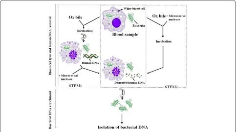
background DNA in the DNA preparations from blood samples, and consequently the sensitivity in PCR detec-tion of SalmonellaTyphi in blood samples is dramatic-ally enhanced. Figure 1 shows the format and principle of STEM.
Materials and methods Strains and culture
Wild-type Salmonella serovar Typhi Quailes strain, obtained from the University of Maryland, was used to spike blood samples in this study. The strain was sub-cultured in tryptone soya broth (TSB) or on tryptone soya agar (TSA) (Oxoid, Basingstoke, UK) as needed.
Preparation of spiked blood samples
A colony of overnight culture of Salmonella Typhi Quailes strain on TSA was inoculated into 2 ml TSB, and cultured in a 37°C incubator with shaking at 200 revolu-tions per minute (RPM) for about 2 hours. The culture was first adjusted to an optical density at 600nm of 0.25 and then prepared for serial 10-fold dilutions in TSB. An aliquot of 100μl each of dilutions was plated onto a TSA plate, and the plates were incubated overnight at 37°C to determine the colony forming unit (CFU) of Salmonella Typhi per millilitre. Blood was obtained from healthy individuals with a sterile syringe and immediately pipetted into tubes containing heparin. 100 μl each of the bacterial dilutions was spiked into a 900 μl blood
sample and 200 μl spiked blood samples were used for DNA isolation.
The blood samples used in the study were obtained from laboratory volunteers with written informed con-sent, in accordance with the university policies - Taking Blood Samples from Colleagues or Students for Research and Teaching OHS Policy Document 1/03 approved by the Central University Research Ethics Committee (CUREC), University of Oxford.
Conventional DNA preparation
Blood DNA was isolated from 200 μl heparinised blood sample using QIAamp DNA mini kit (Qiagen, Crawley, UK) according to the manufacturer’s instruction, except that the DNA was eluted with 50μl Buffer AE and incu-bated at 65°C for 5 minutes before centrifugation. The DNA was stored at−20°C until further use. The aliquots of DNA preparation were run on a 1% agarose gel, stained with ethidium bromide and visualized by UV transllumination. The DNA concentration was deter-mined by UV spectrometry.
Digestion of human blood DNA with micrococcal nuclease
0.5μg human blood DNA was mixed with 103gel units of micrococcal nuclease (New England Biolab, Herts, UK) in 20μl reaction volume containing 1μM CaCl2and
[image:3.595.56.540.449.720.2]0, 1%, 3%, 5%, 7% or 9% ox bile. Following incubation at room temperature for 10 minutes, the reaction mixture
was run on a 1% agarose gel, stained with ethidium bromide, and photographed under UV illuminator.
SelectiveSalmonellaTyphi target DNA enrichment and preparation
A flow diagram of two selective target DNA enrichment methods (STEM1 and STEM2) used in this study for en-richment of Salmonella Typhi bacterial DNA from spiked blood samples was shown in Figure 1.
In STEM1, 200 μl spiked blood sample was gently mixed with equal volume of 10% ox bile (Oxgall, BD Biosciences, Oxford, UK), incubated at room tempera-ture for 10 minutes, and then centrifuged at 13,000 RPM for 5 minutes. The pellet was re-suspended in 100μl 0.1μM CaCl2, and 1μl (103gel units)
micrococ-cal nuclease was added. The mixture was incubated at 37°C for 10 minutes and then centrifuged again at 13,000 RPM for 5 minutes. The pellet contained the enriched Salmonella Typhi bacteria and was re-suspended in 200 μl phosphate buffered saline (PBS) for bacterial DNA isolation.
In STEM2, 200 μl spiked blood was mixed together with 200μl of 10% ox bile, 4μl of 0.1 mM CaCl2and 2μl
(2×10³ gel units) micrococcal nuclease, incubated at room temperature for 20 minutes, and then centrifuged at 13,000 RPM for 5 minutes to collect the pellet containing enriched bacterial cells. The pellet was re-suspended in 200μl PBS for bacterial DNA isolation.
Following the re-suspension of the enriched bacterial pellet obtained in STEM1 or STEM2, bacterial DNA was isolated using QIAamp DNA mini kit in the same way as described in the section of Conventional DNA preparation above, except that the mixture of 20 μl pro-teinase K solution and bacterial pellet suspension was incubated for 5 minutes at room temperature to com-pletely remove any residual activity of micrococcal nuclease before Buffer AL was added.
PCR primers ofSalmonella serovar Typhi Quailes strain
The PCR primers for Salmonella serovar Typhi Quailes strain were designed according to the fliC-d gene sequence of Salmonella serovar Typhi (Accession number L21912): H-for (ACTCAGGCTTCCCGTAACGC) and Hd-rev (GGCTAGTATTGTCCTTATCGG) [15], and synthesized by Sigma Genosys (Sigma-Aldrich, Dorset, England).
PCR protocol
The PCR reaction was carried out in a 50 μl volume, comprising 0.5 U of Taq DNA polymerase (Qiagen, Crawley, UK), 1 x Qiagen PCR buffer, 1.5 mM magnesium chloride, 200μM concentrations of each deoxynucleoside triphosphate, 0.5μM concentrations of the primers H-for and Hd-rev, and 10 μl of DNA template. The following
amplification steps were used: 1 cycle of 95°C for 5 min; 35 cycles of 93°C for 30 sec, 55°C for 30 sec, and 72°C for 40 sec; and 1 cycle of 72°C for 5 min. The PCR amplifica-tion product was separated by electrophoresis on a 1% agarose gel, stained with ethidium bromide, and photo-graphed by a UV transilluminator.
Results
Micrococcal nuclease is active on human DNA in the presence of ox bile
Human blood DNA was incubated with micrococcal nuclease at different concentrations of ox bile. The extent of digestion of human DNA by micrococcal nuclease was compared, as shown in Figure 2. The result showed that micrococcal nuclease remained active at the bile concentration up to 9%.
Selective target DNA enrichment methods reduce the background human DNA prepared from blood samples
DNA prepared by the 2 different STEMs was compared with that isolated through the conventional method from blood samples spiked with Salmonella Typhi, as shown in Figure 3. There was a high background caused by human DNA in the preparations made through the conventional method (lanes 8–14), but not in the pre-parations made by STEM1 (lanes 1–7). Similar results were also obtained with STEM2 (data not shown).
PCR sensitivity for detection ofSalmonellaTyphi is enhanced through the selective removal of background human DNA from blood samples
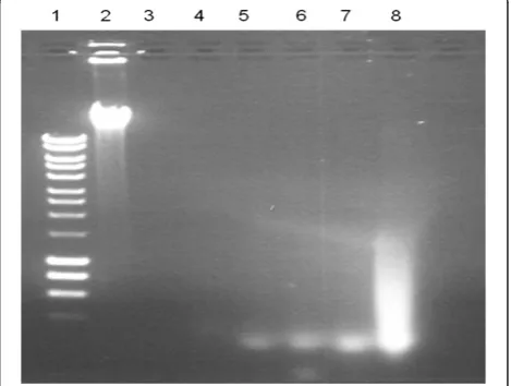
[image:4.595.305.540.498.675.2]The sensitivity of PCR was compared using DNA prepared by STEM and by conventional methods from
the spiked blood samples. Figure 4 shows the fliC-d amplicons from the different DNA preparations. Using the DNA prepared with the conventional method, the fliC-d amplicons were only seen in the sample with 3x10⁵CFU per millilitre of blood (equivalent to approxi-mately 6,000 CFU per PCR). However, the fliC-d ampli-cons were seen in all the samples with 3x10⁵, 3x10⁴, 3x10³and 3x10²CFU per millilitre of blood, with high-est sensitivity equal to approximately 6 CFU per PCR reaction. Independent experiments consistently found that STEM enhanced PCR sensitivity by 1,000 fold through selective removal of background human DNA in the spiked blood samples.
Discussion
We describe a novel method (STEM) that enhances detection of Salmonella Typhi in spiked blood speci-mens. Using this method we were able to show a sub-stantial reduction in dominating human background DNA in clinical samples and consequently increased the sensitivity of PCR detection of Salmonella Typhi from blood samples by 1,000 fold.
Early diagnosis and prompt treatment are essential for optimal management of typhoid patients, and therefore a sensitive and specific PCR assay is useful. Although many SalmonellaTyphi PCR-based assays were studied in last decades, none has been brought to clinical use
[image:5.595.63.538.90.207.2]due to the limitations caused by low bactereamic level in typhoid patients. PCR sensitivity is directly related to the actual number of colony forming units found in the blood [3]. Typical clinical typhoid samples have <1 bac-teria/ml of blood [11,19]. As a result, PCR offers only limited potential for typhoid diagnostics unless a new sample preparation method is developed. A DNA or bacterial capture system or even a culture enrichment step prior to amplification could improve PCR sensitiv-ity. Using a culture enrichment step prior to amplifica-tion Teh et al. demonstrated that the 5 hour broth culture enrichment improved PCR sensitivity by 10 times for spiked blood, and 100 times for spiked stool samples [23]. We have recently developed a blood cul-ture PCR assay which could detect as few as 0.75 bac-teria per millilitre of blood following 3 hours of culture enrichment [22]. But like conventional blood culture, the PCR assay following bacterial culture enrichment still has some practical limitations including availability of culture facilities in disease endemic areas and bacteria being unculturable due to the use of antibiotics. There-fore, a PCR assay method that can be used directly on clinical blood samples is still a better choice. Collecting and then extracting DNA from a large volume of blood may theoretically improve PCR assays, but the presence of dominating human DNA causes false-positive PCR signals due to the non-specific binding of primers and Figure 3DNA preparations made by STEM1 and the conventional method.M: DNA marker; 1–7: DNA prepared by the STEM1; 8–14: DNA prepared by the conventional method. Lanes 1 and 8, 2 and 9, 3 and 10, 4 and 11, 5 and 12, 6 and 13, 7 and 14: DNA prepared from the spiked blood samples containing 3x10⁵, 3x10⁴, 3x10³, 3x10², 30 , 3, 0 CFU/ml ofSalmonellaTyphi, respectively.
[image:5.595.59.538.595.675.2]false-negative results due to reduced sensitivity. Cur-rently two protocols have been commercially marketed for reducing the presence of dominating human DNA: MolYsis (Molzym GmbH & Co. KG, Bremen, Germany) and Pureprove (SIRS-Lab GmbH, Jena, Germany). The former uses a chaotropic buffer containing guanidine hydrochloride to selectively lyze human cells. The released eukaryotic DNA was subsequently degraded by the addition of a chaotropic resistant nuclease, and the intact bacterial cells were collected for bacterial DNA purification. For the latter, total genomic (i.e. human and bacterial) DNA is first conventionally extracted specific bacterial genomic DNA is then isolated using a DNA binding protein that recognizes unmethylated CpG motifs predominantly present in bacterial and fungal genomes at significantly lower frequencies than in human genomes. Horzet al.recently compared these two pro-tocols for DNA preparation, and found that both were able to substantially reduce the human background DNA in most of the cases but loss of bacterial DNA was also a problem [24]. For example, with MolYsis the recovery ofP. gingivalisin periodontal samples was less than 20%. As mentioned above, the MolYsis protocol uses a guanidine hydrochloride chaotropic buffer for lysis of human/animal cells, and bacteria with a thin or labile cell wall (e.g. members of the genusTreponema) or those exposed to cell wall-active antibiotics and/or human immunosystems or bacteria even devoid of a cell wall (e.g. Mycoplasma,Chlamydia) are also lyzed. There-fore, the MolYsis protocol may not be suitable for use in the diagnosis of Gram-negative sepsis. They also found, on the other hand, the Pureprove technology was not efficient in removal of human DNA. This is because it is based on selective binding of bacterial DNA to a DNA binding protein that recognizes unmethylated CpG motifs. CpG islands and motifs are not distributed equally over the entire human genome, and fragments without any 5′-methylcytosin are present in every preparation and will mix with prokaryotic DNA. In addition, as cytosine methylation is negatively correlated with gene expression, in regions of active genes the CpG dinucleotides-motifs are also de-methylated and, if frag-mentized, could possibly mix with prokaryotic DNA as well. It should be emphasized that in Horzet al.study, the mean absolute bacterial gene copy numbers was 2.23×10¹⁰ (saliva and supragingival plaque collected by cotton tampon) and 1.37x10¹⁰(pooled subgingivalsulcus fluid) [24]. Such a high level of bacterial template in a blood sample from a patient with typhoid is unlikely. Therefore, the fact that typical clinical typhoid samples contain <1 organism/ml of blood highlights the big chal-lenges faced for developing PCR diagnostics for typhoid.
Bile is a widely used ingredient in culture media for blood culture and the bile resistance of Salmonella is
well-known. This could be exploited for developing typhoid diagnostics as bile can selectively lyze human blood cells and release both human DNA and intracellu-lar bacteria without damaging bacterial cells [22,25]. In the present study, we have investigated the use of ox bile and micrococcal nuclease for selective lysis of blood cells and removal of background human DNA. We tested the use of micrococcal nuclease for removal of the released human DNA in the presence and absence of ox bile, and found that micrococcal nuclease is bile resistant and ac-tive at a wide range of bile concentrations used up to 9%. This suggests that micrococcal nuclease could be used to degrade human DNA while ox bile is added for the lysis of blood cells, potentially simplifying the design of typhoid diagnostics. We have tried two STEM proto-cols for removal of background human DNA and con-firmed that both methods can substantially reduce background human DNA in the DNA preparations. As a result, the percentage of bacterial DNA in the prepar-ation was increased. When the DNA prepared by STEM was used for PCR assay, we have consistently found that PCR sensitivity increased by at least 1,000 fold, com-pared with that using conventionally precom-pared DNA (which contained a large amount of background human DNA). These promising results indicate that STEM should now be investigated in field trials and to this end the approach is currently being applied in an endemic setting. Although the present study aims to improve current diagnostics for typhoid, the method described herein can be generally applied to sample preparation for diagnosis of infections with other ox bile resistant bacterial and fungal pathogens, in particular, where the low level of a pathogen presents a problem for current molecular diagnostics.
In conclusion, we describe herein a novel method, which we have termed“the selective target DNA enrich-ment method” (STEM). STEM can enrich bacteria from large volume samples, remove background human DNA, and enhance the sensitivity of PCR detection for SalmonellaTyphi in blood samples. Therefore, this novel method of sample preparation offers a better option for improved typhoid PCR assays directly using clinical specimens in diagnosis of this globally important infec-tion disease which we believe could be of importance in improving clinical care and providing effective evalu-ation of novel vaccines.
Competing interests
LZ and AJP are authors on patents in the field of typhoid diagnostics.
Authors' contributions
Acknowledgements
This work and LZ were supported by the Oxford Partnership Comprehensive Biomedical Research Centre Programme with funding from the Department of Health's NIHR Biomedical Research Centres funding scheme. AJP is a Jenner Institute Investigator.SalmonellaTyphi Quailes strain was provided by Professor Myron Levine, University of Maryland.
Received: 24 February 2012 Accepted: 11 July 2012 Published: 27 July 2012
References
1. Crump JA, Mintz ED:Global trends in typhoid and paratyphoid fever. Clinical Infectious Diseases2010,50:241–246.
2. Petit PLC, Wamola IA:Typhoid fever - a review of its impact and diagnostic problems.East African Medical Journal1994,71:183–188. 3. Baker S, Favorov M, Dougan G:Searching for the elusive typhoid
diagnostic.BMC Infect Dis2010,10:45.
4. Levine MM, Grados O, Gilman RH, Woodward WE, Solis-Plaza R, Waldman W: Diagnostic value of the Widal test in areas endemic for typhoid fever. Am J Trop Med Hyg1978,27:795–800.
5. Lim PL, Tam FC, Cheong YM, Jegathesan M:One-step 2-minute test to detect typhoid-specific antibodies based on particle separation in tubes. J Clin Microbiol1998,36:2271–2278.
6. Prakash P, Sen MR, Mishra OP, Gulati AK, Shukla BN, Nath G:Dot enzyme immunoassay (Typhidot) in diagnosis of typhoid fever in children.J Trop Pediatr2007,53:216–217.
7. Dutta S, Sur D, Manna B, Sen B, Deb AK, Deen JL, Wain J, Von Seidlein L, Ochiai L, Clemens JD, Bhattacharyas K:Evaluation of new-generation serologic tests for the diagnosis of typhoid fever: data from a
community-based surveillance in Calcutta, India.Diagn Microbiol Infect Dis 2006,56:359–365.
8. Ochiai RL, Acosta CJ, Danovaro-Holliday MC, Baiqing D, Bhattacharya SK, Agtini MD, Bhutta ZA, Canh do G, Alim S, Wain J, Page AL, Albertm J, Farrar J, Abu-Elyazeed R, Pang T, Galindo CM, Vonseidlein L, Clemens JD: A study of typhoid fever in five Asian countries: disease burden and implications for controls.Bull World Health Organ2008,86:260–268. 9. Parry CM, Hien TT, Dougan G, White NJ, Farrar JJ:Typhoid fever.N Engl J
Med2002,347:1770–1782.
10. Wain J, Hosoglu S:The laboratory diagnosis of enteric fever.J Infect Dev Ctries2008,2:421–425.
11. Wain J, Pham VB, Ha V, Nguyen NM, To SD, Walsh AL, Parry CM,
Hasserjian RP, HoHo VA, Tran TH, Farrar J, White NJ, Day NP:Quantitation of bacteria in bone marrow from patients with typhoid fever: relationship between counts and clinical features.J Clin Microbiol2001,39:1571–1576. 12. Wain J, Diep TS, Bay PV, Walsh AL, Vinh H, Duong NM, Ho VA, Hien TT,
Farrar J, White NJ, Parry CM, Day NP:Specimens and culture media for the laboratory diagnosis of typhoid fever.J Infect Dev Ctries2008,2:469–474. 13. Ali A, Haque A, Sarwar Y, Mohsin M, Bashir S, Tariq A:Multiplex PCR for
differential diagnosis of emerging typhoidal pathogens directly from blood samples.Epidemiol Infect2009,137:102–107.
14. Ali K, Zeynab A, Zahra S, Akbar K, Saeid M:Development of an ultra rapid and simple multiplex polymerase chain reaction technique for detection of salmonella typhi.Saudi Med J2006,27:1134–1138.
15. Levy H, Diallo S, Tennant SM, Livio S, Sow SO, Tapia M, Fields PI, Mikoleit M, Tamboura B, Kotloff KL, Lagos R, Nataro JP, Galen JE, Levine MM:PCR method to identify salmonella entericaserovars Typhi, Paratyphi A, and Paratyphi B among salmonella Isolates from the blood of patients with clinical enteric fever.J Clin Microbiol2008,46:1861–1866.
16. Massi MN, Shirakawa T, Gotoh A, Bishnu A, Hatta M, Kawabata M:Rapid diagnosis of typhoid fever by PCR assay using one pair of primers from flagellin gene of salmonella typhi.J Infect Chemother2003,9:233–237. 17. Ambati SR, Nath G, Das BK:Diagnosis of typhoid fever by polymerase
chain reaction.Indian J Pediatr2007,74:909–913.
18. Song JH, Cho H, Park MY, Na DS, Moon HB, Pai CH:Detection of salmonella typhi in the blood of patients with typhoid fever by polymerase chain reaction.J Clin Microbiol1993,31:1439–1443. 19. Wain J, Diep TS, Ho VA, Walsh AM, Nguyen TT, Parry CM, White NJ:
Quantitation of bacteria in blood of typhoid fever patients and relationship between counts and clinical features, transmissibility, and antibiotic resistance.J Clin Microbiol1998,36:1683–1687.
20. Handschur M, Karlic H, Hertel C, Pfeilstocker M, Haslberger AG:Preanalytic removal of human DNA eliminates false signals in general 16s rDNA PCR monitoring of bacterial pathogens in blood.Comp Immunol Microbiol Infect Dis2009,32:207–219.
21. Horz HP, Scheer S, Huenger F, Vianna ME, Conrads G:Selective isolation of bacterial DNA from human clinical specimens.J Microbiol Methods2008, 72:98–102.
22. Zhou L, Pollard AJ:A fast and highly sensitive blood culture PCR method for clinical detection ofSalmonellaentericaserovar Typhi.Ann Clin Microbiol Antimicrob2010,9:14.
23. Teh CS, Chua KH, Puthucheary SD, Thong KL:Further evaluation of a multiplex PCR for differentiation of salmonella paratyphi A from other salmonellae.Jpn J Infect Dis2008,61:313–314.
24. Horz HP, Scheer S, Vianna ME, Conrads G:New methods for selective isolation of bacterial DNA from human clinical specimens.Anaerobe2010, 16:47–53.
25. Kaye D, Palmieri M, Rocha H:Effect of bile on the action of blood against salmonella.J Bacteriol1966,91:945–952.
doi:10.1186/1471-2334-12-164
Cite this article as:Zhou and Pollard:A novel method of selective removal of human DNA improves PCR sensitivity for detection of
SalmonellaTyphi in blood samples.BMC Infectious Diseases201212:164.
Submit your next manuscript to BioMed Central and take full advantage of:
• Convenient online submission
• Thorough peer review
• No space constraints or color figure charges
• Immediate publication on acceptance
• Inclusion in PubMed, CAS, Scopus and Google Scholar
• Research which is freely available for redistribution