Persistent Replication of a Chikungunya Virus Replicon in
Human Cells Is Associated with Presence of Stable
Cytoplasmic Granules Containing Nonstructural Protein 3
Roland Remenyi,
a,bYanni Gao,
a,bRuth E. Hughes,
a,bAlistair Curd,
a,bCarsten Zothner,
a,bMichelle Peckham,
a,bAndres Merits,
cMark Harris
a,baSchool of Molecular and Cellular Biology, Faculty of Biological Sciences, University of Leeds, Leeds, United
Kingdom
bAstbury Centre for Structural Molecular Biology, University of Leeds, Leeds, United Kingdom
cInstitute of Technology, University of Tartu, Tartu, Estonia
ABSTRACT
Chikungunya virus (CHIKV), a mosquito-borne human pathogen, causes
a disabling disease characterized by severe joint pain that can persist for weeks,
months, or even years in patients. The nonstructural protein 3 (nsP3) plays essential
roles during acute infection, but little is known about the function of nsP3 during
chronic disease. Here, we used subdiffraction multicolor microscopy for spatial and
temporal analysis of CHIKV nsP3 within human cells that persistently replicate
repli-con RNA. Round cytoplasmic granules of various sizes (i) repli-contained nsP3 and stress
granule assembly factors 1 and 2 (G3BP1/2), (ii) were next to double-stranded RNA
foci and nsP1-positive structures, and (iii) were close to the nuclear membrane and
the nuclear pore complex protein Nup98. Analysis of protein turnover and mobility
by live-cell microscopy revealed that the granules could persist for hours to days,
ac-cumulated newly synthesized protein, and moved through the cytoplasm at various
speeds. The granules also had a static internal architecture and were stable in cell
lysates. Refractory cells that had cleared the noncytotoxic replicon regained the
abil-ity to respond to arsenite-induced stress. In summary, nsP3 can form uniquely stable
granular structures that persist long-term within the host cell. This continued
pres-ence of viral and cellular protein complexes has implications for the study of the
pathogenic consequences of lingering CHIKV infection and the development of
strategies to mitigate the burden of chronic musculoskeletal disease brought about
by a medically important arthropod-borne virus (arbovirus).
IMPORTANCE
Chikungunya virus (CHIKV) is a reemerging alphavirus transmitted by
mosquitos and causes transient sickness but also chronic disease affecting muscles
and joints. No approved vaccines or antivirals are available. Thus, a better
under-standing of the viral life cycle and the role of viral proteins can aid in identifying
new therapeutic targets. Advances in microscopy and development of noncytotoxic
replicons (A. Utt, P. K. Das, M. Varjak, V. Lulla, A. Lulla, A. Merits, J Virol 89:3145–
3162, 2015,
https://doi.org/10.1128/JVI.03213-14
) have allowed researchers to study
viral proteins within controlled laboratory environments over extended durations. Here
we established human cells that stably replicate replicon RNA and express tagged
nonstructural protein 3 (nsP3). The ability to track nsP3 within the host cell and during
persistent replication can benefit fundamental research efforts to better understand
long-term consequences of the persistence of viral protein complexes and thereby
provide the foundation for new therapeutic targets to control CHIKV infection and treat
chronic disease symptoms.
KEYWORDS
Airyscan, confocal microscopy, neglected tropical diseases, nonstructural
Received23 March 2018Accepted25 May
2018
Accepted manuscript posted online6 June
2018
CitationRemenyi R, Gao Y, Hughes RE, Curd A,
Zothner C, Peckham M, Merits A, Harris M. 2018. Persistent replication of a chikungunya virus replicon in human cells is associated with presence of stable cytoplasmic granules containing nonstructural protein 3. J Virol 92:e00477-18.https://doi.org/10.1128/JVI .00477-18.
EditorMichael S. Diamond, Washington
University School of Medicine
Copyright© 2018 Remenyi et al. This is an
open-access article distributed under the terms of theCreative Commons Attribution 4.0 International license.
Address correspondence to Mark Harris, [email protected].
crossm
on November 6, 2019 by guest
http://jvi.asm.org/
viral proteins, nucleopore, self-labeling tag, superresolution microscopy, video
microscopy, virus-cell interaction
C
hikungunya virus (CHIKV), a reemerging arbovirus of the
Alphavirus
genus, causes
a transient illness with debilitating symptoms (fever, headache, rash, myalgia, and
arthralgia). Chronic disease is common, and joint pain can persist for months to years
(1–3). Half of the patients from the recent Latin American outbreak may develop
chronic inflammatory rheumatism, raising the health burden of musculoskeletal disease
in areas of endemicity (4, 5). During acute infection, this cytotoxic virus induces
apoptosis, leading to direct tissue injury and local inflammation (6–8). Biopsies have
also revealed the persistence of CHIKV antigens and RNA in synovial macrophages and
muscle tissue (1, 9). CHIKV also persists in mice and nonhuman primate models (10–13).
Chronic disease may be a consequence of persistent, replicating, and transcriptionally
active CHIKV RNA (13), but an understanding of CHIKV’s long-term effect is still
emerging.
The
⬃
12-kb positive-sense RNA genome of CHIKV encodes four nonstructural
proteins, nsP1 to nsP4, which make up the viral replication and transcription complex
(Fig. 1A) (reviewed in reference 14). A subgenomic RNA expresses six structural
pro-teins. Cellular responses to infection include apoptosis, interferon signaling, stress
granule (SG) formation, unfolded protein response, host cell shutoff, and autophagy
(reviewed in reference 15). Previous research on alphaviruses established the vital role
that nsP3 plays in counteracting cellular responses (16–20) and identified essential
protein-protein interactions between nsP3 and host proteins (16, 21–23). However, few
studies have systematically investigated the long-term effect of persistently replicating
CHIKV RNA and continued expression of proteins such as nsP3 on human cells.
Although recent studies characterize the formation of organelles that contain nsP3
during acute infection and transient replication (16, 24–27), a corresponding
charac-terization during persistent CHIKV replication is missing. To address these gaps, we
sought to further develop CHIKV replicons capable of persistent replication in human
cells and to harness this system for analysis by subdiffraction multicolor microscopy.
We previously characterized transient replication of CHIKV replicons in mammalian
and invertebrate cell lines (27) and tagged nsP3 with the versatile SNAP tag for
advanced fluorescence microscopy applications (26). The development of a
noncyto-toxic CHIKV replicon allowed the establishment of persistent replication in a human cell
line (28). Here, we extended the SNAP-based labeling system to this noncytotoxic
CHIKV replicon and generated a human cell line that persistently replicates replicon
RNA and stably expresses SNAP-tagged nsP3. We then characterized nsP3-containing
cytoplasmic granular organelles by subdiffraction multicolor microscopy. We used this
technique to address questions relating to the subcellular localization of
nsP3-containing granules and their stability, composition, and motility. This report is the first
to shed light on the persistence of stable intracellular granules of nsP3 within human
cells. In turn, understanding the link between the persistence of stable viral protein
complexes and pathogenesis has relevance to future studies of chronic CHIKV disease.
(This article was submitted to an online preprint archive [29].)
RESULTS
Development of a stable human-origin cell line carrying a SNAP-tagged CHIKV
replicon and superresolution microscopy of nsP3-G3BP-containing granules.
To
determine the intracellular distribution of nsP3, we previously generated a
SNAP-tagged replicon construct (26). Whereas this replicon is cytotoxic and replicates
tran-siently, noncytotoxic replicons can establish persistent replication in the human cell line
HuH-7 (28). To improve the HuH-7 CHIKV cell line, we added a SNAP-tagged nsP3
to a noncytotoxic replicon (Fig. 1A) and selected puromycin-resistant cells, which are
called stable CHIKV cells throughout this paper. Silicon-rhodamine-conjugated O
6-benzylguanine probes (BG-647-SiR) labeled SNAP-nsP3 and revealed nsP3-containing
granules (Fig. 1B) comparable to those formed by a wild-type virus, CHIKV
WT, and those
August 2018 Volume 92 Issue 16 e00477-18 jvi.asm.org 2
on November 6, 2019 by guest
http://jvi.asm.org/
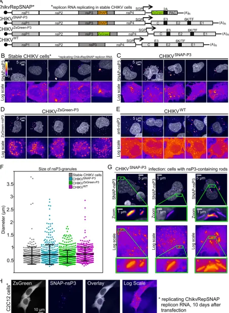
FIG 1nsP3 has a granular distribution in stable CHIKV cells and infected HuH-7 cells. (A) Schematic representation of tagged reporter viruses and noncytotoxic replicon encoding SNAP-nsP3. SGP, subgenomic promoter; PAC, puromycin-N-acetyltransferase; 2A, foot-and-mouth disease virus (FMDV) 2A autoprotease. (B) Subdiffraction confocal microscopy of BG-647-SiR-labeled stable CHIKV cells, imaged in the far-red channel. Cells
(Continued on next page)
on November 6, 2019 by guest
http://jvi.asm.org/
[image:3.585.46.498.76.693.2]formed by CHIKV
SNAP-P3and CHIKV
ZsGreen-P3viruses harboring SNAP- or
ZsGreen-tagged nsP3 (Fig. 1C to E). Analysis of the sizes of nsP3-containing granules formed in
stable CHIKV cells also showed size distributions and diameters comparable to those of
granules formed during infection with tagged and untagged viruses (Fig. 1F). Further
experiments focused on the characterization of these nsP3-containing granules.
Whereas cells infected with CHIKV
ZsGreen-P3displayed only a granular nsP3-ZsGreen
distribution pattern, cells infected with CHIKV
SNAP-P3also made rod-like structures (Fig.
1G), as described previously (26, 27). However, the presence of rods did not correlate
with infectivity, as ZsGreen- and SNAP-tagged viruses replicated to similar titers (Table
1). Although the rest of this study used stable CHIKV cells derived from HuH-7 cells, the
SNAP-tagged noncytotoxic replicon ChikvRepSNAP could also replicate in the C2C12
mouse myoblast cell line (Fig. 1H).
CHIKV nsP3 sequesters G3BP1/2 when expressed alone (17), in the context of a
replicon (16, 26), or during virus infection (24, 25), thereby interfering with SG
re-sponses. Recent subdiffraction microscopy revealed stable substructures of G3BP1
protein within SGs (30, 31). To determine whether nsP3-containing granules also
sequestered G3BP1/2 proteins and contained similar substructures, we imaged stable
CHIKV cells with Airyscan microscopy. Airyscan or image scanning microscopy (32, 33)
relies on array detectors to reassign photon pixels and oversample the pattern from
diffracted light, thereby improving image resolution (1.7-fold) and sensitivity (34).
Airyscan outperformed standard confocal microscopy and was sensitive enough to
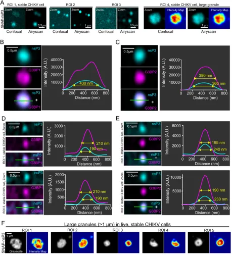
detect small granular structures of nsP3 (Fig. 2A, region of interest [ROI] 1 to 3). Whereas
nsP3 appeared to have a diffuse distribution in confocal images, the improved
resolu-tion of the Airyscan microscope uncovered an uneven distriburesolu-tion in a large (1.2-
m-diameter) granule, consistent with the presence of substructure (Fig. 2A, ROI 4). Both
G3BP1 (Fig. 2B) and G3BP2 (Fig. 2C) were colocalized with these granules, which also
had a high fluorescence intensity. The higher sensitivity of the Airyscan method also
made small clusters of nsP3 more visible; these clusters had full width at half maximum
(FWHM) of 190 to 240 nm and were about 10 times less intense (Fig. 2D and E, see line
profiles of fluorescence intensity) than larger granules (Fig. 2B and C, FWHM of 360 to
430 nm). Lastly, live-cell imaging also confirmed that fluorescent nsP3 signals in large
granules were nonuniformly distributed within each granule (Fig. 2F).
Juxtaposition of nsP3-containing granules, dsRNA foci, nsP1-positive
struc-tures, nuclear membrane, and Nup98.
During the viral life cycle, nsP3-containing
granules sequester G3BP1, thereby blocking SG assembly (16, 17). The relationship
between cytoplasmic nsP3-G3BP1 complexes and CHIKV RNA synthesis is less clear;
FIG 1Legend (Continued)
[image:4.585.40.372.82.132.2]were chemically fixed and stained with fluorescent BG-647-SiR, which irreversibly binds SNAP-tagged proteins. (C to E) Naive HuH-7 cells were infected with viral stocks of CHIKVSNAP-P3, CHIKVZsGreen-P3, or CHIKVWT. For cells infected with CHIKVZsGreen-P3, images were acquired in the green channel. Cells infected with CHIKVWTwere immunostained with anti-nsP3 antibodies to visualize untagged nsP3. Data shown are maximum-intensity projections of Z-stacks acquired on an Airyscan confocal system, operated in the superresolution mode. To enhance the appearance of dim structures, Icy software (84) was used to pseudocolor image channels with the predefined Fire lookup table based on pixel intensity. Color bars indicate the relative range of pixel intensity (white⫽high, purple⫽low, from 0 arbitrary units to 1). Nuclear counterstain (gray) was overlaid as a reference. Images displayed in the Fire view, based on a logarithmic scale, illustrate both high-intensity and low-intensity granules in the same image. (F) Size of nsP3-containing granules. Diameters of granules were extracted from the maximum-intensity projections of a total of 10 fields of view (FOVs; including panels B to E and five additional FOVs). Feret diameters represent the maximum distance between any two points of the extracted surface. (G) Airyscan microscopy of rod-containing cells infected with CHIKVSNAP-P3. Zoomed-in views of nsP3-containing rods are provided in insets. (H) Mouse myoblast cell line (C2C12) replicating the SNAP-tagged noncytotoxic replicon. At 10 days after transfection and puromycin selection, cells were fixed and stained for SNAP-nsP3 with BG-TMR-Star. The ZsGreen channel is presented in grayscale view. TABLE 1Viral titers of CHIKVSNAP-P3and CHIKVZsGreen-P3
CHIKV construct
Virus titer (PFU/ml)a
24 h 48 h 72 h
CHIKVSNAP-P3 5.5⫻105 4.4⫻107 1.4⫻107
CHIKVZsGreen-P3 8.6⫻104 6.1⫻107 1.4⫻107
aInoculum obtained from supernatant at 24, 48, and 72 h after infection.
August 2018 Volume 92 Issue 16 e00477-18 jvi.asm.org 4
on November 6, 2019 by guest
http://jvi.asm.org/
FIG 2Characterization of nsP3-G3BP1/2 interaction by subdiffraction microscopy. (A) Comparison of confocal and Airyscan images. Airyscan provided improved resolution and signal-to-noise ratios (SNRs). Airyscan was able to image faint clusters that did not resolve well with standard confocal microscopy (ROI 1 to 3). Note that we use the term “granules” for these protein clusters. Airyscan also revealed differences in fluorescence intensity within large granules that were not apparent with confocal imaging (ROI 4). (B to E) SNAP-nsP3 (cyan) was stained with BG-647-SiR as described in the legend to Fig. 1. G3BP1 or G3BP2 (magenta) was immunostained with specific antibodies. Images were acquired with an Airyscan microscope operated in superresolution mode. For high-contrast display of nsP3-containing granules, contrast was optimized within each image by adjusting the view range in the histogram viewer window of Icy software. Overlaps between cyan and magenta layers appear in white (panels “nsP3⫹G3BP1/2”). Line profiles that plot the fluorescence intensity along the line of interest (green) are provided as well, along with the full-width at half maximum (FWHM) of granules. Fluorescence intensity was measured in arbitrary units (A.U.). Images represent single slices, which were extracted from Z-stacks. (F) Live-cell Airyscan microscopy of large granules. Stable CHIKV cells were stained with BG-647-SiR and imaged with an Airyscan microscope operated in the Fast Airyscan mode. Fluorescence intensity maps were created in Icy software and represent relative pixel intensity according to the Jet color map.
on November 6, 2019 by guest
http://jvi.asm.org/
[image:5.585.41.500.73.578.2]viral double-stranded RNA (dsRNA) foci overlap minimally with nsP3-G3BP1-positive
clusters in replicons (16). Moreover, few dsRNA foci colocalized with G3BP2 puncta at
6 h postinfection, and even fewer overlapped after 8 h (25). Recently, it was reported
that large cytoplasmic and small plasma-membrane-bound G3BP1-nsP3 complexes
colocalize with viral genomic RNA during CHIKV infection, with dsRNA foci forming
nearby (24). Structural and biochemical data also support a model in which a matrix of
nsP3-G3BP1-containing complexes stabilizes replication complexes and shields viral
replicative intermediates from the RNA degradation machinery or cytosolic dsRNA
sensors (35). Thus, nsP3-G3BP1/2-containing granules appear to play roles in addition
to sequestering SG-related proteins.
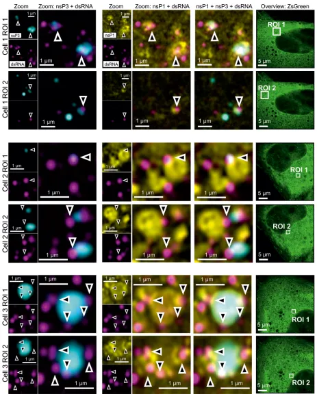
To further explore the spatial relationship between nsP3 and replication sites in
stable CHIKV cells, we visualized SNAP tag-labeled nsP3, together with immunostaining
for nsP1 and dsRNA. The antibody against dsRNA was previously used to identify
alphavirus replication complexes (36). The fluorescence of ZsGreen in stable CHIKV cells
served as an indirect readout of the viral subgenomic RNA (Fig. 1A, cartoon). Rather
than completely overlapping with larger nsP3-containing granules, dsRNA foci were in
a proximal location and often juxtaposed (Fig. 3, arrowheads). In another example, a
dsRNA focus coincided with a smaller nsP3-containing cluster (Fig. 3, cell 2, ROI 1,
arrowhead). Ring-like structures coated with nsP1 were also near these dsRNA foci (Fig.
3). The proximity of dsRNA foci, nsP1-coated structures, and nsP3-containing granules
suggested that nsP3-containing granules not only sequestered G3BP1/2 protein but
also played a role in viral replication.
As described above, nsP3-containing granules were part of a unique
microenviron-ment that also housed dsRNA foci and nsP1. Moreover, a fraction of granules containing
nsP3 and G3BP2 were located close to the nuclear membrane (Fig. 4A and B). The
nuclear transport factor 2 (NTF2)-like domain of G3BP1 has previously been
cocrystal-lized with FXFG (phenylalanine-glycine motif, where X is usually serine) nucleoporin
(Nup) repeat peptides (37) and overexpressed G3BP1 interacts with some nucleoporins,
whereas a mutant of G3BP lacking the binding site does not (38).
To further characterize the environment at the nuclear membrane of stable CHIKV
cells, we probed for the nuclear pore complex protein Nup98. nsP3-containing granules
were detected (i) at the nuclear membrane (Fig. 4C), flanked by Nup98-containing
regions, and (ii) near cytoplasmic clusters of Nup98 (Fig. 4D). Thus, we were able to
visualize a subset of nsP3-containing granules at the nuclear membrane and near
Nup98-containing structures by Airyscan microscopy.
Imaging the dynamics of nsP3-containing granules within stable CHIKV cells.
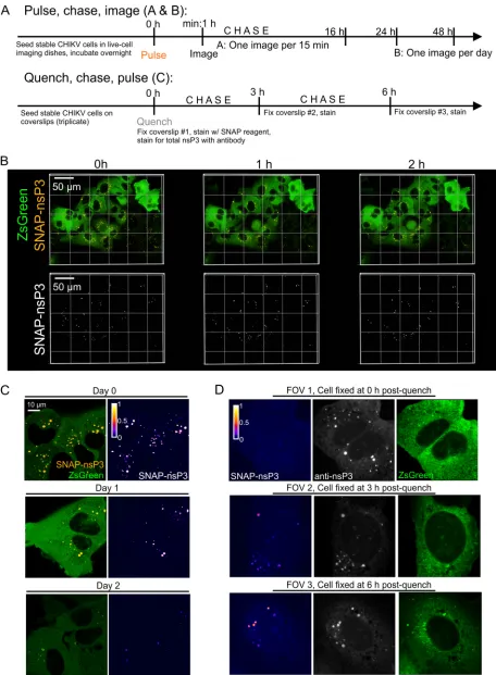
SNAP reagents can label live cells, allowing both the analysis of the movement of
tagged proteins and pulse-chase studies to examine protein turnover. nsP3-containing
granules labeled at the onset of a pulse-chase image experiment (Fig. 5A and B) could
be tracked in live-cell imaging experiments over the entire length of a recording that
lasted 16 h (Fig. 5B; see Video S1 in the supplemental material). Moreover, stable CHIKV
cells still contained “aged” nsP3-containing granules after chase periods of 1 to 2 days
(Fig. 5C). The addition of a nonfluorescent SNAP ligand (i.e., quench) in complementary
quench-pulse-chase experiments (Fig. 5A) blocked all binding sites of the SNAP-tagged
protein pool (Fig. 5D, field of view 1 [FOV1]). After a defined chase period of 3 and 6 h
in unlabeled medium, pulsing with the fluorescent SNAP reagent uncovered an
un-blocked population of nsP3-containing granules, consistent with newly synthesized
protein accumulating in granular structures (Fig. 5D, FOV2 and 3).
To further study the intracellular transport of nsP3-containing granules, we used
instant structured illumination microscopy (iSIM) for live-cell recordings at high frame
rates (39). iSIM increases spatial resolution by a factor of
公
2 compared with wide-field
microscopy and by a further factor of
公
2 with postprocessing, while rapid image
capture provides the temporal resolution needed for dynamic events within cells. A
variety of fast-moving nsP3-containing objects with linear displacements and
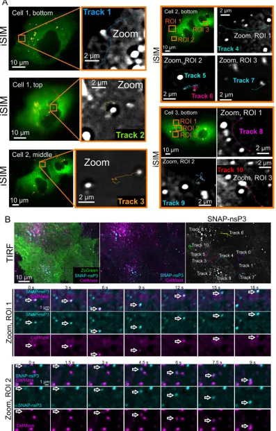
intermit-tent bursts of speed were tracked in live-cell recordings (Fig. 6A; Videos S2 to S5). As can
be seen in the videos, the movement of these nsP3-containing objects differed from
August 2018 Volume 92 Issue 16 e00477-18 jvi.asm.org 6
on November 6, 2019 by guest
http://jvi.asm.org/
FIG 3Four-color microscopy of nsP3, dsRNA, nsP1, and ZsGreen. Stable CHIKV cells were fixed and probed for nsP3 (cyan), dsRNA (magenta), nsP1 (nsP1), and ZsGreen (green) by a combination of SNAP tag labeling and indirect immunofluorescence assays. Images were taken with an Airyscan microscope operated in the superresolution mode. Overlay images are a combination of the nsP3, nsP1, and dsRNA layers as indicated. The zoomed-out ZsGreen channel is shown as a separate reference, with the corresponding ROI marked by a white box. Arrowheads indicate regions of proximity between nsP3, dsRNA, and nsP1.
on November 6, 2019 by guest
http://jvi.asm.org/
[image:7.585.38.501.68.644.2]FIG 4Association of nsP3-containing granules with nuclear membrane and Nup98. (A) Fixed, stable CHIKV cells were examined for the presence of nsP3 (cyan) and G3BP2 (magenta) at the nucleus (stained with DAPI; yellow). As shown in Fig. 2, both G3BP1 and G3BP2 colocalize with nsP3. Here we probed for G3BP2, since immunostaining had a higher SNR than G3BP1 staining. ROIs 1 to 4 are high-magnification views of granules associating with nuclei. (B) Quantification
(Continued on next page)
August 2018 Volume 92 Issue 16 e00477-18 jvi.asm.org 8
on November 6, 2019 by guest
http://jvi.asm.org/
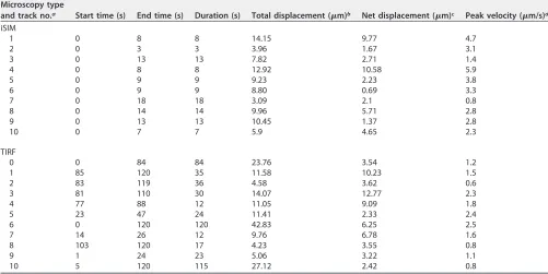
[image:8.585.41.541.76.688.2]static objects containing nsP3, which were imaged in the same recording. Fast-moving
granules containing nsP3 could reach speeds of 0.8 to 5.9
m/s (Table 2) with net
displacements of 9.77 and 10.58
m measured in the longest tracks (Table 2, tracks 1
and 4). Next, to complement our iSIM live-cell imaging analysis, we took advantage of
total internal reflection fluorescence (TIRF) microscopy, which images only fluorescent
molecules located close to the glass/specimen interface. TIRF microscopy produced
images with good signal-to-noise ratio and reduced the background fluorescence from
out-of-focus planes, enabling the observation of nsP3-containing granules located in or
close to the plasma membrane (Fig. 6B; Video S6). Tracks analyzed in iSIM and TIRF
images revealed similar object displacements and peak velocities (Table 2). Prior to TIRF
imaging, we also labeled the plasma membrane with the CellMask orange plasma
membrane stain and captured the images at least 1 h after staining to ensure that some
of the dye was internalized in membrane-containing vesicles. Multicolor TIRF
micros-copy then allowed us to visualize the cotrafficking of nsP3- and CellMask-containing
structures (Fig. 6B, Zoom, ROI 1 and 2). In summary, the dynamic analysis of
nsP3-containing granules showed that they (i) could persist in cells for days, (ii) accumulated
newly synthesized protein, (iii) could be classified into static and motile subclasses with
characteristic displacements and speeds, and (iv) cotrafficked with membrane-containing
structures.
Static internal architecture of nsP3-containing granules during persistent
rep-lication.
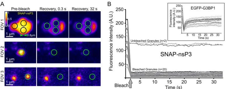
To determine the dynamic behavior of nsP3 in granules, we performed
fluorescence recovery after photobleaching (FRAP) experiments. Stable CHIKV cells
were labeled with BG– 6-carboxytetramethylrhodamine (TMR)–Star, defined regions
with diameters of about 0.8
m were photobleached, and fluorescent recovery was
measured within each region of interest (Fig. 7A). No fluorescence recovery occurred
over the duration of the experiment (Fig. 7A and B), suggesting that nsP3 remained
fixed within the granular architecture and did not undergo dynamic exchange within
each granule or with the surrounding cytoplasm. In contrast, SGs formed during
overexpression of an enhanced green fluorescent protein (EGFP)-G3BP1 fusion in
uninfected HuH-7 cells showed a characteristic rapid recovery of fluorescence after the
photobleaching (Fig. 7B, inset), consistent with G3BP1 rapidly shuttling into and out of
SGs.
Previously, G3BP1-containing SGs were shown to be stable in cell lysates, suggesting
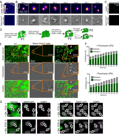
that stable core structures make up these membrane-less organelles (30). To further
test the stability of nsP3-containing granules, we microscopically examined lysates of
stable CHIKV cells (Fig. 8A). Bright-field images of cell lysates indicated the presence of
refractive granules, while fluorescence microscopy identified granules that had
incor-porated the BG-TMR-Star label (Fig. 8B). We also confirmed that the stability of
nsP3-containing granules was not unique to stable CHIKV cells, as we could also detect
ZsGreen-positive granules following lysis of cells infected with CHIKV
ZsGreen-P3(Fig. 8C).
In stable CHIKV cells, ZsGreen serves as a fluorescent marker of subgenomic
replicon-RNA synthesis (Fig. 1A, cartoon). The green fluorescence also allowed us to use
fluorescence-activated cell sorting (FACS) to eliminate any ZsGreen-negative cells.
Although stable CHIKV cells maintained high levels of SNAP-nsP3 and ZsGreen for up
to 2 months, cells with reduced or undetectable ZsGreen fluorescence accumulated in
the absence of puromycin selection after only 1 week of culturing (Fig. 8D and E). These
cells were sensitive to puromycin (Fig. 8D and F), suggesting that they no longer
harbored the replicon. To test whether ZsGreen-negative cells that had emerged during
FIG 4Legend (Continued)
of association of nsP3-containing granules with nucleus. Z-stacks of three FOVs containing cells 1 to 3 were scored for the location of granules in three categories: (i) nuclear rim, (ii) top, bottom, or inside nucleus, and (iii) cytoplasm. (C) Fixed, stable CHIKV cells were examined for the presence of nsP3 (cyan) and Nup98 (magenta) at the nucleus (stained with DAPI; yellow). Arrows serve as digital fiducial markers and point toward regions where nsP3 is close to Nup98-positive cellular structures. Images are single slices extracted from Z-stacks that were taken with an Airyscan microscope operated in the superresolution mode. (D) Samples were prepared as described for panel C. Chosen ROIs are in perinuclear, cytoplasmic areas. Arrows point to nsP3-containing granules that are associated with cytoplasmic Nup98-positive structures.
on November 6, 2019 by guest
http://jvi.asm.org/
FIG 5Long-term imaging of SNAP-nsP3 using pulse-chase and quench-pulse-chase approaches. (A) Schematic drawing of pulse-chase image and quench-pulse-chase approaches. “Pulse” refers to labeling with a SNAP-specific dye; “chase” refers to incubation in standard culture medium free of SNAP-specific dyes. (B) Stable CHIKV cells were plated in glass-bottom dishes. Labeling with BG-647-SiR was carried out the following day. Cells were imaged with a Nikon Ti2-E inverted microscope equipped with a light-emitting diode (LED) light source, a 60⫻oil 1.4-NA objective, and a
(Continued on next page)
August 2018 Volume 92 Issue 16 e00477-18 jvi.asm.org 10
on November 6, 2019 by guest
http://jvi.asm.org/
[image:10.585.41.497.69.690.2]culturing in puromycin-free medium regained some of the characteristics of uninfected
cells, we subjected a mixed population (with both ZsGreen-positive and -negative cells)
to a restress experiment with sodium arsenite. Consistent with previous experiments
that examined the effects of “restressing” alphavirus-infected cells (17, 40),
ZsGreen-positive cells sequestered G3BP2 into nsP3-containing granules even in the absence of
sodium arsenite (Fig. 8G, FOV1), and new G3BP2-containing granules were absent after
arsenite-induced stress (Fig. 8G, FOV3). In contrast, cells lacking ZsGreen did not have
any G3BP2-containing granules in the absence of arsenite stress (Fig. 8G, FOV2) but
were able to form G3BP2-positive clusters after arsenite treatment (Fig. 8G, FOV4).
Therefore, the renewed ability to respond to arsenite-induced stress was associated
with a loss of viral replication and nsP3-containing granules.
DISCUSSION
The objectives of this study were, first, to characterize the interaction between
CHIKV nsP3 and cellular components during persistent replication and, second, to
evaluate the persistence of cytoplasmic granules composed of viral and cellular
pro-teins. To achieve these objectives, we expanded the utility of a noncytotoxic replicon
by combining it with SNAP tag-based fluorescent labeling and subdiffraction multicolor
microscopy to provide unprecedented insights into the substructure of persistent
nsP3-G3BP-containing granules. These studies revealed their relationship with dsRNA,
nsP1-positive structures, and the nuclear membrane. Examining the dynamics of
nsP3-containing granules uncovered a stable population of nsP3-containing granules
along with a subclass of nsP3-positive structures trafficking through the cell cytoplasm.
Importantly, we observed that nsP3-containing granules lacked a dynamic internal
architecture and remained stable in cell lysates. Lastly, we showed that the ability to
respond to oxidative stress was associated with the loss of CHIKV replication and
nsP3-containing granules.
Stable CHIKV cells as a versatile tool for studying cytoplasmic nsP3-containing
granules.
Previous reports on noncytotoxic Old World alphaviruses elucidated the
relationship between cytotoxicity, nsP2, and viral genome replication (28, 41–43). The
typical cytotoxicity of CHIKV replicons precluded long-term studies of a previously
described SNAP-tagged replicon. We have now overcome this limitation with a new
HuH-7 cell line that harbors replicating CHIKV replicon RNA and encodes both
SNAP-tagged nsP3 and ZsGreen as a genetic reporter for subgenomic replicon RNA. Whether
this replicon establishes persistent replication only in specific cell types, as has been
observed for other noncytotoxic replicons (28, 41), remains to be determined. We found
that the SNAP-tagged replicon also persisted in C2C12 mouse myoblasts, albeit less
efficiently.
To our knowledge, the system presented here is the first to allow intracellular
tracking of nsP3 during persistent replication of CHIKV RNA in a replicon system. A
similar accumulation of nsP3 in cytoplasmic granules occurs in transient replicons (16,
26, 27) and during late stages of infection (24, 25, 27). Strikingly, SNAP-nsP3 in stable
CHIKV cells did not form rod-like structures, which were observed in cells infected with
CHIKV
SNAP-P3. Rod-like structures appear not only during transient replication in HuH-7
cells (26) but also in other cell types (mouse myoblasts, glial cells, dermal fibroblasts)
during transient replication and late stages of infection (25, 27). Moreover, mutagenesis
FIG 5Legend (Continued)
heated stage insert (set to 37°C with 5% CO2). Z-stacks of the same position were taken every 30 min for a total of 16 h. The entire time-lapse recording is also provided in Video S1 in the supplemental material. (C) Cells were prepared as described for panel B but imaged with an Airyscan microscope operated in the Fast Airyscan mode after the final wash (0 h), after 24 h (day 1), and after 48 h (day 2). Cell dishes were returned to a heated incubator after separate FOVs were imaged at each time point. (D) Quench-pulse-chase experiment. Stable CHIKV cells were plated in 24-well plates containing glass coverslips. The next day, nonfluorescent bromothenylpteridine was used to block the reactivity of intracellular SNAP-nsP3. Blocked cells were fixed with 4% formaldehyde at the indicated times postblock (0 h, 3 h, 6 h), and newly synthesized SNAP-nsP3 was stained with BG-647-SiR postfixation. Total nsP3 was stained with an indirect immunofluorescence assay using nsP3-specific antibodies. Stained samples were imaged with an LSM880 system operated in the Fast Airyscan mode. One representative field of view (FOV) is shown from each sample. The same laser power and detector settings were used to image each FOV. Z-stacks were acquired to capture all the granules present within cells. Images are maximum-intensity projections. The SNAP-nsP3 channel was pseudocolored with the Fire lookup table.
on November 6, 2019 by guest
http://jvi.asm.org/
FIG 6Live imaging of SNAP-nsP3 in stable CHIKV cells showing movement patterns of nsP3-containing granules. (A) Live imaging of SNAP-nsP3 in stable CHIKV cells by instant structured illumination microscopy (iSIM). Entire recordings are also included in Video S2 to S5 in the supplemental material. The two-dimensional (2-D) time-lapse series consisted of 100 to 200
(Continued on next page)
August 2018 Volume 92 Issue 16 e00477-18 jvi.asm.org 12
on November 6, 2019 by guest
http://jvi.asm.org/
[image:12.585.43.440.76.697.2]of nsP3’s C-terminal domain results in the formation of long rod-like structures (16, 44).
In contrast, CHIKV nsP3 preferentially forms granules in specific cell types, such as
human muscle and epithelial cell lines (27). Surprisingly, infection with CHIKV
ZsGreen-P3was not associated with the presence of rod-like structures. However, we cannot rule
out the possibility that rod-like structures only form transiently and are no longer
present at the observed time point.
Nonetheless, the lack of rods was not accompanied by a reduction in infectious
titers. Thus, our results suggest that the ability to form rod-like structures can be
affected by the sequence of the inserted tag in the C-terminal domain (SNAP versus
ZsGreen) but also by whether nsP3 is expressed during persistent replication or
infection. Interestingly, the noncytotoxic replicon also encodes a leucine residue
in-stead of isoleucine at position 175, in a presumed unstructured region between
predicted domains of nsP3 (28). Although this mutation may primarily stabilize
repli-cation complexes in conjunction with other noncytotoxic mutations (28), we do not
know yet whether it affects the formation of rod-like structures. Taken together,
SNAP-nsP3 can form a cytoplasmic mixture of rod-like and granular structures during
CHIKV infection, but only granules persist in cells that persistently replicate CHIKV
replicon RNA.
Persistence of nsP3-G3BP-containing granules within a microenvironment
con-taining dsRNA, nsP1, and cellular markers.
Subdiffraction multicolor microscopy of
FIG 6Legend (Continued)
[image:13.585.41.542.86.337.2]frames. The original 2-D time-lapse series consisted of 100 to 200 frames but were cropped to the relevant frames in zoomed-in views. Images were acquired at intervals of 88 ms. The relative axial position of each ZsGreen-positive cell is indicated as top, bottom, or middle of the cell. The statistics for tracks 1 to 10 are listed in Table 2. (B) Live imaging of SNAP-nsP3 in stable CHIKV cells by total internal reflection fluorescence (TIRF) microscopy. After labeling SNAP-nsP3 with BG-647-SiR (cyan), the plasma membrane was stained with CellMask dye (magenta). Live-cell images were acquired after at least 1 h to allow for internalization of the CellMask dye via membrane-containing vesicles. Entire recordings are also included in Video S6 in the supplemental material. The statistics for tracks 0 to 10 are listed in Table 2. Arrows serve as digital fiducial markers and point toward nsP3- and CellMask-containing structures showing cotrafficking.
TABLE 2Quantification of tracks
Microscopy type
and track no.a Start time (s) End time (s) Duration (s) Total displacement (m)b Net displacement (m)c Peak velocity (m/s)d
iSIM
1 0 8 8 14.15 9.77 4.7
2 0 3 3 3.96 1.67 3.1
3 0 13 13 7.82 2.71 1.4
4 0 8 8 12.92 10.58 5.9
5 0 9 9 9.23 2.23 3.8
6 0 9 9 8.80 0.69 3.3
7 0 18 18 3.09 2.1 0.8
8 0 14 14 9.96 5.71 2.8
9 0 13 13 10.45 1.37 2.8
10 0 7 7 5.9 4.65 2.3
TIRF
0 0 84 84 23.76 3.54 1.2
1 85 120 35 11.58 10.23 1.5
2 83 119 36 4.58 3.62 0.6
3 81 110 30 14.07 12.77 2.3
4 77 88 12 11.05 9.09 1.8
5 23 47 24 11.41 2.33 2.4
6 0 120 120 42.83 6.25 2.5
7 14 26 12 9.76 6.78 1.6
8 103 120 17 4.23 3.55 0.8
9 1 24 23 5.06 3.22 1.1
10 5 120 115 27.12 2.42 0.8
aiSIM, instant structured illumination microscopy; TIRF, total internal reflection fluorescence microscopy.
bTotal displacement, the sum of all consecutive displacements in each track, which corresponds to the total distance traveled by the object. cNet displacement, the distance between the starting and ending positions of each track.
dPeak velocity, highest velocity of the object over the duration of each track.
on November 6, 2019 by guest
http://jvi.asm.org/
stable cells revealed that nsP3-containing granules were (i) G3BP1 and G3BP2 positive,
(ii) juxtaposed to dsRNA foci and nsP1-positive structures, (iii) associated with the
nuclear membrane, and (iv) proximal to Nup98-positive organelles. Alphavirus nsP3
forms cytoplasmic granules with vertebrate G3BP1/2 and the mosquito homolog
Rasputin (16, 17, 21, 24, 25, 45–47). The noncytotoxic replicon preserved this interaction
in cytoplasmic granules whose diameters and protein contents varied. Moreover,
Airyscan microscopy allowed us to address the internal substructure of larger granules
(
⬎
1
m), which had detectable differences in fluorescence intensity within the granule,
suggesting intragranular variations in the density of nsP3. In the future, stochastic
optical reconstruction microscopy (STORM), which can provide an even higher
resolu-tion to Airyscan microscopy, may be necessary to reveal the detailed substructure of
smaller granules (
⬍
500 nm). For example, STORM revealed that G3BP-containing SGs
had stable core structures with diameters of
⬃
200 nm (30).
Multicolor Airyscan microscopy provided a convenient workflow to examine
ZsGreen-expressing stable cells for interactions between nsP3, dsRNA, and nsP1.
Al-phavirus nsP1 can bind membranes (48, 49) and may use its membrane-binding
domain to tether replication complexes to cellular membranes (50). During infection of
the related Semliki Forest virus (SFV), nsP1 colocalizes with G3BPs in putative
replica-tion complexes (17). However, the nsP1-nsP3 and nsP1-G3BP associareplica-tions could not be
clearly detected during transient CHIKV replication and CHIKV infection (16, 25, 26). We
were able to image a partial overlap of nsP1-positive structures with nsP3 granules in
stable cells. Occasionally, nsP1 coated ring-like structures, which may represent
virus-induced membranous organelles. Furthermore, we could detect dsRNA-positive foci in
contact with nsP3-containing granules. Large cytoplasmic G3BP-nsP3 structures contain
viral genomic RNA but not dsRNA, and these complexes grow over the course of an
infection (24). During SFV infection, replication complexes initially form at the plasma
membrane within so-called spherules, which have a characteristic bulb shape and a
diameter of about 50 nm. Later, spherules are internalized and incorporated in large
intracellular cytopathic vacuoles, which are derived from endolysosomal membranes
and about 0.6 to 2
m in diameter (51–54). Replication complexes are comprised not
only of the complementary negative strand but also the full-length positive strand and
the subgenomic mRNA. However, negative-strand synthesis occurs only during the first
few hours of SFV infection (55, 56). During Sindbis virus (SINV) infection, dsRNA
intermediates are packed into membrane spherules at the plasma membrane and also
FIG 7Static internal architecture of nsP3-containing granules. (A) FRAP of stable CHIKV cells stained with BG-TMR-Star. Single-channel images of three FOVs were pseudocolored according to the Fire predefined color map in Icy software. Black and green circles mark photobleached areas (diameters of⬃0.8m). Graphs plot the mean fluorescence intensity within each bleached area over time. Intensity values from 20 ROIs were pooled from a total of 10 FOVs. Two unbleached ROIs of the same size were included as controls. Insets show FRAP experiments of uninfected HuH-7 cells overexpressing an EGFP-G3BP1 plasmid. Ten FOVs of cells overexpressing EGFP-G3BP1 granules were selected, and 20 ROIs were pooled to create graphs. All FRAP experiments were done with an LSM700 microscope with a Plan-Apochromat 63⫻1.4-NA oil objective. Images of cells were recorded every 0.32 s after the photobleaching for a total of 100 cycles.
August 2018 Volume 92 Issue 16 e00477-18 jvi.asm.org 14
on November 6, 2019 by guest
http://jvi.asm.org/
[image:14.585.41.428.70.223.2]FIG 8(A) Cell lysates from stable CHIKV cells. Live cells were stained with BG-TMR-Star and lysed with Glasgow lysis buffer. The lysate was then bound to plastic chamber slides overnight and imaged the following day. Images were acquired with an LSM880 microscope operated in Fast Airyscan mode. Cell lysates from uninfected, naive HuH-7 cells are shown as a control. (B) Zoomed-in views of the sample shown in panel A, providing higher-magnification views of nsP3-containing granules and corresponding bright-field images (transmission). (C) Cell lysates from HuH-7 cells infected with CHIKVZsGreen-P3. Samples were prepared as described for panel A and imaged in the green channel. (D) Schematic overview of different populations (P1 to P4) obtained during culture of stable CHIKV cells. (E) Wide-field microscopy of stable CHIKV cells passaged for 1 week in the absence of puromycin (P2). Naive HuH-7 cells and cells treated for 1 week with puromycin (P1) are shown as controls. Images were obtained with an IncuCyte Zoom live-cell imaging system. (F) Effect of puromycin treatment on mixed populations containing both ZsGreen-positive and ZsGreen-negative cells. Confluence of the two populations was determined from images taken with an IncuCyte Zoom live-cell imaging system. (G) Effect of sodium arsenite treatment on mixed populations containing both ZsGreen-positive and ZsGreen-negative cells. To induce cellular stress granules, sodium arsenite was added for at least 30 min. Cells were fixed and then stained for SNAP-nsP3 (cyan) and G3BP2
(Continued on next page)
on November 6, 2019 by guest
http://jvi.asm.org/
[image:15.585.42.542.70.633.2]contain nsP1 early during infection (2 h), whereas nsP1-nsP3-dsRNA cytoplasmic
com-plexes appear later in infection (36). Thus, nsP3 structures that are associated with
dsRNA and ring-like structures of nsP1 in this study may be related to cytopathic
vacuoles. The fraction of nsP3 associated with active RNA replication complexes varies
between alphaviruses (reviewed in reference 57), and a smaller fraction of
nsP3-containing structures colocalizes with dsRNA in CHIKV-infected cells than in
SFV-infected cells, where most of the staining colocalizes. Moreover, replication complexes
are efficiently internalized from the cell periphery for SFV but not CHIKV, and this
reduction in CHIKV replication complex internalization correlates with a reduced
stim-ulation of the prosurvival PI3K-Akt-mTOR pathway in comparison to that in SFV
infection. Hence, persistent replication of the noncytotoxic replicon may also be
associated with an increased internalization of membrane-bound replication
com-plexes and increased colocalization of large, cytoplasmic nsP3-containing granules with
dsRNA. Ultimately, correlative light and electron microscopy (CLEM) of stable CHIKV
cells can elucidate the ultrastructure of nsP3-containing granules and their relationship
with membranous organelles, as was done for SFV (58). Stable CHIKV cells offer
particular advantages during CLEM sample preparation: (i)
tetramethylrhodamine-coupled SNAP ligands are compatible with CLEM approaches (59), (ii) every
puromycin-selected cell is guaranteed to harbor the replicon, and (iii) ZsGreen fluorescence marks
the cytoplasm of imaged cells.
We also captured high-resolution images of an association between
nsP3-containing granules and the nuclear membrane. Moreover, we investigated the
previ-ously unexplored relationship between nsP3 and the nucleoporin Nup98. Little is
known about the nuclear transport of nsP3, while the localization of nsP2 to the
nucleus is well documented (16, 46, 60, 61). Intriguingly, a role for G3BP1 as a nuclear
transport factor has been proposed, and SINV nsP3 has been identified at the nuclear
membrane (21). Our results imply that nsP3-containing granules are associated with a
nucleoporin during persistent replication and may connect to RNA transport pathways
at the nuclear membrane. Viral proteins that bind to Nups or RNA transport factors have
been shown to stimulate remodeling of the nuclear membrane and affect the nuclear
transport of cellular mRNA and proteins (62, 63). During SFV infection, many nuclear
proteins relocate to the cytoplasm, where they play both proviral and antiviral roles
(64). We also observed an association of nsP3 granules with cytoplasmic Nup98. During
hepatitis C virus (HCV) infection, cytoplasmic nucleoporins accumulate at sites rich in
viral proteins, including virus-induced membranous organelles and cytosolic lipid
droplets (65, 66). In summary, Nups may play a role in persistent replication of CHIKV,
which could hijack the physiological functions of nucleoporins to transport CHIKV
nonstructural protein components, mRNA, viral RNA, or cellular proteins. Our data
warrant a further investigation of this hypothesis.
Stable CHIKV cells contain a mixture of static and dynamic nsP3-containing
granules, which lack a dynamic internal architecture and are stable in cell lysates.
Self-labeling enzyme tags such as the SNAP tag provide experimental control over the
time of labeling, thereby allowing us to study protein turnover. nsP3-containing
granules were stable for hours and persisted for days. Granules were also the site where
newly synthesized nsP3 accumulated. Thus, old and new populations of nsP3 may
continuously mix within cytoplasmic granules, as was seen during transient replication
(26). Live-cell microscopy also provided the first real-time tracking of CHIKV
nsP3-containing granules and in-depth view of granule dynamics in mammalian cells.
Previous live-cell microscopy revealed three subclasses of nsP3 structures during SFV
infection: (i) small, nonacidic, nsP3-positive vesicles undergoing multidirectional and
short-distance (2-
m) movement reminiscent of actin-based movement, (ii) large,
FIG 8Legend (Continued)
(magenta). Stained cells were imaged by Airyscan microscopy. FOVs 1 and 3 were centered on cells expressing ZsGreen, whereas FOVs 2 and 4 focused on cells that were ZsGreen negative.
August 2018 Volume 92 Issue 16 e00477-18 jvi.asm.org 16
on November 6, 2019 by guest
http://jvi.asm.org/
acidic vesicles displaying less-frequent jumps over distances of
⬎
10
m, and (iii) large,
acidic vesicles that were immobile and concentrated in the perinuclear area (51).
Blebbistatin, an inhibitor of the actin motor protein myosin II, inhibited the dynamic
movements of small vesicles, while nocodazole, a tubulin-disrupting agent, inhibited
saltatory movements (51). We report similar movement patterns, including (i) the
presence of immobile granules within perinuclear regions and (ii) granules moving over
short (1- to 3-
m) and long (
⬎
4-
m) distances at maximum speeds between 0.8 and
5.9
m/s. We also visualized cotrafficking of nsP3- and membrane-containing
struc-tures, which suggests that nsP3 moves through the cell by hijacking components of the
cellular secretory machinery. In the future, stable CHIKV cells can provide invaluable
real-time insight into interactions between CHIKV and the host through multicolor
imaging of ZsGreen, far-red-fluorescent SNAP-nsP3 labeling, and a third, blue or red,
fluorescent marker.
FRAP experiments revealed the static internal architecture of nsP3-containing
gran-ules, whereas arsenite-induced G3BP-containing granules had a fluorescence recovery
similar to that seen in human osteosarcoma cells (67). The absence of a rapid exchange
in CHIKV-induced granules implies that nsP3 may play a role that differs biochemically
from the dynamic role of G3BP1 in SGs (30, 31, 68). For example, nsP3 may create a
scaffold similar to the one formed by Fas-activated serine/threonine kinase (FASTK) in
SGs (68). Although we cannot rule out that G3BP1 or G3BP2 shuttles in and out of
nsP3-containing granules, we predict that G3BP1/2 would be similarly fixed in granules:
nsP3 completely overlapped G3BP, and nsP3-containing granules were stable enough
to be preserved in cell lysates. Moreover, previous studies demonstrated that alphavirus
nsP3-G3BP-containing granules lack canonical SG markers (16, 17) and remain stable
during cycloheximide treatment (16), which dissolves SGs (69). FRAP experiments of
membrane-associated foci containing nonstructural proteins of another RNA virus, HCV,
also found a limited exchange between clusters of nonstructural proteins and the
periphery (70–72). Thus, some of the nsP3 structures may represent cytopathic
vacu-oles, in which nsP3 has a limited exchange with the surrounding cytoplasm.
Unlike cytopathic vacuoles, which would be sensitive to detergents, a population of
nsP3-containing granules was detergent resistant and stable in cell lysates. This
per-sistence in lysates mimics that of mammalian SG cores, where a dynamic shell around
core structures gives SGs biochemical qualities akin to liquid-liquid phase separations
(30). We propose that similar stable core structures might make up
nsP3-G3BP-containing granules. Recent studies show that environmental conditions can cause
proteins bearing intrinsically disordered protein regions to undergo liquid-liquid phase
separation and assemble droplets, hydrogels, and aggregates; this concentration of
proteins into discrete subcellular domains appears to be essential for cellular
metab-olism and stress responses (for a recent review, see reference 73). In turn, defects in the
regulation of such membrane-less organelles could impair cellular functions, alter stress
responses, and form the basis of pathogenic inclusions linked with neurodegenerative
disease (74). Intriguingly, the C terminus of alphavirus nsP3 itself is unstructured, which
is a prerequisite not only for proteins to undergo phase separation but also to form
more solid gel-like granules. Moreover, viral genomic RNA colocalizes with nsP3-G3BP
(24), providing evidence that nsP3-G3BP-containing granules are made up not only of
protein but also of RNA, a key component of cellular ribonucleoprotein granules (for a
review on ribonucleoprotein granules, see reference 75). The link between liquid-liquid
phase separation, membrane-less organelles, stress responses, and toxic protein
clus-ters forms the basis of a new hypothesis that nsP3-containing granules can perturb
cellular responses to environmental conditions. However, more experiments are
needed to (i) further characterize persistent nsP3-containing granules biochemically, (ii)
identify other cellular or viral proteins within granules, and (iii) induce granular
disas-sembly. Clearing cells of these stable cytoplasmic complexes could be essential for
preventing any toxicity that emerges during prolonged exposure to CHIKV proteins.
Moreover, directly targeting persistent nsP3-containing granules could lead to new
approaches to combat chikungunya virus infections. Cells that had turned ZsGreen
on November 6, 2019 by guest
http://jvi.asm.org/
negative during culturing in puromycin-free medium were not irreversibly perturbed
but rather had regained the ability to form SGs in response to arsenite treatment.
In summary, our results present the first evidence that granules containing the viral
protein nsP3 and cellular protein G3BP persist in human cells with autonomously
replicating CHIKV replicon RNA. Generation of a cell line harboring a persistently
replicating SNAP-tagged replicon and advances in microscopy technology allowed us
to reveal interactions between SNAP-nsP3, viral components (nsP1, dsRNA), and the
nuclear membrane. Overall, nsP3-containing granules were stable, differed in their
mobility, lacked a dynamic internal architecture, and were stable in cell lysates. These
findings may also have clinical relevance, as CHIKV can cause chronic infection and
persist in various cell types, such as macrophages, muscle, and liver cells. However,
whether prolonged exposure to nsP3-containing granules causes pathogenic changes
within the cell and can contribute to chronic chikungunya disease remains to be
determined. Lastly, the reagent presented in this study adds a new dimension for future
explorations of host-pathogen interactions, in particular as they relate to nsP3, and for
the search for inhibitors that specifically target nsP3.
MATERIALS AND METHODS
CHIKV constructs.The replicon CHIKVRepRLuc-FL-5A-PG-IL was described previously and allows for stable, noncytotoxic growth in HuH-7 cells (28). It contains a cassette encoding a puromycin-N -acetyltransferase (Pac)-FMDV 2A autoprotease-ZsGreen fusion under the control of the subgenomic promoter. In the CHIKVRepRLuc-FL-5A-PG-IL replicon, aRenillaluciferase (Rluc) flanked by SpeI restriction sites was inserted into nsP3. The SNAP-tagged replicon, which has a SNAP sequence (also flanked by SpeI restriction sites) inserted into nsP3, has also been described previously (26). The parental replicon used in the generation of the SNAP-tagged replicon was originally assembled from DNA constructs containing the CHIKV replicon cDNA from the LR2006 OPY1 strain, which was isolated from the serum of a febrile patient traveling from La Réunion (76); cDNA fragments (Geneart) were synthesized based on the published sequence of the LR2006 OPY1 strain and assembled in vitroto generate fully synthetic replicons. To generate a noncytotoxic SNAP-tagged replicon (CHIKVRepSnap), we ligated a DNA frag-ment corresponding to the region encoding the SNAP tag (excised by SpeI digestion of SNAP-tagged nsP3) to SpeI-digested CHIKVRepRLuc-FL-5A-PG-IL vector.
Restriction site cloning via SpeI was used to replace a gene encoding the green fluorescent ZsGreen protein (originally derived from anAnthozoaspecies of reef corals [77]) with the SNAP sequence, in the context of an infectious CHIKV virus (CHIKVZsGreen-P3). This infectious clone was synthesized previously based on the sequence from CHIKV LR2006 OPY1 (43).
CHIKV constructs were verified by DNA sequencing of nsP3 regions (to confirm correct orientation of SNAP tag after ligation at SpeI sites) and the subgenomic region, as well as analysis of EcoRI/BamHI restriction digest patterns to test for the overall integrity of CHIKV replicons and infectious constructs. Cells, media, transfection, and infection. HuH-7 cells were maintained in complete medium (Dulbecco’s modified Eagle’s medium supplemented with fetal calf serum, penicillin, streptomycin, nonessential amino acids, and HEPES buffer) as described previously (26). HuH-7 is a well-differentiated hepatocyte-derived cellular carcinoma cell line taken from the liver tumor of a male Japanese patient in 1982 (78); these cells were from John McLauchlan (Centre for Virus Research, Glasgow). Growth medium supplemented with puromycin (final concentration, 5g/ml) was used for antibiotic selection.
In vitrotranscription and electroporation of CHIKV RNA.Plasmids containing cDNA of SNAP-tagged noncytotoxic CHIKV replicon were linearized by NotI digestion. Purified DNA was used as the template for anin vitrotranscription reaction using the mMESSAGE mMACHINE SP6 transcription kit (Ambion). RNA was purified with the PureLink RNA minikit (Thermo Fisher Scientific) and stored in aliquots of distilled water at⫺80°C until the day of electroporation. RNA was transfected into cells via electroporation as described before (26). Electroporated cells were seeded in 10-cm dishes. Cells were incubated in puromycin-free medium for a minimum of 2 days before starting puromycin selection. During puromycin selection, cells were monitored with a wide-field fluorescence microscope and a fluorescein isothiocyanate (FITC) filter setup for ZsGreen fluorescence. After ZsGreen-positive cells reached a high proportion (2 to 5 days), cells were expanded in puromycin-free medium. Heterogeneous populations of ZsGreen-positive cells, which we call stable CHIKV cells, were collected from confluent T75 flasks to make frozen cell stocks in fetal calf serum supplemented with 10% dimethyl sulfoxide (DMSO) (about 2 weeks after electroporation). At the same time, stable CHIKV cells were passaged under standard cell culture conditions and used in microscopy experiments. To study the appearance of a subpopulation of ZsGreen-negative cells, a pure population of ZsGreen-positive cells was obtained with fluorescence-activated cell sorting (FACS) of live cells. Cell populations were sorted with a FACsMelody instrument (BD Biosciences) based on green fluorescence (488-nm laser and 527/32 filter). Only singlets were picked to avoid artificially high fluorescence. Following a cell sort of 1 million cells, cells were plated into a T25 flask and allowed to expand in puromycin-free medium to allow for the appearance of ZsGreen-negative cells. For infection experiments, plasmids containing cDNA of CHIKVSNAP-P3 and CHIKVZsGreen-P3 were linearized by NotI digestion.In vitrotranscription was carried out as described above. HuH-7 cells were harvested from T175 flasks, electroporated at 0.5⫻107cells/ml using a square-wave protocol at 260 V
August 2018 Volume 92 Issue 16 e00477-18 jvi.asm.org 18
on November 6, 2019 by guest
http://jvi.asm.org/
for 25 ms, seeded into T175 flasks, and allowed to incubate for multiple days. Supernatants were frozen and used as virus stocks. The working stock of CHIKV was plaque titrated in BHK-21 cells (ATCC CCL10). For microscopy analysis of viral infection, this working stock was added to naive HuH-7 cells at a multiplicity of infection (MOI) of 10 and fixed 24 h later. To compare the multiplication of ZsGreen- and SNAP-tagged virus, HuH-7 cells were infected with viral stocks at the same MOI; supernatants were collected at 24, 48, and 72 h postinfection and plaque titrated in BHK-21 cells.
Primary and secondary antibodies.Polyclonal anti-G3BP2 was obtained from Bethyl Laboratories, mouse anti-G3BP1 antibody was from BD Biosciences, and antibodies detecting Nup98 were from Cell Signaling. For immunofluorescence labeling of dsRNA, mouse monoclonal anti-dsRNA (J2; Scicons) was used. J2 specifically recognizes dsRNA of more than 40 bp in length (79). Polyclonal rabbit antibodies against CHIKV nsP3 and nsP1 were produced in-house (Merits laboratory). Whole species-specific IgG secondary antibodies were either anti-rabbit Alexa Fluor 594-conjugated IgG (Thermo Fisher Scientific) (Fig. 2 and 4), anti-rabbit DyLight 405 IgG (to detect nsP1 in Fig. 3), or anti-mouse Alexa Fluor 594-conjugated IgG (to detect J2 in Fig. 3).
Intracellular SNAP tag staining.To stain intracellular SNAP-tagged proteins with the standard protocol, benzylguanine (BG), conjugated to fluorophores (silicon rhodamine [SiR], or TMR-Star, com-mercially available as SNAP-Cell 647-SiR and SNAP-Cell TMR-Star [NEB]), was added to live cells and incubated for at least 15 min at 37°C, 5% CO2. This was followed by three washes in complete medium and an extended incubation in complete medium for at least 30 min to remove background fluores-cence. For fixed-cell microscopy analysis shown in Fig. 1, cells were fixed at room temperature with 4% formaldehyde for 30 min. Cells were then counterstained with 4=,6-diamidino-2-phenylindole (DAPI) and mounted onto glass slides by the addition of ProLong diamond antifade mountant (Thermo Fisher Scientific).
IFAs.For indirect immunofluorescence assay (IFA) and staining with G3BP1, G3BP2, or J2 antibodies, formaldehyde-fixed cells were permeabilized with 100% methanol for 10 min at⫺20°C. For all other antibodies, cells were permeabilized with a buffer containing 5% fetal calf serum and 0.3% Triton X-100. Cells were incubated with primary antibody solution containing 1% bovine serum albumin (BSA) overnight at 4°C, except the mouse J2 antibody, which was incubated for 2 h at room temperature in diethylpyrocarbonate-treated phosphate-buffered saline (PBS). After three washes in PBS, secondary antibody (anti-rabbit Alexa Fluor 594-conjugated IgG or anti-mouse Alexa Fluor 594-conjugated IgG; Molecular Probes) was added. For nsP3/J2/nsP1 triple staining, rabbit nsP1 antibody was added over-night at 4°C to cells already stained with BG-647-SiR (benzylguanine-silicon-rhodamine) and mouse J2. The following day, cells were washed three times in PBS, and secondary antibody (anti-rabbit Alexa Fluor DyLight 405) was added. These cells were not counterstained with DAPI. However, where indicated (Fig. 1, 2, and 4), DAPI was added to visualize nuclei. Coverslips were mounted onto glass slides by the addition of ProLong diamond antifade mountant (Molecular Probes).
Pulse-chase and quench-pulse-chase experiments.For long-term pulse-chase experiments (Fig. 5B and C), stable CHIKV cells were plated in 35-mm glass-bottom dishes with a no 1.5 gridded coverslip (Nunc); labeling with BG-SiR was carried out the following day using the live-cell protocol described above. Live-cell imaging solution (supplemented with HEPES, 10% FBS, nonessential amino acids, and ProLong live antifade reagent) was added after the final wash. A Nikon Ti2-E inverted microscope was used to image the turnover of SNAP-nsP3 over 16 h. To image live cells after 24-h and 48-h chase periods, an LSM880 imaging system was operated in Fast Airyscan mode. Different fields of view were taken with the same imaging settings as those for the 0-h time point.
For quench-pulse-chase experiments, stable CHIKV cells were plated in 24-well plates containing 13-mm glass coverslips. The next day, 10M nonfluorescent bromothenylpteridine (SNAP-Cell block; NEB) was used to block the reactivity of intracellular SNAP-nsP3 in stable CHIKV cells. After a 45-min incubation, blocked cells were washed three times with complete medium, followed by a 30-min incubation in complete medium. Cells were fixed with 4% formaldehyde at the indicated times (Fig. 5) postblock (0 h, 3 h, 6 h), and newly synthesized SNAP-nsP3 was stained with BG-SiR. Total nsP3 was stained with a rabbit antiserum against nsP3 and dye-conjugated secondary antibodies (anti-rabbit Alexa Fluor DyLight 405). Stained coverslips were mounted onto glass slides in ProLong diamond (Molecular Probes), and Z-stacks were acquired with an LSM880 system (Zeiss) operated in the Fast Airyscan mode. A Plan-Apochromat 63⫻/1.4 oil Ph3 M27 objective was used for these experiments.
Subdiffraction light microscopy.An LSM880 upright confocal microscope with Airyscan (Zeiss) was used to acquire subdiffraction microscopy images as described previously (26, 80). This microscope provides a maximum lateral resolution of 140 nm and an axial resolution of 400 nm for a fluorophore emitting at 480 nm. Z-stacks were acquired with a 63⫻/1.4NA Plan-Apochromat oil objective at a step size of 0.16m. Pixel size was 40 nm by 40 nm by 160 nm. Sequential scans (scan zoom⫽4 in frame mode, 1-s frame time, averaging set to 1 or 2) were acquired in four channels, as follows: channel 1⫽ 633 nm laser, channel 2⫽561 nm laser, channel 3⫽488 nm, channel 4⫽405 laser. Z-stacks in Fig. 2, 5C, 5D, 8A, 8B, and 8G and single-slice images of live cells (Fig. 2F) were acquired with the Fast Airyscan mode. To increase signal-to-noise ratio and resolution, image stacks were processed by Airyscan processing within Zen Black. Single-slice images were extracted to produce panels in Fig. 2, 3, and 4.
Live-cell microscopy of stable CHIKV cells.Live-cell wide-field imaging in Fig. 5B was done with a Nikon Ti2-E inverted microscope equipped with a Lumencor Spectra X LED light source, CFI Plan Apo Lambda 60⫻oil/1.4NA objective, a photometric Prime 95B sCMOS monochrome camera, and a heated stage insert (set to 37°C with 5% CO2). Z-stacks were taken every 30 min for a total of 16 h. Cells were grown in 35-mm glass (no. 1.5)-bottom dishes with a 27-mm viewing area (Nunc). Stable CHIKV cells were stained with BG-647-SiR and then maintained at 37°C in an optically clear, physiological, and CO2
on November 6, 2019 by guest
http://jvi.asm.org/
independent imaging buffer (Molecular Probes; live-cell imaging solution supplemented with 10% fetal calf serum, nonessential amino acids, and buffered with 10 mM HEPES). To suppress photobleaching, ProLong live antifade reagent was added according to the manufacturer’s instructions (Molecular Probes).
A home-built instant structured illumination microscope (iSIM) was used to acquire additional subdiffraction time-lapse series at high frame rates (one image every 88 ms) (Fig. 6A and B). This instrument is fitted with an Olympus water immersion objective 1.2-numerical aperture (NA) UPLSAPO 60XW and 488-nm and 561-nm lasers (81). Stable CHIKV cells were stained with red-fluorescent BG-TMR-Star before image acquisition. The heated stage was set to 37°C. The same live-cell imaging medium described above was used, supplemented with ProLong live antifade reagent. Regions of interest were found using the live iSIM display in the green channel (ZsGreen) to avoid bleaching of the red channel (nsP3). A single-slice two-color image of the green and red channels was taken as a reference. Two-color reference images were processed to remove striped scanning artifacts (39) with the stripes filter in ImageJ plugin Xlib (82). The Z-position corresponded to either the bottom, middle, or top of the cell. Time-lapse series were acquired by taking images of the red channel at intervals of 88 ms for 100 to 200 cycles. Cropped ROIs from these time-lapse series were processed using the Richardson-Lucy algorithm in the ImageJ plugin DeconvolutionLab (six iterations) (83) and a Gaussian filter (⫽1 pixel). Image contrast was adjusted for each cropped time-lapse series within the Icy (http://icy.bioimageanalysis .org) platform (84) by dragging the adjustable bounds of the histogram viewer, which enhances the contrast in the selected channel without altering the data (84). A viewing range that provided the best contrast for the moving objects within the time lapse was selected.
For TIRF (total internal reflection fluorescence) microscopy, a Ti2-E inverted microscope equipped with an LU-N4 laser bed (405, 488, 561, 647), a CFI Apochromat SR TIRF 100⫻oil 1.5-NA objective, and a photometric Prime 95B sCMOS monochrome camera was used. Stable CHIKV cells were stained with BG-SiR. The plasma membrane was stained with red-fluorescent CellMask orange (Molecular Probes). Three-color images (green, red, far-red) of the ZsGreen, CellMask orange, and BG-SiR signals were taken. Time-lapse images were acquired at least 1 h after staining with CellMask orange to allow for internal-ization of this plasma membrane-specific dye. The total duration of the three-color TIRF time lapse was 2 min, with 167 cycles.
Images of ZsGreen-positive and ZsGreen-negative cells (Fig. 8E) were acquired with an IncuCyte Zoom system (Essen BioScience), which consists of an automated phase-contrast and fluorescence microscope housed within a humidifying incubator connected to a 5% CO2line. For quantification of ZsGreen-positive and ZsGreen-negative populations in response to puromycin (Fig. 8F), cells cultured for at least 1 week in puromycin-free medium were plated in six-well plates. The next day, medium was changed to either puromycin-containing or puromycin-free medium and microscopy images were taken at 3-h intervals for a total time of 30 h. In each well (well 1, puromycin treated; well 2, puromycin free), the system was set to take nine images with the following software settings: Nikon 10⫻objective, dual-color-filter module (model 4459), two image channels (green and phase with 1,392 by 1,040 pixels at 1.22m per pixel, acquisition times of 400 ms and 1,000 ms, respectively). Cell confluence (in %) and green object confluence (in %) were determined with the basic analyzer module of the IncuCyte Zoom software.
FRAP analysis.Stable CHIKV cells, stained with BG-TMR-Star with the live-cell protocol, were used for experiments imaging SNAP-nsP3. An LSM700 imaging system (Zeiss) was used for FRAP experiments. Circular bleach areas were drawn within the Zen Black software (diameter of about 0.8m). Analyzed ROIs were pooled from recordings of 10 FOVs. One reference region of identical size was drawn over a granule and left unbleached. Another reference region was drawn within the cytoplasm to measure fluorescence background. Bleaching was set with the 405-nm, 488-nm, and 555-nm laser lines at 100% output. Bleaching was started after 3 frames, and another 97 frames were taken every 320 ms during the recovery period. Values of mean ROI intensities were extracted with Zen Black software, exported to Microsoft Excel, and graphed with GraphPad Prism. To induce genuine stress granules in HuH-7 cells, the plasmid pEGFP-G3BP (kindly provided by Richard Lloyd, Baylor University), encoding an EGFP-G3BP1 fusion protein (85), was transfected with Lipofectamine 2000 reagent (Thermo Fischer Scientific) in cells plated in a 35-mm glass (no. 1.5)-bottom dish with a 27-mm viewing area (Nunc). After 24 h, cells containing G3BP1 granules were identified by live-cell microscopy on an LSM700 confocal system set to 37°C.
Isolation of SNAP-nsP3 from cell lysates.Stable CHIKV cells grown in six-well plates were labeled with BG-TMR-Star according to the live-cell staining protocol outlined above. Cells were collected by scraping them into PBS using plastic cell scrapers, followed by centrifugation in 1.5-ml microcentrifuge tubes. Cell pellets were lysed with 300l ice-cold Glasgow lysis buffer [1% Triton X-100, 120 mM KCl, 30 mM NaCl, 5 mM MgCl2, 10% glycerol, and 10 mM piperazine-N,N=-bis(2-ethanesulfonic acid) (PIPES)-NaOH, pH 7.2] containing protease inhibitors. Lysates were vortexed for 30 s for four cycles and returned to ice between cycles. A final spin at 850⫻gwas included to remove the remaining cellular debris. The final supernatant was added to a two-well Ibidi plastic slide with an Ibitreat surface for optimal cell adhesion (Ibidi). After an overnight incubation at 4°C, 1 ml of 4% formaldehyde was added to each well for 1 h at room temperature. Wells were washed with PBS, and images were captured with an LSM880 system operated in Fast Airyscan mode. The same protocol was used to analyze lysates from HuH-7 cells infected with CHIKVZsGreen-P3.
Bioimage analysis.Data sets from Fig. 5B and 6B and from Video S1 in the supplemental material were processed with NIS-Elements AR imaging software (Nikon). Wide-field images were deconvolved within the Elements softw