0022-538X/10/$12.00 doi:10.1128/JVI.00725-10
Copyright © 2010, American Society for Microbiology. All Rights Reserved.
Identification of Rep-Associated Factors in Herpes Simplex Virus Type
1-Induced Adeno-Associated Virus Type 2 Replication Compartments
䌤
Armel Nicolas,
1,2,3Nathalie Alazard-Dany,
1,2,3Coline Biollay,
3,4Loredana Arata,
3,4Nelly Jolinon,
1,2,3Lauriane Kuhn,
5,6,7Myriam Ferro,
5,6,7Sandra K. Weller,
8Alberto L. Epstein,
3,4Anna Salvetti,
1,2,3†* and Anna Greco
3,4†*
INSERM U758, Lyon F-69007, France1; Ecole Normale Supe´rieure de Lyon, Lyon F-69007, France2; Universite´ de Lyon, UCB-Lyon 1, Lyon F-69003, France3; CNRS UMR5534, Centre de Ge´ne´tique Mole´culaire et Cellulaire, Villeurbanne F-69622, France4; INSERM,
U880, Grenoble F-38054, France5; CEA, iRTSV, Laboratoire d’Etude de la Dynamique des Prote´omes, Grenoble F-38054, France6; Universite´ Joseph Fourier, Grenoble F-38054, France7; and Department of Molecular, Microbial and
Structural Biology, University of Connecticut Health Center, Farmington, Connecticut 06030-32058
Received 6 April 2010/Accepted 11 June 2010
Adeno-associated virus (AAV) is a human parvovirus that replicates only in cells coinfected with a helper virus, such as adenovirus or herpes simplex virus type 1 (HSV-1). We previously showed that nine HSV-1
factors are able to support AAVrepgene expression and genome replication. To elucidate the strategy of AAV
replication in the presence of HSV-1, we undertook a proteomic analysis of cellular and HSV-1 factors associated with Rep proteins and thus potentially recruited within AAV replication compartments (AAV RCs). This study resulted in the identification of approximately 60 cellular proteins, among which factors involved in DNA and RNA metabolism represented the largest functional categories. Validation analyses indicated that the cellular DNA replication enzymes RPA, RFC, and PCNA were recruited within HSV-1-induced AAV RCs.
Polymerase␦was not identified but subsequently was shown to colocalize with Rep within AAV RCs even in
the presence of the HSV-1 polymerase complex. In addition, we found that AAV replication is associated with the recruitment of components of the Mre11/Rad50/Nbs1 complex, Ku70 and -86, and the mismatch repair proteins MSH2, -3, and -6. Finally, several HSV-1 factors were also found to be associated with Rep, including UL12. We demonstrated for the first time that this protein plays a role during AAV replication by enhancing the resolution of AAV replicative forms and AAV particle production. Altogether, these analyses provide the basis to understand how AAV adapts its replication strategy to the nuclear environment induced by the helper virus.
Adeno-associated virus (AAV) is a human parvovirus that is currently used as a gene transfer vector (14). AAV particles consist of a small icosahedral capsid protecting a single 4.7-kb single-stranded DNA (ssDNA) genome with two open reading frames,repandcap, surrounded by inverted terminal repeats (ITRs). The ITRs are the only sequences required incisfor genome replication and packaging. Therepgene encodes four nonstructural Rep proteins: Rep78, -68, -52, and -40. The two larger isoforms, Rep78 and -68, have origin binding, helicase, and site-specific endonuclease activities and are involved in AAV gene expression and genome processing, including rep-lication and site-specific integration (39). The two smaller Rep isoforms are not required for AAV DNA replication but are involved in the control of viral gene expression and packaging of viral DNA (30).
When wild-type (wt) AAV infects a cell in the absence of a helper virus, it enters latency. Latent AAV genomes persist in
cells either as episomes or as integrated genomes, preferen-tially at a specific locus (named AAVS1) on human chromo-some 19. In most instances, no detectable viral gene expression or genome replication occurs unless the cell is co- or superin-fected by a helper virus, such as adenovirus, herpes simplex virus type 1 (HSV-1), or HSV-2. Under these conditions, AAV replication and assembly take place in large intranuclear do-mains called replication compartments (RCs) that frequently colocalize with replication domains formed by the helper virus itself (81). The viral genome replicates by leading-strand syn-thesis and generates new ssDNA molecules by a strand dis-placement mechanism that occurs after strand- and site-spe-cific cleavage of viral DNA by Rep78/68 within the ITRs (39). Studies conducted on the relationship between AAV and its helper viruses are important not only to identify helper activ-ities that can be used to produce recombinant AAV vectors but also to understand how AAV adapts its replication strategy to the helper virus and to the nuclear environment in general. Adenovirus helper functions have historically been the first and most extensively studied functions. These studies have shown that adenovirus helps AAV by stimulating viral gene expression and by enhancing AAV genome replication, mostly indirectly (19). Indeed, early studies showed that the adenovi-rus polymerase (E2b) is dispensable for AAV replication (8) and that the viral DNA-binding protein (DBP), the product of the E2a gene, is able to modestly enhance the processivity of * Corresponding author. Mailing address for Anna Salvetti:
INSERM U758, ENS Lyon, 46 Alle´e d’Italie, 69007 Lyon, France. Phone: 33472728980. Fax: 33472728137. E-mail: anna.salvetti@ens -lyon.fr. Mailing address for Anna Greco: CNRS UMR5534, Centre de Ge´ne´tique Mole´culaire et Cellulaire, 16 rue Raphae¨l Dubois, 69622 Villeurbanne, France. Phone: 33472431325. Fax: 33472432685. E-mail: [email protected].
† A.S. and A.G. are co-senior authors.
䌤Published ahead of print on 23 June 2010.
8871
on November 8, 2019 by guest
http://jvi.asm.org/
AAV genome replication in vitro (77). More recently, the adenovirus proteins E1b55k and E4orf6 were shown to stimu-late AAV genome replication by degrading the cellular Mre11/ Rad50/Nbs1 (MRN) complex that restricts AAV genome rep-lication during adenovirus coinfection (32). The concept that AAV genome replication can rely mostly, if not uniquely, on direct help from cellular factors was further strengthened by the demonstration that purified proteins such as replication protein A (RPA), replication factor C (RFC), proliferating cell nuclear antigen (PCNA), minichromosome mainte-nance (MCM) proteins, and DNA polymerase␦(Pol␦) were sufficient to replicate the AAV genomein vitroin the pres-ence of Rep (40–41, 43). The involvement of these cellular proteins during AAV genome replication was also con-firmed by the proteomic analysis of factors associated with Rep proteins during adenovirus-induced AAV replication (42).
Interestingly, studies conducted on HSV-1 helper activities suggest that the strategy of AAV replication may vary depend-ing on the helper virus. Indeed, previous studies showed that the HSV-1 helicase-primase (HP) complex (UL5/8/52) and DBP (ICP8) could replicate transfected AAV-2 plasmids (80) and that the helicase activity, but not primase activity, of the HP complex was required for this effect (62, 66). More re-cently, a comprehensive study of HSV-1 helper activities dem-onstrated that the HSV-1 immediate-early proteins ICP0, ICP4, and ICP22 could stimulaterepgene expression, probably by diminishing intrinsic antiviral effects (1, 18). In addition, the HSV-1 DNA polymerase encoded by UL30, along with its associated processivity factor (UL42), although not strictly re-quired, was demonstrated to significantly increase AAV repli-cation levels induced in the presence of the HP complex and ICP8. Interestingly, the HSV-1 HP complex, DBP, and poly-merase were also shown to be sufficient to replicate AAV DNA in vitroin the presence of Rep proteins without any cellular protein (78). Altogether, these observations indicate that in the context of an HSV-1 coinfection, AAV relies extensively on viral activities provided by the helper that directly participate in AAV genome replication.
To further elucidate the strategy of AAV replication in the presence of HSV-1, we undertook a proteomic analysis to identify the cellular and HSV-1 factors associated with Rep proteins and, consequently, potentially recruited within AAV RCs. To analyze Rep-associated proteins in the presence and absence of HSV-1 DNA replication, this analysis was per-formed using wt HSV-1 and an HSV-1 mutant in which the DNA polymerase encoded by the UL30 gene is absent (HSV⌬UL30). This study resulted in the identification of ap-proximately 60 cellular proteins, among which the largest func-tional categories corresponded to factors involved in DNA and RNA metabolism. Immunofluorescence analyses confirmed that in the presence of HSV-1, a basal set of cellular DNA replication enzymes, including RPA, RFC, and PCNA, was recruited within AAV RCs, with the exception of the MCM helicases. The cellular DNA polymerases, in particular Pol␦, were not identified by this analysis but subsequently were shown to be recruited in AAV RCs even in the presence of the HSV-1 polymerase complex. In addition, our results indicate that AAV replication induced by HSV-1 is associated with the recruitment of DNA repair factors, including components of
the MRN complex, the Ku proteins, PARP-1, and factors of the mismatch repair (MMR) pathway. Finally, several HSV-1 proteins, most notably the UL12 protein, were also identified within AAV RCs. Our analyses confirmed the association be-tween UL12 and Rep and demonstrated for the first time that this viral exonuclease plays a critical role during AAV replica-tion by enhancing the formareplica-tion of discrete AAV replicative forms and the production of AAV particles.
Altogether, these results indicate that in the presence of HSV-1, AAV may replicate by using a basal set of cellular DNA replication enzymes but also relies extensively on HSV-1-derived proteins for its replication, including UL12, a newly discovered helper factor. These results suggest that AAV may be able to differentially adapt its replication strategy to the nuclear environment induced by the helper virus.
MATERIALS AND METHODS
Cell lines, virus strains, and infection of cells.African green monkey kidney fibroblasts (Vero), human cervical epithelial cells (HeLa), and derived cell lines were maintained in Dulbecco’s modified Eagle’s medium (DMEM; Sigma) sup-plemented with 10% fetal calf serum (FCS; HyClone) and 1% penicillin-strep-tomycin (5,000 U/ml; Invitrogen). The HeLaAAVtCR cellular clone used in this study has been described previously (1). Briefly, it contains a modified AAV-2
genome (AAVtCR) in which thecapgene has been removed and the sequence
coding for AAV Rep proteins fused in frame at its N terminus with coding sequence for the mCherry fluorescent protein and the streptavidin and calmod-ulin binding peptide (SBP and CBP) tags derived from the pNTAP-A plasmid (Interplay mammalian tandem affinity purification [TAP] system; Stratagene).
The HSV-1 strains used in the study were wt HSV-1 (17 syn⫹), HSV⌬UL30
(HP66; derived from the KOS strain), provided by D. Coen (Harvard University,
Boston, MA), and HSV⌬UL12 (AN-1) (82). The wt,⌬UL30, and⌬UL12 HSV-1
stocks were produced and titrated by standard procedures on Vero PolB3 cells expressing the HSV-1 UL30 gene (provided by C. Hwang, SUNY Health Science Center, Syracuse, NY) or Vero 6.5 cells expressing the UL12 gene. HeLa and HeLaAAVtCR cells were infected just before confluence with HSV-1 at the indicated multiplicity of infection (MOI) in DMEM–2% FCS. Times postinfec-tion (p.i.) were calculated from the time of addipostinfec-tion of the virus.
Plasmids.The HSV-1 plasmids used were described previously (1). Briefly, the pRF plasmid encodes the viral HP complex (UL5/8/52) and the ssDBP ICP8 (UL29). The pTF3pol plasmid codes for the HSV-1 proteins ICP0, ICP4, and ICP22 and the viral polymerase complex (UL30/UL42). The pSAKUL12 and pSAKUL12.5 plasmids contain the UL12 open reading frame (ORF) under the control of the cytomegalovirus promoter (36). The pSAKUL12 plasmid has a
mutation of the internal UL12.5 start codon (35). The pUL12exo⫺mutant
construct encodes a mutated UL12 protein (D340E) that has lost its exonuclease activity (21).
Optimized TAP procedure. Nuclear extracts were prepared from
approxi-mately 3⫻108HeLa and HeLaAAVtCR cells infected with either wt HSV-1 or
HSV⌬UL30 for 20 h at an MOI of 5 PFU/cell. Cells were resuspended in a cold
hypotonic buffer (10 mM Tris-HCl, pH 7.4, 10 mM NaCl, 2 mM MgCl2, and 5
mM dithioerythritol [DTE]) in the presence of a protease inhibitor mixture (Roche Applied Science) and then gently lysed by addition of 0.7% Nonidet P-40. The nuclei were pelleted by centrifugation at 4°C, resuspended in lysis buffer (TAP system; Stratagene, La Jolla, CA), and disrupted by ultrasonication on ice. Debris was removed by centrifugation, and supernatants containing the soluble nuclear proteins were harvested.
Purification of AAVtCRep fusion proteins and potential interactors was per-formed as suggested by the supplier (Stratagene, La Jolla, CA), with some modifications. All purification steps were conducted at 4°C in the presence of a protease inhibitor cocktail (Sigma) and phenylmethylsulfonyl fluoride (PMSF). Cleared nuclear protein extracts were incubated overnight under gentle rotation with streptavidin beads previously saturated with bovine serum albumin (BSA). Beads were washed extensively with streptavidin buffer. Bound complexes were eluted with streptavidin elution buffer, adjusted with 4 volumes of calmodulin buffer, and incubated overnight under gentle rotation with calmodulin beads previously saturated with BSA. Beads were washed with calmodulin buffer, and protein complexes bound to calmodulin beads were eluted with SDS-PAGE loading buffer. The presence of AAV Rep protein in the purified complexes was verified by Western blot analysis using the anti-Rep 303.9 antibody (87).
on November 8, 2019 by guest
http://jvi.asm.org/
Sample preparation for mass spectrometry (in-gel trypsin digestion).The proteins present in the complexes purified from nuclear extracts prepared from
80⫻106cells were subsequently separated by a short 1-cm migration in a 12%
SDS-PAGE gel. The gel was then stained with colloidal Coomassie blue, and each lane was manually cut into 10 1-mm-wide slices. Each gel slice was incu-bated with trypsin for in-gel protein digestion and prepared automatically (EVO150; Tecan). Samples were washed several times by incubation in 25 mM
NH4HCO3for 15 min and then in 50% (vol/vol) acetonitrile containing 25 mM
NH4HCO3for 15 min. Gel pieces were dehydrated with 100% acetonitrile and
then incubated with 7% H2O2for 15 min before being washed again with the
destaining solutions described above. A total of 0.15g of modified trypsin
(sequencing grade; Promega) in 25 mM NH4HCO3was added to the dehydrated
gel pieces for overnight incubation at 37°C. Peptides were then extracted from
gel pieces in three 15-min sequential extraction steps in 30l of 50% acetonitrile,
30l of 5% formic acid, and finally, 30l of 100% acetonitrile. The pooled
supernatants were then dried under vacuum.
Protein identification by nano-LC–MS/MS and data analysis.The dried ex-tracted peptides were resuspended in 4% acetonitrile and 0.5% trifluoroacetic acid and analyzed by online nano-liquid chromatography–tandem mass spec-trometry (nano-LC–MS/MS) (Ultimate 3000 [Dionex] and LTQ-Orbitrap [Thermo Fischer Scientific] instruments). The nano-LC method consisted of a 40-min gradient ranging from 5% to 55% acetonitrile in 0.1% formic acid at a
flow rate of 300 nl/min. Peptides were sampled on a 300-m by 5-mm PepMap
C18precolumn and separated on a 75-m by 150-mm C18column (Gemini C18;
Phenomenex). MS and MS/MS data were acquired using Xcalibur (Thermo Fischer Scientific) and were processed automatically using Mascot Daemon software (Matrix Science). Consecutive searches against the SwissProt/Trembl database were performed for each sample, using an in-house version of Mascot 2.0. Peptide modifications allowed during the search were acetylation (N-termi-nal), dioxidation (M), oxidation (M), and trioxidation (C). Proteins were
iden-tified by a score higher than the query threshold (P⬍0.05) and were
automat-ically validated with in-house software (IRMa; peptides with a rank superior to
1 and a score of⬍40 were discarded) (15). Raw MS/MS data were screened
manually to remove misidentified proteins and redundancies and then analyzed
using the VirHostNet interactome database (http://pbildb1.univ-lyon1.fr
/virhostnet) in order to reconstitute likely protein-protein interaction networks. Some additional information was derived from the UniProtKB/SwissProt data-base and specialized literature.
Immunofluorescence analysis.Analyses were performed as described previ-ously, using Alexa Fluor 488-conjugated secondary antibodies (Molecular Probes) (1). Images were collected on an Axioplan2 LSM510 confocal micro-scope (Zeiss) and processed using LSM Image Browser software (Zeiss). The primary antibodies used (see Table 2) were DEK (610948; BD Biosciences), DNA-PKcs (SC 9051), hnRNP C1/C2 (10294; Abcam), Hsc70 (ab19136), ICP8 (39-S; ATCC), Ku70 (ab10878), MCM2 (sc-56321), MCM7 (ab2360), MRE11 (ab397), mtSSB (HPA002866; Sigma), MSH2 (ab52266), MSH3 (BD61139), MSH6 (sc1243), NBS1 (ab398), PARP1 (P7605; Sigma), PCNA (ab2426), PHB
(ab28172), Pol␦(ab38338), RAD50 (ab89), RFC2 (ab3615), RPA2 (ab2175),
RuvBL2 (ab36569), SMC1A (ab9262), UL12/12.5 (BWp12; a gift from Joel Bronstein and Peter Weber [6]), UL30 (mABC-4; a gift from C. Knopf), and UL42 (sc-53331) antibodies. Each experiment was reproduced several times, and the images shown are representative of the overall effects observed under each condition.
Immuno-FISH analysis.Fluorescencein situhybridization (FISH) analyses were conducted as previously described (1), using a mixture of four
nonoverlap-ping digoxigenin (DIG)-labeled probes of 400 to 500 bp covering therepgene to
detect AAV sequences. HSV-1 DNA was detected using a mixture of three biotin-labeled probes produced by nick translation (nick translation kit; Roche) of HSV-1 cosmids (a gift from F. Catez). Labeled HSV and AAV probes were detected using streptavidin-Alexa Fluor 647 (Molecular Probes) diluted 1/1,000
and sheep anti-DIG–fluorescein antibody (4g/l; Roche). Rep proteins were
detected using undiluted mouse anti-Rep 303.9 monoclonal antibody (87) and then Alexa Fluor 555-conjugated anti-mouse secondary antibody (Molecular Probes), used at a 1/1,000 dilution. Images were collected using a Spectral TCS SP5 AOBS DM6000 confocal microscope (Leica) and were processed with Im-ageJ software (NIH).
Western blot analysis.Proteins from cell lysates and from purified complexes were resolved by 12% SDS-PAGE. Proteins in the gels were then transferred to nitrocellulose membranes. Membranes were incubated with a primary antibody, and proteins were revealed by chemiluminescence (Super Signal WestDura sub-strate; Pierce), using a peroxidase-conjugated rabbit, mouse, or anti-goat antibody at a 1/5,000 dilution (Sigma).
Coimmunoprecipitation.Nuclear extracts in 50 mM Tris-HCl, pH 7.2, 150 mM NaCl, and protease inhibitors (Complete; Roche Molecular Biochemicals) were incubated overnight at 4°C with antibodies directed against several of the pro-teins identified by mass spectrometry in this study (6). Immune complexes were immobilized on protein A Sepharose beads (CL4B; Amersham Pharmacia Bio-tech) for 30 min at 4°C. The beads were collected and washed three times with the previous buffer. Bound proteins were eluted by addition of Laemmli buffer and 5 min of warming at 95°C. Proteins were then separated by SDS-PAGE and analyzed by Western blotting, using the antibodies used for coimmunoprecipi-tation or anti-Rep antibodies.
qPCR.Primers used for quantitative PCRs (qPCRs) were Rep-F (5⬘-GCAA
GACCGGATGTTCAAAT-3⬘), Rep-R (5⬘
-CCTCAACCACGTGATCCTTT-3⬘),-globin-F (5⬘-CCCTTGGACCCAGAGGTTCT-3⬘), and-globin-R (5⬘-C
GAGCACTTTCTTGCCATGA-3⬘). For all reaction mixtures, 10l of FastStart
Universal SYBR green master mix (Rox; Roche) was used in a final volume of
20l (in a 96-well tray; Eurogentec), with a final concentration of 300 nM for
each primer. Approximately 12.5 ng of DNA was added in a 5-l volume.
Reaction mixtures were always set up in duplicate. Each qPCR was performed under the following conditions: 10-min hot-start denaturation at 95°C and 40 amplification cycles (15 s at 95°C, 40 s at 60°C). The melting temperatures of the final double-stranded DNA (dsDNA) products were determined by gradual heating from 60°C to 95°C over 20 min. All qPCRs were performed with a StepOnePlus real-time PCR system (Applied Biosystems) and associated
soft-ware. Absolute amounts of Rep and-globin amplicons, in arbitrary units,
were determined using serial dilutions of genomic DNA from uninfected
HeLaAAVtCR cells as a standard. The data are expressed as Rep/-globin
ratios, fixed at 1 for uninfected HeLaAAVtCR cells.
RESULTS AND DISCUSSION
Purification of Rep-associated factors in HSV-1-induced
AAV replication compartments.In order to identify the
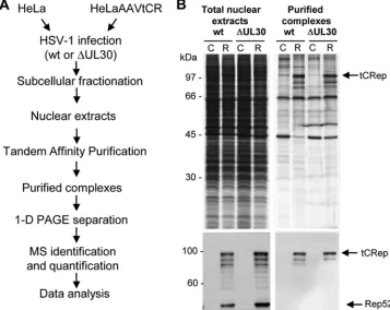
cellu-lar and viral factors recruited in AAV RCs, we developed a strategy based on the purification of protein complexes asso-ciated with the two larger AAV Rep proteins. Indeed, Rep78 and -68 are involved in AAV genome replication, through their DNA binding, helicase, and endonuclease activities (27), whereas the two smaller Rep isoforms are dispensable for AAV genome replication (11, 44). Previous studies have also shown that Rep68 and -78 can remain covalently attached to AAV genomes after cleavage (64). Therefore, we reasoned that by pulling down the two larger Rep proteins under non-denaturing conditions, we should also recover DNA-protein complexes containing factors that are directly or indirectly associated with Rep and/or AAV DNA. To efficiently purify Rep proteins and their nuclear interaction partners, we used the previously described HeLaAAVtCR cells (1), which con-tain approximately three integrated copies of a modified AAV-2 genome in which therepORF is fused at its 5⬘terminus to sequences coding for two affinity purification tags (strepta-vidin and calmodulin binding peptides) and the fluorescent mCherry protein (Fig. 1A). The AAVtCR genome thus en-codes two unmodified proteins, Rep52 and -40, and two fusion proteins, Rep78 and -68 (designated tCRep), that were previ-ously demonstrated to be fully functional (1). Importantly, the capgene was removed from this construct in order to focus the analysis on proteins involved in early replication events (i.e., viral gene expression and DNA replication) and to prevent sequestration of single-stranded AAV genomes within the viral capsids (71).
Studies performed to visualize AAV Rep proteins upon infection with wt HSV-1 or HSV⌬UL30 revealed the presence of fluorescent tCRep proteins as early as 8 h p.i. and their colocalization with HSV-1 ssDBP ICP8 protein, thus confirm-ing previous observations (66) (Fig. 1B). The Rep pattern was
on November 8, 2019 by guest
http://jvi.asm.org/
different depending on the helper virus used. In the case of wt HSV-1, Rep and ICP8 accumulated in a large compartment that increased in size over time. With HSV⌬UL30, Rep and ICP8 formed small foci early after infection and a larger but compact compartment at later times. These larger foci were previously demonstrated to contain AAV DNA in addition to Rep and ICP8 proteins (1). Because a previous study had suggested that AAV RCs are distinct from those formed by HSV-1 (20), we additionally analyzed AAV and HSV-1 DNA localization by immuno-FISH. At 20 h p.i., in cells where both HSV-1 and AAV DNAs were detected, two main patterns were observed: in some cells, the two viruses were found to replicate in partially overlapping domains (Fig. 1C, upper pan-els), and in other cells, the two replication domains coincided. The latter pattern was observed in nearly all cells at 24 h p.i. (Fig. 1C, lower panels). These observations suggest that as previously documented, HSV-1 and AAV replicate initially in adjacent domains that progressively fuse to form a unique replication compartment. Kinetic studies associated with a pre-cise quantification of each phenotype will indicate whether these two phenotypes correspond to different stages during HSV-1 and AAV coinfection.
For TAP of Rep-associated complexes, HeLaAAVtCR cells were infected with either HSV⌬UL30 or wt HSV-1. The HSV⌬UL30 mutant was chosen to analyze Rep-associated proteins in the absence of HSV-1 DNA replication. The wt HSV-1 strain was used to perform the same analysis under conditions in which AAV replication is optimal (1). HeLa cells infected with the same HSV-1 strains were used as the
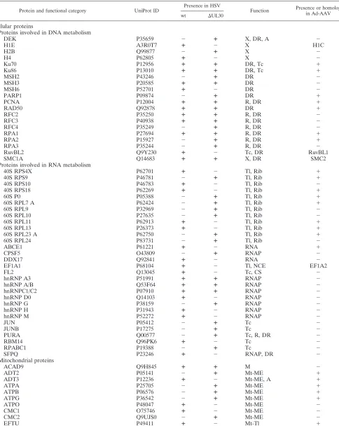
corre-sponding negative controls. Our previous analysis indicated that with both HSV-1 strains, AAV DNA synthesis reached its peak between 20 and 24 h p.i. (data not shown). Therefore, infected cells were collected at 20 h p.i., and nuclear extracts were subjected to two consecutive purifications, first on streptavidin and then on calmodulin resin (Fig. 2A). Compar-ison of the protein profiles for a fraction of the purified ma-terial (Fig. 2B, upper panel) revealed that during this purifi-cation process, there was a progressive clearance of the extracts associated with an enrichment in tCRep78/68 proteins, as shown by silver staining and Western blotting, specifically in the HeLaAAVtCR-derived extracts (Fig. 2B).
Identification of Rep-associated factors by mass
spectrom-etry analysis and validation studies.Proteins eluted from the
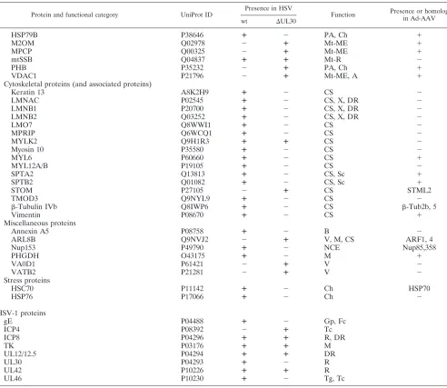
calmodulin resin at the end of the large-scale TAP procedure were subjected to a semiquantitative LC-MS/MS analysis after separation in a preparative gel by a short migration. This re-sulted in the identification of 138 (112 cellular and 26 viral) and 140 (129 cellular and 11 viral) proteins in the Rep-con-taining complexes purified from HeLaAAVtCR cells infected with wt HSV-1 and HSV⌬UL30, respectively. The lists were first double checked manually for redundant proteins, and then the proteins purified from HeLaAAVtCR cells were compared with those purified from the corresponding infected HeLa con-trol cells. The identified proteins were classified according to their known functions and included in the final list (Table 1) if they met the following criteria: (i) the number of peptides found in the corresponding HeLa control sample was either null or at least four times smaller than that in the Rep-con-FIG. 1. (A) Schematic view of the AAVtCR genome. p5 and p19 are the wt AAV-2 promoters, and C and S refer to the sequences coding for the calmodulin and streptavidin binding peptide tags. The dotted line indicates the position of alternative splicing in therepgene. (B) Immuno-fluorescence analysis of HSV-1-infected HeLaAAVtCR cells to examine colocalization of tCRep and HSV-1 DBP ICP8 proteins. Infected cells were fixed at 8 and 20 h p.i., stained with an anti-ICP8 antibody, and analyzed by confocal microscopy. Bars, 5m. (C) Immuno-FISH analysis of HSV-1-infected HeLaAAVtCR cells. HeLaAAVtCR cells were infected for 20 or 24 h with wt HSV-1 (5 PFU/cell) and then analyzed using a biotin-labeled HSV-1 probe and a DIG-labeled AAV probe. After hybridization and washes, cells were additionally stained with an anti-Rep antibody and DAPI (4⬘,6-diamidino-2-phenylindole) and were analyzed by confocal microscopy. Bars, 5m.
on November 8, 2019 by guest
http://jvi.asm.org/
taining sample infected with the same HSV-1 strain, (ii) the protein was identified by at least two different specific peptides, and (iii) the protein was not known to be associated closely with the calmodulin interaction network. This resulted in a list of 57 and 47 cellular proteins, as well as 8 and 5 viral proteins, identified from complexes purified from HeLaAAVtCR cells infected with wt HSV-1 and HSV⌬UL30, respectively. For both conditions, the largest functional categories corresponded to cellular factors involved in DNA metabolism, including DNA replication, repair, and chromatin modification, and RNA metabolism, with proteins involved in transcription, splicing/export, and translation (Fig. 3). Importantly, the final list of proteins also included several HSV-1 proteins, some of which were previously described as implicated in HSV-1-in-duced AAV replication. Interestingly, a significant number of proteins listed in Table 1 were previously found in a similar proteomic analysis performed during AAV and adenovirus coinfection, and some of them were shown to be involved directly in AAV replication (40–42).
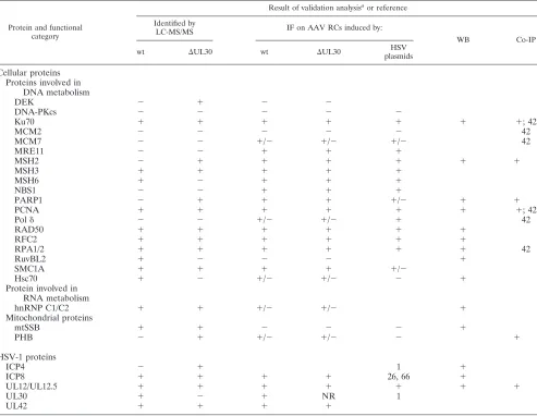
For this study, Western blot analyses performed on the pu-rified Rep-containing complexes validated the presence of 15 selected proteins (Fig. 4A and Table 2). In addition, reverse coimmunoprecipitations were also conducted with antibodies recognizing several cellular and viral factors to verify their association with Rep (Fig. 4B and Table 2). These analyses confirmed the association of Rep with PCNA and Ku70 (42) and demonstrated, for the first time, its presence in purified
complexes containing PARP-1, PHB, MSH2, and UL12. It remains to be determined whether these interactions are direct or mediated by other factors and/or DNA.
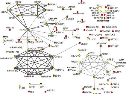
In silicoanalysis of cellular interaction pathways.In order to reconstitute likely interaction networks between cellular proteins identified in AAV RCs, the final list of cellular pro-teins specific to complexes purified from HeLaAAVtCR ex-tracts (Table 1) was submitted to the VirHostNet database (http://pbildb1.univ-lyon1.fr/virhostnet/login.php), which lists direct and experimentally validated interactions between hu-man proteins (Fig. 5). Not surprisingly, the two main networks that emerged from this analysis were the DNA replication and repair network (upper left) and the RNA splicing network (lower left). Other minor networks identified included ribo-somal proteins (upper right), mitochondrial factors (lower right), and cytoskeleton constituents.
Only minimal differences were found between the two HSV-1 helper strains. Notably, a larger number of cytoskeletal proteins were recovered in the presence of wt HSV-1. Their predominant identification from cells infected with wt HSV-1 might be the consequence of the more pronounced cytopathic effect induced by this helper virus than that with the replica-tion-defective HSV⌬UL30 strain.
Regarding the DNA replication machinery, components of RPA and RFC complexes were recovered, as well as PCNA, but no cellular DNA polymerase was recovered, even in the case of HSV⌬UL30-induced AAV replication, where a cellular FIG. 2. (A) Outline of the experimental procedure used to purify nuclear complexes associated with AAV Rep proteins. See Materials and Methods for details of the TAP steps with streptavidin and calmodulin resins. (B) Sequential analysis of the purified extracts. Proteins present either in total nuclear extracts or in the Rep-containing complexes after elution from the second calmodulin resin were resolved by SDS-PAGE, followed by silver staining (upper panels) or Western blotting using an anti-Rep antibody (lower panels). C, control infected HeLa cells; R, HeLaAAVtCR cells expressing Rep upon HSV-1 infection; wt, wt HSV-1;⌬UL30, HSV⌬UL30.
on November 8, 2019 by guest
http://jvi.asm.org/
[image:5.585.113.470.71.355.2]TABLE 1. Identification and classification of proteins in Rep-associated complexesa
Protein and functional category UniProt ID
Presence in HSV
Function Presence or homolog
in Ad-AAV
wt ⌬UL30
Cellular proteins
Proteins involved in DNA metabolism
DEK P35659 ⫺ ⴙ X, DR, A ⫺
H1E A3R0T7 ⴙ ⫺ X H1C
H2B Q99877 ⫺ ⴙ X ⫺
H4 P62805 ⴙ ⫺ X ⫺
Ku70 P12956 ⴙ ⴙ DR, Tc ⫹
Ku86 P13010 ⴙ ⴙ DR, Tc ⫹
MSH2 P43246 ⫺ ⴙ DR ⫺
MSH3 P20585 ⴙ ⴙ DR ⫺
MSH6 P52701 ⴙ ⫺ DR ⫺
PARP1 P09874 ⫺ ⴙ DR ⫹
PCNA P12004 ⴙ ⴙ R, DR ⫹
RAD50 Q92878 ⴙ ⴙ DR ⫹
RFC2 P35250 ⴙ ⴙ R, DR ⫺
RFC3 P40938 ⴙ ⴙ R, DR ⫺
RFC4 P35249 ⫺ ⴙ R, DR ⫺
RPA1 P27694 ⴙ ⴙ R, DR ⫹
RPA2 P15927 ⫺ ⴙ R, DR ⫹
RPA3 P35244 ⫺ ⴙ R, DR ⫺
RuvBL2 Q9Y230 ⴙ ⫺ Tc, DR RuvBL1
SMC1A Q14683 ⴙ ⴙ X, DR SMC2
Proteins involved in RNA metabolism
40S RPS4X P62701 ⴙ ⫺ Tl, Rib ⫹
40S RPS9 P46781 ⫺ ⴙ Tl, Rib ⫹
40S RPS10 P46783 ⴙ ⫺ Tl, Rib ⫺
40S RPS18 P62269 ⴙ ⫺ Tl, Rib ⫹
60S P0 P05388 ⫺ ⴙ Tl, Rib ⫹
60S RPL7 A P62424 ⫺ ⴙ Tl, Rib ⫹
60S RPL9 P32969 ⫺ ⴙ Tl, Rib ⫺
60S RPL10 P27635 ⫺ ⴙ Tl, Rib ⫺
60S RPL11 P62913 ⴙ ⫺ Tl, Rib ⫹
60S RPL13 P26373 ⴙ ⫺ Tl, Rib ⫹
60S RPL23 A P62750 ⫺ ⴙ Tl, Rib ⫹
60S RPL24 P83731 ⫺ ⴙ Tl, Rib ⫺
ABCE1 P61221 ⴙ ⫺ RNA ⫹
CPSF5 O43809 ⫺ ⴙ RNAP ⫺
DDX17 Q92841 ⴙ ⫺ RNA ⫺
EF1A1 P68104 ⴙ ⫺ Tl, NCE EF1A2
FL2 Q13045 ⴙ ⫺ Tc, CS ⫺
hnRNP A3 P51991 ⴙ ⴙ RNAP ⫺
hnRNP A/B Q53F64 ⴙ ⴙ RNAP ⫺
hnRNPC1/C2 P07910 ⴙ ⴙ RNAP ⫺
hnRNP D0 Q14103 ⴙ ⫺ RNAP ⫺
hnRNP G P38159 ⫺ ⴙ RNAP ⫺
hnRNP H P31943 ⴙ ⫺ RNAP ⫺
hnRNP M P52272 ⴙ ⫺ RNAP ⫺
JUN P05412 ⫺ ⴙ Tc ⫺
JUNB P17275 ⫺ ⴙ Tc ⫺
PURA Q00577 ⫺ ⴙ Tc, R, DR ⫺
RBM14 Q96PK6 ⴙ ⫺ Tc ⫺
RPABC1 P19388 ⫺ ⴙ Tc ⫺
SFPQ P23246 ⴙ ⫺ RNAP, DR ⫺
Mitochondrial proteins
ACAD9 Q9H845 ⴙ ⴙ M ⫺
ADT2 P05141 ⫺ ⴙ Mt-ME ⫹
ADT3 P12236 ⴙ ⫺ Mt-ME, A ⫹
ATPA P25705 ⫺ ⴙ Mt-ME ⫹
ATPB P06576 ⫺ ⴙ Mt-ME ⫹
ATPG P36542 ⫺ ⴙ Mt-ME ⫹
ATPO P48047 ⴙ ⫺ Mt-ME ⫺
CMC1 O75746 ⴙ ⫺ Mt-ME ⫺
CMC2 Q9UJS0 ⫺ ⴙ Mt-ME ⫺
EFTU P49411 ⴙ ⫺ Mt-Tl ⫹
Continued on following page
on November 8, 2019 by guest
http://jvi.asm.org/
enzyme is necessarily required. RNA metabolism factors iden-tified could be divided into two major categories, i.e., factors involved in splicing and ribosomal proteins; a few transcription factors were also identified. Most of the splicing factors iden-tified are hnRNPs. Since most of these factors associate with mRNAs during the process of transcription and mark imma-ture mRNAs (37), this finding was not unexpected, as viral RCs are active sites of transcription, and thus newly synthe-sized viral mRNAs and associated complexes are probably major components of these intranuclear domains (81). Rep proteins themselves are involved in AAV genome transcription and have been shown to affect the ratio of spliced versus unspliced AAV mRNAs (47). An increasing number of studies
[image:7.585.47.539.79.506.2]have suggested that various hnRNPs are involved in the rep-lication of numerous viruses, including herpesviruses (7, 23, 24, 33). Interestingly, some of the hnRNPs recovered were also reported to be able to associate with DNA by recognizing structural motifs and to regulate several aspects of DNA me-tabolism, such as chromosome maintenance and DNA repli-cation and repair (25). For instance, hnRNP A3 was recently reported to bind the single-stranded telomeric repeat, thus contributing to its protection from nuclease attacks (67). In-terestingly, an hnRNP A/B-related protein was shown to bind the single-stranded genome of feline parvovirus and to modu-late virus replication (73). Among the various hnRNPs found in this study, the association of hnRNP C1/C2 with Rep was TABLE 1—Continued
Protein and functional category UniProt ID
Presence in HSV
Function Presence or homolog
in Ad-AAV
wt ⌬UL30
HSP79B P38646 ⴙ ⫺ PA, Ch ⫹
M2OM Q02978 ⫺ ⴙ Mt-ME ⫹
MPCP Q00325 ⫺ ⴙ Mt-ME ⫹
mtSSB Q04837 ⴙ ⴙ Mt-R ⫺
PHB P35232 ⫺ ⴙ PA, Ch ⫹
VDAC1 P21796 ⫺ ⴙ Mt-ME, A ⫹
Cytoskeletal proteins (and associated proteins)
Keratin 13 A8K2H9 ⴙ ⫺ CS ⫺
LMNAC P02545 ⴙ ⫺ CS, X, DR ⫺
LMNB1 P20700 ⴙ ⫺ CS, X, DR ⫺
LMNB2 Q03252 ⴙ ⫺ CS, X, DR ⫺
LMO7 Q8WWI1 ⴙ ⫺ CS ⫺
MPRIP Q6WCQ1 ⴙ ⫺ CS ⫺
MYLK2 Q9H1R3 ⴙ ⴙ CS ⫺
Myosin 10 P35580 ⴙ ⫺ CS ⫺
MYL6 P60660 ⴙ ⫺ CS ⫹
MYL12A/B P19105 ⴙ ⫺ CS ⫺
SPTA2 Q13813 ⴙ ⫺ CS, Sc ⫹
SPTB2 Q01082 ⴙ ⫺ CS, Sc ⫹
STOM P27105 ⫺ ⴙ CS STML2
TMOD3 Q9NYL9 ⴙ ⫺ CS ⫺
-Tubulin IVb Q8IWP6 ⴙ ⫺ CS -Tub2b, 5
Vimentin P08670 ⴙ ⫺ CS ⫹
Miscellaneous proteins
Annexin A5 P08758 ⴙ ⫺ B ⫺
ARL8B Q9NVJ2 ⫺ ⴙ V, M, CS ARF1, 4
Nup153 P49790 ⴙ ⫺ NCE Nup85,358
PHGDH O43175 ⴙ ⫺ M ⫹
VA0D1 P61421 ⫺ ⴙ V ⫺
VATB2 P21281 ⫺ ⴙ V ⫺
Stress proteins
HSC70 P11142 ⴙ ⫺ Ch HSP70
HSP76 P17066 ⴙ ⫺ Ch ⫺
HSV-1 proteins
gE P04488 ⴙ ⫺ Gp, Fc
ICP4 P08392 ⫺ ⴙ Tc
ICP8 P04296 ⴙ ⴙ R, DR
TK P03176 ⴙ ⴙ M
UL12/12.5 P04294 ⴙ ⴙ DR
UL30 P04293 ⴙ ⫺ R
UL42 P10226 ⴙ ⴙ R
UL46 P10230 ⴙ ⫺ Tg, Tc
aThe table indicates the specific proteins identified in HeLaAAVtCR cells infected with wt HSV-1 or HSV⌬UL30, their known functions, and whether they were
previously found in adenovirus- and AAV-coinfected cells (42). A, apoptosis; B, blood; Ch, chaperone; CS, cytoskeleton; DR, DNA repair and recombination; Fc, Fc receptor; Gp, glycoprotein; M, metabolism; ME, membranes and transmembrane exchanges; Mt, mitochondrion; NCE, nucleocytoplasmic exchange; PA, control of cell proliferation and aging; R, DNA replication; Rib, ribosome; RNA, RNA metabolism, unspecified; RNAP, mRNA processing; Sc, secretion; Tc, transcription; Tg, tegument; Tl, translation; V, vesicles; X, chromatin and chromosomes. The list of identified proteins is also available on the VirHostNet website (http://pbildb1 .univ-lyon1.fr/virhostnet/login.php).
on November 8, 2019 by guest
http://jvi.asm.org/
validated by Western blotting (Fig. 4A). The other main cat-egory of RNA metabolism factors was composed of ribosomal proteins. Association of ribosomal constituents with AAV RCs could be mediated through the known interaction between Rep and the major nucleolar component B23/NPM (4). How-ever, NPM was not recovered, and since only a minority of the ribosomal proteins was identified, it is likely that these proteins were derived from contamination of the purified extracts with these highly prevalent nuclear proteins. Accordingly, several
ribosomal proteins were also identified in complexes purified from cells coinfected with AAV and adenovirus (42).
[image:8.585.111.473.63.212.2]Another significant network was that of mitochondrial pro-teins. In particular, several mitochondrial factors involved in ATP synthesis were specifically retrieved from cells infected with HSV⌬UL30, similar to what was previously reported in the case of adenovirus- and AAV-coinfected cells (42). This is surprising, since no specific localization of Rep to mitochon-dria has ever been reported. Since AAV, like other parvovi-FIG. 3. Functional classification of proteins copurified with Rep from HeLaAAVtCR cells infected with wt HSV-1 or HSV⌬UL30 and identified by mass spectrometry analysis. See Table 1 for definitions of the functional categories.
FIG. 4. Western blot analyses of purified complexes. (A) Protein complexes were purified from HeLa (C) or HeLaAAVtCR (R) cells infected with wt HSV-1 and then analyzed by Western blotting, using antibodies against a panel of cellular and viral proteins. (B) Reverse coimmuno-precipitation of tCRep in purified complexes containing selected cellular and viral proteins. Control HeLa (C) cells and HeLaAAVtCR (R) cells were infected with wt HSV-1 for 20 h (MOI⫽5 PFU/cell). Nuclear extracts from 2⫻106infected cells were subjected to coimmunoprecipitation,
using UL12, Ku70, MSH2, PARP-1, PCNA, and PHB polyclonal antibodies as indicated. The anti-HA antibody was used as a negative control. The immunoprecipitates were then immunoblotted with an anti-Rep antibody (303.9). The arrows indicate the positions of the tCRep and Rep52 proteins, and the asterisk shows a nonspecific band.
on November 8, 2019 by guest
http://jvi.asm.org/
[image:8.585.110.473.396.662.2]ruses, has been shown to induce apoptosis (55, 72), it is tempt-ing to speculate that the identification of these factors may be linked to a possible interaction of AAV Rep proteins with mitochondrial constituents.
Because we were interested mainly in the analysis of factors involved in the early steps of AAV replication, further analyses were focused on only a subset of cellular and viral factors that were found in this screen.
Factors involved in AAV DNA replication.
Adenovirus-in-duced AAV replication is performed mainly by cellular factors, and previous studies have shown that PCNA, RFC, Pol ␦, RPA, and MCM constitute the minimal set of cellular proteins able to drive AAV DNA replicationin vitro(40, 41, 44). Most of these factors were also found in a recent proteomic analysis of Rep-associated proteins in adenovirus- and AAV-coin-fected cells (42) (Fig. 5 and Table 1). However, at the begin-ning of this study, it was unknown whether the same set of cellular proteins, with the exception of RPA, was also involved during HSV-1-induced AAV replication.
[image:9.585.46.539.81.463.2]In our proteomic analysis using wt HSV and HSV⌬UL30 as helpers, we identified members of the RPA and RFC com-plexes as well as PCNA as Rep-interacting partners (Table 1 and Fig. 4). In contrast, neither the MCM proteins nor Pol␦or any other cellular DNA polymerase was identified. To further analyze these data, immunofluorescence studies were con-ducted to determine whether these proteins were recruited in AAV RCs (Fig. 6 and Table 2). To discriminate between fac-tors recruited by replicating HSV-1 and AAV DNAs, these analyses were performed in cells infected with HSV⌬UL30. However, HSV-1 is known to carry a protein that binds to the Fc domain of human, rabbit, and goat antibodies (29, 83), thus resulting in significant nonspecific cytoplasmic staining in anal-yses performed at late stages of infection (14 to 20 h p.i.). Thus, additional analyses were also performed on cells transfected with a set of HSV-1 helper plasmids previously reported to be able to induce AAV replication as efficiently as that induced by wt HSV-1 (1). These analyses showed a colocalization of RFC, PCNA (Fig. 6), and RPA (not shown) with Rep. These results TABLE 2. Summary of validation analyses
Protein and functional category
Result of validation analysisaor reference
Identified by
LC-MS/MS IF on AAV RCs induced by:
WB Co-IP
wt ⌬UL30 wt ⌬UL30 HSV
plasmids
Cellular proteins Proteins involved in
DNA metabolism
DEK ⫺ ⫹ ⫺ ⫺
DNA-PKcs ⫺ ⫺ ⫺ ⫺ ⫺
Ku70 ⫹ ⫹ ⫹ ⫹ ⫹ ⫹ ⫹; 42
MCM2 ⫺ ⫺ ⫺ ⫺ ⫺ 42
MCM7 ⫺ ⫺ ⫹/⫺ ⫹/⫺ ⫹/⫺ 42
MRE11 ⫺ ⫺ ⫹ ⫹ ⫹
MSH2 ⫺ ⫹ ⫹ ⫹ ⫹ ⫹ ⫹
MSH3 ⫹ ⫹ ⫹ ⫹ ⫹
MSH6 ⫹ ⫺ ⫹ ⫹ ⫹
NBS1 ⫺ ⫺ ⫹ ⫹ ⫹
PARP1 ⫺ ⫹ ⫹ ⫹ ⫹/⫺ ⫹ ⫹
PCNA ⫹ ⫹ ⫹ ⫹ ⫹ ⫹ ⫹; 42
Pol␦ ⫺ ⫺ ⫹/⫺ ⫹/⫺ ⫹ 42
RAD50 ⫹ ⫹ ⫹ ⫹ ⫹ ⫹
RFC2 ⫹ ⫹ ⫹ ⫹ ⫹ ⫹
RPA1/2 ⫹ ⫹ ⫹ ⫹ ⫹ ⫹ 42
RuvBL2 ⫹ ⫺ ⫺ ⫺ ⫹
SMC1A ⫹ ⫹ ⫹ ⫹ ⫹/⫺
Hsc70 ⫹ ⫺ ⫹/⫺ ⫹/⫺ ⫺ ⫹
Protein involved in RNA metabolism
hnRNP C1/C2 ⫹ ⫹ ⫹/⫺ ⫹/⫺ ⫹
Mitochondrial proteins
mtSSB ⫹ ⫹ ⫺ ⫺ ⫺ ⫹
PHB ⫺ ⫹ ⫹/⫺ ⫹/⫺ ⫺ ⫹
HSV-1 proteins
ICP4 ⫺ ⫹ 1 ⫹
ICP8 ⫹ ⫹ ⫹ ⫹ 26, 66 ⫹
UL12/UL12.5 ⫹ ⫹ ⫹ ⫹ ⫹ ⫹ ⫹
UL30 ⫹ ⫺ ⫹ NR 1
UL42 ⫹ ⫹ ⫹ ⫹
a
Summary of validation analyses, performed by immunofluorescence (IF) assay of infected cells, Western blotting (WB) of TAP complexes, and reverse
coimmu-noprecipitation (co-IP). For IF analyses, “⫹” indicates colocalization with AAV RCs, “⫹/⫺” indicates unclear or faint colocalization with AAV RCs, and “⫺” indicates
absence from AAV RCs. NR, not relevant.
on November 8, 2019 by guest
http://jvi.asm.org/
did not demonstrate a direct or indirect interaction with Rep but indicated that these cellular factors were recruited in HSV-1-induced AAV RCs. In contrast, our immunofluorescence study showed that MCM remained more uniformly distributed in the nucleus and did not clearly colocalize with Rep (Fig. 6 and Table 2). This is in agreement with the fact that MCM proteins were not identified by MS in this study. This result suggests that the MCM helicase complex does not participate in HSV-1-induced AAV replication, in contrast to what was recently shown in the presence of adenovirus (41, 42). Similar activities could be provided by other viral or cellular proteins. The previous study on adenovirus-induced AAV replication showed that the Ku proteins, although not essential, can par-tially substitute for MCM inin vitro replication assays (42). Therefore, it is possible that the Ku proteins directly or indi-rectly provide strand displacement activity during HSV-1-in-duced AAV DNA replication. Indeed, Ku86 and -70 were found in Rep-associated complexes (Table 1 and Fig. 4) and colocalized in AAV RCs (Fig. 7 and Table 2). Alternatively, this activity could also be provided by the HSV-1 HP complex (UL5/8/52), which was previously demonstrated to be essential
[image:10.585.80.501.70.384.2]for HSV-1 helper activity, in particular via its helicase subunit (62, 80). However, our analysis did not retrieve any of the members of the HP complex. It is possible either that the association of the HP complex with AAV genomes is not stable enough for it to be recovered during the purification procedure or that this complex is required only at early replication steps. It was surprising that no cellular polymerase was identified by MS, even in the case of cells infected with HSV⌬UL30, where a cellular enzyme is necessarily required (Table 1). Pre-vious studies have indicated that Pol␦is the cellular enzyme involved in AAV genome replication in the presence of ade-novirus and that this enzyme is also able to drive AAV repli-cationin vitro(40, 41). In the case of HSV-1, the situation is more complex. Although the viral polymerase complex (UL30/ 42) is not strictly essential, it was shown to be able to replicate AAV DNAin vitroand to potently enhance AAV replication in live cells (1, 78, 80). In this regard, it is interesting that both UL30 and UL42 were identified by MS (Table 1) and colocal-ized within AAV RCs (Table 2). Therefore, we examined whether Pol␦could be visualized in AAV RCs. The recruitment of Pol␦within AAV RCs was observed in HeLaAAVtCR cells FIG. 5.In silicoreconstitution of Rep-associated interaction networks. Likely protein-protein interactions were reconstituted using the cellular protein list from Table 1 and analysis tools provided by the VirHostNet interactome website (http://pbildb1.univ-lyon1.fr/virhostnet/login.php), complemented with additional information derived from the UniProtKB/SwissProt database and specialized literature. The color codes refer to proteins specifically identified from HeLaAAVtCR cells infected with wt HSV-1 (red), HSV⌬UL30 (yellow), or both helper viruses (orange). Green circles, proteins for which a physical or functional interaction with a protein from another virus has already been reported; black lines, VirHostNet-derived interactions, with the link width correlating with the number of different reports describing the interaction found in the database (black circles correspond to self-interacting proteins); dashed brown arrows, UniProtKB/SwissProt-derived interactions; asterisks, proteins identified in Rep-containing complexes purified from adenovirus- and AAV-coinfected cells (42); dashed brown circles, proteins constituting a known functional complex.
on November 8, 2019 by guest
http://jvi.asm.org/
not only infected with HSV⌬UL30 but also transfected with HSV-1 helper plasmids (Fig. 6) or infected with wt HSV-1 (data not shown). Altogether, these findings suggest that Pol␦ is present within HSV-1-induced AAV RCs, even in the pres-ence of the viral polymerase. This finding raises the questions of the relative extent to which AAV uses the cellular polymer-ase versus its viral analog when both are present and of the mode of AAV replication with these enzymes.
Finally, two additional cellular factors were of particular interest: prohibitin (PHB1) and mtSSB/SSBP1, the mitochon-drial ssDBP. Prohibitin is a mitochonmitochon-drial and nuclear protein which displays several functions, including inhibition of DNA synthesis (68, 74). Interestingly, a recent study indicates that this protein negatively controls the onset of cellular DNA replication by physically interacting with MCM proteins (53). In our study, PHB1 was identified in Rep-containing com-plexes purified from cells infected with HSV⌬UL30 (Table 1) and was also found to be associated with Rep in reverse co-immunoprecipitation experiments (Fig. 4B). Immunofluores-cence analysis indicated that in HeLaAAVtCR cells infected with either HSV-1 strain, the protein was present in the nu-cleus, but not exclusively within AAV RCs (Table 2). However, a similar pattern was not observed in HeLaAAVtCR cells transfected with HSV-1 helper plasmids, in which the protein was localized mostly in the cytoplasm. The second interesting factor that was identified in samples from cells infected with wt and⌬UL30 HSV-1 strains was mtSSB (Table 1 and Fig. 4A). Following infection with Epstein-Barr virus, this protein has
been shown to be relocalized to the nucleus, where it stimu-lates viral replication (84). Surprisingly, however, the protein was not detected by immunofluorescence in the nuclei of either infected or transfected HeLaAAVtCR cells (Table 2). Both of these discrepancies could be linked to lower sensitivities of the PHB and mtSSB antibodies used for the immunofluorescence analysis. Additional studies will be needed to determine whether mtSSB and PHB can modulate AAV replication.
Factors involved in DNA repair.Viruses that replicate in the
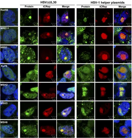
nucleus, such as adenovirus and HSV-1, have been shown to significantly interfere with the DNA repair machinery and the DNA damage response (DDR) to inhibit the cellular defenses and to usurp some cellular functions for their own replication (10). In particular, several studies have highlighted that with respect to DNA repair, adenovirus and HSV-1 use different strategies to accomplish their replicative cycles. HSV-1 notably recruits and exploits the MRN complex to accomplish its rep-licative cycle (31, 85). This complex is involved in the recogni-tion of dsDNA breaks and in downstream signaling to mole-cules involved in DNA repair through both homologous and nonhomologous recombination pathways (61). In addition, many other DNA repair proteins were found in ICP8-associ-ated complexes, including the DNA-PK complex, XRCC4, Rad50, PARP1, the MMR proteins MSH2, -3, and -6, and the repair helicases WRN and BLM (69). In contrast, adenovirus degrades and/or inactivates MRN and ligase IV in order to prevent the concatemerization of the viral genomes and the inhibition of its replication (2, 3, 9, 48, 65, 79). Importantly, FIG. 6. Colocalization of cellular DNA replication factors with Rep in HSV-1-induced AAV RCs. HeLaAAVtCR cells were either infected with HSV⌬UL30 (MOI⫽5 PFU/cell) and fixed at 9 or 14 h p.i. or transfected with 0.25 pmol/35-mm well of each HSV-1 helper plasmid (pTF3pol and pRF) and fixed at 48 h posttransfection. Alexa Fluor 488-conjugated donkey anti-mouse, anti-rabbit, and anti-goat secondary antibodies were used. DNA was stained with TO-PRO-3 iodide (Invitrogen). Images were taken using an Axioplan2 LSM510 confocal microscope (Zeiss). Green, protein of interest; red, tCRep; blue, TO-PRO-3 staining. The left panels correspond to results for noninfected and nontransfected control HeLaAAVtCR cells. Bars, 5m. The cytoplasmic background visible for some antibodies in HSV-infected cells is due to antibody binding to the HSV-encoded Fc receptor (29, 83).
on November 8, 2019 by guest
http://jvi.asm.org/
the ability of adenoviral proteins to degrade and/or delocalize the MRN complex contributes to the helper activities required for AAV replication (58).
Our proteomic analysis retrieved several factors involved in the DNA damage response, including the Rad50 protein, Ku70 and -86, PARP1, MSH2/3/6, RuvBL2, and SMC1A (Table 1 and Fig. 4). The identification of Rad50 within Rep-containing complexes is of particular interest given the previously docu-mented positive role of the MRN complex during HSV-1 rep-lication (31, 60, 85; N. Balasubramanian and S. K. Weller, unpublished data). Our immunofluorescence analysis con-firmed the colocalization of Rad50 with Rep and showed that both Mre11 and Nbs1, the two other members of the MRN complex, were similarly recruited within AAV RCs induced upon infection with HSV⌬UL30 (Fig. 7). Similar results were
also observed when the cells were transfected with HSV-1 helper plasmids, further indicating that the recruitment of this complex is not dependent upon the presence of a replication-competent HSV-1 genome. It is not possible at the moment to conclude whether the MRN complex displays a positive or negative effect on AAV replication induced by HSV-1. Never-theless, it is tempting to speculate that the same complex differentially regulates AAV genome replication depending on the nature of the helper virus used.
[image:12.585.81.503.67.508.2]Other factors of interest identified in our study were the MMR factors MSH2, -3, and -6. These three proteins partici-pate in the formation of two distinct complexes that remove mismatches arising during DNA synthesis (28). The MSH2/6 complex (MutS␣) is the most abundant and is involved mainly in the recognition of base-base mismatches generated by errors FIG. 7. Colocalization of cellular DNA repair factors with Rep in AAV RCs. Cells were processed as described in the legend to Fig. 6 and then stained with antibodies recognizing the indicated cellular DNA repair factors. Green, protein of interest; red, tCRep; blue, TO-PRO-3 staining. The left panels indicate the staining observed in noninfected and nontransfected HeLaAAVtCR cells. Bars, 5m.
on November 8, 2019 by guest
http://jvi.asm.org/
of DNA polymerases. The MSH2/3 complex (MutS) recog-nizes longer insertion/deletion loops generated, notably, dur-ing replication of microsatellite regions (28). In addition, this pathway is also involved in the cellular DNA damage response and in the control of homologous recombination (HR) events. Indeed, in mammalian cells, MSH2, -3, and -6 prevent recom-bination between divergent sequences (16, 63). Our analyses confirmed the recruitment of MSH2, -3, and -6 in AAV RCs (Table 2 and Fig. 7) and the association of MSH2 with Rep (Fig. 4). These results are in agreement with previous studies suggesting that HSV-1 replication may be dependent upon HR events requiring both viral and cellular proteins (86). Indeed, MSH proteins have been identified in the replication compart-ments of various herpesviruses (13, 69, 75). Factors such as MRN and MSH are both involved in the control of HR and most likely present in AAV RCs because they are recruited by HSV-1-encoded factors for its own replication. However, this does not exclude the possibility that these factors may also be involved in the control of AAV replication induced by HSV-1. Data from a recent study on adenovirus-induced AAV rep-lication indicated the presence of both Ku70/80 and DNA-PKcs within Rep-associated complexes (42), and further anal-yses showed that DNA-PKcs mediates AAV-induced DDR in the presence of adenovirus (12, 57). DNA-PKcs was not iden-tified in our proteomic analysis, despite the presence of Ku (Table 1). In addition, immunofluorescence analyses con-firmed the absence of DNA-PKcs in HSV-1-induced AAV RCs (data not shown). This result was not surprising, since previous studies have shown that ICP0, an HSV-1 immediate-early factor, can induce the proteasomal degradation of DNA-PKcs in a cell line-dependent manner (45). This event is con-sidered central to the strategy used by HSV-1 to inactivate the nonhomologous end-joining (NHEJ) DNA repair pathway ini-tiated by the recognition of dsDNA breaks by the DNA-PK complex. However, recent studies have also raised the possi-bility of end-joining events in the absence of classical NHEJ factors, including joining of broken ends by the direct recruit-ment of XRCC4 and ligase IV by Ku70/80 in the absence of DNA-PKcs (34). Interestingly, knockdown of either ligase IV or XRCC4 has been shown to affect HSV-1 genome replica-tion and viral yields (38). Therefore, it is possible that the Ku proteins found in AAV RCs (Fig. 7 and Table 2) may be implicated in the recruitment of downstream factors involved in NHEJ. Further studies will indicate whether these factors are indeed recruited within AAV RCs as well as the impact of potential NHEJ events on AAV genome replication. Finally, another important DNA repair signaling protein found in our analysis was PARP-1, which was found to be recruited in AAV RCs and associated with Rep (Table 2 and Fig. 4). This mul-tifunctional factor, previously found associated with several viruses that replicate in the nucleus, has also been implicated in the regulation of dsDNA break repair pathway choice, in particular by promoting alternative NHEJ mechanisms (56, 61). Interestingly, Mre11, a component of MRN, was also recently found to be involved in alternative NHEJ (49, 88). The potential role of this alternative repair pathway during the AAV life cycle, and particularly during AAV DNA replication, remains to be defined.
Identification of HSV-1 helper proteins. As expected,
sev-eral known HSV-1 helper factors were identified in this study.
In contrast, a previous analysis of adenovirus-induced AAV replication resulted in the identification of a single adenoviral helper protein (42). This finding additionally illustrates the more direct role of HSV-1 in AAV replication than that of adenovirus. Among the helper factors retrieved in our study were the ICP8, ICP4, UL30, and UL42 proteins. Other factors known to help AAV, such as ICP0 and the HP complex, were not identified, despite previous evidence of their biological effect (1, 18, 80). It is possible, as noted above, that these factors intervene only at early steps of the AAV replicative cycle, further explaining why they were not identified from complexes purified from cells at 20 h postinfection. Alterna-tively, helper factors such as ICP0 might stimulate AAV rep-lication in an indirect way without actually physically associat-ing with Rep complexes. Other HSV-1 factors not already known to exert any helper activity were also identified in the present study. In particular, the UL12 protein was identified for both helper viruses by the presence of a significant number of peptides. This early factor is a 5⬘-3⬘ exonuclease that has been shown to be involved in HSV-1 genome maturation. Indeed, previous studies have shown that this exonuclease forms a complex with the HSV-1 ICP8 protein that is able to mediate a robust strand exchange activityin vitro(51, 52). This recombinase activity plays an important role in the generation of mature progeny viral DNA that can be packaged efficiently into newly formed virions (21, 22, 46). The UL12 ORF also encodes a truncated protein, UL12.5, that similarly displays 5⬘-3⬘ exonuclease and strand exchange activities but is local-ized predominantly in the mitochondria, where it induces deg-radation of host mitochondrial DNA (50, 54). The MS analysis did not indicate which UL12 isoform was part of the purified complexes, since all identified peptides mapped in the region common to the two proteins. However, both UL12 and UL12.5 were detected in Rep-associated complexes by Western blot-ting (Fig. 4A). Reverse coimmunopurification experiments confirmed the interaction between UL12 and Rep (Fig. 4B). It is likely that this interaction is mediated by HSV-1 DBP, since this protein is able to interact directly with both UL12 and Rep (26, 66, 70). Also, immunofluorescence studies showed that UL12 colocalized with Rep in AAV RCs induced by infection with either wt HSV-1 or HSV⌬UL30 (Fig. 8A). To determine whether UL12 had a helper activity, we analyzed its effect on AAV genome replication in HeLaAAVtCR cells either in-fected with a wt or UL12-defective (HSV⌬UL12) HSV-1 strain or transfected with the set of HSV-1 helper plasmids together with a plasmid encoding UL12. Quantitative PCR analysis indicated that UL12 did not affect AAV genome replication in terms of the amount of viral DNA synthesized in infected or transfected cells (Fig. 8B). In contrast, Southern blot analysis indicated an altered pattern of AAV replication in the absence of UL12 (Fig. 8C). Indeed, the absence of this factor resulted in a reduction of the typical AAV replicative forms, the dou-ble-stranded monomer and dimer forms, as well as an increase in the smear-like background associated with high-molecular-weight forms that suggested the presence of a ladder of repli-cation intermediates. Interestingly, a typical replicative pattern could be restored fully by complementation with a plasmid encoding either UL12 or UL12.5 but not with a plasmid en-coding an exonuclease-negative UL12 mutant.
These results are reminiscent of a report by Blumel et al.
on November 8, 2019 by guest
http://jvi.asm.org/
showing that the six core HSV-encoded replication factors can induce concatemer formation on simian virus 40 (SV40) ge-nomes if SV40 large T antigen is also provided (5). Since SV40 replication normally results in the appearance of two circular daughter molecules, it is noteworthy that the presence of HSV replication proteins can alter the mode of replication to gen-erate concatemeric DNA. Thus, when HSV is used as the helper, it is possible that the mechanism of AAV DNA repli-cation is altered and that UL12 may be required to effectively produce progeny genomes suitable for packaging.
To further analyze the effect of UL12 on the AAV life cycle,
[image:14.585.82.501.66.463.2]we finally examined its effect on the production of infectious AAV particles. For this purpose, recombinant AAV vector particles coding for green fluorescent protein (rAAV-GFP) were produced in HeLa cells by using either the set of helper HSV-1 plasmids or the HSV⌬UL12 virus complemented or not with the UL12-expressing plasmid. As a positive control, rAAV-GFP particles were also produced using either adeno-viral helper plasmids or wt HSV-1. Cell lysates from trans-fected/infected cells were then used to infect HeLa cells, which were analyzed by flow cytometry to count the number of GFP-positive cells. The results shown in Fig. 8D indicate that HSV-1 FIG. 8. Involvement of HSV-1 UL12 protein in AAV replication. (A) Colocalization of UL12 with Rep within AAV RCs. HeLaAAVtCR cells were either infected with the indicated HSV-1 strain or transfected with the HSV-1 helper plasmids and pSAKUL12 plasmid and then stained with an anti-UL12 antibody and TO-PRO-3. Bars, 5m. (B) Analysis of the effect of UL12 on AAV DNA replication. Total DNA was extracted from HeLaAAVtCR cells either infected with wt HSV-1 or HSV⌬UL12 or transfected with HSV-1 helper plasmids and a plasmid encoding either UL12, UL12.5, or an exonuclease-negative UL12 mutant (pUL12exo⫺) and then quantified by qPCR, usingrep-specific primers. Data presented are means with error bars calculated for three independent experiments. (C) The same samples used in panel B were analyzed by Southern blotting, using a DIG-labeledrepprobe. mRF, monomer replicative form; dRF, dimer replicative form. (D) Effect of UL12 on rAAV-GFP particle production. HeLa cells transfected with rAAV-GFP andrep-cap-expressing plasmids were either cotransfected with HSV-1 helper plasmids or infected with HSV⌬UL12 in the absence or presence of a plasmid encoding UL12. As a control, the cells were cotransfected with adenoviral helper plasmids (pXX6 and pGKE1) or infected with wt HSV-1. Cell lysates were then used to infect HeLa cells in the presence of wt adenovirus, and GFP-positive cells were counted by flow cytometry at 24 h p.i. Mean values with error bars, calculated for at least three independent experiments, are presented.
on November 8, 2019 by guest
http://jvi.asm.org/
helper plasmids induced the formation of infectious rAAV-GFP particles at a level that was approximately 10-fold lower than that found with adenoviral helper plasmids. Addition of UL12 reproducibly resulted in a 5-fold increase in the number of infectious rAAV-GFP particles produced. Interestingly, UL12 also led to an increase in the number of infectious rAAV-GFP particles when it was used to complement the HSV⌬UL12 virus. These results clearly indicate that UL12 can increase the number of infectious AAV particles produced, further suggesting that its effect on AAV DNA replication enhances the production of DNA forms that are suitable for encapsidation. Altogether, these data indicate that UL12 can be considered an additional HSV-1 helper factor that, al-though not essential, can critically enhance the AAV produc-tive cycle.
Several lines of evidence suggest that HSV-1 may replicate its genome in two stages, involving UL9-dependent initiation from a viral origin of replication and then replication via a recombination-driven and origin-independent mechanism (86). The UL12/ICP8 recombinase certainly plays an impor-tant role in this process. The participation of UL12 in HR was also suggested by recent data indicating that this viral factor is able to bind to some components of the MRN complex (Bala-subramanian and Weller, unpublished data). In contrast, the current model of AAV genome replication does not involve any recombination event, despite previous indirect evidence for HRin vitro, in cultured cells, andin vivo, in tissues. Indeed, recombination events have been observed between mutant AAV genomes under conditions enabling replication, and mul-tiple AAV variants have been detected in individual tissues from nonhuman primates (17, 59, 76). On the basis of these data, it is tempting to speculate that in the context of an HSV-1 infection, HR may control AAV genome replication, in par-ticular by enhancing the resolution of high-molecular-weight concatemers into discrete replicative forms, thus providing more DNA substrates for packaging into assembled capsids.
Altogether, these results indicate that in the presence of HSV-1, AAV may replicate by using a basal set of cellular DNA replication enzymes but also relies on HSV-1-derived proteins for its replication, including UL12, a newly discovered helper factor that intervenes with AAV genome replication at a qualitative level. These findings provide the basis to further understand how AAV adapts its replication strategy to the nuclear environment induced by the helper virus.
ACKNOWLEDGMENTS
We thank Bernard Lopez for critically reading the manuscript, Juer-gen Kleinschmidt for providing the anti-Rep antibodies, and Fre´de´ric Catez for providing the HSV probes used for FISH analyses. We are also grateful to the PLATIM and qPCR teams of IFR128 for excellent technical assistance and to Vincent Lotteau, Benoît De Chassey, and Laure`ne Meyniel for helpful discussions and for help with data analysis on the VirHostNet website.
This work was supported by CNRS, INSERM, Universite´ Claude Bernard Lyon-1, and Ecole Normale Supe´rieure de Lyon. It was funded by grants from the Association Franc¸aise contre les Myopa-thies (AFM), to A.S. and A.G., and from the NIH (R01 AI069146), to S.K.W.
REFERENCES
1.Alazard-Dany, N., A. Nicolas, A. Ploquin, R. Strasser, A. Greco, A. L. Epstein, C. Fraefel, and A. Salvetti.2009. Definition of herpes simplex virus
type 1 helper activities for adeno-associated virus early replication events.
PLoS Pathog.5:e1000340.
2.Araujo, F. D., T. H. Stracker, C. T. Carson, D. V. Lee, and M. D. Weitzman.
2005. Adenovirus type 5 E4orf3 protein targets the Mre11 complex to
cyto-plasmic aggresomes. J. Virol.79:11382–11391.
3.Baker, A., K. J. Rohleder, L. A. Hanakahi, and G. Ketner.2007. Adenovirus E4 34k and E1b 55k oncoproteins target host DNA ligase IV for proteasomal
degradation. J. Virol.81:7034–7040.
4.Bevington, J. M., P. G. Needham, K. C. Verrill, R. F. Collaco, V. Basrur, and J. P. Trempe.2007. Adeno-associated virus interactions with
B23/nucleo-phosmin: identification of sub-nucleolar virion regions. Virology357:102–
113.
5.Blumel, J., S. Graper, and B. Matz.2000. Structure of simian virus 40 DNA
replicated by herpes simplex virus type 1. Virology276:445–454.
6.Bronstein, J. C., and P. C. Weber.1996. Purification and characterization of herpes simplex virus type 1 alkaline exonuclease expressed in Escherichia
coli. J. Virol.70:2008–2013.
7.Brunner, J. E., K. J. Ertel, J. M. Rozovics, and B. L. Semler.2010. Delayed kinetics of poliovirus RNA synthesis in a human cell line with reduced levels
of hnRNP C proteins. Virology400:240–247.
8.Carter, B. J.1990. Adeno-associated virus helper functions, p. 255–282.InP. Tijssen (ed.), Handbook of parvoviruses, vol. 1. CRC Press, Boca Raton, FL. 9.Cathomen, T., and M. D. Weitzman.2000. A functional complex of adeno-virus proteins E1B-55kDa and E4orf6 is necessary to modulate the
expres-sion level of p53 but not its transcriptional activity. J. Virol.74:11407–11412.
10.Chaurushiya, M. S., and M. D. Weitzman.2009. Viral manipulation of DNA
repair and cell cycle checkpoints. DNA Repair (Amsterdam)8:1166–1176.
11.Chejanovsky, N., and B. J. Carter.1989. Mutagenesis of an AUG codon in the adeno-associated virus rep gene: effects on viral DNA replication.
Vi-rology173:120–128.
12.Collaco, R. F., J. M. Bevington, V. Bhrigu, V. Kalman-Maltese, and J. P. Trempe.2009. Adeno-associated virus and adenovirus coinfection induces a cellular DNA damage and repair response via redundant
phosphatidylino-sitol 3-like kinase pathways. Virology392:24–33.
13.Daikoku, T., A. Kudoh, Y. Sugaya, S. Iwahori, N. Shirata, H. Isomura, and T. Tsurumi.2006. Postreplicative mismatch repair factors are recruited to
Epstein-Barr virus replication compartments. J. Biol. Chem. 281:11422–
11430.
14.Daya, S., and K. I. Berns.2008. Gene therapy using adeno-associated virus
vectors. Clin. Microbiol. Rev.21:583–593.
15.Dupierris, V., C. Masselon, M. Court, S. Kieffer-Jaquinod, and C. Bruley.
2009. A toolbox for validation of mass spectrometry peptides identification
and generation of database: IRMa. Bioinformatics25:1980–1981.
16.Elliott, B., and M. Jasin.2001. Repair of double-strand breaks by homolo-gous recombination in mismatch repair-defective mammalian cells. Mol.
Cell. Biol.21:2671–2682.
17.Gao, G., M. R. Alvira, S. Somanathan, Y. Lu, L. H. Vandenberghe, J. J. Rux, R. Calcedo, J. Sanmiguel, Z. Abbas, and J. M. Wilson.2003. Adeno-associ-ated viruses undergo substantial evolution in primates during natural
infec-tion. Proc. Natl. Acad. Sci. U. S. A.100:6081–6086.
18.Geoffroy, M. C., A. L. Epstein, E. Toublanc, P. Moullier, and A. Salvetti.
2004. Herpes simplex virus type 1 ICP0 protein mediates activation of adeno-associated virus type 2 rep gene expression from a latent integrated form.
J. Virol.78:10977–10986.
19.Geoffroy, M. C., and A. Salvetti.2005. Helper functions required for wild type and recombinant adeno-associated virus growth. Curr. Gene Ther.
5:265–271.
20.Glauser, D. L., R. Strasser, A. S. Laimbacher, O. Saydam, N. Clement, R. M. Linden, M. Ackermann, and C. Fraefel.2007. Live covisualization of com-peting adeno-associated virus and herpes simplex virus type 1 DNA
replica-tion: molecular mechanisms of interaction. J. Virol.81:4732–4743.
21.Goldstein, J. N., and S. K. Weller.1998. The exonuclease activity of HSV-1
UL12 is required for in vivo function. Virology244:442–457.
22.Goldstein, J. N., and S. K. Weller.1998. In vitro processing of herpes simplex virus type 1 DNA replication intermediates by the viral alkaline nuclease,
UL12. J. Virol.72:8772–8781.
23.Guang, S., A. M. Felthauser, and J. E. Mertz.2005. Binding of hnRNP L to the pre-mRNA processing enhancer of the herpes simplex virus thymidine kinase gene enhances both polyadenylation and nucleocytoplasmic export of
intronless mRNAs. Mol. Cell. Biol.25:6303–6313.
24.Hadian, K., M. Vincendeau, N. Mausbacher, D. Nagel, S. M. Hauck, M. Ueffing, A. Loyter, T. Werner, H. Wolff, and R. Brack-Werner.2009. Iden-tification of a heterogeneous nuclear ribonucleoprotein-recognition region
in the HIV Rev protein. J. Biol. Chem.284:33384–33391.
25.He, Y., and R. Smith.2009. Nuclear functions of heterogeneous nuclear
ribonucleoproteins A/B. Cell. Mol. Life Sci.66:1239–1256.
26.Heilbronn, R., M. Engstler, S. Weger, A. Krahn, C. Schetter, and M. Boshart.2003. ssDNA-dependent colocalization of adeno-associated virus Rep and herpes simplex virus ICP8 in nuclear replication domains. Nucleic
Acids Res.31:6206–6213.
27.Im, D. S., and N. Muzyczka.1990. The AAV origin binding protein Rep68