0022-538X/07/$08.00⫹0 doi:10.1128/JVI.01213-07
Copyright © 2007, American Society for Microbiology. All Rights Reserved.
Herpes Simplex Virus Type 1 C-Terminal Variants of the Origin Binding
Protein (OBP), OBPC-1 and OBPC-2, Cooperatively Regulate Viral
DNA Levels In Vitro, and OBPC-2 Affects Mortality in Mice
䌤
Malen A. Link
2and Priscilla A. Schaffer
1,2*
Department of Medicine1and Department of Microbiology and Molecular Genetics, Program in Virology,2
Harvard Medical School at Beth Israel Deaconess Medical Center, Boston, Massachusetts 02215
Received 2 June 2007/Accepted 10 July 2007
Two in-frame, C-terminal isoforms of the herpes simplex virus type 1 (HSV-1) origin binding protein (OBP), OBPC-1 and OBPC-2, and a unique C-terminal transcript, UL8.5, are specified by HSV-1 DNA. As the first isoform identified, OBPC-1 was initially assumed to be the product of the UL8.5 transcript. Recent evidence has demonstrated, however, that OBPC-1 is a cathepsin B-mediated cleavage product of OBP, suggesting that OBPC-2 is the product of the UL8.5 transcript. Because both OBPC-1 and -2 contain the majority of the OBP DNA binding domain, we hypothesized that both may be involved in regulating origin-dependent, OBP-mediated viral DNA replication. In this paper, we demonstrate that OBPC-2 is, indeed, the product of the UL8.5 transcript. The translational start site of OBPC-2 was mapped, and a virus (M571A) that does not express this protein efficiently was constructed. Using M571A, we have shown that OBPC-2 is able to bind origin DNA, even though it lacks seven N-terminal amino acid residues of the previously mapped OBP DNA binding domain, resulting in a revision of the limits of the OBP DNA binding domain. Consistent with their proposed roles in regulating viral DNA replication, OBPC-1 and -2 act together to down-regulate viral DNA replication in vitro. During functional studies in vivo, OBPC-2 was identified as a factor that increases mortality in the mouse ocular model of HSV-1 infection.
Herpes simplex virus (HSV) DNA replication has long been believed to progress through two distinct stages, initiating by an origin-dependent mechanism (stage I) and progressing to an origin-independent mechanism (stage II) (7, 56, 65). As viral DNA circularizes upon infection (22, 46, 62), stage I replication likely proceeds by a theta-like mechanism begin-ning with the binding of the HSV type 1 (HSV-1) origin bind-ing protein (OBP) to specific sites in viral origins. Bindbind-ing results in the distortion and partial unwinding of the A-T-rich apex of the origin. Other viral proteins involved in viral DNA replication bind to OBP, further distorting the A-T-rich apex, completing the initiation process, and beginning the elongation process.
Although stage II DNA replication is origin independent and appears to proceed via a rolling circle mechanism, recent evidence has shown that two viral proteins expressed with early (E) kinetics, the single-stranded DNA binding protein, ICP8, and the viral DNase (the products of the unique long 29 [UL29] and UL12 genes, respectively) are able to mediate strand exchange (48–51). This observation, as well as the pres-ence of cellular recombination and repair enzymes in viral replication compartments, suggests that viral DNA replication may also be initiated by a recombination-dependent mecha-nism (41–44, 49, 66) during the origin-independent stage of DNA replication (stage II). OBP is not required for viral DNA
elongation or replication at late times postinfection. On the contrary, overexpression of OBP is inhibitory to (i) plasmid DNA replication mediated by viral proteins and (ii) viral rep-lication (4, 7, 34, 36, 56, 58). Therefore, stage I viral DNA replication is OBP dependent, whereas stage II is OBP inde-pendent. To date, the mechanism that mediates the switch from stage I to stage II has not been determined. Because overexpression of OBP is inhibitory to viral DNA replication (5, 7, 34, 36, 56), however, this mechanism must eliminate OBP itself or OBP functions required for origin-dependent DNA replication.
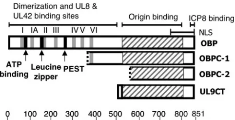
The N-terminal half of OBP exhibits helicase and ATPase activities and contains a leucine zipper as well as binding sites for the proteins specified by UL8, a component of the viral helicase/primase complex, and UL42, the viral DNA polymer-ase accessory factor, both of which are essential for HSV-1 DNA replication (Fig. 1). C-terminal amino acids 824 to 851 of OBP are required for binding to ICP8 (8). The domain re-quired to bind to specific sites in HSV-1 origins of DNA rep-lication has been mapped to the C-terminal amino acids 564 to 818 (1, 15, 37). The nuclear localization signal of OBP is located in the C-terminal 107 amino acids (35). OBP is thought to begin the viral DNA replication process by binding to spe-cific DNA sequences in viral origins and serving as a docking protein to which other viral proteins necessary for viral DNA replication bind.
In 1994, Baradaran et al. identified a novel transcript, des-ignated UL8.5, that is expressed with delayed early (DE) ki-netics and comprises the C terminus of the UL9 transcript, which specifies OBP (4). In vitro transcription/translation of the UL8.5 transcript yielded a protein of approximately the same size (⬃53 kDa) as a protein detected in infected cell
* Corresponding author. Present address: Department of Molecular and Cellular Biology, The University of Arizona, Life Sciences South Building, 1007 E. Lowell St., P.O. Box 210106, Tucson, AZ 85712-0106. Phone: (520) 626-3185. Fax: (520) 621-3709. E-mail: pas@email .arizona.edu.
䌤Published ahead of print on 18 July 2007.
10699
on November 8, 2019 by guest
http://jvi.asm.org/
extracts. The protein was designated OBPC, and it was con-cluded that OBPC was expressed from the UL8.5 transcript. The recent discovery of a second protein product that is in frame with and comprises the C-terminal portion of OBP, however, prompted the designation of OBPC as OBPC-1 and the new protein (⬃35 kDa) as OBPC-2 (32a). Moreover, re-cent evidence indicating that OBPC-1 is a cathepsin B-medi-ated cleavage product of OBP (32a) led to the hypothesis that OBPC-2, rather than OBPC-1, is expressed from the UL8.5 transcript.
A possible mechanism for facilitating the switch from stage I to stage II was suggested by the discovery of the two C-terminal variants of OBP. Proteins comprising the C-C-terminal portion of OBP would be predicted to retain the nuclear lo-calization signal as well as the ICP8 and origin binding capa-bilities of OBP but lack helicase and ATPase activities and possibly the ability to bind UL8 and UL42. Consistent with this prediction, synthetic C-terminal forms of OBP are able to bind to origin DNA (1, 5, 11, 15, 28, 37, 59). Of special interest were studies investigating the function of the C terminus of OBP with the synthetic peptide UL9CT (Fig. 1) (59), which is com-prised of the 10 N-terminal amino acids of OBP fused to the 317 C-terminal amino acids, and an OBPC-1 like protein (5), which likely includes the C-terminal 487 amino acids of OBP. Not surprisingly, these experiments demonstrated that the C terminus of OBP alone is insufficient to complement viral DNA replication or growth of an OBP null virus (5, 28, 45, 60, 61). Importantly, these and other studies have also shown that overexpression of the C terminus of OBP is inhibitory to viral DNA synthesis (and consequently viral replication) and that this inhibition is dependent upon the ability of the C terminus to bind origin DNA (5, 11, 28, 45, 61). Because OBPC-1 and OBPC-2 likely share the origin binding domain of OBP, these observations suggest that OBPC-1 and/or -2 may play a role in regulating stage I origin-dependent DNA replication by
block-ing bindblock-ing of OBP to viral origins, thus facilitatblock-ing the switch to OBP- and origin-independent viral DNA replication.
The present study was designed to distinguish the func-tion(s) of OBPC-2 from the functions of OBPC-1 and OBP using a mutational approach. This objective was complicated by the fact that any mutation introduced into the OBPC-2 regulatory region or open reading frame (ORF) may also affect the functions of overlapping proteins OBPC-1 and OBP. De-spite this complication, we have succeeded in demonstrating that OBPC-2 is the product of the UL8.5 transcript and have identified the translational start site of the protein by site-directed mutagenesis. A mutant virus that expresses only min-imal levels of OBPC-2 while leaving the DNA binding and initiator functions of OBP intact was constructed. Despite the reduced size of its DNA binding domain, OBPC-2 is able to bind site I origin DNA, redefining the limits of the OBP origin binding domain. In infected cells expressing only OBPC-1, OBPC-2, or neither protein, we have demonstrated that OBPC-1 and OBPC-2 act in concert to down-regulate levels of viral DNA synthesis in vitro. In vivo, we have shown that although OBPC-2 is not essential for viral replication in mice or for the establishment of or reactivation from latency in the mouse ocular model, it plays a crucial role in determining the lethality of HSV-1 in this model.
MATERIALS AND METHODS
Cells and viruses.Vero cells (ATCC CCL-81; American Type Culture Col-lection, Rockville, MD) were cultured in Dulbecco’s modified Eagle’s medium (DMEM) supplemented with 5% fetal bovine serum (FBS), and all experiments in Vero cells were performed using DMEM supplemented with 10% FBS. The UL9 complementing cell line, 2B.11 (34), was cultured in DMEM supplemented
with 10% FBS and 300g/ml G418. PC12 cells (ATCC CRL-1721) were cultured
in DMEM supplemented with 5% FBS and 10% horse serum. All media
con-tained penicillin (100 units/ml), streptomycin (100g/ml), and 2 mML
-glu-tamine. All cells were maintained at 37°C in 5% CO2.
The wild-type virus used in these studies was HSV-1 strain KOS and was propagated as previously described (54). The hr94 virus, whose genome contains
a-galactosidase expression cassette inserted at codon 534 of the OBP open
reading frame, was isolated and kindly provided by Sandra Weller (University of Connecticut Health Center, Farmington) (34).
Plasmids. (i) UL8.5 promoter truncation plasmids.To determine whether the UL9 or UL8.5 transcript specifies OBPC-2, the promoter truncation plasmids pSbfI, pBbvCI, and pBamHI were generated by digesting pUL9H with the SbfI and BglII, BbvCI and BglII, or BamHI and BglII restriction enzymes, respec-tively. The SbfI-to-BglII fragment (nucleotides [nt] 22481 to 25147 of the HSV-1 genome) was removed from pUL9H to create pSbfI. This deletion removed the UL9 promoter and transcriptional start site but it retained the putative UL8.5 promoter and transcription start site. The BbvCI-to-BglII deletion (nt 22193 to 25147 of the HSV-1 genome) and BamHI-to-BglII deletion (nt 21653 to 25147 of the HSV-1 genome) produced pBbvCI and pBamHI, respectively. Both plasmids lack both the UL9 and UL8.5 promoters and transcription start sites.
(ii) Nonsense mutations in the OBP ORF.To identify the region of the OBP ORF that contains the OBPC-2 translational start site, plasmids containing nonsense mutations at multiple sites in the OBP ORF were constructed (see Fig. 3C, below). Nonsense mutations were introduced at amino acids 101, 245, 368, 494, and 597 of the OBP ORF (nt 22957 to 22959, 22525 to 22526, 22156, 21778 to 21780, and 21469 to 21471 of the HSV-1 genome) in plasmid pADE (32a), which contains the promoter regions and coding sequences for UL8.5 and UL9, by using the pAlter system of mutagenesis with the following primers: 101,
5⬘-CGG GAA GCG ATC CAC TAACCG GAC ACG AGT-3⬘; 245, 5⬘-GAG
TAC GCC ATG CCC GGG TTT TAAGCG CGC-3⬘; 368, 5⬘-GGC ATG TTC
GCCTAGGTAAAA CCC ATG AAC-3⬘; 494, 5⬘-G ACC CTA AAC TGCTAG
CGC GTG CGC TTC TGG-3⬘; 597, 5⬘-C TGT CTC CGC GTT CCCTAGGCC
ACC CGC AG-3⬘. Each primer was designed to remove a restriction enzyme site
[image:2.585.43.284.67.190.2](italics), BspEI, AscI, SnaBI, MluI, and MscI, respectively, to facilitate screening for mutant clones by restriction digest and analysis by agarose gel electrophore-sis. The presence of the mutation was confirmed by sequencing of the plasmid.
FIG. 1. Diagrams of OBP, OBPC-1, OBPC-2, and UL9CT. The DNA binding domains of the four proteins are represented by the hatched regions. Helicase domains are shown in gray and numbered I to VI. The ATPase domain, leucine zipper, and putative PEST se-quence of OBP are shown in black. The N-terminal half of OBP confers dimerization ability and contains domains that interact with the proteins encoded by the UL8 (member of the helicase/primase complex) and UL42 (the polymerase accessory factor) genes. The nuclear localization signal (NLS) and ICP8 binding domain are located in the C-terminal half of OBP and are present in all four proteins. Hatched lines at the beginning of OBPC-1 and OBPC-2 indicate that at the inception of these studies, the translational start sites were unknown. The scale beneath the figure represents amino acids.
on November 8, 2019 by guest
http://jvi.asm.org/
Individual mutations are shown in bold. Plasmids of interest were digested with DraIII, and the fragment (nt 21078 to 24037 of the HSV-1 genome) was ligated into pUL9H (4) from which the wild-type DraIII fragment had been removed.
(iii) p⌬UL9GFP.To construct the plasmid to generate the mutant virus
⌬UL9GFP, the cassette containing the EGFP-N1 gene driven by the
cytomeg-alovirus immediate-early (IE) promoter was digested from the pEGFP-N1 vector (Clontech, Mountain View, CA) with restriction enzymes BstBI and NcoI. This fragment was used to replace the BstBI-NcoI fragment of pUL9n24 to create
p⌬UL9GFP (see Fig. 3B and E, below). The 5⬘ end of the UL10 transcript
overlaps the 5⬘end of the OBP ORF. In order to preserve UL10 expression, the
EGFP-N1 cassette was inserted in a way that it did not replace the entire OBP
ORF in p⌬UL9GFP. Thus, the pUL9n24 plasmid, which contains a nonsense
mutation (see Fig. 3E, below) 24 amino acids downstream of the OBP
transla-tional start site, was used to construct p⌬UL9GFP to ensure that minimal
N-terminal sequences of OBP would be expressed from the p⌬UL9GFP plasmid.
(iv) Mutation of the OBPC-2 putative translational start site.To identify the OBPC-2 translational start site, each potential translational start site between amino acids 494 and 597 of the OBP ORF that would yield a product of the
expected size (⬃35 kDa) was mutated. The translational start site mutant
plas-mids were created in the same manner as the nonsense mutant plasplas-mids using
the following primers: prM571A, 5⬘-GAA ATT GTC GCG CTCGCG CGC
AAC CTC AAC-3⬘; prM578R, 5⬘-CTC AAC AGC CTG AGGGGA CGC ACG
CGG TTT ATT TAC-3⬘; prM571A:M578R, 5⬘-GTC GCG CTCGCG CGC
AAC CTC AAC AGC CTG AGG GGA CGC ACG-3⬘; prM571A:L577L:
M578A, 5⬘-GTC GCG CTCGCG CGC AAC CTC AAC AGC CTA GCG GGA
CGC ACG-3⬘. Novel restriction enzyme sites either removed (FspI) or
intro-duced (AxyI) using these primers are in italics, and mutations are shown in bold. Mutations were identified by restriction digest and analysis by agarose gel elec-trophoresis. Mutations were confirmed by sequencing. Plasmids containing the correct mutations were digested with DraIII, and the fragment (nt 21078 to 24037 of the HSV-1 genome) was ligated into pUL9H (4) from which the wild-type DraIII fragment had been removed.
Northern blot analysis.Northern blot analysis was performed to determine the levels of the UL8.5 transcript as described previously (2). Briefly, 1-day-old Vero
cells (3⫻106cells) were transfected with the indicated plasmid using
Lipo-fectamine 2000 (Invitrogen, Carlsbad, CA) according to the manufacturer’s protocol. Transfected cells were infected 18 h later with the indicated virus at a multiplicity of infection (MOI) of 10 PFU/cell. Total cell RNA was harvested 6 h later using TRIzol reagent (Invitrogen). RNA was resuspended in diethyl
pyro-carbonate-treated H2O, and RNA concentrations were determined by
spectro-photometry (A260). Sample RNA (7.5g) was denatured in 1⫻MOPS [20 mM
3-N-(morpholino)propane sulfonic acid, 1 mM sodium acetate, 1 mM EDTA]
containing 17.5% formaldehyde and 50% formamide for 10 min at 65°C. RNA
was separated on a 1% agarose gel containing 6.7% formaldehyde and 1⫻
MOPS and transferred to a nylon membrane. RNAs were UV cross-linked to the
membrane (200 mJ/cm2) using a UV Stratalinker 1800 (Stratagene, La Jolla,
CA) and prehybridized for 30 min at 60°C in prehybridization buffer (50%
formamide, 5⫻Denhardt’s solution [Eppendorf, Hamburg, Germany], 6⫻SSPE
[0.9 M sodium chloride, 60 mM sodium phosphate, monobasic, 6 mM EDTA, pH 7.0], and 100 mg/ml of salmon testis DNA). Blots were hybridized in
prehybrid-ization buffer with a32
P-labeled riboprobe synthesized from the linearized
SP68R-3⬘plasmid, which recognizes the UL8, -8.5, -9, and -9.5 transcripts (4).
Membranes were rinsed three times with 2⫻SSC (1⫻SSC is 0.15 M NaCl plus
0.015 M sodium citrate) containing 0.05% sodium dodecyl sulfate (SDS) at room
temperature and then washed three times for 20 min at 85°C with 0.1⫻SSC
containing 0.1% SDS. The hybridized blots were exposed on a PhosphorImager cassette (Molecular Dynamics, Sunnyvale, CA) overnight and the signal analyzed using ImageQuant 3.3 software (Molecular Dynamics).
Whole-cell extracts.Cells were seeded at 3⫻106
cells/dish in a 100-mm dish and transfected 1 day later with the indicated plasmid using Lipofectamine 2000 according to the manufacturer’s protocol. At 18 h posttransfection (p.t.), cells were infected at an MOI of 10 PFU/cell. At 8 h postinfection (p.i.), cells were washed twice with phosphate-buffered saline (PBS) and harvested by scraping
into 5 ml of ice-cold PBS. Cells were pelleted and resuspended in 100l of
ice-cold NET buffer (50 mM Tris pH 7.8, 100 mM NaCl, 1 mM EDTA) supple-mented with aprotinin (Roche, Indianapolis, IN), leupeptin, pepstatin, phenyl-methylsulfonyl fluoride, and dithiothreitol (DTT; Sigma, St. Louis, MO). Cells were snap-frozen in liquid nitrogen and quickly thawed at 37°C. Cells were then sonicated twice for 30 seconds, cellular debris was pelleted, and the supernatant fluid was retained, aliquoted, and snap-frozen in liquid nitrogen.
Nuclear extracts.Nuclear extracts were prepared as previously described from
1⫻106
cells transfected with the plasmid indicated using Lipofectamine 2000 and infected 18 h later with 10 PFU/cell of the indicated virus or mock infected
(27). Cells were harvested at 8 h p.i. unless otherwise noted. Briefly, cells were harvested by scraping into medium, washed twice with ice-cold PBS, and resus-pended in ice-cold resuspension buffer (10 mM Tris-HCl pH 7.5, 10 mM NaCl,
and 5 mM MgCl2) supplemented with 0.5 mM DTT and 1g/ml of the following
protease inhibitors: leupeptin, aprotinin, pepstatin, and phenylmethylsulfonyl fluoride. Cells were then pelleted at 4°C by low-speed centrifugation (5 min at
3,000⫻g) and resuspended in resuspension buffer containing 0.5% NP-40, DTT,
and protease inhibitors. Cells were centrifuged at 4°C (5 min at 3,000⫻g) and
resuspended in ice-cold 20 mM HEPES pH 7.9, 25% glycerol, 0.42 M KCl, and 0.2 mM EDTA containing protease inhibitors and incubated at 4°C for 30 min.
Cell debris was cleared by centrifugation at 13,000⫻gfor 30 min, and the
supernatant fluid was dialyzed at 4°C in 20 mM HEPES pH 7.9, 20% glycerol, 0.1 M KCl, and 0.2 mM EDTA containing protease inhibitors and DTT. Nuclear
extracts were aliquoted, snap-frozen in liquid nitrogen, and stored at⫺80°C.
Western blot analysis.Laemmli buffer was added to 10l of whole-cell extract
or 10g of nuclear extracts, and samples were boiled for 5 min. Proteins were
separated by polyacrylamide gel electrophoresis in 8% acrylamide gels and transferred to polyvinylidene difluoride membranes (Millipore, Billerica, MA) using a Mini Trans-Blot cell (Bio-Rad Laboratories, Hercules, CA). Membranes were blocked in Tris-buffered saline (TBS) with 5% milk overnight. Primary rabbit antibody specific for the C-terminal 20 amino acids of OBP, anti-OBPCT (Bethyl Laboratories, Montgomery, TX), was added to the membrane at a 1:500 dilution in TBS with 0.05% Tween and 5% milk and incubated for 1 h at room temperature. Membranes were washed three times for 20 min with TBS con-taining 0.05% Tween. Goat anti-rabbit secondary antibody conjugated to horse-radish peroxidase (Jackson ImmunoResearch Laboratories, West Grove, PA) was added to the blot at a 1:10,000 dilution in block for 1 h at room temperature. The membrane was washed as described above. Membranes were incubated with Immobilon Western chemiluminescent horseradish peroxidase substrate (Milli-pore, Billerica, MA) according to the manufacturer’s instructions and exposed on CL-X Posure film (Pierce, Rockford, IL).
Mutant virus construction. (i)⌬UL9GFP.The mutant virus⌬UL9GFP was constructed using standard marker transfer techniques (16). Briefly, 2B.11 cells
were transfected with the 3.2-kb DraIII-DraIII fragment of p⌬UL9GFP (nt
21078 to 24037 of the HSV-1 genome [see Fig. 3E, below]) containing the EGFP-N1 cassette and infectious KOS DNA at a molar ratio of 1:50 using Lipofectamine 2000 according to the manufacturer’s protocols. At 4 h p.t., monolayers were overlaid with 2% methylcellulose and plaque formation was monitored. Plaques were screened for green fluorescent protein (GFP) expres-sion using a Nikon Eclipse TE300 fluorescence microscope (Diagnostic Instru-ments, Sterling Heights, MI). Green fluorescent plaques were picked and plaque purified three times on 2B.11 cells. Correct insertion of the fragment in the KOS genome was confirmed by Southern blot analysis (described below) as indicated by the presence of a 1,652-bp PstI fragment and the loss of an 1,174-bp PstI fragment (see Fig. 3G, below). The absence of OBP, OBPC-1, and OBPC-2 expression was verified by Western blot analysis (see Fig. 4B, below).
(ii) M571A.OBPC-2 translational start site mutant viruses were constructed using standard marker transfer techniques (16). Vero cells were transfected with pM571A using Lipofectamine 2000 according to the manufacturer’s protocol. At
18 h p.t., cells were infected with⌬UL9GFP at an MOI of 2.5 PFU/cell.
Mono-layers were overlaid with 2% methylcellulose, and plaque formation was moni-tored. Plaques were screened using a Nikon Eclipse TE300 fluorescence micro-scope, white recombinant plaques were picked, and plaques were purified three times on Vero cell monolayers. Correct insertion of the mutation was verified by Southern blot analysis (described below) confirming the loss of a 723-bp FspI fragment and the gain of a 1,272-bp FspI fragment (see Fig. 3G, below). The absence of OBPC-2 expression was verified by Western blot analysis (see Fig. 4B, below).
(iii) M571AR. A rescuant virus (M571AR) was constructed by standard marker rescue procedures using a wild-type 1-kb EcoNI-DraIII fragment from plasmid pUL9H (nt 22084 to 21078 of the HSV-1 genome) (see Fig. 3F, below) and infectious M571A DNA. Plaques were screened by restriction digest analysis and Southern blot analysis was performed, as described below, to confirm the loss of a 1,272-bp FspI fragment and the gain of a 723-bp FspI fragment (see Fig. 3G). The proper expression of OBP, OBPC-1, and OBPC-2 was verified by Western blot analysis (see Fig. 4B, below).
Southern blot analysis.To establish the genotypes of mutant viruses, Vero or
2B.11 cells (5.5⫻105
) were plated in 35-mm dishes. Twenty-four h later, cells were infected with the individual plaque isolates described above at an MOI of 2.5 PFU/cell. When cytopathic effects were generalized, cells were harvested by
scraping into medium. Cells were pelleted by centrifugation at 2,000⫻gat 4°C,
washed twice with cold PBS, and resuspended in 200l lysis buffer (10 mM
Tris-HCl pH 8.0, 1 mM EDTA pH 8.0, 0.2% SDS, and 400g/l proteinase K).
on November 8, 2019 by guest
http://jvi.asm.org/
The cell lysates were incubated at 55°C for 18 h. DNA was isolated by extracting once with phenol-chloroform-isoamyl alcohol (25:24:1) followed by ethanol
pre-cipitation. DNA was quantified by spectrophotometry (A260), and approximately
100 ng was used for Southern blot analysis. The DNA was digested with PstI (to
screen⌬UL9GFP) or FspI (to screen M571A and M571AR), and DNA
frag-ments were separated by agarose gel electrophoresis and transferred to a nylon membrane (GE Osmonics, Inc., Minnetonka, MN). DNAs were UV cross-linked
(200 mJ/cm2) using a UV Stratalinker 1800, prehybridized for 30 min at 60°C in
ExpressHyb solution, and hybridized in ExpressHyb solution for 30 min at 60°C
using a32P-labeled random-primed probe consisting of the PstI fragment from nt
22481 to 23655 of the HSV-1 genome (to screen for⌬UL9GFP) or the DraIII
fragment from nt 24037 to 21078 of the HSV-1 genome (to screen for M571A and M571AR) (see Fig. 2B and C, below). Membranes were rinsed three times
with 2⫻SSC containing 0.05% SDS at room temperature and then washed three
times for 20 min at 68°C with 0.1⫻SSC containing 0.1% SDS. The hybridized
blots were exposed overnight on a PhosphorImager cassette.
Gel shift analysis.The gel shift analysis procedure has been described
previ-ously (29). The wild-type site I probes used were as follows:⫹strand, 5⬘-GAA
GCG TTC GCA CTT CGT CCC AAT-3⬘;⫺strand, 3⬘-CTT CGC AAG CGT
GAA GCA GGG TTA-5⬘.
Briefly, 5g of whole-cell extract was incubated with 105cpm of32P-labeled
probe (1 ng), 3g of poly(dA-dT) in DNA binding buffer (10% glycerol, 50 mM
HEPES [pH 7.9], 100 mM NaCl, 0.5 mM DTT) in a final volume of 10l for 30
min on ice. Antibody supershifts were performed by adding 5l of antibody
specific to the C terminus of OBP, RH7 (kindly provided by Deborah Parris, Ohio State University, Columbus) (40) to the binding reaction mixture after 5 min of incubation. Protein-DNA complexes were resolved on 6% nondenaturing polyacrylamide gels. Gels were dried and exposed overnight on a PhosphorImager cassette.
Viral DNA synthesis.Vero cells were infected at an MOI of 2.5 PFU/cell with
wild-type or mutant virus in the presence or absence of 1M Ca074Me (Sigma,
St. Louis, MO). Following 1 h of adsorption, cells were washed three times with PBS, and DMEM was added to the cultures. Cells were harvested at 3-h intervals for 18 h p.i. by scraping into the medium. Cells were washed twice with PBS and pelleted. DNA was harvested as described for Southern blot analysis, and 100 to
500 ng (1l) was used for real-time PCR analysis. Real-time PCR was
per-formed with an Mx3005P quantitative PCR system (Stratagene) using SYBR Green PCR master mix (Applied Biosystems, Warrington, United Kingdom) as described previously (64). Briefly, DNA was amplified in duplicate using a probe specific to the thymidine kinase gene for 40 cycles (30 s at 95°C and then 60s at 60°C). The amount of DNA amplified was determined by comparison to a standard curve of infectious KOS DNA and standardized to the amount of DNA (in ng) loaded in the reaction mixture as determined by spectrophotometry
(A260).
Viral growth curves.Vero cells were infected at an MOI of 2.5 PFU/cell with wild-type or mutant virus. Following a 1-h adsorption, cells were washed three times with PBS, and growth medium was added to the cultures. Cells were harvested at 3-h intervals for 18 h p.i. by scraping into the medium. After
scraping, cells were frozen at⫺80°C, thawed, and sonicated, and the cell debris
was pelleted. Infectious virus in the supernatant was quantified by standard plaque assay on Vero cell monolayers.
Inoculation of mice. Five- to six-week-old male CD-1 mice (⬃30 g body weight; Charles River, Wilmington, MA) were handled in accordance with the
Guide for the Care and Use of Laboratory Animals(39). All animal protocols have been previously described (3, 26). Briefly, following anesthetization with xylazine (9 mg/kg of body weight) and ketamine (100 mg/kg), mouse corneas were
scar-ified with a 26-gauge needle, tear film was blotted, and 2⫻105
PFU of virus in
3l of complete DMEM was added to each eye. The actual titers of the viral
inocula were verified by plaque assays on Vero cell monolayers and varied less than twofold from one another.
Measurement of viral titers in tear film, TG, and cerebellum.Measurement of viral titers in tear film, trigeminal ganglia (TG), and cerebellum has been pre-viously described (3). To determine viral titers in tear film, both eyes were swabbed with a single cotton-tipped applicator, which was immediately
trans-ferred to 500l of complete DMEM and frozen at⫺80°C. Samples were thawed
and mixed thoroughly, and virus titers were determined by standard plaque assays on Vero cell monolayers. Viral titers for each cotton tip were then divided by 2 to determine the number of PFU per eye.
To determine viral titers in TG and cerebella, mice were euthanized by CO2
asphyxiation. TG and cerebella were removed, placed in microcentrifuge tubes
with 500l DMEM containing 10% FBS and 100l of 1-mm-diameter glass
beads and frozen at⫺80°C. TG and cerebella were then thawed and
homoge-nized with a Mini-Bead beater (Biospec Products, Bartlesville, OK) and frozen
at⫺80°C. Samples were then thawed, sonicated, and clarified by centrifugation
(2,000⫻gfor 10 min). Viral titers were determined by standard plaque assays on
Vero cell monolayers.
Measurement of viral genome loads in TG by real-time PCR.To quantify the amount of viral DNA present in TG of latently infected mice, total DNA from each TG was harvested at 30 to 35 days p.i. using the QiaAmp DNA mini kit (QIAGEN, Hilden, Germany) as per the manufacturer’s protocol. Real-time PCR was performed as described above for viral DNA synthesis (64).
Measurement of ex vivo reactivation efficiency.Measurement of reactivation efficiency by explant cocultivation in Vero cells has been described previously
(24, 25). Briefly, mice were euthanized by CO2asphyxiation on day 28 to 32
postinoculation, and TG were removed and washed with Hanks’ balanced salt
solution. TG were cut into four equal-sized pieces and cocultivated with 3⫻
104
Vero cells in 1.5 ml of complete DMEM, yielding 1 TG/well of a 24-well plate. Reactivation was monitored by transferring 10% of culture medium to fresh Vero cell monolayers and scoring for cytopathic effects daily for 9 days postplating.
RESULTS
OBPC-2 is expressed from UL8.5.As described above, fol-lowing the identification of OBPC-1 by Baradaran et al. (4), a second protein in frame with and comprising the C terminus of OBP, designated OBPC-2, was identified when Western blot analysis of whole-cell extracts of KOS-infected Vero cells re-vealed three proteins reactive with anti-OBPCT: OBP (⬃95 kDa), OBPC-1 (⬃53 kDa), and OBPC-2 (⬃35 kDa) (32a). All three proteins were expressed by three HSV-1 lab strains and two low-passage clinical isolates, suggesting that expression of OBPC-1 and OBPC-2 is a common feature of all strains of HSV-1 (32a).
In those same studies, we demonstrated that OBPC-1 is not a product of the UL8.5 transcript and that OBPC-2 is not a degradation product of OBP. In contrast, OBPC-2 was ex-pressed with DE kinetics, as is the UL8.5 transcript, rather than with E kinetics, as is the UL9 transcript. These observa-tions suggested that OBPC-2 is likely the product of the DE UL8.5 transcript; however, this possibility was not formally tested in that study (32a). In order to determine whether OBPC-2 is expressed from the UL9, UL8.5, or neither tran-script, the pUL9H plasmid, which contains the UL8 to -10 genes and their promoters (see Fig. 3B, below), was digested with specific restriction enzymes to remove (i) the UL9 pro-moter region as well as the UL9 transcriptional and transla-tional start sites (pSbfI) or (ii) the UL9 and UL8.5 promoter regions and transcriptional start sites as well as the UL9 trans-lational start site (to yield pBbvCI and pBamHI).
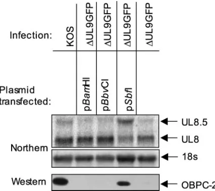
Northern blot analysis of cells transfected with pSbfI, pBbvCI, or pBamHI and superinfected with⌬UL9GFP indi-cated that, as expected, all plasmids expressed the UL8 tran-script but only the wild-type and pSbfI constructs expressed the UL8.5 transcript (Fig. 2). Western blot analysis demonstrated that OBPC-2 was detected only when cells were transfected with the wild-type or pSbfI plasmids and not when transfected with the pBbvCI or pBamHI plasmids (Fig. 2), demonstrating that OBPC-2 is present only when the UL8.5 transcript is detectable and indicating that OBPC-2 is expressed from the UL8.5 transcript.
Identification of the OBPC-2 translational start site.In or-der to determine the location of the OBPC-2 translational start site within the UL8.5 transcript, nonsense mutations were in-troduced at intervals in the OBP ORF in pUL9H (Fig. 3C) at locations that eliminated unique restriction enzyme sites.
on November 8, 2019 by guest
http://jvi.asm.org/
pression of OBPC-2 was detectable by Western blot analysis when stop codons were introduced at amino acids 101, 245, 368, and 494 of the OBP ORF (Fig. 3D). Introduction of a stop codon at amino acid 597, however, eliminated expression of OBPC-2 (Fig. 3D), indicating that OBPC-2 translation is ini-tiated between amino acids 494 and 597 of the OBP ORF. Importantly, the-galactosidase insertion used to generate the UL9 null virus, hr94, was inserted at amino acid 534, possibly upstream of the OBPC-2 translational start site. To eliminate the possibility that hr94 may express low levels of OBPC-2, a new UL9 virus,⌬UL9GFP, was constructed which contained a stop codon at amino acids 24 and 29 of the OBP ORF and in which amino acids 210 to 688 of the OBP ORF were replaced by a GFP expression cassette (Fig. 3E). Western blot analysis confirmed that this virus does not express OBP, OBPC-1, or OBPC-2 (Fig. 4B). This virus was used as the UL9 null virus for the remainder of these studies.
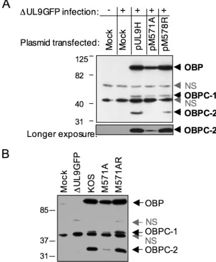
Two in-frame methionine codons that would yield a protein of the expected size are located in the region between amino acids 494 and 597 of the OBP ORF. Mutations were intro-duced into pUL9H to change both methionines to alanine (pM571A) or arginine (pM578R), and the mutant plasmids were tested for expression of OBPC-2. While mutation of the second methionine did not eliminate OBPC-2 expression, mu-tation of the first methionine greatly reduced expression of OBPC-2 (Fig. 4A). These observations indicated that, contrary to transcription/translation assays performed with the UL8.5 transcript (4), translation of OBPC-2 initiates at amino acid 571 of the OBP ORF (nt 21391 to 21393 of the HSV-1 ge-nome) and not at amino acid 365 as previously concluded.
Notably, while OBPC-2 expression was significantly de-creased by the M571A mutation, overexposure of the Western blot revealed that it was not completely eliminated (Fig. 4A).
One possible explanation for this observation is that when the first methionine is mutated, the downstream AUG can be utilized. It is also possible that a cryptic translational start site (CUG), which lies between the first and second methionines, contributes to translation of OBPC-2.
To determine if these putative translational start sites play a role in the translation of OBPC-2 during viral infection and to further characterize the function(s) of OBPC-2, viruses mu-tated in the first methionine codon (M571A), the first and second methionine codons (M571A:M578R), and the first and second methionine codons together with the putative cryptic translational start site (M571A:L577L:M578A) were con-structed by marker transfer with the⌬UL9GFP virus serving as the recipient genome. Western blot analysis demonstrated that, as for the pM571A plasmid, the M571A virus expressed minimal levels of OBPC-2 (Fig. 4B). Moreover, neither muta-tion of the putative second translamuta-tion start site (M571A: M578R) nor mutation of the putative cryptic start site in com-bination with mutation of the second in-frame AUG (M571A: L577L:M578A) further decreased OBPC-2 expression (data not shown), demonstrating that these codons do not play a role in OBPC-2 translation. Since the mutation in the first methi-onine was common to all three viruses, we concluded that this methionine serves as the principal translational start site for OBPC-2.
OBPC-2 binds to origin DNA. Baradaran et al. first pro-posed potential roles for OBPC in viral replication (5). Those authors postulated that (i) OBPC might bind to origins block-ing bindblock-ing of OBP, thereby facilitatblock-ing the switch from stage I to stage II DNA replication, and (ii) OBPC might bind to origins in newly synthesized genomes, thereby blocking initia-tion and facilitating encapsidainitia-tion.
Because OBPC-2 is significantly smaller than OBPC-1 and lacks the first seven N-terminal amino acids of the mapped origin DNA binding domain of OBP (1, 15, 37), we were interested in determining whether OBPC-2 retained origin binding capability. For these tests, whole-cell extracts were prepared from undifferentiated PC12 cells infected with KOS or the M571A mutant virus. Undifferentiated PC12 cell ex-tracts form complexes in a manner similar to Vero cell exex-tracts in gel shift experiments but produce fewer nonspecific com-plexes (28). Extracts were incubated with radiolabeled probe containing site 1 origin DNA sequences in the presence or absence of antibody specific to the C-terminal 10 amino acids of OBP (RH7) (40), and the complexes were resolved on a nondenaturing gel. Two specific complexes, A and C, as de-fined by Isler and Schaffer (28), were readily detected when the site I probe was incubated with KOS-infected cell extracts (Fig. 5). Both complexes were supershifted with RH7 antibody, demonstrating that they contain OBP, OBPC-1, and/or OBPC-2 (Fig. 5). Extracts of M571A-infected cells, however, produced barely detectable levels of complex A (Fig. 5), sug-gesting that low levels of OBPC-2 expression resulted in the inefficient formation of complex A. This observation indicates that OBPC-2 binds to site 1 DNA to produce complex A. The low level of complex A formed with M571A-infected cell ex-tracts is likely due to the small amount of OBPC-2 produced by this virus (Fig. 4C). Notably, complex C was formed as effi-ciently with M571A-infected cell extracts as with wild-type-infected cell extracts (Fig. 5), demonstrating that the
methi-FIG. 2. OBPC-2 is expressed from the UL8.5 transcript. Results shown are from a Northern blot analysis of total cellular RNA isolated from Vero cells transfected with the indicated plasmid and/or infected with the indicated virus and using the 8R3⬘riboprobe (4). The UL8 and UL8.5 transcripts are shown, as is the 18S band. Also shown are Western blot results using anti-OBPCT to detect OBPC-2 in whole-cell extracts of Vero whole-cells transfected with the indicated plasmid and/or infected with the indicated virus.
on November 8, 2019 by guest
http://jvi.asm.org/
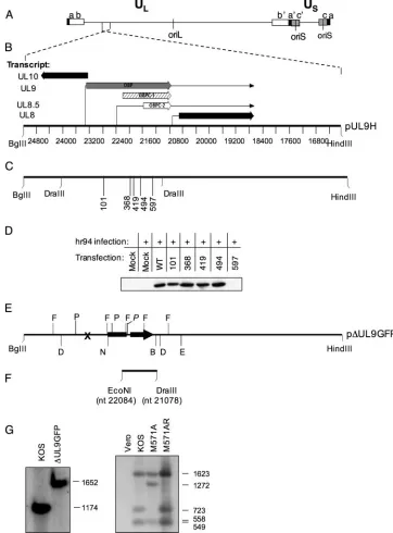
[image:5.585.56.269.69.258.2]FIG. 3. Construction of mutant plasmids and viruses. (A) Diagram of the HSV-1 genome showing the ULregion flanked by inverted repeat
sequences (a b and b⬘a⬘) and the USregion flanked by inverted repeat sequences a⬘c⬘and c a. The locations of oriL and oriS are shown. (B) The
9.5-kb HSV-1 sequences between the BglII and HindIII sites included in the plasmid pUL9H are shown. The directions and locations of the UL8 to -10 transcripts and their directions of transcription are indicated. The scale represents the nucleotides of the HSV-1 genome. (C) The locations of the amino acid codons (101 to 597) into which nonsense mutations were introduced into pUL9H. The DraIII restriction sites (nt 24037 and 21078 in the HSV-1 genome) used to clone the mutant sequences from pADE to pUL9H are indicated. (D) Western blot analysis using anti-OBPCT to detect OBPC-2 in nuclear extracts of Vero cells transfected with plasmids containing the indicated nonsense mutations and infected with hr94 or mock infected. (E) Plasmid p⌬UL9FP. The location of the nonsense mutation (X) in pUL9n24 is shown. The cytomegalovirus IE promoter is represented as a black rectangle, and the EGFP-N1 gene is shown as a black arrow. The DraIII restriction sites (D) (nt 24037 to 21078 in the HSV-1 genome) used to generate the⌬UL9GFP virus and construct the probe for Southern blot analysis are indicated. Locations of the FspI (F) and PstI (P) restriction sites used to screen mutant viruses are also shown. The P site in the GFP cassette is denoted in italics. The NcoI (N) and BstBI (B) restriction enzyme sites used to clone the EGFP-N1 cassettes into pUL9H are shown. The DraIII (D)-EcoRV (E) sites that comprise the DNA fragment in pADE are indicated. (F) The location of the⬃1-kb wild-type EcoNI-DraIII fragment (nt 22084 to 21078 of the HSV-1 genome) used to generate the rescuant virus M571AR is shown. (G) Southern blot analysis of wild-type, mutant, and rescuant viruses. Blots were probed with either a radiolabeled DraIII fragment or radiolabeled PstI fragment. Fragment sizes are indicated to the right of the blot.
on November 8, 2019 by guest
http://jvi.asm.org/
onine-to-alanine mutation did not disrupt the DNA binding capability of OBP and eliminating the possibility that complex A is lost due to the loss of DNA binding ability conferred by the mutation. Consistent with these observations, extracts of cells transfected with a plasmid which expresses only OBPC-2 and not OBP or OBPC-1 and infected with⌬UL9GFP form complex A (data not shown), again demonstrating that OBPC-2 is able to bind site I DNA to form complex A. These observations also demonstrate that amino acids 564 to 571 are not essential for binding of the C terminus of OBP to site I DNA, further narrowing the boundary of the OBP DNA bind-ing domain from amino acids 564 to 818 to amino acids 572 to 818.
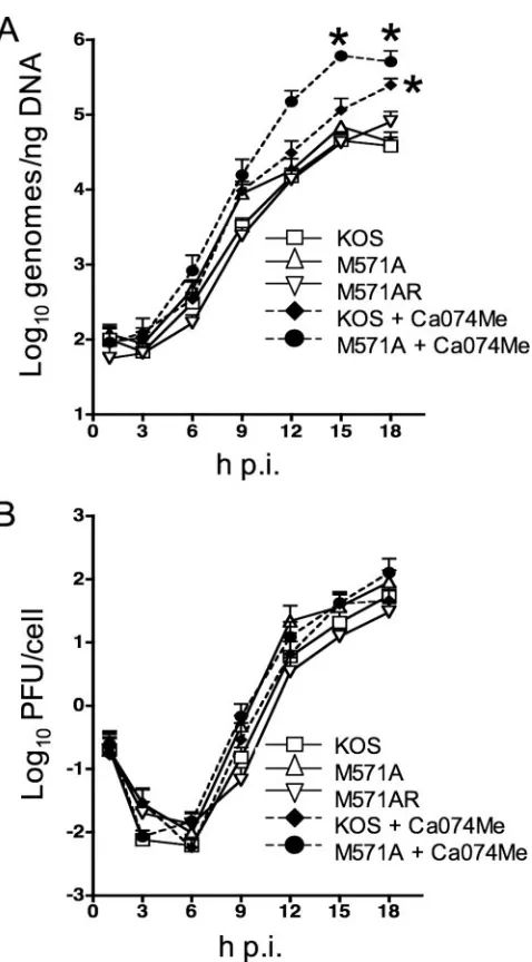
OBPC-1 and OBPC-2 are essential for wild-type levels of viral DNA synthesis in cell culture. To determine whether OBPC-2 is necessary to achieve wild-type levels of viral DNA replication and viral growth in cell culture, levels of viral DNA and infectious virus were monitored in a one-step growth curve. The M571A mutant was similar to wild-type and rescu-ant viruses in both the efficiency of viral DNA synthesis (Fig. 6A) and viral growth (Fig. 6B) in all cell types tested (Vero cells [Fig. 6] and undifferentiated PC12, 293, and ARPE-19 cells [data not shown]), demonstrating that OBPC-2 is not required for viral DNA synthesis or viral replication in a spec-trum of cell types.
Because OBPC-2 and possibly OBPC-1 retain the DNA binding ability of OBP, it is possible that they have similar or
cooperative functions during viral growth. In order to deter-mine if the absence of both OBPC-1 and OBPC-2 has an effect on viral DNA levels or virus replication, cells were infected with either KOS or M571A in the presence or absence of the cathepsin B inhibitor Ca074Me, which blocks cleavage of OBP and therefore inhibits production of OBPC-1 (32a). Amounts of viral DNA synthesized and infectious virus produced were monitored. At 18 h p.i. the levels of viral DNA in KOS-infected cells in the presence of Ca074Me, and therefore in the absence of OBPC-1, were significantly higher (⬃5-fold;P⬍0.05, Bon-ferroni’s posttest) than the amounts of DNA synthesized from cells infected with KOS, M571A, or M571AR in the absence of the inhibitor and therefore in the presence of OBPC-1 (Fig. 6A). Notably, at 15 and 18 h p.i. the amounts of viral DNA in M571A-infected cells in the presence of Ca074Me, and there-fore in the absence of both OBPC-1 and OBPC-2, were sig-nificantly higher than in KOS-infected cells in the presence (⬃5-fold at 15 h p.i. and⬃2-fold at 18 h p.i.;P⬍ 0.001) or absence (⬃14-fold at 15 h p.i. and⬃13-fold at 18 h p.i.;P⬍
0.001) of the inhibitor (Fig. 6A). Collectively, elimination of OBPC-1 expression resulted in increased amounts of viral DNA, while elimination of OBPC-2 expression alone had no effect on the amount of viral DNA produced. Intriguingly, the simultaneous elimination of both OBPC-1 and OBPC-2 had a synergistic effect on the level of viral DNA synthesis and sug-gests that together these proteins down-regulate the level of viral DNA produced in KOS-infected cells.
[image:7.585.53.270.71.335.2]Importantly, cathepsin B likely has multiple targets in the cell whose cleavage may affect viral DNA replication. While it would be advantageous to construct a virus in which the ca-thepsin B cleavage site is mutated, to date, efforts to identify the cathepsin B cleavage site on OBP have been unsuccessful
FIG. 4. Identification of the OBPC-2 translational start site. (A) Western blot analysis to detect OBPC-2 using anti-OBPCT in nuclear extracts of Vero cells transfected with the indicated plasmids and infected with⌬UL9GFP. The locations of OBP, OBPC-1, and OBPC-2 bands as well as nonspecific (NS) bands are indicated. (B) Western blot analysis of nuclear extracts of cells infected with wild-type, mutant, or rescuant viruses.
FIG. 5. OBPC-2 binds site I DNA in viral origins. Gel shift analysis of whole-cell extracts of undifferentiated PC12 cells infected with the indicated viruses or mock infected and incubated with a radiolabeled 24-mer whose sequence spans site I of oriS is shown in the presence or absence of RH7 antibody. Complexes A and C, as defined by Isler and Schaffer (28), are denoted. The presence of a nonspecific (NS) band and the location of the free probe are also indicated.
on November 8, 2019 by guest
http://jvi.asm.org/
[image:7.585.324.515.71.279.2](32a). Extensive evidence exists, however, demonstrating that overexpression of OBP itself is inhibitory to viral DNA syn-thesis (5, 7, 34, 36, 56). We have also determined that addition of the cathepsin B inhibitor does not inhibit replication com-partment formation in infected cells at 8 h p.i. (data not shown). Together, these observations suggest that the
inhibi-tion of cleavage of OBP by cathepsin B is responsible for the increase in viral DNA levels.
The levels of infectious virus did not vary significantly among the three viruses with or without the cathepsin B inhibitor (Fig. 6B), suggesting that the increased levels of viral DNA did not affect the efficiency of virus production, at least in Vero cells, and that OBPC-1 and OBPC-2 function(s) is not essential for viral replication in these cells.
Acute replication of the M571A mutation in mice.To deter-mine whether OBPC-2 plays a role in viral replication in vivo, mice were infected following corneal scarification with 2⫻105
PFU/eye of KOS, M571A, or M571AR (the rescuant virus), and viral replication was monitored in tear film, TG, and cer-ebellum.
To examine virus replication at the site of infection, viral tear film titers were determined daily for 9 days p.i. Mutant virus replicated as efficiently as wild-type and rescuant viruses in the eye throughout the 9-day test period (P⫽0.9890, two-way analysis of variance [ANOVA]), demonstrating that the loss of OBPC-2 expression does not impair the ability of the virus to replicate in the cornea (Fig. 7A).
To examine the ability of M571A to replicate in TG and spread to and replicate in the cerebellum, these tissues were harvested daily for 9 days p.i. and the amount of infectious virus present at these times was determined. The levels of infectious virus present in both the TG and cerebellum were similar (P ⫽ 0.6463 and P ⫽ 0.6025 by two-way ANOVA, respectively) for KOS, M571A, and M571AR at each day postinfection (Fig. 7B and C). These findings show that OBPC-2 is not essential for virus spread from the cornea to the TG and cerebellum or for acute replication at these sites.
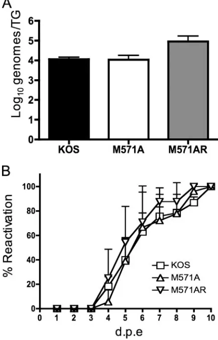
Establishment and reactivation of M571A from latency.We next tested the ability of M571A to establish and reactivate from latency. For this purpose, mice were infected following corneal scarification with 2⫻105PFU/eye. On day 30 p.i. mice
were euthanized and TG were removed and assayed for (i) viral genome loads to determine the efficiency of establishment of latency and (ii) the appearance of infectious virus following explant to determine reactivation kinetics.
The number of genomes detected by real-time PCR in the TG of mice infected with M571A or M571AR did not differ significantly from the number detected in the TG of KOS-infected mice (P⫽0.3077, one-way ANOVA) (Fig. 8A). This finding suggests that the absence of OBPC-2 does not affect the ability of HSV-1 to establish latency.
[image:8.585.45.284.63.495.2]The kinetics and efficiency of reactivation of the mutant virus in explanted TG were measured by explant cocultivation of TG. The mean times to first detection of reactivating virus were as follows: KOS, 4.0 ⫾ 1.0 days postexplant (mean ⫾ standard error), M571A, 4.7 ⫾ 0.6 days postexplant, and M571AR, 3.7⫾0.6 days postexplant, indicating that there was no significant difference among the three viruses (P⫽0.0983, one-way ANOVA) in mean time to reactivation. The final percentage of reactivating TG and the kinetics of reactivation were similar for all three viruses (P ⫽ 0.9993, two-way ANOVA) (Fig. 8B), indicating that efficient expression of OBPC-2 is not necessary for efficient viral reactivation. South-ern blot analysis of M571A DNA prepared from TG induced to reactivate by explant and subjected to restriction enzyme digestion confirmed the absence of the FspI site removed by
FIG. 6. OBPC-1 and OBPC-2 are required to achieve wild-type levels of viral DNA synthesis. Vero cells were infected at an MOI of 2.5 PFU/cell with KOS, M571A, or M571AR in the presence or absence of the cathepsin B inhibitor Ca074Me. Cells were harvested at 1 h p.i. and every 3 h p.i. thereafter for 18 h by scraping into medium. (A) Total cell DNA was isolated, and DNA amounts were measured by real-time PCR analysis using a probe specific for the TK promoter. (B) Cells were frozen, thawed, and sonicated. Cell debris was pelleted, and the titer of infectious virus in the supernatant was determined on Vero cells by standard plaque assay. The results of three experiments are shown as the means ⫾the standard errors. Statistical comparisons were performed using a two-way ANOVA. Asterisks indicate signifi-cant differences between KOS with and without drug and M571A with and without drug.
on November 8, 2019 by guest
http://jvi.asm.org/
the M571A mutation (data not shown), indicating that the results obtained with M571A were generated by the mutant and not a spontaneous revertant virus.
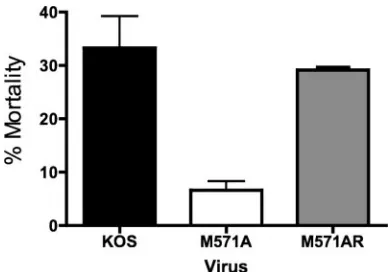
[image:9.585.64.260.66.611.2]OBPC-2 affects the mortality rate of HSV-1-infected mice. The gross pathology of mice infected with wild-type, mutant, or rescuant viruses was similar throughout the 30-day infection period. Mice infected with each virus exhibited moderate to severe symptoms of blepharoconjunctivitis, ulcerative lesions, periocular hair loss, and encephalitis. Many were lethargic, disoriented, or hyperactive beginning on day 7 p.i., with symp-toms resolving by day 14 p.i. By contrast, the mortality rate of
FIG. 7. Viral titers in tear film, TG, and cerebella of mice infected with KOS, M571A, or M571AR. Following corneal scarification, 5- to 6-week-old male CD-1 mice were inoculated in both eyes with 2⫻105
PFU/eye of the indicated virus. (A) Each day for 9 days p.i. (d.p.i.), viral titers in the tear film of eight mice per virus were determined by standard plaque assay on Vero cell monolayers. (B and C) Four mice per virus were euthanized by CO2asphyxiation, and the TG and cerebella were
[image:9.585.311.528.73.412.2]har-vested and processed. Viral titers were determined by standard plaque assay on Vero cell monolayers. The results shown represent the average of three experiments for the tear film titers and two experiments for titers in TG and cerebella. The mean titers⫾ the standard deviations are graphed. Statistical analysis was performed using a two-way ANOVA.
FIG. 8. Establishment and reactivation of KOS, M571A, and M571AR from latency. (A) Genome loads in TG of latently infected mice. Following corneal scarification, four 5- to 6-week-old male CD-1 mice per virus were infected in both eyes with 2⫻105PFU/eye of the
indicated virus. At 30 days p.i., mice were euthanized by CO2
asphyx-iation and TG were removed and processed. Viral DNA was measured by real-time PCR using primers specific for the thymidine kinase gene. The results shown are the averages of three experiments and are presented as the means⫾the standard deviations. Statistical compar-ison was performed using a one-way ANOVA. (B) Reactivation effi-ciencies of KOS, M571A, and M571AR from TG explants. TG from 6 mice per virus (12 TG per virus) were removed on day 30 p.i. Medium (150l) from each cultured TG was removed daily for 9 days postex-plant (d.p.e.) to detect reactivated virus. Specifically, medium samples were transferred to indicator plates of Vero cells, which were moni-tored daily for cytopathic effects. The results shown are the averages of two or three experiments⫾the standard deviations. A statistical com-parison was performed using a two-way ANOVA.
on November 8, 2019 by guest
http://jvi.asm.org/
mice infected with M571A (6.6% ⫾ 2.3%) was significantly lower (P ⫽ 0.02, one-way ANOVA) in these tests than the mortality rates of mice infected with either KOS (33.3% ⫾ 5.9%) or M571AR (29.2% ⫾0.1%) in two independent ex-periments of 24 mice each (i.e.,n ⫽ 48 mice) (Fig. 9). The mortality rates of mice infected with KOS or M571AR were similar to mortality rates observed in other studies with KOS (3). This observation demonstrates that the ability of the virus to express OBPC-2 significantly affects the mortality of mice infected with HSV-1 via the ocular route. The molecular and biological bases for this property of OBPC-2 are currently being examined.
DISCUSSION
Overlapping transcripts are a common feature of the HSV-1 transcriptional program and include transcripts from each ki-netic class. US1.5 is expressed with IE kiki-netics, UL12.5 is expressed with E kinetics, UL8.5 and US8.5 are expressed with DE kinetics, and UL9.5, UL26.5, UL49.5, and US3.5 are ex-pressed with L kinetics (4–6, 9, 10, 23, 28, 33, 38, 47, 48, 67, 68). Each of these overlapping transcripts encodes a protein that comprises the C terminus of and is in frame with the ORF encoded by the larger transcript it overlaps. Because these N-terminally truncated C-terminal proteins share a subset of C-terminal functional domains with their larger counterparts, the possibility that they regulate the levels and/or functions of these larger proteins is intriguing but has been addressed only with UL12.5, UL15.5, and UL26.5. These studies showed that while the smaller proteins share some functions of the larger proteins, only UL26.5 is essential for viral growth in vitro (9, 38, 48, 55, 57, 67). The minimal efforts undertaken to date to elucidate the functions of these overlapping C-terminal pro-teins is due in large part to the challenge of introducing mu-tations that eliminate expression of the truncated protein with-out affecting the expression or function of the full-length protein. The prevalence of these genes, however, suggests that they are functionally important for virus replication.
The UL8.5 transcript encodes OBPC-2.The identification of a second protein (OBPC-2) that comprises a C-terminal portion of OBP, as well as recent evidence that OBPC-1 is a cathepsin B-mediated cleavage product of OBP (32a) led us to examine the molecular origin of OBPC-2. Evidence presented above demon-strates that OBPC-2 and not OBPC-1 is a product of the DE UL8.5 transcript. Interestingly, the first in-frame methionine codon of this transcript (nt 22351 of the HSV-1 genome [4]) does not appear to be utilized for initiation of translation of OBPC-2 in Vero cells. Rather, evidence presented here sug-gests that the seventh in-frame methionine codon (nt 21549 of the HSV-1 genome) is used as the OBPC-2 translation initia-tion site. This is contradictory to previous data from in vitro transcription/translation assays performed with the UL8.5 transcript, which suggested that the first methionine codon was likely used (4). These observations suggest that in systems with complex transcriptional and translational strategies, such as HSV-1, in vitro transcription/translation data should be sup-ported by data from infected cells.
Based on the data presented here and the previously mapped UL8.5 transcriptional start site (4), the 5⬘ -untrans-lated region (5⬘UTR) of the UL8.5 transcript is approximately 800 nucleotides in length. While a 5⬘UTR of this size seems unusual, it is not unique. In the human genome, genes involved in regulatory processes, such as growth factors and growth factor receptors, are often inefficiently transcribed under nor-mal conditions. Genes of this type in the human genome typ-ically have 5⬘UTRs of 100 bp or greater as well as upstream AUGs and an extensive predicted secondary structure (14). DE genes of HSV-1 are not efficiently expressed in the absence of viral DNA replication. Similar to the structure of genes in the human genome, the 5⬘UTR of UL8.5, a DE gene, has many upstream AUGs and a highly stable predicted secondary struc-ture (⌬G⫽ ⫺340 kcal/mol [69]). Many organisms, including viruses (53), use a method termed ribosomal shunting (30), in which the secondary structure of the 5⬘UTR results in the initiation of translation much further down the transcript than would be expected. One or all of these factors may contribute to the use of the downstream translational start site.
Although the translational start site of OBPC-2 has been successfully identified, mutation of this start site did not com-pletely eliminate synthesis of the protein. We hypothesized that this low level of OBPC-2 is initiated from a downstream codon. However, evidence presented here suggests that this is not the case, as mutation of the initiation codon combined with mutation of (i) the AUG immediately downstream of the ini-tiation codon and (ii) a possible cryptic translational start site had no further effect on the translation of OBPC-2. It is also possible that secondary structure transcripts that may initiate low levels of translation from this transcript form in the exten-sive 5⬘UTR of the UL8.5.
[image:10.585.65.259.67.203.2]Revision of the limits of the OBP origin binding domain. The origin binding domain of OBP was mapped previously to amino acids 564 to 818 (1, 15, 37). Data presented in this paper indicate that OBPC-2 translation begins at amino acid 571 of the OBP ORF, well within the previously mapped DNA bind-ing domain, yet it retained the ability to bind HSV-1 origin DNA in gel shift assays. These findings establish the N-termi-nal limit of the DNA binding domain of OBP to amino acid 571 of the OBP ORF. These findings also imply that the
FIG. 9. Mortality rates of mice infected with KOS, M571A, or M571AR. Mortality rates were tabulated during latency experiments. Specifically, the number of mice that died during each experiment was counted. The results represent the average of two experiments, with 24 mice per virus per experiment. The results are shown are the means⫾ the standard deviations. Statistical significance of the differences was determined by a one-way ANOVA and Bonferroni’s multiple compar-isons test.
on November 8, 2019 by guest
http://jvi.asm.org/
M571A mutation did not impair the ability of full-length OBP to bind to site I DNA. The ability of OBPC-2 to bind to origins is consistent with the hypothesis that OBPC-2 may act to reg-ulate OBP activity by binding to origin DNA. However, affinity binding studies will be required to test this possibility.
Roles of OBPC-1 and OBPC-2 in viral DNA replication.As OBPC-1 and OBPC-2 are both able to bind origin DNA, it was of interest to determine if they have similar functions during viral replication. Herein, we have presented evidence to sug-gest that OBPC-2 and cathepsin B cleavage of OBP to yield OBPC-1 act in concert to down-regulate levels of viral DNA synthesis in vitro, as the absence of both proteins resulted in significantly higher levels of viral DNA compared to results in the absence of OBPC-1 alone. It is important to note that while some C-terminal forms of OBP that are similar in size to OBPC-1 are able to bind origins (5), some are not (11). There-fore, it will be important to determine if OBPC-1 is also able to bind to DNA to begin to understand if it may act similarly to OBPC-2 during infection. It is possible that OBP cleavage alone is more important in regulating OBP activity than OBPC-1 itself. It will be necessary to map the cathepsin B cleavage site of OBP to begin to distinguish these possibilities. Notably, in the absence of only OBPC-2, no increase in the amount of viral DNA was observed. It is possible that cleavage of OBP and/or OBPC-1 alone is sufficient to efficiently regulate the levels of viral DNA synthesized. It is also possible, how-ever, that the low levels of OBPC-2 produced by the M571A virus are sufficient, in the presence of OBP cleavage and/or OBPC-1, to properly regulate viral DNA synthesis.
Possible functions of OBPC-1 and OBPC-2.Based on work with temperature-sensitive mutants of OBP (7, 56), Weller et al. have proposed that HSV-1 DNA replication occurs in two stages (65). Stage I viral DNA replication is believed to be origin dependent and, therefore, OBP dependent. Stage II viral DNA replication is believed to be origin independent and, therefore, OBP independent. As noted in the introduction, it has been proposed that the change from OBP-dependent to OBP-independent replication likely involves the removal of OBP from the origin replication complex and/or the loss of the OBP initiator function (11, 36).
OBP function may be regulated in a manner similar to pro-teins involved in DNA replication in bacteriophage systems. For example, gene II of bacteriophage f1 is a site-specific endonuclease required for initiation of DNA replication. Gene X is in frame with and comprises the C terminus of gene II and inhibits phage-specific DNA replication in vivo but is required for phage replication and phage DNA synthesis (19, 20). The A protein of bacteriophageX174 is a site-specific endonuclease required for initiation of DNA replication at the origin. The A* protein is in frame with and comprises the C terminus of A and can inhibit host DNA replication as well as facilitate the switch from semiconservative replicative-form DNA synthesis to plus-strand single-stranded DNA synthesis in vitro (18). The A* protein is not essential for viral replication but increases the replication efficiency of the virus (13).
We propose that OBPC-1 and OBPC-2 are involved in reg-ulating OBP function in a manner similar to the splice variants of Epstein-Barr virus RAZ (21), bovine papillomavirus E2 (12, 31), and adenovirus E1A (32), which inhibit the function of the larger protein they overlap. The C-terminal proteins of HSV-1
appear to substitute for splice variants in HSV-1 in which the splicing mechanism is inhibited at early times in infection. Cleavage of OBP to yield OBPC-1 may play a role in the loss of the OBP initiator function or some other OBP-associated function at viral origins. Additionally, we propose that OBPC-1 and/or OBPC-2 bind to origin sequences and potentially block OBP binding to origin sequences. Because OBPC-1 and OBPC-2 both lack helicase domains as well as the domains necessary to bind most of the essential viral DNA replication proteins, binding of these proteins to origins would likely in-hibit origin-dependent initiation. It is possible that when these proteins are absent in vitro, both dependent and origin-independent DNA replication are permitted to occur concur-rently. As origin-dependent DNA replication is thought to proceed in a theta-like manner, while origin-independent DNA replication is thought to produce long concatemers, it will be of interest to examine the structure of replicating DNA in the absence of both OBPC-1 and OBPC-2.
Function of OBPC-2 in vivo.Although the functions of some C-terminal HSV-1 proteins have been characterized in vitro (4–6, 9, 10, 23, 28, 33, 38, 47, 48, 67, 68), to our knowledge none has yet been characterized in vivo. Therefore, the phenotype of the OBPC-2 null virus, M571A, was investigated in the mouse ocular model of infection. The M571A virus was observed to replicate as efficiently as the wild-type virus in the cornea and TG of infected mice. It was also able to migrate to and repli-cate in the cerebellum as efficiently as the wild-type and res-cuant viruses. These observations demonstrate that OBPC-2 is not essential for wild-type levels of acute replication and spread in infected mice.
Recent evidence has suggested that cathepsin B is active mainly in proliferating cells and is inactive in differentiated neuronal cells (52). Because cathepsin B activity is essential for the expression of OBPC-1 (32a), we hypothesized that, similar to the in vitro studies in which the absence of both OBPC-1 and OBPC-2 led to an increase in viral DNA replication, viral DNA levels may be higher in TG of M571A-infected mice than wild-type-infected mice, as OBPC-1 levels may be lower in TG. No differences were observed, however, in viral genome loads in TG at 30 days p.i., indicating that the ability of the mutant virus to establish latency was not affected by the loss of OBPC-2. This demonstrates that OBPC-2 is not essential to achieve wild-type levels of viral DNA in TG. We propose three possible explanations for this observation. (i) OBPC-1 is ex-pressed in TG and can compensate for the loss of OBPC-2. (ii) The low levels of OBPC-2 produced by the M571A virus are sufficient to repress viral DNA replication in vivo. (iii) There is a threshold level of viral genomes able to establish latency in mouse TG, and the amount of viral DNA produced by the M571A virus exceeds this level. This last possibility is sup-ported by previous studies using replication-impaired viruses or different amounts of viral inoculum in which a threshold level of viral genomes was established in mice TG regardless of increasing viral titers (3, 26, 63).
The M571A virus was also able to reactivate from latency as efficiently as the wild-type virus. This indicates that OBPC-2 is not essential for efficient reactivation from latency. Because OBPC-1 and OBPC-2 may play partially redundant roles dur-ing viral replication, it will be interestdur-ing to study the