Open Access
Short Report
Ocular changes in primary hypothyroidism
Banu T Ozturk*
1, Hurkan Kerimoglu
1, Oguz Dikbas
2, Hamiyet Pekel
1and
Mustafa S Gonen
3Address: 1Department of Ophthalmology, Meram Faculty of Medicine, Selcuk University, Konya, Turkey, 2Department of Endocrinology and
Metabolism Diseases, Sakarya Education and Research Hospital of Ministry of Health, Sakarya, Turkey and 3Department of Endocrinology and
Metabolism Diseases, Meram Faculty of Medicine, Selcuk University, Konya, Turkey
Email: Banu T Ozturk* - [email protected]; Hurkan Kerimoglu - [email protected]; Oguz Dikbas - [email protected]; Hamiyet Pekel - [email protected]; Mustafa S Gonen - [email protected]
* Corresponding author
Abstract
Background: To determine the ocular changes related to hypothyrodism in newly diagnosed patients without orbitopathy.
Findings: Thirty-three patients diagnosed to have primary overt hypothyroidism were enrolled in the study. All subjects were assigned to underwent central corneal thickness (CCT), anterior chamber volume, depth and angle measurements with the Scheimpflug camera (Pentacam, Oculus) and cup to disc ratio (C/D), mean retinal thickness and mean retinal nerve fiber layer (RNFL) thickness measurements with optical coherence tomography (OCT) in addition to ophthalmological examination preceeding the replacement therapy and at the 1st, 3rd and 6th
months of treatment.
The mean age of the patients included in the study were 40.58 ± 1.32 years. The thyroid hormone levels return to normal levels in all patients during the follow-up period, however the mean intraocular pressure (IOP) revealed no significant change. The mean CCT was 538.05 ± 3.85 μ initially and demonstrated no statistically significant change as the anterior chamber volume, depth and angle measurements did. The mean C/D ratio was 0.29 ± 0.03 and the mean retinal thickness was 255.83 ± 19.49 μ initially and the treatment did not give rise to any significant change. The mean RNFL thickness was also stable during the control visits, so no statistically significant change was encountered.
Conclusions: Neither hypothyroidism, nor its replacement therapy gave rise to any change of IOP, CCT, anterior chamber parameters, RNFL, retinal thickness and C/D ratio.
Introduction
The eye is a unique sensory organ that is prone to the effects of various systemic disorders. Hypothyroidism is one of these, presenting frequently with chemosis, perior-bital edema and blepharoptosis. These changes, described as orbitopathy are attributed to the accumulation of
hydrophilic mucopolysaccharides in the ground sub-stance of dermis and other tissues leading to thickening which is called myxoedema [1].
Another proposed, ocular finding of hypothyroidism is the intraocular pressure (IOP) rise. There has been a
Published: 29 December 2009
BMC Research Notes 2009, 2:266 doi:10.1186/1756-0500-2-266
Received: 14 September 2009 Accepted: 29 December 2009
This article is available from: http://www.biomedcentral.com/1756-0500/2/266
© 2009 Ozturk et al; licensee BioMed Central Ltd.
number of reports regarding the higher prevalance of pri-mary open angle glaucoma (POAG) among hypothyroid individuals, however contoversy stil exists as some others have failed to demonstrate this [2-5]. In a study of Smith et al [2] 23.4% of POAG patients had hypothyroidism. As they noted decrease of IOP with treatment of hypothy-roidism in a patient with POAG, IOP rise was attributed to the reduction in facility of outflow in the hypothyroid state [6,7]. Another study of Centanni et al [8] demon-strated a reversible increase of IOP even in subclinical hypothyroidism which raised the question about whether some microscopic findings precede the macroscopic signs of hypothyroidism. The study of Bahçeci et al [9] sup-ported this further by demonstrating a reversible increase of the central corneal thickness (CCT) which lead to a reversible IOP rise and a decreased CCT correlated with the decrease of thyroid-stimulating hormone (TSH).
In an attempt to elucidate the precise effect of hypothy-roidism on ocular structures including cornea, anterior chamber, lens and retina, we conducted a study on newly diagnosed, overt hypothyroid patients without orbitopa-thy comparing the pre- and postreplacement findings.
Materials and methods
This prospective, single-center, clinical study was con-ducted at the department of Ophthalmology, Selcuk Uni-versity, Meram Faculty of Medicine with the collaboration of the department of Endocrinology. Sixtysix eyes of 33 patients who were diagnosed to have acquired primary hypothyroidism in the outpatient clinic of the Endo-crinology department were enrolled in the study. The ini-tial ophthalmological examination was conducted preceding the reatment of hypothyroidism to exclude any history or finding of an ocular disease and for the approval of the informed consent. A complete ophthal-mic examination including visual acuity, IOP with Gold-mann applanation tonometer, anterior segment and fundus examination was performed and patients with any sign of orbitopathy, corneal pathology, glaucoma and ret-inal vascular disease were excluded from the study. Eligi-ble patients were assigned to undergo CCT, anterior chamber volume, depth and angle with the Scheimpflug camera (Pentacam, Oculus) in addition to cup to disc ratio (C/D), retinal thickness and retinal nerve fiber layer (RNFL) thickness measurements with optical coherence tomography [(OCT), (Stratus OCT-3) Carl Zeiss Meditec, Inc., CA]. After pupillary dilatation, fast optic disc and fast RNFL scans were performed by OCT. C/D ratios were recorded after optic disc analysis was carried out. The ret-ina thickness in the superior, nasal, inferior and temporal quadrants, calculated automatically by OCT device were recorded and average RNFL thickness values are obtained from the retinal nerve fiber analysis of OCT.
Control visits were scheduled for the first, 3rd and 6th months after initiation of the medical therapy for hypothyrodism and included the same ophthalmic exam-ination procedures performed initially together with Pen-tacam and OCT measurements following control examination in the outpatient clinic of the Endocrinology department and thyroid hormone level measurements including thyroid-stimulating hormone (TSH), free T3 and free T4 in the clinic of endocrinology. To avoid the effect of diurnal and personel changes, both the inital examination and control visits were performed at the same time in the morning and by the same doctor (BTO).
The data was analyzed by using the Statistical Package for Social Science (SPSS) programme (Worldwide Headquar-ters SPSS Inc. 15.0 Windows package program). According to the normality tests, parameters showing a normal dis-tribution were analyzed using the repeated measures test and the remaining parameters showing an abnormal dis-tribution were analyzed using the nonparametric k related sample test. Additionally the correlation of the TSH change with all of the study parameters were analyzed by Pearson correlation test and Spearman's correlation test depending on the distribution type of the data. As TSH has been demonstrated to be an excellent screening test for hypothyroidism correlation with thyroid hormones has not been analyzed [1]. A p value of < 0.05 was consid-ered as statistically significant.
Results
Sixty-six eyes of 32 female patients and 1 male patient who was diagnosed to have primary clinical hypothy-roidism and completed the sixth month follow-up pro-gram were enrolled in the study. The mean age was 40.58 ± 1.32 years ranging between 19-61 years. The initial mean TSH level of 16.02 ± 2.38 μIU/ml decreased to 3.36 ± 0.54 μIU/ml at the end of the sixth months, while mean free T3 level increased from 2.21 ± 0.19 pg/ml to 4.05 ± 0.07 pg/ml and mean free T4 level achieved a normal level
of 1.06 ± 0.06 ng/dl by ascending from the initial level of 0.57 ± 0.02 ng/dl (Table 1). Hypothyroidism was related to Hashimoto thyroiditis in 15 out of 33 patients.
The mean CCT obtained via Pentacam Scheimpflug cam-era was 538.05 ± 31.29 μ before the treatment. It demon-strated a slight decrease at the end of the first month of therapy, followed by a slight increase at the 3rd month
control and remained quite stabile at the 6th month
con-trol at a level of 537.64 ± 33,37 μ. Statistical analysis revealed no significant change of the CCT (p = 0.82). The mean anterior chamber depth changed from the initial level of 2.90 ± 0.38 mm to 2.89 ± 0.38 mm at the end of the first month, 2.89 ± 0.38 mm at the third month con-trol and 2.87 ± 0.30 mm at the sixth month concon-trol. This gradual decrease was also found to be statistically insignif-icant (p = 0.31). In contrast, both the mean volume of anterior chamber which was 159.67 ± 43.91 mm3 at the
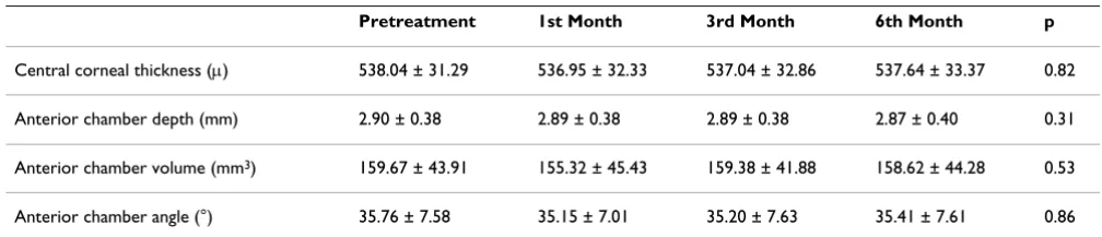
beginning and the mean anterior chamber angle meas-ured as 35.76 ± 7.58° at the initial visit demonstrated var-iation. However the statistical analysis revealed no significant change for both parameters (p = 0,53, p = 0.86, respectively) (Table 2).
The correlation between the change of the TSH level and the change of the CCT, anterior chamber depth, volume and angle was evaluated by comparing the initial and the final (6th month) measurements however no significant
correlation could be estimated (p = 0.43, p = 0.31, p = 0.40, p = 0.49 respectively).
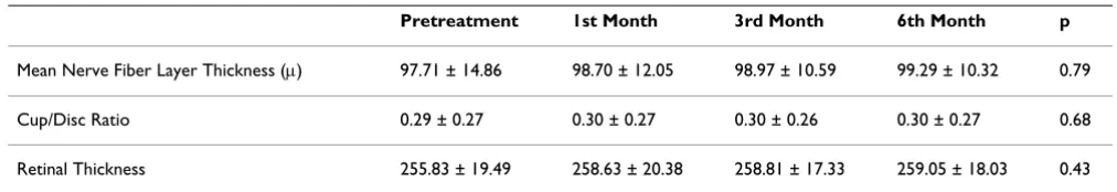
The mean retinal thickness, mean RNFL thickness and the mean C/D ratio was 255.83 ± 19.49 μ, 97.71 ± 1.83 μ and 0.29 ± 0.03 respectively at the beginning of the study and no significant change could be detected after treatment according to the statistical analysis (p = 0.79, p = 0.68, p = 0.43, respectively) (Table 3). The correlation of retinal
thickness, RNFL thickness and C/D ratio measurement with the decrease of TSH was also insignificant (p = 0.14, p = 0.13, p = 0.39)
Discussion
Thyroid hormone plays a pivotal role in the neural devel-opment of the eye especially for normal develdevel-opment of retina and attainment of color vision. It regulates intrinsic mechanisms for controlling retinal cytoarchitecture and layering [10]. As Gamborino et al. [11] demonstrated in a rat model, the photoreceptor and ganglion cell layer thick-ness displayed significantly lower values in congenital-neonatal hypothyroidism. In contrast acquired deficiency of thyroid hormones is reported to mainly affect the IOP beside findings of orbitopathy like periorbital edema and chemosis related to myxedema [1].
The first reports regarding the IOP increase in hypothy-roidism are dating back to 1897 and has been ascribed to hypothalamic disturbance either directly or via the pitui-tary gland acting on the thyroid and the eye at the same time and glaucoma has been associated at times with thy-rotoxicosis and at times with myxoedema. Another specu-lation of Cheng & Perkins is the genetic predisposition to both conditions [12]. In 1965, McLenachan and Davies postulated that the deposition of glycosaminoglycan in trabecular meshwork might lead to a decrease of aqueus humor outflow [13], however it has not been demon-strated so far. Later Becker et al [14] raised the question whether hypothyroidism induce myxedema of the trabec-ular meshwork and Smith et al [15] proposed vasculopa-thy altering ocular bloodflow as the mechanism of IOP increase in hypothyroidism. The glycosaminoglycan
dep-Table 1: Pre- and posttreatment thyroid hormone levels
Pretreatment 1st Month 3rd Month 6th Month Normal range
TSH (μIU/ml) 16.02 ± 2.38 6.63 ± 0.83 4.14 ± 0.70 3.36 ± 0.54 0.4-4.0
Free T3 (pg/ml) 2.21 ± 0.19 3.45 ± 0.14 3.85 ± 0.19 4.05 ± 0.07 1.57-4.71
[image:3.612.52.556.100.187.2]Free T4 (ng/dl) 0.57 ± 0.02 0.75 ± 0.03 1.02 ± 0.07 1.06 ± 0.06 0.8-1.90
Table 2: Change of the follow-up parameters measured with Pentacam Scheimpflug camera at control visits
Pretreatment 1st Month 3rd Month 6th Month p
Central corneal thickness (μ) 538.04 ± 31.29 536.95 ± 32.33 537.04 ± 32.86 537.64 ± 33.37 0.82
Anterior chamber depth (mm) 2.90 ± 0.38 2.89 ± 0.38 2.89 ± 0.38 2.87 ± 0.40 0.31
Anterior chamber volume (mm3) 159.67 ± 43.91 155.32 ± 45.43 159.38 ± 41.88 158.62 ± 44.28 0.53
[image:3.612.52.556.621.729.2]osition in trabecular meshwork seems to gain most atten-tion among these, though it could not been demonstrated histopathologically so far.
The literature contains a number of conflicting reports [2-4,15] that determine the incidence of hypothyroidism among POAG patients to decide whether screening of thy-roid hormones are necessary for open-angle glaucoma patients or not. Smith et al [2] and Girkin et al [4] found a high incidence of hypothyroidism among subjects with POAG, whereas Gillow [15] and Munoz-Negrete [3] failed to show any evidence of a clinically important association between hypothyroidism and glaucoma as Cheng and Perkins [13].
Other type of studies designed to estimate the prevalance of open-angle glaucoma in acquired hypothyroidism patients found no relationship between these disorders, except Tahat et al [16] who reported a positive relation-ship, though only 3 of 60 patients was diagnosed to have glaucoma. Karadimas et al [5] examined 100 hypothyroid patients and none of them had glaucoma. In our study, we found ocular hypertension in only one patient. As it is to our knowledge glaucoma incidence increases over 40 years [17]. Studies evaluating the incidence of hypothyro-dism among glaucoma patients have usually older age ranges, in contrast studies designed to determine the inci-dence of glaucoma among hypothyroid patients have younger subjects which may explain the lower glaucoma incidence found in this type of studies including ours.
As estimating only the prevalance of glaucoma may cause to overlook the true effect of hypothyroidism on IOP because changes under 21 mmHg would not be taken into account; we investigated the correlation of IOP with the change of TSH and found no decrease of IOP parallel to the decrease of TSH with treatment. Bahçeci et al [9] found significant decrease of IOP with treatment, however it was also not correlated with the changes in the thyroid hormone levels. They also reported a significant decrease of CCT after replacement therapy and stated that these reversible changes may be related to mucopolysaccharide deposition in corneal stroma as corneal thickness decreased following the replacement therapy, however the mean CCT revealed no significant change in our study.
Both studies included newly detected hypothyroid patients however the period of hypothyroidism is unknown and the method for corneal thickness measure-ments are different. Bahceci et al used ultrasonic pachym-etry while we used the Scheimpflug camera. These may be an explanation of the conflicting results.
In our study we aimed to sort out the effect of hypothy-roidism on ocular tissues including cornea, anterior chamber, lens and retina in search for an answer to these contradictory results and evaluated the pre- and posttreat-ment measureposttreat-ments of the anterior chamber depth, vol-ume, angle of hypothyroid patients. The initial measurements were not significantly different from the ones at the 1st, 3rd and 6th months of replacement ther-apy. This finding may be explained by the lack of orbitop-athy and unknown duration of the disease. However as our study is the first evaluating these parameters, further studies comparing these findings with that of the hypothyroid patients with orbitopathy are necessary.
The changes in RNFL thickness, C/D ratio and retinal thickness were also evaluated in this study and found steady in all measurements during the follow-up period. In the literature RNFL thickness of hypothyroid patients was obtained just in the study of Bahçeci et al [9] and they also found no change in RNFL thickness between pretreat-ment and postreplacepretreat-ment measurepretreat-ments.
Conclusions
Regarding these findings together, this survey suggest that a direct influence of thyroid hormones on ocular struc-tures is unlikely. Though the unknown period of the hypothyroid state, the younger mean age and lack of orbi-topathy might be responsible for the conflicting results, it may also support the speculation about the predisposi-tion of individuals with inclinapredisposi-tion to auto-immunity to several diseases including hypothyroidism and glaucoma [18]. Further studies evaluating hypothyroid patients according to etiology (Hashimoto disease, Graves dis-ease), period of the disease and presence of orbitopathy seperately may enlighten this dilemma.
Competing interests
[image:4.612.51.557.99.186.2]The authors declare that they have no competing interests.
Table 3: Change of the follow-up parameters measured with OCT at control visits
Pretreatment 1st Month 3rd Month 6th Month p
Mean Nerve Fiber Layer Thickness (μ) 97.71 ± 14.86 98.70 ± 12.05 98.97 ± 10.59 99.29 ± 10.32 0.79
Cup/Disc Ratio 0.29 ± 0.27 0.30 ± 0.27 0.30 ± 0.26 0.30 ± 0.27 0.68
Publish with BioMed Central and every scientist can read your work free of charge
"BioMed Central will be the most significant development for disseminating the results of biomedical researc h in our lifetime."
Sir Paul Nurse, Cancer Research UK
Your research papers will be:
available free of charge to the entire biomedical community
peer reviewed and published immediately upon acceptance
cited in PubMed and archived on PubMed Central
yours — you keep the copyright
Submit your manuscript here:
http://www.biomedcentral.com/info/publishing_adv.asp
BioMedcentral
Authors' contributions
BTO carried out the ophthalmological examinations and drafted the manuscript. HK participated in its design and coordination. OD carried out the endocrinological exam-inations. SG participated in its design and coordination. HP revised the manuscript for publication. All authors read and approved the final manuscript.
Acknowledgements
We gratefully thank to Dr. Banu Bozkurt for her kind help in statistical anal-ysis. We would also like to thank all participants enrolled in the present study.
References
1. Wartofsky L: Diseases of the thyroid. In Harrison's Principles of Internal MedicineVolume Chapter 331. 14th edition. Edited by: Fauci SA, Braunwald E. Philadelphia: The McGraw Hill Companies; 1998:2012-35.
2. Smith KD, Arthurs BP, Saheb N: An association between hypothyroidism and primary open-angle glaucoma. Ophthal-mology 1993, 100:1580-84.
3. Munoz-Negrete FJ, Rebodella G, Almodovar F, Diaz B, Varela C:
Hypothyroidism and primary open-angle glaucoma. Ophthal-mologica 2000, 214:347-9.
4. Girkin CA, McGwin G Jr, McNeal SF, Lee PP, Owsley C: Hypothy-roidism and the development of open-angle glaucoma in a male population. Ophthalmology 2004, 111:1649-52.
5. Karadimas P, Bouzas EA, Topouzis F, Koutras DA, Mastorakos G:
Hypothyrodisim and glaucoma. A study of 100 hypothyroid patients. Am J Ophthalmol 2001, 131:126-128.
6. Smith KD, Tevaarwerk GJ, Alen LH: Reversal of poorly controlled glaucoma on diagnosis and treatment of hypothyroidism.
Can JOphthalmol 1992, 27:345-47.
7. Smith KD, Tevaarwerk GJ, Alen LH: An ocular dynamic study suporting the hypothesis that hypothyroidism is a treatable cause of secondary open-angle glaucoma. Can J Ophthalmol
1992, 27:341-44.
8. Centanni M, Cesareo R, Verallo O, et al.: Reversible increase of intraocular pressure in subclinical hypothyroid patients. Eur J Endocrinol 1997, 136:595-8.
9. Bahçeci UA, Özdek Ş, Pehlivanlı Z, Yetkin İ, Önol M: Changes in intraocular pressure and corneal and retinal nerve fiber layer thicknesses in hypothyroidism. Eur J Ophthalmol
15:556-61.
10. Pinazo-Durán MD, Iborra FJ, Pons S, Sevilla-Romero E, Gallego-Pinazo R, Muňoz A: Postnatal thyroid hormone supplementa-tion rescues developmental abnormalities induced by con-genital-neonatal hypothyroidism in the rat retina. Ophthalmic Res 2005, 37:225-34.
11. Gamborino MJ, Sevilla-Romero E, Muňoz A, Hernández Yago J, Renau-Piqueras J, Pinazo-Durán MD: Role of thyroid hormone in craniofacial and eye development using a rat model. Opthal-mic Res 2001, 33:283-91.
12. Cheng H, Perkins ES: Thyroid disease and glaucoma. Br J Oph-thalmol 1967, 51:547-553.
13. McLenachan J, Davies DM: Glaucoma and the thyroid. Br J Oph-thalmol 1965, 49:441-448.
14. Becker B, Holker AE, Ballin N: Thyroid function and glaucoma.
Am J Ophthalmol 1966, 61:997-999.
15. Gillow JT, Shah P, O'Neill EC: Primary open angle glaucoma and hypothyroidism: chance or true association? Eye 1997, 11(Pt 1):113-114.
16. Tahat AA, al-Khawaldeh AM: Hypothyroidism and open-angle glaucoma: an accidental or an essential coexistence. EastMed-iterr Health J 2000, 6:299-303.
17. Coleman AL, Miglior S: Risc factors for glaucoma onset and pro-gression. Surv Ophthalmol 2008, 53(Supll 1):3-10.
18. Cartwright MJ, Grajewski AL, Friesberg ML, et al.: Immune-related disease and normal-tension glaucoma: a case-control study.