0022-538X/95/$04.0010
Copyrightq1995, American Society for Microbiology
Lack of Detection of Negative-Strand Hepatitis C Virus RNA in
Peripheral Blood Mononuclear Cells and Other Extrahepatic
Tissues by the Highly Strand-Specific rTth Reverse
Transcriptase PCR
ROBERT E. LANFORD,1* DEBORAH CHAVEZ,1FRANCIS V. CHISARI,2ANDCAMILLE SUREAU1†
Department of Virology and Immunology, Southwest Foundation for Biomedical Research, San Antonio, Texas 78228,1
and Department of Molecular and Experimental Medicine, The Scripps Research Institute, La Jolla, California 920372
Received 26 May 1995/Accepted 30 August 1995
To further explore the controversial potential for extrahepatic replication of hepatitis C virus (HCV), the highly strand-specific rTth method of reverse transcriptase PCR was used to examine sera, liver, peripheral blood mononuclear cells, and other extrahepatic tissues from HCV-infected chimpanzees and humans. Posi-tive-strand HCV RNA was present in the liver at approximately 10-fold-higher levels than negaPosi-tive-strand HCV RNA. No negative-strand RNA was detected in peripheral blood mononuclear cells or other extrahepatic tissues despite the presence of abundant positive-strand RNA. These data demonstrate that within the limits of sensitivity of this highly strand-specific reverse transcriptase PCR method, no extrahepatic replication of HCV was detected.
The replication of hepatitis C virus (HCV) in extrahepatic tissues, especially peripheral blood mononuclear cells (PBMC), is highly controversial, and yet whether extrahepatic replication occurs is a critical question with regard to transmission and pathogenesis. One of the problems of determining the tropism of HCV for other tissues is the very low levels of virus in infected individuals. HCV can be routinely detected only by reverse transcriptase PCR (RT-PCR). The presence of HCV RNA is not sufficient to demonstrate replication of HCV in other tissues, since viral RNA may be present because of con-tamination with plasma and/or adherence of circulating virus. Methods for the detection of negative-strand HCV RNA are controversial. Several reports indicate that the standard method for detection of negative-strand HCV RNA lacks suf-ficient strand specificity to answer biological questions (5, 8, 9, 14). The lack of strand specificity is due to false priming of the incorrect strand during the cDNA step. With the high level of amplification provided by PCR, detection of the incorrect strand is not easily avoided.
We have recently described two methods to circumvent the problem of false priming and have used the methods to dem-onstrate the appearance of positive- and negative-strand HCV RNA following in vitro infection of primary chimpanzee hepa-tocytes (8). The rTth method of RT-PCR provided 10,000- to 100,000-fold differentials between the detection of the correct and incorrect strands in assays for HCV positive- and negative-strand synthetic RNA. To further explore the possibility of extrahepatic replication of HCV, we have examined serum, liver, PBMC, and other extrahepatic tissues from
HCV-in-fected chimpanzees and humans by using rTth RT-PCR for negative- and positive-strand HCV RNA.
Strand-specific RT-PCR.In our earlier studies, tagged
RT-PCR was used for detection of negative-strand RNA and rTth RT-PCR was used for detection of positive-strand RNA (8). In this study, the rTth method was used for the detection of both strands, because it is easier to perform and yields better strand specificity than tagged RT-PCR. With rTth RT-PCR, false priming of the incorrect strand is avoided by conducting cDNA
synthesis at 708C with the thermostable rTth RT. Strand
spec-ificity requires that no RT activity remain after cDNA synthesis when both primers are added for PCR amplification. In the
rTth method, this was accomplished by chelating Mn21and
adding Mg21, which results in DNA polymerase activity
with-out significant RT activity.
To demonstrate the sensitivity and specificity of the assays, synthetic positive- and negative-strand HCV RNAs (nucleo-tides 1 to 582) were transcribed in vitro and purified exten-sively to remove DNA. RNA was diluted in normal cell RNA
such that all samples contained 1mg of cell RNA. RT-PCR was
performed by a modification of the previously described rTth (8) procedure for both positive- and negative-strand RNA.
RNA in 10ml was layered with mineral oil and heated to 958C
for 1 min. The temperature was lowered to 708C, and 10ml of
preheated cDNA reaction mixture was added. The reaction mixture consisted of 10 mM Tris (pH 8.3), 90 mM KCl, 1 mM
MnCl2, 200mM each deoxynucleoside triphosphate, 50 ng of
cDNA primer, and 5 U of rTth (Perkin-Elmer Cetus). The
temperature was dropped to 608C for 2 min for annealing and
then raised to 708C for 15 min for the cDNA reaction. The
tem-perature was held at 708C until 40ml of a prewarmed buffer
con-taining ethylene glycol-bis (b-aminoethyl ether)-N,N,N9,N9
-tetraacetic acid (EGTA) (20 mM Tris [pH 8.3], 200 mM KCl, 1.5 mM EGTA, 0.1% Tween 20, and 10% glycerol) was added
to chelate the Mn21 and inactivate the RT activity of rTth.
Reaction tubes were held at 708C while 40ml of prewarmed
PCR mixture (50 ng of forward PCR primer in 3.75 mM
MgCl2) was added. The PCR conditions consisted of an initial
* Corresponding author. Mailing address: Department of Virology and Immunology, Southwest Foundation for Biomedical Research, 7620 N. W. Loop 410, San Antonio, TX 78228. Phone: (210) 674-1410. Fax: (210) 670-3329. Electronic mail address: [email protected].
† Present address: Institut De Biologie, URA Centre National de la Recherche Scientifique, 34060 Montpellier Cedex, France.
8079
on November 9, 2019 by guest
http://jvi.asm.org/
cycle at 948C for 3.5 min; 35 to 45 cycles at 948C for 1.3 min,
608C for 2 min, and 728C for 3 min; and a final extension
cy-cle at 728C for 7 min. For positive-strand RNA, the cDNA
reverse primer was 59-TCGCGACCCAACACTACTC-39and
the forward primer was 59-GGGGGCGACACTCCACCA-39.
The same primers were used in reverse order for detection of negative-strand RNA. The amplified product spans
nucleo-tides 15 to 274 of the 59noncoding region of HCV. One-fifth
of the first-round product was analyzed by agarose gel
electro-phoresis and Southern hybridization with a32P-labeled probe
internal to the PCR primers.
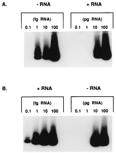
Analysis of highly purified synthetic HCV RNA with the rTth RT-PCR assays for positive- and negative-strand RNA revealed 10,000- to 100,000-fold differentials between detec-tion of the correct and incorrect strands of RNA. In the ex-periment whose results are depicted in Fig. 1, the negative-strand assay detected negative-negative-strand RNA at 1 fg and strand RNA at 10 pg (Fig. 1A), while the positive-strand assay detected positive-positive-strand RNA at 0.1 fg and neg-ative-strand RNA at 10 pg (Fig. 1B). Either the detection of the incorrect strand is due to a lapse of strand specificity at levels 10,000-fold higher than required to detect the correct strand of RNA or it may reflect a trace contamination with the DNA of the transcriptional vector not removed by two rounds of DNase treatment and RNAzol extraction. This assay rou-tinely yields a minimum of a 1,000-fold differential with syn-thetic RNA.
Analysis of chimpanzee and human sera for HCV
positive-and negative-strpositive-and RNA. Sera from two acutely infected
(3186 and3187) and three chronically infected (3059,3341,
and3174) chimpanzees were examined for HCV positive- and
negative-strand RNA. The PCR titers for positive-strand RNA
ranged from 102to 105PCR units per ml. The titers of RNA
were extrapolated from the last dilution in which 10 ml was
positive multiplied by 100 for PCR units per milliliter. No negative-strand HCV RNA was detected in the chimpanzee sera even with undiluted sera (Table 1). To determine wheth-er negative-strand RNA might be present in swheth-era having very high HCV RNA titers, two high-titer human sera were
examined. One serum sample had titers of 106and 102PCR
units per ml for positive- and negative-strand RNA, respec-tively. This serum sample was not routinely positive for
neg-ative-strand HCV RNA even with 10 ml of undiluted sera.
The other serum sample had titers of 107and 104PCR units
per ml for positive- and negative strand RNA, respectively; however, for this serum sample, the negative-strand signal
was very weak at both of the dilutions tested (1021and 1022
dilutions). Since the differentials in titer between the posi-tive- and negaposi-tive-strand-RNA assays for these sera were of the same order of magnitude as was observed with positive-strand synthetic RNA, it cannot be concluded that negative-strand RNA was definitively present in sera (see ‘‘Conclu-sions’’).
Detection of HCV positive- and negative-strand RNA in the
livers of HCV-infected chimpanzees.To determine the level of
positive- and negative-strand HCV RNA in the liver, biopsy samples were obtained from the five chimpanzees mentioned above. Dilutions of the liver RNA were assayed for positive-and negative-strpositive-and HCV RNA. For each liver sample, posi-tive-strand HCV RNA was detected at a 10-fold-higher level than negative-strand HCV RNA (Fig. 2). Higher levels of positive-strand RNA than negative-strand RNA have been observed in cells infected with other positive-strand RNA vi-ruses.
Lack of detection of HCV negative-strand RNA in PBMC
derived from HCV-infected chimpanzees and humans.To
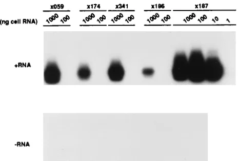
ex-amine PBMC for the presence of negative-strand HCV RNA, PBMC were purified from the plasma of the five chimpanzees mentioned above. Serial dilutions of PBMC RNA were tested for the presence of positive- and negative-strand RNA. Posi-tive-strand RNA could be detected in the dilution of PBMC
RNA containing 10 ng of RNA for3187 and 1mg of RNA for
the remaining four animals (Fig. 3). No negative-strand RNA was detected in any of the dilutions of PBMC RNA. Since a 10-fold-lower level of HCV negative-strand RNA than posi-tive-strand RNA was detected in the liver RNA of the same animals, detection of negative-strand RNA may have been
[image:2.612.83.270.69.316.2]expected only in the PBMC RNA of3187, in which case the
FIG. 1. Analysis of synthetic HCV RNA by strand-specific rTth RT-PCR. (A) Negative-strand-RNA assay of negative-strand (0.1 to 100 fg) and positive-strand (0.1 to 100 pg) RNA (2RNA and1RNA, respectively). (B) Positive-strand-RNA assay of positive-strand (0.1 to 100 fg) and negative-strand (0.1 to 100 pg) RNA.
TABLE 1. Positive- and negative-strand HCV RNA in sera
Serum samplea PCR titer (units/ml of sera) with b
:
1RNA 2RNA
Chx059 105 ,102
Chx174 103 ,102
Chx341 104 ,102
Chx186 102 ,102
Chx187 104 ,102
HS1 106 102
HS2 107 104
a
RNA was purified from 10-fold dilutions of chimpanzee (Ch) and human (HS) sera prepared in fetal bovine serum. HS1 was a gift from Paul Holland and Ken Quramoto and has been previously described (8). HS2 was a gift from Robert Purcell and is the original H strain inoculum of HCV (2).
b
rTth RT-PCR was performed for both positive-strand (1RNA) and nega-tive-strand (2RNA) HCV RNA by using RNA prepared from 10ml of each serial dilution. The PCR products were detected by Southern hybridization. Titers were extrapolated from the last dilution in which 10ml was positive for PCR multiplied by 100 for a PCR titer per milliliter of sera.
on November 9, 2019 by guest
http://jvi.asm.org/
[image:2.612.315.555.83.185.2]level of positive-strand RNA was sufficiently high to expect detection of negative-strand RNA.
To extend these observations to HCV-infected humans, PBMC RNA was purified from 10 HCV-infected individuals. In general, higher levels of positive-strand RNA were detected in human PBMC than in chimpanzee PBMC. Positive-strand
HCV RNA was detected at titers of 103PCR units permg of
PBMC RNA for two of the PBMC samples, 102PCR units per
mg of PBMC RNA for three of the PBMC samples, and 101
PCR units per mg of PBMC RNA for two of the PBMC
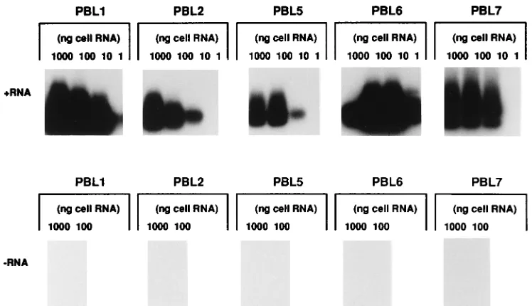
samples. No positive-strand RNA was detected in the RNA from three of the PBMC preparations. No negative-strand RNA was detected in any of the PBMC RNA preparations (Table 2 and Fig. 4). If positive-strand RNA were present at 10-fold-higher levels than negative-strand RNA, as in the liver, negative-strand RNA should have been detectable in five of the PBMC samples. If present, negative-strand RNA must
have been at a level at least 1,000-fold lower than that of positive-strand RNA for the two PBMC samples with the high-est titers of positive-strand RNA.
Analysis of extrahepatic tissues for the presence of HCV
positive- and negative-strand RNA.To extend the observations
to other extrahepatic tissues, multiple tissues were obtained from a chimpanzee that was euthanized for humanitarian rea-sons because of kidney failure. The kidney failure may have been due to long-term HCV infection (6, 7, 12). Tissues were obtained precisely at the time of death and frozen in liquid nitrogen. Serial dilutions of liver RNA were examined, while
all other tissues were analyzed at 1mg of RNA per PCR. An
intense reaction for positive-strand RNA was obtained with 10 ng of liver RNA. For all other tissues, positive-strand RNA was
detected from 1mg of RNA with various degrees of intensity
(Fig. 5). The reactions for the spleen, muscle, and lymph nodes were the most intense. The pancreas and kidney were weakly positive with a short exposure of the autoradiogram (4 h [Fig.
[image:3.612.113.485.70.245.2]FIG. 2. Detection of positive- and negative-strand HCV RNA in chimpanzee liver RNA. Total cell RNA was purified from liver biopsy samples from three chronically HCV-infected chimpanzees (3059,3174, and3341) and two chimpanzees during acute infection (3186 and3187). Tenfold dilutions of liver RNA were prepared in normal cell RNA to yield samples with 1,000 to 1 ng of liver RNA and 1mg of normal cell RNA per 10ml. All assays included several negative controls consisting of normal cellular RNA. In addition, assays were controlled by amplification of 1 or 10 fg of the correct strand (sensitivity control) and 100 fg of the incorrect strand (specificity control) of synthetic HCV RNA (Syn RNA). Serum, plasma, and liver needle biopsy samples were taken by standard methods under a protocol approved by the institutional animal research committee.
[image:3.612.59.292.517.674.2]FIG. 3. Lack of detection of negative-strand HCV RNA in PBMC from chimpanzees infected with HCV. PBMC were prepared from the same chim-panzees described in the legend to Fig. 2, and dilutions of the PBMC RNA were subjected to strand-specific RT-PCR for HCV positive-strand (1RNA) and negative-strand (2RNA) RNA. PBMC were isolated on Histopaque 1077, and the PBMC layer was washed three times with serum-free RPMI 1640.
TABLE 2. Lack of detection of negative-strand HCV RNA in human PBMC
PBMC samplea PCR titer (units/mg of RNA) with b
:
1RNA 2RNA
1 103 2
2 102 2
3 2 2
4 2 2
5 102 2
6 103 2
7 102 2
8 101 2
9 101 2
10 2 2
a
Total cell RNA was prepared from purified PBMC, and 10-fold dilutions of 1mg of cell RNA were prepared in normal cell RNA.
b
rTth RT-PCR was performed for positive-strand (1RNA) and negative-strand (2RNA) HCV RNA. The PCR titer represents the highest positive dilution. The largest amount of RNA tested was 1mg; samples negative at this level are designated by a minus sign.
on November 9, 2019 by guest
http://jvi.asm.org/
5A]), while detection of the reaction for PBMC and bone marrow required a longer exposure (data not shown). Nega-tive-strand RNA was not detected in any tissue other than liver, which was positive with 100 ng of RNA (Fig. 5B). These
data are consistent with the virus being present in other tissues because of contamination with serum.
Conclusions.Whether extrahepatic replication occurs is an
important question with regard to the transmission and patho-genesis of HCV infections. Several investigations have re-ported the detection of HCV negative-strand RNA in PBMC (3, 4, 10, 11, 13, 15), with detection of high levels of negative-strand RNA in plasma in approximately 50% of the studies. In most instances, the differentials, if determined, between nega-tive- and posinega-tive-strand RNA in PBMC and sera were 10- to 100-fold. In this study, PBMC from 10 humans chronically infected with HCV were examined. For the PBMC from five of these patients, the level of positive-strand RNA was quite high yet no negative-strand RNA could be detected. The data sug-gest that if negative-strand RNA were present in these PBMC, it was present at 100- to 1,000-fold-lower levels than positive-strand RNA.
The potential for extrahepatic replication was also examined in a series of tissues from a chimpanzee chronically infected with HCV. Low levels of positive-strand RNA were detected in all tissues examined, but no negative-strand RNA was de-tected. The detection of positive-strand RNA in all tissues in the absence of detectable negative-strand RNA suggests that the positive-strand RNA may be due to contamination with circulating virus.
Although these data do not exclude the potential for repli-cation of HCV in extrahepatic tissue, they do suggest that several criteria should be met in order to determine whether replication occurs. First, titration assays should be performed with synthetic RNA in the presence of a high level of heterol-ogous RNA to determine the degree of strand specificity and the level of sensitivity for the RT-PCR assay being employed. Second, the cellular RNA samples should be titrated to deter-mine the endpoints of detection for positive- and negative-strand RNA and thus the ratio of negative- to positive-negative-strand RNA. Negative-strand RNA must be detected at a level higher
[image:4.612.116.490.69.284.2]FIG. 4. Lack of detection of negative-strand HCV RNA in PBMC from humans infected with HCV. PBMC RNA was purified, diluted in normal cell RNA, and subjected to strand-specific RT-PCR for positive-strand (1RNA) and negative-strand (2RNA) HCV RNA. PBMC were obtained from 10 patients with chronic HCV infection. The diagnosis of chronic infection was based on standard clinical parameters and serological assays. All patients were anti-HCV antibody positive as determined by the second-generation (c200/c22-3) HCV ELISA test system (Ortho Diagnostics, Inc., Raritan, N.J.) as previously described (1). PBMC were separated on Ficoll-Hypaque density gradients, washed three times in Hank’s balanced salt solution, and resuspended with RPMI 1640 as previously described (1). Peripheral blood lymphocyte (PBL) samples correspond to the PBMC samples in Table 2.
FIG. 5. Lack of detection of negative-strand HCV RNA in extrahepatic tissues from a chronically HCV-infected chimpanzee. (A) Positive-strand-RNA assay. (B) Negative-strand-RNA assay. Tissues were obtained from a chimpan-zee immediately at the time of death and were frozen in liquid nitrogen. Tenfold dilutions of RNA from each tissue were prepared in normal cell RNA as de-scribed in Fig. 2. Liver RNA (Lv) was examined at 100 and 10 ng for positive-strand HCV RNA (A) and at 1,000 to 10 ng for negative-positive-strand HCV RNA (B). RNA samples from other tissues were examined at 1,000 ng, including spleen (Sp), pancreas (Pn), kidney (Kd), muscle (Mu), lymph node (LN), bone marrow (BM), and PBL. Positive-strand (1) and negative-strand (2) synthetic HCV RNAs (Syn RNA) were included as controls for strand specificity and sensitivity, as described in the legend to Fig. 2.
on November 9, 2019 by guest
http://jvi.asm.org/
than can be accounted for by false priming of the incorrect strand before replication can be definitively documented. In this respect, our examination of sera from HCV-infected hu-mans is a good example of borderline results. Low levels of negative-strand RNA were detected in two human serum sam-ples that had exceptionally high titers of positive-strand RNA. Since the level of negative-strand RNA was 1,000- to 10,000-fold lower than the level of positive-strand RNA, and the differential in strand specificity with synthetic RNA was in the same order of magnitude, it cannot be concluded that nega-tive-strand RNA was detected in plasma. The detection of negative-strand RNA in plasma has been attributed to the release of replicative complexes from damaged hepatocytes. However, this argument also suggests that any negative-strand RNA detected in PBMC could be accounted for by adherence of circulating negative-strand RNA. Thus, in this case, to doc-ument replication of HCV in PBMC would require detection of a higher ratio of negative- to positive-strand RNA in PBMC than is detected in plasma from the same samples.
Certainly, resolution of the dilemma created by the need for RT-PCR to detect HCV, and of the problem of using RT-PCR for strand-specific detection of RNA, will require additional studies to resolve the problem of extrahepatic HCV replica-tion.
This study was supported by grant CA57955 from the National Cancer Institute and grant AI200001 from the National Institute for Allergy and Infectious Disease. C.S. was supported in part by the ‘‘Association pour la Recherche sur le Cancer.’’
REFERENCES
1. Cerny, A., J. G. McHutchison, C. Pasquinelli, M. E. Brown, M. A. Brothers, B. Grabscheid, P. Fowler, M. Houghton, and F. V. Chisari.1995. Cytotoxic T lymphocyte response to hepatitis C virus-derived peptides containing the HLA A2.1 binding motif. J. Clin. Invest. 95:521–530.
2. Feinstone, S. M., H. J. Alter, H. P. Dienes, Y. Shimizu, H. Popper, D.
Blackmore, D. Sly, W. T. London, and R. H. Purcell.1981. Non-A, non-B hepatitis in chimpanzees and marmosets. J. Infect. Dis. 144:588–598. 3. Gabrielli, A., A. Manzin, M. Candela, M. L. Caniglia, S. Paolucci, M. G.
Danieli, and M. Clementi.1994. Active hepatitis C virus infection in bone marrow and peripheral blood mononuclear cells from patients with mixed cryoglobulinaemia. Clin. Exp. Immunol. 97:87–93.
4. Gil, B., C. Qian, J. I. Riezu-Boj, M. P. Civeira, and J. Prieto. 1993. Hepatic and extrahepatic HCV RNA strands in chronic hepatitis C: different patterns of response to interferon treatment. Hepatology 18:1050–1054.
5. Gunji, T., N. Kato, M. Hijikata, K. Hayashi, S. Saitoh, and K. Shimotohno. 1994. Specific detection of positive and negative stranded hepatitis C viral RNA using chemical RNA modification. Arch. Virol. 134:293–302. 6. Johnson, R. J., D. R. Gretch, W. G. Couser, C. E. Alpers, J. Wilson, M.
Chung, J. Hart, and R. Willson.1994. Hepatitis C virus-associated glomer-ulonephritis. Effect ofa-interferon therapy. Kidney Int. 46:1700–1704. 7. Johnson, R. J., R. Willson, H. Yamabe, W. Couser, C. E. Alpers, M. H.
Wener, C. Davis, and D. R. Gretch.1994. Renal manifestations of hepatitis C virus infection. Kidney Int. 46:1255–1263.
8. Lanford, R. E., C. Sureau, J. R. Jacob, R. White, and T. R. Fuerst. 1994. Demonstration of in vitro infection of chimpanzee hepatocytes with hepatitis C virus using strand-specific RT/PCR. Virology 202:606–614.
9. McGuinness, P. H., G. A. Bishop, G. W. McCaughan, R. Trowbridge, and E. J. Gowans.1994. False detection of negative-strand hepatitis C virus RNA. Lancet 343:551–552.
10. Muller, H. M., E. Pfaff, T. Goeser, B. Kallinowski, C. Solbach, and L. Theilmann.1993. Peripheral blood leukocytes serve as a possible extrahe-patic site for hepatitis virus replication. J. Gen. Virol. 74:669–676. 11. Navas, S., I. Castillo, J. Bartolome´, E. Marriott, M. Herrero, and V.
Car-ren˜o.1994. Positive and negative hepatitis C virus RNA strands in serum, liver and peripheral blood mononuclear cells in anti-HCV patients: relation with the liver lesion. J. Hepatol. 21:182–186.
12. Roth, D. 1995. Hepatitis C virus: the nephrologist’s view. Am. J. Kidney Dis. 25:3–16.
13. Saleh, M. G., C. J. Tibbs, J. Koskinas, L. M. M. B. Pereira, A. B. Bomford, B. C. Portmann, I. G. McFarlane, and R. Williams.1994. Hepatic and extrahepatic hepatitis C virus replication in relation to response to interferon therapy. Hepatology 20:1399–1404.
14. Willems, M., H. Moshage, and S. H. Yap. 1993. PCR and detection of negative HCV RNA strands. Hepatology 17:526.
15. Yun, Z.-B., A. So¨nnerborg, and O. Weiland.1994. Hepatitis C virus replica-tion in liver and peripheral blood mononuclear cells of interferon-a-treated and untreated patients with chronic hepatitis C. Scand. J. Gastroenterol. 29:82–86.