JOURNAL OFVIROLOGY, Aug. 1996, p. 5405–5413 Vol. 70, No. 8 0022-538X/96/$04.0010
Copyrightq1996, American Society for Microbiology
Anterograde, Transneuronal Transport of Herpes Simplex Virus
Type 1 Strain H129 in the Murine Visual System
NING SUN,1M. D. CASSELL,2
ANDSTANLEY PERLMAN1,3*
Departments of Pediatrics,1Anatomy,2and Microbiology,3University of Iowa, Iowa City, Iowa 52242
Received 15 March 1996/Accepted 2 May 1996
Herpes simplex virus (HSV) undergoes retrograde and anterograde axonal transport as it establishes latency and later intermittently reactivates. Most strains of HSV show preferential retrograde transport within the central nervous system (CNS), however. Previous experiments suggest that an exception to this is HSV type 1 (HSV-1) strain H129, since this virus appears to spread primarily in the CNS via anterograde, transneuronal movement. The objective of the present study was to test how specifically this virus spreads in the visual system, a system with well-described neuronal connections. In the present study, the pattern of viral spread was examined following inoculation into the murine vitreous body. Virus was initially detected in the retina and optic tract. Virus then appeared in all known primary targets of the retina, including those in the thalamus (e.g., lateral geniculate complex), hypothalamus (suprachiasmatic nucleus), and superior colliculus (superfi-cial layers). In previous studies, many strains of HSV were shown to infect these structures, even though they spread predominantly in a retrograde direction. However, the H129 strain was unique in then spreading, via anterograde transport, to the primary visual cortex (layer 4 of area 17) via thalamocortical connections. At later times after infection, specific labeling was also detected in other cortical and subcortical areas known to receive projections from the visual cortex. No labeling was ever detected in the contralateral retina, which is consistent with a lack of retrograde spread of HSV-1 strain H129. These results demonstrate the specific anterograde movement of this virus from the retina to subcortical and cortical regions, with no clear evidence for retrograde spread. HSV-1 strain H129 should be generally useful for tracing sensory pathways and may provide the basis for designing a virus vector capable of delivering genetic material via anterograde pathways within the CNS.
Herpes simplex virus type 1 (HSV-1), the most common cause of sporadic acute encephalitis in the United States (44), has been shown in many studies to spread from a peripheral or central site of inoculation transneuronally to distant connec-tions within the central nervous system (CNS) (2, 3, 5, 13, 17, 21, 27, 37, 45). Only structures neuroanatomically linked to the site of inoculation are infected, and the pattern of spread is determined by many factors, one of which is whether the virus spreads predominantly in an anterograde (from the cell body to the axon) or retrograde (from the axon to the cell body) direction. Nearly all nonherpesviruses, pseudorabies virus, and most strains of HSV spread primarily in a retrograde manner (from the axon to the cell body), although some degree of anterograde spread has been detected with most of these vi-ruses (6, 19). A strain of virus which moves primarily in the anterograde direction would be useful for mapping pathways in the CNS utilized in the relay of sensory information. In addi-tion, HSV is potentially useful for delivering genetic informa-tion to the CNS. For both of these reasons, identifying a strain of HSV-1 which moves in the anterograde direction is a nec-essary step for determining the factors underlying the spread of HSV-1 through CNS pathways.
HSV-1 strain H129 has been preliminarily identified as spreading primarily if not solely in the anterograde direction. This was suggested initially by experiments in which the virus was inoculated centrally into the primate motor cortex (45) and shown to spread to cortical, subcortical, and spinal cord
regions known to receive input from the motor cortex (45). In more recent experiments, the virus was injected peripherally into the murine tooth pulp and was observed to specifically infect ascending CNS systems associated with the processing of trigeminal pain (3).
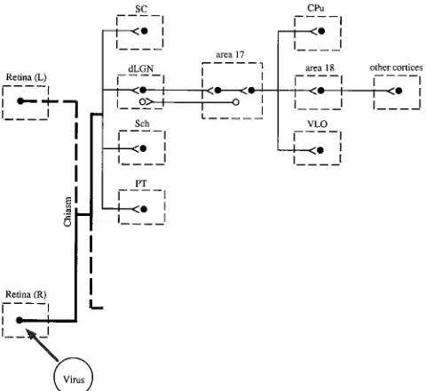
The visual system is a well-defined sensory pathway which possesses both centripetal and centrifugal nonreciprocal con-nections (Fig. 1) (29). In rodents, most projections from the retina go contralaterally to the thalamus, to the superior col-liculus (SC), and to several structures in the hypothalamus, including the suprachiasmatic nucleus (Sch) (14, 23). Within the thalamus, several specific thalamic nuclei, including the dorsal lateral geniculate nucleus (dLGN), are primary retino-recipient areas. From the dLGN, visual information is relayed to the primary and secondary visual cortices (areas 17 and 18) (9, 15, 32). Thalamocortical projections terminate in specific layers of the visual cortex, whereas efferent projections from the cortex to the thalamus originate in a different subset of layers (12, 36). These connections between the thalamus and visual cortex are therefore very useful for analyzing the di-rection of spread of a virus. Visual information is further transmitted from the visual cortex to many other cortical and subcortical regions, such as the temporal, frontal, and ventro-lateral orbital cortices and the basal ganglia, for further pro-cessing. A careful study of the sequential spread of HSV-1 strain H129 after intraocular inoculation should provide infor-mation about the direction of spread of the virus and should also demonstrate the temporal movement of the virus from the retina to its primary, secondary, and tertiary targets. The latter has not been possible with traditional tracing techniques, largely because too many synapses are involved. Preliminary data from this study have been published in abstract form (35).
* Corresponding author. Mailing address: 207 Med. Lab., Dept. of Pediatrics, University of Iowa, Iowa City, IA 52242. Phone: (319) 335-8549. Fax: (319) 356-4855. Electronic mail address: [email protected].
5405
on November 9, 2019 by guest
http://jvi.asm.org/
MATERIALS AND METHODS
Animals.Thirty-one male 6-week-old BALB/c mice, purchased from Sasco Laboratories (Omaha, Nebr.), were used in this study. For surgery, mice were anesthetized by intraperitoneal injections (52.5 mg/kg of body weight) of a sodium pentobarbital solution (31). All surgical procedures were approved by the Institutional Animal Care and Use Committee at the University of Iowa.
Virus.HSV-1 strain H129 was kindly provided by William Stroop, University of Arkansas. The virus was grown on RK13 cells, and titers were determined on Vero cells.
Inoculation and tissue processing.Each mouse was deeply anesthetized, and 2ml of virus (23104PFU) was injected into the vitreous body of one eye.
Animals were killed at 3, 4, 5, 6, and 7 days postinoculation (p.i.) by transcardiac perfusion of phosphate-buffered saline under deep pentobarbital anesthesia. Brains were removed, embedded in Tissue-Tek O.C.T. compound (Miles Lab-oratory, Elkhart, Ind.), and quickly frozen in dry ice before further processing.
In situ hybridization.In situ hybridization was used to detect viral RNA and DNA in brain tissue sections. An antisense35S-labeled RNA probe for HSV-1
was synthesized from a plasmid encoding the VP5 gene of HSV-1 (kindly pro-vided by E. Wagner, University of California—Irvine). In situ hybridization was performed as previously described (3). Briefly, 35-mm coronal brain sections were cut at 100- to 200-mm intervals on a cryostat. Sections were collected on silane-treated slides and fixed with 2.5% paraformaldehyde for 45 min. After treatment with proteinase K and acetylation, 106cpm of the probe was applied
to each slide. After overnight incubation, slides were treated with RNase and washed with solutions of increasing stringency. Slides were dipped in NTB-2 photographic emulsion (Kodak, Rochester, N.Y.) and exposed for 2 weeks. The slides were developed, stained with cresyl violet, and examined by bright- and dark-field light microscopy.
Immunohistochemistry.Immunohistochemistry was also used to detect HSV antigen. Frozen brain sections were cut at 25- to 30-mm intervals on a cryostat and fixed for 20 min with 4% paraformaldehyde. Sections were then washed and incubated with primary antibody, rabbit anti-HSV-1 (Dako Corp., Carpinteria, Calif.), at a 1:200 dilution for 24 h at 48C. After incubation with biotinylated goat anti-rabbit antibody, sections were treated with Vectastain ABC (Vector Labo-ratory, Burlingame, Calif.), used according to the manufacturer, with 3,39 -di-aminobenzidine as the final substrate. Finally, sections were dehydrated, mounted on coverslips, and examined with a microscope.
Controls.Several experiments were performed to ensure that the spread which was observed in this study was not due to nonspecific infection through nonvisual pathways. To assess further the possible role of olfactory spread after leakage via
the nasolacrimal duct, different amounts of virus were tested to determine the maximum amount of virus which could be administered without olfactory-path-way involvement. The possibility of hematogenous spread was examined by inoculation of virus (23104PFU) into the tail vein. In addition, to assess the
sensitivity of the in situ hybridization method, some sections were processed by immunohistochemistry as described above. In other control experiments, mice were inoculated in the vitreous body with HSV-1 strain 17 (23104PFU). HSV-1
strain 17 spreads primarily in the retrograde direction (2, 4). Finally, no signal was detected if uninfected mice were analyzed with the HSV-1-specific probe.
RESULTS
The temporal movement of virus within the brain was ex-amined following intraocular inoculation into the vitreous body of one eye. For the purpose of determining whether HSV-1 strain H129 moved in an anterograde or retrograde direction, it was necessary to analyze CNS structures at the earliest times that they showed signs of virus infection. The site of virus entry into a particular structure would indicate the direction of spread of the virus. At later times, virus was noted to spread to many sites within a specific infected structure, making it impossible to determine where the virus first ap-peared. Brains were examined from days 3 to 7 p.i. There was some variability from animal to animal in the rate of spread, but this did not affect identification of the basic pattern of virus spread within the infected brain. (Detailed descriptions of the neuroanatomic structures infected by HSV-1 strain H129 are available from S.P.)
As expected, the infection was confined initially to the ret-ina. Although inoculation was directed at the fovea, the entire retina was eventually infected (Fig. 2A). Virus was detected next in the optic nerve, optic chiasm, and optic tract beginning at day 3. Only the ipsilateral optic nerve was infected, with no evidence of infection of the contralateral optic nerve. Labeling in the contralateral optic chiasm reflected the decussation of most fibers to the contralateral side. The majority of labeling was found in the contralateral optic tract in all cases (Fig. 3), although a small amount of labeling was also found in the ipsilateral optic tract in some animals. No viral infection of the contralateral retina was ever detected, even as late as 6 to 7 days p.i., which is consistent with the lack of retrograde spread of HSV-1 strain H129.
By days 4 to 5 p.i., virus was detected in two of seven animals in three visually associated brain regions, the thalamus (Fig. 2B), tectum (Fig. 2C and D), and hypothalamus (Fig. 3). These structures receive direct anterograde projections from the ret-ina. Labeled hypothalamic structures, including the Sch, were detected in two animals. Both the ipsilateral and contralateral Sch were infected, although the ipsilateral Sch was often la-beled first (Fig. 3). This was unexpected, since most fibers are reported to cross over to the contralateral Sch (16).
Thalamic labeling was first detected in the dLGN, ventral lateral geniculate nucleus (vLGN), and intergeniculate leaflet (IGL) on the contralateral side (Fig. 2B). The earliest labeling observed in the dLGN was insubstantial and limited to the dorsal region of the nucleus. In the vLGN, only the lateral portion, the magnocellular division, was labeled. Labeling was less abundant in the ipsilateral thalamus, although early infec-tion of the IGL and magnocellular division of the vLGN were found in one case. In the tectum, only the contralateral SC was labeled at day 4. The earliest evidence of virus infection was present in the superficial gray and optic nerve layers, the su-perficial layers of the SC (Fig. 2C and D). These are the layers which receive projections from the retina (14). In the cortex, the earliest labeling was detected in layer 4 of the contralateral primary visual cortex (area 17) of one animal at day 4. No other cortical labeling was detected at this time.
[image:2.612.60.300.71.291.2]By day 5 p.i., virus spread along thalamocortical pathways
FIG. 1. Schematic drawing of primary, secondary, and distal connections of the retina. Only connections related to this study are shown. Most projections from the retina cross over at the optic chiasma and project to the contralateral side of the brain, although a minority project to the same structures on the ipsilateral side. For the sake of simplicity, only the contralateral structures are shown. The dLGN of the thalamus projects to layer 4 of area 17 (closed circles) of the visual cortex, whereas neurons in layers 5 and 6 of this area project back to the thalamus (open circles). PT, pretectal nucleus. Closed circles, anterograde connection; open circles, retrograde connection. L and R, left and right, respec-tively.
5406 SUN ET AL. J. VIROL.
on November 9, 2019 by guest
http://jvi.asm.org/
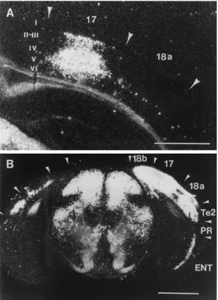
was indicated by prominent infection of the contralateral visual cortex in seven of eight mice. The earliest labeling was de-tected only in layer 4 of the primary visual cortex (area 17) (Fig. 4A and C). This layer receives monosynaptic projections from the dLGN in the thalamus. Consistent with only antero-grade spread of the virus, no labeling was found at this time in the deeper cortical layers (layers 5 and 6), which send projec-tions back to the thalamus (12, 36). In cases in which the infection spread more rapidly, virus could be detected at day 5 p.i. in other layers of area 17 (Fig. 5A) and in area 18 (Fig. 4B and D). Labeling in the secondary visual cortex (area 18) was first detected in layers 2, 3, and 5 (Fig. 4B and D), which is consistent with corticocortical spread from the primary vi-sual cortex (22) and in contrast to the initial appearance of virus in layer 4 of area 17. Although labeling was observed in rostral aspects of area 18 without a simultaneous appearance of virus in layers 2 and 3 of the adjacent part of area 17 (Fig. 4B and D), all layers of the caudal primary visual cortex were infected prior to infection of area 18 (Fig. 5A). This suggests that virus spread to area 18 originated from the caudal part of layer 4 of area 17.
Heavier and more extensive infection of the hypothalamus, thalamus, and deeper layers of the SC was observed at this time, in contrast to what was observed at day 4 p.i. In the hypothalamus, virus was present in large amounts bilaterally in most cases at day 5 p.i. This pattern of labeling was consistent
with known connections between the retina and Sch (8, 42). In addition to labeling in the Sch, heavy labeling was also de-tected in the subparaventricular zone of the hypothalamus, an area which primarily receives projections from the Sch (40, 41) (data not shown). In the contralateral thalamus, the dLGN, vLGN, and IGL were completely covered by intense labeling. On the ipsilateral side, most cases showed extensive labeling in the vLGN and IGL, with smaller amounts of labeling present in the dLGN. In the SC, labeling was found in both deeper and superficial layers on the contralateral side.
Another finding at day 5 p.i. was labeling in the caudate putamen (CPu). This region receives direct projections from the deeper layers of both the primary and secondary visual cortices. There is no evidence that the CPu projects back to the visual cortices (29). Labeling in the CPu was first detected in the dorsal region at the caudal level and extended to the rostral CPu (Fig. 6A). Labeling in the dorsal CPu always followed labeling in the visual cortex, and in no case was labeling noted without extensive infection of deeper layers of the visual cor-tex.
[image:3.612.62.555.71.402.2]Specific labeling in other regions of the contralateral cortex was also apparent. Labeling was found in layers 2 and 3 of the contralateral ventrolateral orbital cortex (VLO) (area 11) (Fig. 6B) in four of eight cases and in the frontal cortex (area 8) in one of eight cases. These areas receive projections from the visual cortex, and consistent with this, cases in which areas 11
FIG. 2. Viral infection of the retina, thalamus, and SC. Virus was inoculated into the vitreous body of one eye. (A) Widespread infection of the retina was observed by days 4 to 5 p.i. (B) Infection in the contralateral thalamus was first detected at day 4 p.i. and, as shown, several thalamic structures, including the lateral posterior (LP) nucleus, dLGN, IGL, and vLGN were infected by day 5 p.i. Only the magnocellular part of the vLGN was labeled, which is consistent with neuroanatomical data (25). (C and D) Initial infection of the contralateral SC at rostral (C) and middle (D) levels was limited to the superficial layers, including the superficial gray (SuG) and optic nerve (Op) layers. The deeper gray layer (DpG) was not infected at this time. Arrows in panel C indicate viral labeling in the rostral SC. Bar, 1 mm.
VOL. 70, 1996 TRANSPORT OF HSV-1 IN THE MURINE VISUAL SYSTEM 5407
on November 9, 2019 by guest
http://jvi.asm.org/
FIG. 3. Viral infection of the hypothalamus. Bilateral infection of the Sch was noted at days 4 to 5 p.i. In the case shown, more labeling on the ipsilateral side was observed. This occurred in several cases but was unexpected, since most fibers project to the contralateral Sch (16). The contralateral optic tract (ot) was also heavily labeled. Bar, 0.5 mm.
FIG. 4. Viral infection of the visual cortex. Corresponding bright- and dark-field photomicrographs are shown. (A and C) Infection of the primary visual cortex was first observed in layer 4 of area 17 at days 4 to 5 p.i. (arrows). (B and D) In cases in which virus spread more rapidly, labeling was observed in other layers of area 17 and in area 18 at day 5. Rostral areas 17 and 18 are shown. Although a minor amount of labeling of layer 5 and none in layers 2 and 3 of area 17 is present in this case, infection of all layers of caudal parts of area 17 had already occurred by this time (Fig. 5A and 7A and B). This suggests that infection of layer 5 originated from caudal layers 2 and 3 of area 17. Similarly, labeling in area 18 was observed only after infection had spread to all layers of caudal area 17. The neuroanatomical location of the labeling shown in panels A and B is visualized in panels C and D. Layer numbers are expressed as Roman numerals. Bar, 0.25 mm.
5408
on November 9, 2019 by guest
http://jvi.asm.org/
[image:4.612.65.556.347.675.2]and 8 were labeled also had extensive labeling in the visual cortex. The primary and secondary ipsilateral visual cortices were also infected in two of eight cases at day 5.
A similar distribution of labeling was observed at days 6 and 7. At these times, heavy labeling of both the contralateral and ipsilateral cortices was detected. The pattern of ipsilateral cor-tical labeling, while delayed and less extensive, was similar to that of the contralateral cortex. As with infection of the con-tralateral visual cortex, virus could first be detected in layer 4 of area 17, with extension vertically to other layers within area 17 and to area 18 (Fig. 5B). Contralateral labeling was detected in the temporal visual area in most cases and extended to the entorhinal cortex, although the perirhinal cortex was never infected (Fig. 5B).
At this time, strong labeling was found in layers 2 and 3 of both the contralateral and ipsilateral VLO. The ipsilateral CPu also contained heavy labeling similar to that observed at earlier
times on the contralateral side. This labeling paralleled label-ing of the ipsilateral visual cortex.
Controls. Samples from the brains of mice infected with HSV-1 strain H129 were also examined by immunohistochem-istry in order to assess the sensitivity of the in situ hybridization method. The same pattern of infection was observed as in mice analyzed by in situ hybridization, with extensive infection of the Sch, the dLGN, the SC, and the visual cortices. After intrave-nous inoculation, no virus was detected in the brains at day 6, 7, or 8 p.i. Intranasal inoculation with HSV-1 strain H129 resulted in labeling of structures associated with the olfactory system but not those associated with the visual system. The pattern of labeling detected in the olfactory system after infec-tion with the H129 strain was different from what was observed after intranasal and intrabulbar inoculation with HSV-1 strain 17 (2).
[image:5.612.151.461.71.497.2]In another set of controls, HSV-1 strain 17 was inoculated
FIG. 5. Dark-field micrograph demonstrating infection in the caudal visual cortex and other subcortical and cortical structures. (A) Infection of caudal visual cortex at day 4 p.i. Note that all layers of area 17 were infected, with little labeling observed in area 18 at this time. (B) By day 7 p.i., virus spread to all layers of the visual cortex (17, 18a, and 18b), to the visual temporal cortex (Te2), and to the entorhinal cortex (ENT) on the contralateral side. The perirhinal cortex (PR) was never infected. Ipsilaterally, the visual cortex was also infected. Layer numbers are expressed as Roman numerals. Bars, 1 mm (A) and 2 mm (B).
VOL. 70, 1996 TRANSPORT OF HSV-1 IN THE MURINE VISUAL SYSTEM 5409
on November 9, 2019 by guest
http://jvi.asm.org/
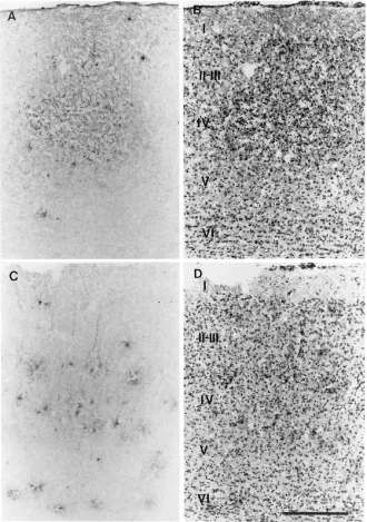
into the vitreous body of one eye. Mice developed acute en-cephalitis at days 4 to 5 p.i., earlier than was observed in the animals infected with HSV-1 strain H129. Examination of the brains of these mice by immunohistochemistry revealed infec-tion of the hypothalamus, thalamus, tectum, and visual cortex. The hypothalamus, thalamus, and tectum were infected in the same sites as found in mice after inoculation with HSV-1 strain H129 (data not shown). In marked contrast to what was ob-served in H129-infected mice (Fig. 7A and B), however, cor-tical labeling was detected only in layers 5 and 6 and not in layer 4 in animals infected with HSV-1 strain 17 (Fig. 7C and D). Also, in contrast to findings for mice infected with HSV-1 strain H129, labeling was detected in both the ipsilateral and contralateral retina at day 5 p.i. (data not shown).
DISCUSSION
The objective of this study was to determine the neuroana-tomic structures infected following intraocular inoculation. The neuroanatomic connections of the visual system have been well characterized for the most part and are very useful for examining the capacity of a virus to spread preferentially in the anterograde or retrograde direction. In this study, we showed that intraocular inoculation of HSV-1 strain H129 resulted in a specific and sequential infection of structures associated with vision. Virus inoculated into the vitreous body of the eye could potentially spread to the anterior chamber or the nose and from there to the CNS via olfactory, trigeminal, or autonomic pathways. The control experiments showed, however, that with
the amount and volume of virus used in this study, no spread occurred via the trigeminal or olfactory system and labeling was confined to the visual pathway.
After inoculation, retinal ganglion cells become labeled. Since different parts of the retina project to different subdivi-sions of the primary retinoceptive targets, it was possible that only a subset of retinal cells and, consequently, their targets, would be infected by the virus. In this study, although we attempted to inoculate the posterior part of the retina, subse-quent examination revealed that all parts of the retina were equally infected (Fig. 2A) and that there was no selective labeling within the retinoceptive areas. The nonselective infec-tion of the retina was also supported by the consistent pattern of infection observed in all animals. As a consequence, the complete array of neurons in the visual cortex associated with monocular vision was infected.
The spread of virus after infection of the retina provided strong evidence for the anterograde transneuronal transport of HSV-1 strain H129 and for the ability of the virus to cross several synapses. First, virus was detected in the ipsilateral optic nerve, the optic chiasm, and, primarily, the contralateral optic tract. These results are consistent with previous studies showing that the majority of retinal projections in rats (and, presumably, mice) are directed to the contralateral tract (30). HSV-1 strain H129 was never detected in the contralateral retina, as would be expected if this virus spread in the retro-grade direction. In addition, in control experiments, HSV-1 strain 17, a strain with preferential retrograde spread (2, 4), did spread to the contralateral retina from primary retinorecipient areas (data not shown). In a previous report, pseudorabies virus, a virus which spreads predominantly in the retrograde direction within the CNS, was also shown to infect the con-tralateral retina after intraocular inoculation (24).
Second, the Sch, dLGN, and SC receive monosynaptic pro-jections from retinal ganglion cells, and these areas were in-fected by HSV-1 strain H129 after intraocular inoculation. In addition to labeling in the Sch, heavy labeling was also de-tected in specific hypothalamic structures, such as the subpara-ventricular zone. This region has been reported to receive a heavy direct projection from the Sch (40, 41). Labeling in the SC first appeared in the superficial layers, again as expected if the virus spread in an anterograde direction (33).
Third, following infection of the thalamus, virus appeared in the primary visual cortex (area 17). The dLGN is reciprocally connected to the visual cortex. However, of major importance for this study, projections from the thalamus to the cortex terminate in layer 4 of the primary visual cortex whereas pro-jections from the cortex to the thalamus originate mostly in layer 6. As shown in Fig. 4A and C, infection with HSV-1 strain H129 results in specific labeling of layer 4 at early times p.i. Other layers of the primary visual cortex were labeled only at later times p.i.; this pattern of labeling suggested that virus spread within the cortex. In marked contrast, in our control experiments, infection with HSV-1 strain 17 resulted in initial labeling in layers 5 and 6, which is consistent with retrograde spread.
[image:6.612.59.296.70.384.2]Fourth, following infection of the primary visual cortex, la-beling was detected in the secondary visual cortex (area 18), ventral orbital cortex, temporal visual cortex, and basal ganglia. The pattern of labeling in area 18 was most consistent with spread from area 17 rather than from the thalamus, since layers 2, 3, and 5 were first infected (Fig. 4B and D) and since labeling appeared after all layers of caudal area 17 were in-fected (Fig. 5A). Since the connections between layers 2, 3, and 5 of areas 17 and 18 are reciprocal (22), this labeling does not provide additional data about the direction of spread of strain
FIG. 6. Infection of contralateral CPu and VLO at days 5 to 6 p.i. (A) Early infection of the CPu was limited to the dorsal part (arrow). (B) Infection of the VLO first appeared in layers 2 and 3 (arrow). Bar, 1 mm.
5410 SUN ET AL. J. VIROL.
on November 9, 2019 by guest
http://jvi.asm.org/
H129. Labeling in the dorsal CPu did not occur in the absence of labeling of the deeper layers of the visual cortex, even if substantial infection of subcortical structures known to receive projections from the CPu was detected. This suggested that retrograde spread from structures other than the visual cortex was unlikely. Previous reports have documented the presence of projections from the visual cortex to this part of the CPu as well as the absence of reciprocal projections (11). These results together again suggest that HSV-1 strain H129 spreads in the anterograde and not retrograde direction.
Fifth, several additional cortical areas, including the VLO (area 11) and frontal (area 8) cortices, are known to receive
projections from the visual cortex (22). In the present study, labeling was detected in layers 2 and 3 of these cortices (Fig. 6B), the layers shown previously to receive direct input from area 18 (22). The specific progressive spread of virus from a small area of the primary visual cortex to larger areas of the primary and secondary visual cortices and to the temporal and frontal cortices is consistent with anterograde, transneuronal spread.
[image:7.612.141.471.73.542.2]These results clearly show that HSV-1 strain H129 spreads primarily if not solely in the anterograde direction and are in agreement with the results of Zemanick et al. (45) and Barnett et al. (3). Most viruses, including HSV, pseudorabies virus,
FIG. 7. Different patterns of infection in the primary visual cortex (area 17) following inoculation with HSV-1 strain H129 (A and B) or strain 17 (C and D). Viral antigen was detected by immunohistochemistry as described in Materials and Methods. (A) Viral infection in area 17 following intraocular inoculation of HSV-1 strain H129 at day 5 to 6 p.i. (B) Counterstain of the section shown in panel A demonstrates labeling in layers 2, 3, and 4, with no labeling detected in deeper layers. (C) Labeling in area 17 following intraocular inoculation with HSV-1 strain 17 at days 4 to 5 p.i. (D) Counterstain of the section shown in panel C demonstrates labeling in layers 2, 3, 5, and 6, with no labeling in layer 4. Layer numbers are expressed as Roman numerals. Bar, 0.25 mm.
VOL. 70, 1996 TRANSPORT OF HSV-1 IN THE MURINE VISUAL SYSTEM 5411
on November 9, 2019 by guest
http://jvi.asm.org/
rabies virus, vesicular stomatitis virus, and mouse hepatitis virus, appear to spread predominantly in the retrograde direc-tion within the CNS (2, 4–6, 18–21, 26, 27, 39). Although evidence for some degree of anterograde spread exists for most of these viruses (e.g., in the visual system), this type of spread is slower and less efficient than retrograde spread (5–7, 21, 27, 39). Consistent with this, the results of our study showed that HSV-1 strain H129 spread more slowly to the cortex than did strain 17. The previous results with other viruses make the anterograde spread of HSV-1 strain H129 to cortical structures even more striking.
Several factors might contribute to the ability of HSV-1 strain H129 to spread in the anterograde direction with no evidence for retrograde spread. First, although the HSV-1 proteins required for retrograde spread have not been identi-fied, these might be defective in the H129 strain. Thus, HSV-1 strain H129 would lack the ability to spread in the retrograde direction and would only retain the ability, shared with other viruses, to spread in the anterograde direction. If the H129 strain spreads at all in the retrograde direction, its movement must be slower than anterograde movement and, at most, represent a very minor component of spread. We have been unable to detect any evidence of retrograde spread, even after inoculation into the hindlimb musculature and olfactory sys-tems (unpublished observations). In contrast, other strains of HSV-1 (including strain 17) readily spread to layer V of the sensorimotor cortex (33a, 37, 38), which is consistent with the ability of these strains to spread in the retrograde direction.
A second possibility is that a complex of two glycoproteins (HSV gE-gI and pseudorabies virus gE-gI), required for effi-cient transport within the CNS (1, 10, 43), differ between HSV-1 strain H129 and other strains of HSV and pseudorabies virus. Pseudorabies virus mutants lacking gE or gI spread only to the Sch and IGL after intraocular inoculation and not to other primary retinorecipient areas (7). In contrast, HSV mu-tants lacking gE or gI spread normally to the SC but showed reduced levels of spread to the Sch and LGN (10). The gE-gI glycoprotein complex might be similarly involved in direction of spread within the CNS. Characterization of HSV-1 strain H129 gE-gI will show whether these proteins are in fact major determinants of anterograde spread.
ACKNOWLEDGMENTS
This work was supported (in part) by research grant 5 RO1 DC 01711-03 from the National Institute on Deafness and Other Commu-nication Disorders. S.P. was supported by a Research Career Devel-opment Award from NIH (NS 01369), and N.S. was supported by an NIH postdoctoral training grant (T32AI07343).
We thank M. Miller for helpful discussion and R. Roller and M. Stinski for critical review of the manuscript.
REFERENCES
1. Balan, P., N. Davis-Poynter, S. Bell, H. Atkinson, H. Browne, and T. Minson. 1994. An analysis of the in vitro and in vivo phenotypes of mutants of herpes simplex virus type 1 lacking glycoproteins gG, gE, gI or the putative gJ. J. Gen. Virol. 75:1245–1258.
2. Barnett, E., M. Cassell, and S. Perlman. 1993. Two neurotropic viruses, herpes simplex virus type I and mouse hepatitis virus, spread along different neural pathways from the main olfactory bulb. Neuroscience 57:1007–1025. 3. Barnett, E. M., G. D. Evans, S. Perlman, and M. D. Cassell. 1995. Antero-grade tracing of trigeminal nociceptive pathways from the murine tooth pulp to cortex using herpes simplex virus type 1. J. Neurosci 15:2972–2984. 4. Barnett, E. M., G. Jacobsen, G. D. Evans, M. D. Cassell, and S. Perlman.
1994. Herpes simplex encephalitis of the temporal cortex and limbic system after trigeminal nerve inoculation. J. Infect. Dis. 169:782–786.
5. Blessing, W., Z. Ding, Y. Li, Z. Gieroba, A. Wilson, P. Hallsworth, and S.
Wesselingh.1994. Transneuronal labeling of CNS neurons with herpes sim-plex virus. Prog. Neurobiol. 44:37–53.
6. Card, J., L. Rinaman, J. Schwaber, R. Miselis, M. Whealy, A. Robbins, and
L. Enquist.1990. Neurotropic properties of pseudorabies virus: uptake and transneuronal passage in the rat central nervous system. J. Neurosci. 10: 1974–1994.
7. Card, J., M. Whealy, A. Robbins, R. Moore, and L. Enquist. 1991. Two alpha-herpesvirus strains are transported differentially in the rodent visual system. Neuron 6:957–969.
8. Cassone, V. M., J. C. Speh, J. P. Card, and R. Y. Moore. 1988. Comparative anatomical analysis of the mammalian hypothalamic suprachiasmatic nu-cleus. J. Biol. Rhythms 3:71–92.
9. Caviness, V. S., Jr., and D. O. Frost. 1983. Thalamocortical projections in the reeler mutant mouse. J. Comp. Neurol. 219:182–202.
10. Dingwell, K. S., L. C. Doering, and D. C. Johnson. 1995. Glycoproteins E and I facilitate neuron-to-neuron spread of herpes simplex virus. J. Virol. 69: 7087–7098.
11. Faull, R., W. Nauta, and V. Domesick. 1986. The visual cortico-striato-nigral pathway in the rat. Neuroscience 19:1119–1132.
12. Herkenham, M. 1980. Laminar organization of thalamic projections to the rat neocortex. Science 207:532–535.
13. Hoover, J. E., and P. L. Strick. 1993. Multiple output channels in the basal ganglia. Science 259:819–821.
14. Huerta, M., and J. Harting. 1984. Connectional organization of the superior colliculus. Trends Neurosci. 7:286–289.
15. Hughes, H. C. 1977. Anatomical and neurobehavorial investigations con-cerning the thalamo-cortical organization of the rat’s visual system. J. Comp. Neurol. 175:311–336.
16. Johnson, R. F., L. P. Morin, and R. Y. Moore. 1988. Retinohypothalamic projections in the hamster and rat demonstrated using cholera toxin. Brain Res. 462:301–312.
17. Kristensson, K., B. Ghetti, and H. Wisniewski. 1974. Study on the propaga-tion of herpes simplex virus (type 2) into the brain after intraocular injecpropaga-tion. Brain Res. 69:189–201.
18. Kucˇera, P., M. Dolivo, P. Coulon, and A. Flamand. 1985. Pathways of the early propagation of virulent and avirulent rabies strains from the eye to the brain. J. Virol. 55:158–162.
19. Kuypers, H. G. J. M., and G. Ugolini. 1990. Viruses as transneuronal tracers. Trends Neurosci. 13:71–76.
20. Lavi, E., P. S. Fishman, M. K. Highkin, and S. R. Weiss. 1988. Limbic encephalitis after inhalation of a murine coronavirus. Lab. Invest. 58:31–36. 21. McLean, J. H., M. T. Shipley, and D. Bernstein. 1989. Golgi-like transneu-ronal retrograde labeling with CNS injections of herpes simplex virus type 1. Brain Res. Bull. 22:867–881.
22. Miller, M., and B. Vogt. 1984. Direct connections of rat visual cortex with sensory, motor, and association cortices. J. Comp. Neurol. 226:184–202. 23. Millhouse, O. 1977. Optic chiasm collaterals afferent to the suprachiasmatic
nucleus. Brain Res. 137:351–355.
24. Moore, R. Y., J. C. Speh, and J. P. Card. 1995. The retinohypothalamic tract originates from a distinct subset of retinal ganglion cells. J. Comp. Neurol.
352:351–366.
25. Nagata, T., and Y. Hayashi. 1984. The visual field representation of the rat ventral geniculate nucleus. J. Comp. Neurol. 227:582–588.
26. Norgren, R., and M. Lehman. 1989. Retrograde transneuronal transport of herpes simplex virus in the retina after injection in the superior colliculus, hypothalamus and optic chiasm. Brain Res. 479:374–378.
27. Norgren, R., J. McLean, H. Bubel, A. Wander, D. Berstein, and M. Lehman. 1992. Anterograde transport of HSV-1 and HSV-2 in the visual system. Brain Res. Bull. 28:393–399.
28. Paxinos, G., and C. Watson. 1986. The rat brain in stereotaxic coordinates. Academic Press, Inc., San Diego, Calif.
29. Peters, A. 1985. The visual cortex of the rat, p. 19–80. In A. Peters and E. G. Jones (ed.), Cerebral cortex, vol. 3. Plenum Press, New York.
30. Sefton, A. J., and B. Dreher. 1985. Visual system, p. 169–221. In G. Paxinos (ed.), The rat nervous system, vol. 1. Forebrain and midbrain. Academic Press, Inc., Sydney, Australia.
31. Shipley, M. T., and Y. Geinisman. 1984. Anatomical evidence for conver-gence of olfactory, gustatory, and visceral afferent pathways in mouse cere-bral cortex. Brain Res. Bull. 12:221–226.
32. Simmons, P. A., V. Lemmon, and A. Pearlman. 1982. Afferent and efferent connections of the striate and extrastriate visual cortex of the normal and reeler mouse. J. Comp. Neurol. 211:295–308.
33. Stein, B. 1981. Organization of the rodent superior colliculus: some com-parisons with other mammals. Behav. Brain Res. 3:175–188.
33a.Sun, N. Unpublished observations.
34. Sun, N., D. Grzybicki, R. Castro, S. Murphy, and S. Perlman. 1995. Acti-vation of astrocytes in the spinal cord of mice chronically infected with a neurotropic coronavirus. Virology 213:482–493.
35. Sun, N., S. Perlman, and M. D. Cassell. 1994. Anterograde transneuronal mapping of visual pathways from mouse retina to subcortical and cortical regions using herpes simplex virus type-1 (HSV-H129). Proc. Soc. Neurosci.
20(1):311.
36. Thompson, S., and R. Robertson. 1987. Organization of subcortical pathways for sensory projections to the limbic cortex. II. Afferent projections to the thalamic lateral dorsal nucleus in the rat. J. Comp. Neurol. 265:189–202.
5412 SUN ET AL. J. VIROL.
on November 9, 2019 by guest
http://jvi.asm.org/
37. Ugolini, G. 1992. Transneuronal transfer of herpes simplex virus type 1 (HSV 1) from mixed limb nerves to the CNS. I. Sequence of transfer from sensory, motor, and sympathetic nerve fibres to the spinal cord. J. Comp. Neurol. 326:527–548.
38. Ugolini, G., H. G. J. M. Kuypers, and P. L. Strick. 1989. Transneuronal transfer of herpes virus from peripheral nerves to cortex and brainstem. Science 243:89–91.
39. Vann, V., and S. Atherton. 1991. Neural spread of herpes simplex virus after anterior chamber inoculation. Invest. Ophthalmol. Visual Sci. 32:2462–2472. 40. Vrang, N., P. Larsen, M. Moller, and J. Mikkelsen. 1995. Topographical organization of the rat suprachiasmatic-paraventricular projection. J. Comp. Neurol. 353:585–603.
41. Watts, A., L. Swanson, and G. Sanchez-Watts. 1987. Efferent projections of
the suprachiasmatic nucleus. I. Studies using anterograde transport of Phaseolus vulgaris leucoagglutinin in the rat. J. Comp. Neurol. 258:204–229. 42. Wenisch, H. 1976. Retinohypothalamic projection in the mouse: electron microscopic and iontophoretic investigations of hypothalamic and optic cen-ters. Cell Tissue Res. 167:547–561.
43. Whealy, M., J. Card, A. Robbins, J. Dubin, H. Rziha, and L. Enquist. 1993. Specific pseudorabies virus infection of the rat visual system requires both gI and gE glycoproteins. J. Virol. 67:3786–3797.
44. Whitley, R. J. 1990. Viral encephalitis. N. Engl. J. Med. 323:242–250. 45. Zemanick, M. C., P. L. Strick, and R. D. Dix. 1991. Direction of
transneu-ronal transport of herpes simplex virus 1 in the primate motor system is strain-dependent. Proc. Natl. Acad. Sci. USA 88:8048–8051.
VOL. 70, 1996 TRANSPORT OF HSV-1 IN THE MURINE VISUAL SYSTEM 5413