Copyright © 2003, American Society for Microbiology. All Rights Reserved.
Functional Dissection of a Poliovirus
cis
-Acting Replication Element
[PV-
cre
(2C)]: Analysis of Single- and Dual-
cre
Viral Genomes and Proteins That Bind Specifically to
PV-
cre
RNA
Jiang Yin, Aniko V. Paul, Eckard Wimmer, and Elizabeth Rieder*
Department of Molecular Genetics and Microbiology, School of Medicine, State University of New York at Stony Brook, Stony Brook, New York 11794
Received 11 September 2002/Accepted 5 February 2003
The role of thecisreplication element (cre) in the 2CATPasecoding region of the poliovirus (PV) genome has
been studied with a series of mutants derived from either a PV1 full-length genome or a replicon (P/L) containing the firefly luciferase reporter gene in place of the capsid region. Using the P/L replicon we have inserted cre elements at three different locations in the genome including the 5ⴕ nontranslated region and within the open reading frame. The successful recovery of replication of a nonviable P/L (A5C) mutant replicon
with an artificialcreelement as “rescuer,” in addition to the results of site-directed mutagenesis and exper-iments with truncated forms of PV-cre(2C), indicated that (i) the sequence within the upper stem and loop regions contains the minimalcreRNA required for VPg uridylylation in vitro, (ii) the location of thecreRNA in the poliovirus genome is not relevant to RNA infectivity, and (iii) specific binding of 3CDproto PV-cre(2C)
occurs within the upper stem region and probably involves several contact residues. The role of a 14-nucleotide conserved “core” sequence among known cre structures in picornaviruses was examined by site-directed mutagenesis of individual nucleotides. In addition to a conserved AAA (4472 to 4474) triplet previously shown to be the primary RNA template for VPg uridylylation by the PV RNA polymerase 3Dpol(E. Rieder, A. V. Paul,
D. W. Kim, J. H. van Boom, and E. Wimmer, J. Virol. 74:10371-10380, 2000), we have now shown that important residues (G4468and A4481) are contained in a predicted internal bulge at the upper stem-loop of PV-cre(2C).
We have further demonstrated that the viral proteins 3CDproand 3Cproform stable complexes with a transcript
PV-cre(2C) RNA that can be considered critical for VPg uridylylation.
The interplay of viral proteins and host factors in virus rep-lication within thePicornaviridaehas been the subject of stud-ies for 2 decades. Although important discoverstud-ies emerged from these studies, the steps required to successfully replicate picornavirus RNA in host cells are not yet fully understood.
During the course of infection poliovirus RNA serves as mRNA and directs the synthesis of a large polyprotein, which is processed by virus-encoded proteinases into structural and nonstructural proteins. The genome of poliovirus possesses a 5⬘ end characteristic of picornaviruses, consisting of a long nontranslated region (5⬘NTR) that is linked to a small viral peptide, VPg. The linkage is between the 5⬘phosphate of the terminal UMP and the hydroxyl group of a tyrosine in VPg (2, 32). The 5⬘NTR contains two highly structured RNA signals: the cloverleaf, which is required for genome replication (3, 4), and the internal ribosome entry site (IRES; (12, 28), which directs translation initiation (Fig. 1). The cloverleaf has been shown to interact with viral protein 3CDproand either 3AB or
cellular poly(rC) binding protein (PCBP2) to perform its func-tion in RNA synthesis (4, 6, 7, 10, 21, 31, 39). More recently, a model of the genome in a circular form aided by the interac-tion between CL, 3CDpro, and PCBP2 at the 5⬘ NTR and
poly(A) and poly(A) binding protein on the 3⬘NTR has been proposed (5, 11).
The poly(A) tail and the heteropolymeric region of the 3⬘
NTR (Fig. 1) contain signals for replication of the poliovirus genome. Despite the observed deleterious effects of mutations within the heteropolymeric region, the role of this highly struc-tured segment of the poliovirus genome in virus replication remains unsolved (1). Truncated forms of the genome lacking the 3⬘ NTR have been shown to replicate, albeit not as effi-ciently as the wild-type genome (35).
A new structural element,cre(cis replication element,oriI), has been identified in the genomes of several picornaviruses.
cres have been described for human rhinovirus 14 (HRV14) (18), cardioviruses (Theiler’s murine encephalomyelitis virus 15), poliovirus types 3 and 1 (9, 26, 30), HRV2 (8) (Fig. 2), and foot-and-mouth disease virus (17). Interestingly, the locations of these elements in the genomes of different picornaviruses vary, although all seem to perform the same function. In assays where the uridylylation partners are reconstituted from puri-fied viral proteins, thecreelements act entirely independently of other RNA signals of the genomic RNA (26, 30) (E. Rieder et al., submitted for publication). This extends to a variant of poliovirus RNA, whose cloverleaf cannot bind 3CDpro yet
functions with wild-type (wt) efficiency in uridylylation (Rieder et al., submitted). In contrast, in studies of the translation/ replication system reported by Lyons et al. (16), the cloverleaf was found to be essential for uridylylation of VPg. This
appar-* Corresponding author. Mailing address: Department of Molecular Genetics and Microbiology, School of Medicine, State University of New York at Stony Brook, Stony Brook, NY 11794. Phone: (631) 632-8804. Fax: (631) 632-8891. E-mail: [email protected] .edu.
5152
on November 8, 2019 by guest
http://jvi.asm.org/
ent conflict in the results remains to be resolved and may be due to differences in the experimental approach.
cre RNAs so far described for picornaviruses consist of a hairpin structure composed of a stem that is heterogeneous in length and that includes internal bulges and a terminal loop of variable size (Fig. 2). Recently, we have shown that poliovirus RNA containing a mutation (underlined) of AAA to CAA (4472 to 4474) in the loop of PV-cre(2C) is nonviable (30). Moreover, we have demonstrated that the PV-cre(2C), in par-ticular the first two As of the AAA triplet, serves as an efficient template for VPg-pUpU synthesis in vitro, which, in turn, is the primer for RNA synthesis (26, 30). Uridylylation of VPg occurs via catalysis by the RNA-dependent RNA polymerase 3Dpolin
a reaction that is greatly stimulated by 3CDpro(26).
Our previous studies have also suggested that a slide-back mechanism is involved in the nucleotidylylation of VPg, where the first UMP in VPg-pUpU is linked to VPg to form VPg-pU by using A4472as the template. This is followed by a
translo-cation of the uridylylation complex to, and base pairing with, A4473and the addition of a second UMP to form VPg-pUpU
(8, 30; A. V. Paul et al., unpublished data).
Available biochemical and genetic evidence has led to the suggestion thatcreRNAs in picornaviruses are required in the sense strand for negative-strand RNA synthesis, acting in cis
(9, 19, 30). A minigenome transcript RNA of poliovirus was used to show that the sense, but not the antisense, PV-cre(2C) is the functional template for the in vitro VPg uridylylation reaction (26). Given these observations, it appeared likely that poliovirus and probably all picornaviruses have acquired a mechanism to efficiently replicate their own genomic RNA via an exquisite interplay of regulatory mechanisms involving vi-rally encoded proteins (precursors and their end products), a number of host factors, and at least three structural RNA elements.
To determine the critical structural features required forcre
function, we have generated dual-crepoliovirus genomic con-structs as well as a dual-cre(luciferase) replicon system. This
strategy was based on previous observations thatcreelements can be moved within replicons of HRV14 (18) or poliovirus (9). In our experiments, we placed secondcres in three differ-ent locations of the poliovirus genome. Moreover, we usedcres from two different picornaviruses (HRV14 and HRV2) to as-sess their ability to rescue the lethal phenotype caused by mutations in the native cre (nucleotides [nt] 4445 to 4504), mapping to the coding region of poliovirus 2CATPase. In
addi-tion, we have analyzed the effect on in vitro VPg uridylylation of partial truncations in PV-cre(2C) and mutations within the core sequence in the upper stem and terminal loop of this hairpin. Our data confirm and extend recent data obtained with the HRV14 cre by Yang et al. (40). We also present evidence for the specific interaction between PV-cre(2C) and 3CDpro(or its cleavage product 3Cpro).
MATERIALS AND METHODS
Enzymes.Poliovirus RNA polymerase was expressed inEscherichia colifrom plasmid pT5T-3Dpol, which encodes the wt sequence of 3Dpolpreceded by an
ATG, and was purified by the method of Pata et al. (22).
For His tag expression of 3CDpro, we used the plasmid pET21b/3CDpro(3Cpro/
H40A). The protein was expressed inE. coliand purified by Ni2⫹ chromatog-raphy as described elsewhere (26). The untagged 3CDpro(H40A) and expression
vectors for His-tagged 3Cprowere gifts from C. E. Cameron (23).
Cellular PCBP2 containing a His tag was expressed inE. colifrom plasmid pET21b/PCBP2 (this is a derivative of the plasmid pQE30-PCBP2, a gift from B. L. Semler) (21) and purified as previously described (26).
The expression of 3AB inE. coliand the purification of the protein were described by Lama et al. (13).
Construction of plasmids and DNA manipulation.pT7PV1(M) (36) refers to the full-length PV1(M) cDNA. Site-directed mutagenesis was performed with the QuikChange mutagenesis kit from Stratagene, and mutagenic oligonucleo-tides are described in Tables 1 and 2. Mutations and final constructs were verified through sequencing using the ABI Prism DNA sequencing kit.
For simplification of the nomenclature, we have numbered a functionally important sequence (consensus sequence, see below) of thecre element as depicted in Fig. 2. Thus, the highly conserved AAA triplet is numbered A5A6A7.
The A5C (AAA to CAA) mutation (underlined) in PV-cre(2C) has been
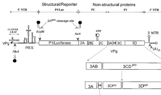
[image:2.603.134.452.70.258.2]char-acterized previously and is referred to by Rieder et al. (30) asmut7. The no-menclature for dual-creconstructs is as follows: PV or P/L (poliovirus-luciferase) refers to the parental (full-length or replicon) plasmid, followed by the genotype
FIG. 1. Schematic diagram of replicon and full-length poliovirus genomes used in this study. Open circle, wt PV-cre(2C). Arrows, positions of unique restriction endonuclease sites engineered for the insertion of the second copy ofcreof various origins. In rescue experiments, the native cre(2C) was inactivated through a point mutation (A5C) in the conserved AAA sequence of the PV-creloop (X) and a second copy ofcre(filled
hairpin loop) from poliovirus, HRV2, or HRV14 or an artificialcrewas inserted at locations indicated by arrows.
on November 8, 2019 by guest
http://jvi.asm.org/
of the native PV-cre(A5C or SLtm), followed by the genotype of the “rescuer”
and its insertion site in the genome (N,NheI; E,EcoRI; S,SacI).
P/L (A5C) is a derivative of replicon P/L (wt) (14) carrying a lethal AAA to
CAA (A5C) mutation (underlined) in the cognate PV-cre(2C) (see above) (30)
and three unique restriction sites that are used in this study: anNheI site between the cloverleaf and IRES, aSacI site at the beginning of the viral 2Aproprotein,
and anEcoRI site next to the initiating AUG (Fig. 1). Dual-creplasmids were constructed by ligation of PCR-amplified DNA fragments carrying either a point mutant PV-cre, an artificialcre, or foreigncreelements to the inactivated P/L (A5C) or triple mutant G4462A A4472C C4465U (named SLtm for stem and loop
triple mutant) replicons at eitherSacI,EcoRI, orNheI restriction enzyme sites. To introduce a point mutation (U11C) in the native HRV2-cre(2A), site-directed
mutagenesis was performed using pT7HRV2 full-length cDNA (33) as the tem-plate and oligonucleotides described in Table 1. HRV14-crewas amplified with pT7HRV14 cDNA (34) as the template.
pT7PVM(S) is a full-length poliovirus cDNA clone used as a parental con-struct that contains an engineered restriction site (SacI) between P1 and 2Apro
coding sequences (14). DNA fragments carrying the mutations in PV-, HRV2-, or HRV14-credescribed above were generated by PCR amplification from the corresponding templates and inserted at theSacI site. To assemble a dual-cre
PV1(M) with a rescuercreinserted between the cloverleaf and the IRES, a fragment betweenPmlI andAgeI in pT7PV(M) was replaced with that of the plasmid P/L (A5C)/PVwt(N).
cDNA templates corresponding to the PV-cre(2C) sequence (nt 4445 to 4504; Fig. 2) were obtained by PCR amplification from plasmid pT7PVM as described previously (30).
In vitro transcription, transfection, and plaque assay.All plasmids were linearized withDraI. Truncated derivatives of PV-creRNA used in in vitro uridylylation experiments were generated by PCR amplification using oligonu-cleotides described in Table 2. RNAs were synthesized with phage T7 RNA polymerase, and the RNA transcripts were transfected into HeLa R19 cell monolayers by the DEAE-dextran method as described previously (36). The incubation time was 12 h for luciferase replicons and up to 3 days for full-length viral constructs. Virus titer was determined by a plaque assay (20).
Analysis of recombinant viruses.To measure one-step growth kinetics, we used HeLa R19 monolayers (5⫻106cells) infected with virus at a multiplicity of
[image:3.603.54.545.75.447.2]infection of 5 to 10. The plates were incubated at 37°C, and the infected cells were harvested at 0, 1, 2.5, 3, 5, 8, and 12 h postinfection. The virus yield of the cell lysates was determined by plaque assay.
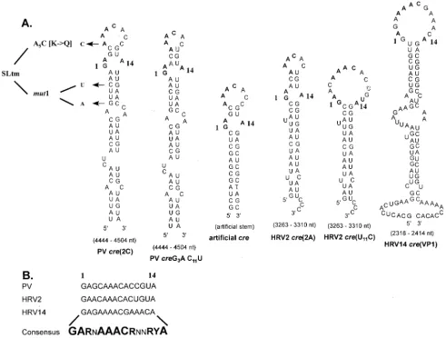
FIG. 2. Comparison of predictedcrestructures of various picornaviruses used in this study. (A) Hairpin structures predicted by the MFOLD RNA program (Michael Zuker) for wt PV-, HRV14-, HRV2-cre, HRV2-crecarrying a U11C mutation which enlarges the top loop structure, and
acreconstructed to contain 14 nt from the PV-cretop loop and an unrelated artificial stem. Arrows, locations of the substitutions in mutant PV-cres corresponding to A5C, G4462A C4465U, and SLtm. Letters in shadow style, mutations introduced in PV-creand HRV2-creto alter their loop sizes.
(B) Sequences of the top 14 nt of three naturally occurring picornaviruscres. The consensus sequence referred to in the text as the core sequence is in boldface. The degree of conservation at each nucleotide position is indicated by the size of the letter. N, any ribonucleotide; R, purine; Y, pyrimidine.
on November 8, 2019 by guest
http://jvi.asm.org/
VPg uridylylation assay.The reaction mixture contained 50 mM HEPES, pH 7.5, 3.5 mM magnesium acetate, 1 g of purified 3Dpol, 2g of synthetic
poliovirus VPg,⬃0.75Ci of [32P]UTP (3,000 Ci/mmol; DuPont NEN), 10M
unlabeled UTP, and 0.5g of template RNA transcribed in vitro from a PCR product containingcre stem-loop RNA or various PV1(M) constructs (26). Unless otherwise indicated either 1g of 3CDproor 0.5g of 3Cprowas added
to the reaction mixture. After incubation at 34°C for 30 min, the reaction was stopped by the addition of 5l of gel loading buffer. The samples were then analyzed on Tris-Tricine sodium dodecyl sulfate-polyacrylamide gel electro-phoresis gel (Bio-Rad) with 13.5% polyacrylamide. Gels were dried without fixing and autoradiographed. The incorporation of [32P]UMP into VPg-pU and
VPg-pUpU was measured on a phosphorimager (Molecular Dynamics; Storm 860).
Luciferase assay.After being transfected with replicon RNA, the monolayer cultures (35-mm-diameter dishes) of HeLa R19 cells were incubated at 37°C in standard tissue culture medium. After 12 h, the growth medium was removed from the dishes, and the cells were washed gently with 2 ml of phosphate-buffered saline. HeLa cell dishes were overlaid with 300l of “passive” lysis buffer supplied by Promega (catalog no. E194A) and rocked at room tempera-ture for 15 min, after which the lysate was transferred to a tube. Fifty microliters of luciferase assay reagent (Promega; luciferase assay system catalog no. E1501) was mixed with 20l of lysate, and the firefly luciferase activity was measured in an Optocomp I luminometer (MGM Instruments, Inc.).
Electrophoretic mobility shift and filter binding assays.Assays were per-formed as described previously with some modifications (10, 39). The radiola-beled RNAcreprobes were generated by in vitro transcription in the presence of [32P]UTP by using templates generated through PCR with primers encoding the
T7 polymerase promoter. To prevent nonspecific binding of the proteins to the RNA, an excess of nonspecific RNA (10g of tRNA) was added to all reaction
mixtures. Radiolabeled PV-cre(2C) RNA (7 nM; nt 4445 to 4504; Fig. 2) was added to the binding buffer (5 mM MOPS [pH 7.4], 25 mM KCl, 2 mM MgCl2,
20 mM dithiothreitol) containing either HeLa S10 extract or various amounts of recombinant protein, i.e., His-tagged 3Cpro, 3CDprowith or without the His tag,
3Dpol, 3AB, or PCBP2), and incubated for 10 min at 30°C. Reactions were
stopped by the addition of glycerol to a final concentration of 10%, and reaction products were loaded on a native 0.5⫻Tris-borate-EDTA–5% polyacrylamide (40:1) gel containing 5% glycerol. The cellular extracts (cytoplasmic S10 extract) were prepared as described previously (20). In filter binding assays, the reactions were performed in duplicate. A 20-l reaction mixture containing various amounts of proteins plus 1l of radiolabeled RNA (7 nM) was incubated for 15 min at room temperature, after which aliquots were filtered at low negative pressure through prewetted nitrocellulose membranes (pore size, 0.45 m; Schleicher & Schuell). Beneath the nitrocellulose membrane a nylon membrane (Boehringer Mannheim) and then a 3MM cellulose membrane were placed (23). Filters were washed once with wash buffer (20 mM HEPES-KOH [pH 6.8], 1 mM magnesium acetate, 10 mM 2-mercaptoethanol) and dried, and the radioactivity was measured by liquid scintillation counting in the presence of EcoLite scintil-lation cocktail (ICN).
RNA structure prediction.We used the MFOLD program designed by Mi-chael Zuker (http:/www.bioinfo.rpi.edu/applications/mfold).
RESULTS
Rescue of a replication-incompetent P/L (A5C) replicon with
a second copy of cre of different picornavirus origin. Previ-ously, we demonstrated that the conserved A5A6A7sequence
[image:4.603.45.540.78.314.2]of the PV-cre(2C) plays a role in the replication of poliovirus
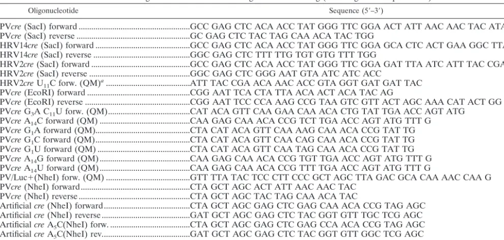
TABLE 1. Oligonucleotides used for mutagenesis and cloning (involving PCR amplification)
Oligonucleotide Sequence (5⬘–3⬘)
PVcre(SacI) forward ...GCC GAG CTC ACA ACC TAT GGG TTC GGA ACT ATT AAC AAC TAC ATA C PVcre(SacI) reverse ...GC GAG CTC TAC TAG CAA ACA TAC TGG
HRV14cre(SacI) forward ...GCC GAG CTC ACA ACC TAT GGG TTC GGA GCA CTC ACT GAA GGC TTA G HRV14cre(SacI) reverse ...GGC GAG CTC TTT TTG TGT GTG TTT TGG
HRV2cre(SacI) forward ...GCC GAG CTC ACA ACC TAT GGG TTC GGA GAT TTA ATC ATT TAC CGA AC HRV2cre(SacI) reverse ...GGC GAG CTC GGG AAT GTA ATC ATC ACC
HRV2creU11C forw. (QM)a...ATT TAC CGA ACA AAC ACC GTA GGT GAT GAT TAC
PVcre(EcoRI) forward ...CGG AAT TCA CTA TTA ACA ACT ACA TAC AG
PVcre(EcoRI) reverse ...CGG AAT TCC CCA AAG CCG TAA GTC GTT ACT AGC AAA CAT ACT GG PVcreG3A C11U forw. (QM)...CAT ACA GTT CAA GAA CAA ACA CTG TAT TGA ACC AGT ATG
PVcreA14C forward (QM) ...CAA GAG CAA ACA CCG TCT TGA ACC AGT ATG TTT G
PVcreG1A forward (QM)...CTA CAT ACA GTT CAA AAG CAA ACA CCG TAT TG
PVcreG1C forward (QM)...CTA CAT ACA GTT CAA CAG CAA ACA CCG TAT TG
PVcreG1U forward (QM) ...CTA CAT ACA GTT CAA TAG CAA ACA CCG TAT TG
PVcreA14G forward (QM) ...CAA GAG CAA ACA CCG TGT TGA ACC AGT ATG TTT G
PVcreA14U forward (QM)...CAA GAG CAA ACA CCG TTT TGA ACC AGT ATG TTT G
PV/Luc⫹(NheI) forw. (QM) ...GTT TTA TAC TCC CTT CCC GCT AGC TTA GAC GCA CAA AAC CAA G PVcre(NheI) forward...CTA GCT AGC ACT ATT AAC AAC TAC
PVcre(NheI) reverse ...CTA GCT AGC TAC TAG CAA ACA TAC
Artificialcre(NheI) forward...CTA GCT AGC GAG CTC GAG CAA ACA CCG TAG AGC Artificialcre(NheI) reverse ...GAT GCT AGC GAG CTC TAC GGT GTT TGC TCG AGC ArtificialcreA5C(NheI) forw. ...CTA GCT AGC GAG CTC GAG CCA ACA CCG TAG AGC
ArtificialcreA5C(NheI) rev...GAT GCT AGC GAG CTC TAC GGT GTT GGC TCG AGC
PVcre(SLtm) forward (QM)...CTA TTA ACA ACT ACA TAC AAT TTA AGA GCC AAC ACC GTA TCG AAC
aQM, oligonucleotide used for QuikChange mutagenesis.
TABLE 2. Oligonucleotides used for the synthesis of truncated PV-cre(2C)
Oligonucleotide Sequence (5⬘–3⬘)
T7⌬1creforward ...GGT GCC GGC TAA TAC GAC TCA CTA TAG GCT ACA TAC AGT TCA AGA GCA AAC ⌬1crereverse...AAG AAT TCC AAA CAT ACT GGT TCA ATA CGG TGT TTG CTC TTG AAC TGT T7⌬2creforward ...GGT GCC GGC TAA TAC GAC TCA CTA TAG GTT CAA GAG CAA AC
⌬1crereverse...CAA GAA TTC CTA GGG TTC AAT ACG GTG TTT GCT CTT GAA CCT ATA G T7⌬3creforward ...GTG CCG GCT AAT ACG ACT CAC TAT AGG CAA ACA CCG TAT TG ⌬3crereverse...AAG AAT TCC TAG GGT TCA ATA CGG TGT TTG CCT ATA GT
on November 8, 2019 by guest
http://jvi.asm.org/
[image:4.603.42.545.650.724.2]RNA (30). For example, the A5C mutation in the loop of
PV-cre(2C) rendered the resulting virus nonviable, and no revertants could be recovered even after five blind passages in HeLa R19 cells. In agreement with these findings a P/L (A5C)
replicon carrying the same mutation in the endogenous
PV-cre(2C) showed little luciferase activity after transfection (see below). The A5C mutation leads to amino acid change K117Q
in 2CATPase. Available evidence suggests that this change does
not significantly interfere with the 2CATPaseactivity (29, 30)
and that the effect of the mutation is due to the inactivation of the cre signal. To provide experimental evidence for this as-sumption and to allow for extensive genetic analysis of thecre
element, we introduced a second copy of wt PV-cre(63 nt from 4444 to 4506) at different locations (Fig. 1). For example, the wt cre was inserted between the coding sequence for the C terminus of P1/luciferase and that for the N terminus of 2Apro
(Fig. 1, SacI site inserts). To facilitate the processing of the modified polyprotein, a 2Apro cleavage site was introduced
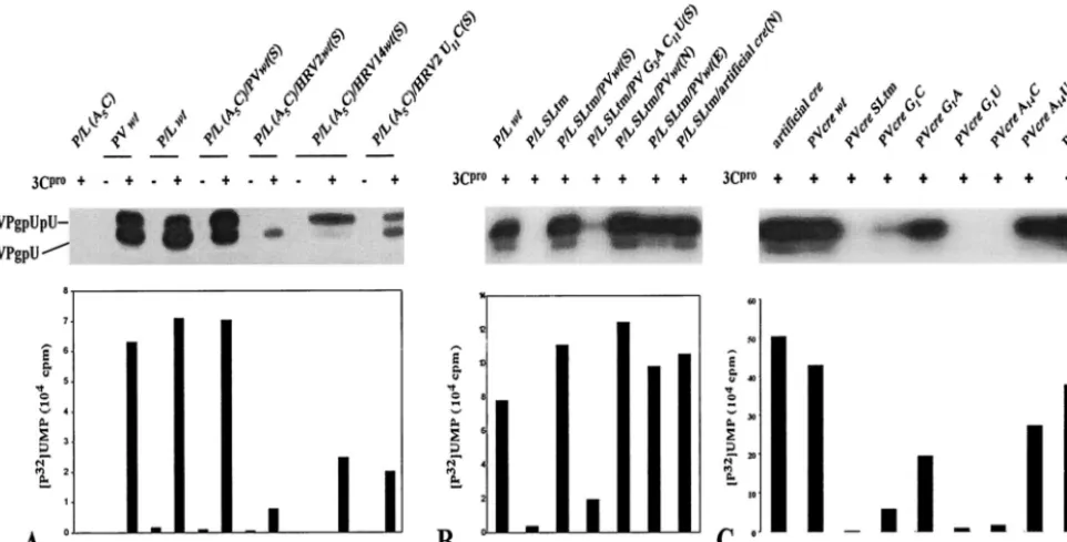
between the translation product of the new cre and the P1/ luciferase so that after translation the extra 21 amino acids encoded by thecrewould be cleaved out of the polyprotein. As shown in Fig. 3A, the second copy of wt PV-creat this position can restore the replication of an inactive P/L (A5C) replicon to
that of the wt P/L (Fig. 3A, compare lanes 3 and 5 with lane 1). Furthermore, a dual-crereplicon carrying two mutant copies of A5Ccre(Fig. 3A, lanes 21 and 22) failed to show any luciferase
activity above the background signal. The background signal was determined by repeating the experiments in the presence of 2 mM guanidine-HCl, an inhibitor of poliovirus RNA syn-thesis (Fig. 3A, even-numbered lanes). Taken together, the data, in agreement with previous results, suggest that (i) the 63-nt PV-creis a functional signal for the replication of polio-virus RNA in vivo (9, 26, 30), (ii) the function of PV-cre is independent of the cognate amino acid sequence of the viral 2CATPase protein, and (iii) a functional cre element can be
moved to a different location within the poliovirus genome (9). We have recently shown thatcreelements from HRV14 and poliovirus can be functionally exchanged in our in vitro uridy-lylation assays using poliovirus 3Dpol(26). Therefore, we were
interested in determining whether heterologouscre elements could rescue the replication of a P/L (A5C) replicon carrying a
lethal mutation in the native PV-cre(2C) by engineering the foreigncreelements into dual-creRNAs. As shown in Fig. 3A (lane 13) HRV14-crerestored the replication level of this rep-licon to 70% of that of wt dual-cre(lane 5). HRV2-cre, on the other hand, barely (3%) rescued the replication of the dual-cre
replicon (Fig. 3A, compare lanes 5 and 9). This is in agreement with our findings that HRV2-cre(2A) is a very poor template for poliovirus 3Dpol in VPg uridylylation (A. V. Paul and E.
Wimmer, unpublished data).
Rescue by genetically altered PV-cre(2C), HRV2-cre(2A), or artificialcre’s.As shown in Fig. 2A, PV-, HRV2-, and
HRV14-crediffer significantly from each other in their primary nucle-otide sequences. However, we have highlighted a consensus sequence of 14 nt (named core in this paper; Fig. 2B) that is highly conserved among allcrestructures. Curiously, although HRV2-cre appears to be more similar to PV-cre than to HRV14-crein its core sequence, HRV14-cre was much more efficient in rescuing a debilitated (A5C) dual-crereplicon than
HRV2-cre (Fig. 3A, compare lane 5 with lanes 9 and 13). A
major difference between the upper portions of HRV2-creand HRV14-creis the size of the loop. We were interested to test the hypothesis that a predicted “tight” structure in the core sequence of HRV2-cre that leads to base pairing of A5
pre-vented it from being an efficient rescuer in the context of the poliovirus genome. Therefore, a point mutation (U11C) was
engineered into the core sequence of wt HRV2-creso that the predicted secondary structure for the altered HRV2-cre resem-bles that of the HRV14-cre top loop. Indeed, the altered HRV2-cre was about 10-fold more efficient in rescuing a
de-FIG. 3. Luciferase activities of various P/L replicons as a measure of RNA replication. (A) RNAs from dual-creP/L replicons with sec-ond rescuercreelements derived from poliovirus, HRV2, or HRV14. Transfection of RNA and luciferase activity measurements are de-scribed in Materials and Methods. Even-numbered lanes, transfections repeated in the presence of 2 mM guanidine-HCl (GnHCl). arts, artificial. (B) Dual-creluciferase replicons with the rescuercrefrom poliovirus carrying point mutations in G1, A2, and A14in the corecre
sequence were tested for their ability to replicate in HeLa R19 cells. The nomenclature is as follows. P/L refers to the parental replicon plasmid followed by A5C when it carries an inactivating mutation in
the 2CATPasecoding sequence. This is followed by the sequences of the
rescuercreand the insertion site (N,NheI; E,EcoRI; S,SacI).
on November 8, 2019 by guest
http://jvi.asm.org/
bilitated (A5C) PV-crethan the wt HRV2-cre(Fig. 3A,
com-pare lane 5 with lanes 9 and 11). The reason why the recovery of luciferase activity is still low compared to that for PV-creor HRV14-creis not known but is undoubtedly related to thecre
structure and its interaction with the proteins involved in uri-dylylation. Perhaps the presence of an unpaired U residue in the upper region of the stem is responsible for the low activity in the context of a poliovirus genome (30). In some cases, a relationship between higher-order structure and function can be predicted. For example, a mutant PV-crecarrying two point mutations (G3A and C11U) in its core sequence has a predicted
loop structure identical to that of wt HRV2-cre. Indeed, this variant PV-creproduced only 8% luciferase activity when used as a rescuercrein the dual-crereplicon (Fig. 3A; compare lanes 5 and 7).
The core sequences of PV-, HRV2-, and HRV14-cre are relatively more conserved than the rest of the hairpin sequence (Fig. 2B). Along with the A5A6A7sequence, the first two (G1
and A2) nucleotides and the last (A14) nucleotide were among
the most conserved. Mutations A2C and A2U did not influence
the ability of the secondcreelement to rescue a lethal mutation in the cognatecre(data not shown). On the other hand, some G1 and A14mutants [P/L (A5C)/PV G1A(E), P/L (A5C)/PV
A14G(E), and P/L (A5C)/PV A14U(E)] displayed significant
reduction in the level of replication compared to wt P/L (Fig. 3B, compare lane 7 with lanes 1, 4, and 6). Interestingly, other G1 mutants [P/L (A5C)/PV G1C(E), P/L (A5C)/PV G1U(E),
and A14(P/L) A5C/PV A14C(E)] exhibited very little if any
replication signal (Fig. 3B, compare lane 7 with lanes 2, 3, and 5). Currently, we have no explanation for the remarkable in-tolerance for transversions at the G1position, but it is likely
related to the reduced ability of this mutant to bind 3CDpro
(see Fig. 9C). A possible explanation for the relatively high activity of the A14U replicon (lane 6), compared to that of the
A14C replicon (lane 5) will be given in Discussion.
The stem structures beneath the core sequence display the biggest diversity among thecrestructures studied. To evaluate the role of sequences located in the stem region beneath the core sequence, we constructed an artificialcreconsisting of an authentic PV-cre core (G4468AGCAAACACCGUA4481)
se-quence and an artificial stem of unrelated nucleotide sese-quence (Fig. 2A). This stem was designed to accommodate an NheI restriction site for easy insertion between the cloverleaf struc-ture and the IRES element in the 5⬘NTR (Fig. 1) and will be discussed below. As seen in Fig. 3A, this artificial cre, when inserted into the P/L (A5C) replicon genome, efficiently
res-cued the lethal phenotype caused by an A5C mutation in the
native PV-cre. This observation suggests that the structure (i.e., scaffolding) of the stem beneath the core sequence of various
cres is more important than its nucleotide sequence.
Rescue of replication by wt PV-cre(2C) placed at different locations in the P/L replicon. To determine if the genomic position of PV-creaffects its role in poliovirus replication, we tested the effect of a rescuing acreelement at three locations in the dual-cre replicon genome: inside the polyprotein open reading frame [ORF; P/L (A5C)/PVwt(S)], in the beginning of
the ORF [P/L (A5C)/PVwt(E)], and in the noncoding region in
the 5⬘NTR between the cloverleaf and the IRES element [P/L (A5C)/PVwt(N)]. The result showed that the location of thecre
elements has very little effect on the replication of the genome (Fig. 3A, compare lane 1 with lanes 5, 15, and 17).
The efficient rescuing function of the polioviruscreelement outside the ORF was surprising. As mentioned above, even an artificialcreelement inserted into theNheI site allowed repli-cation of the RNA carrying a lethal A5C mutation in the
cognate cre element (Fig. 3A, compare lanes 17 and 19). In contrast, an artificial cre with an A5C mutation in the core
sequence did not rescue replication (Fig. 3A, compare lanes 17 and 21), an observation proving that RNA synthesis strictly depended on a functional cre, regardless of its locus in the genome.
In vitro uridylylation of VPg using dual-crepoliovirus RNAs as templates.We have previously developed an in vitro assay in which the viral RNA-dependent RNA polymerase 3Dpol
trans-fers a UMP to the tyrosine residue in the small viral protein VPg, by using poly(A) as the template (27). We further made the discovery that such a reaction was stimulated (by 1 order of magnitude) by the presence of viral protein 3CDpro when
ge-nome RNA or thecresequence was used as the template (26, 30). Enhancement of uridylylation was observed also with 3Cpro (23). Since the stimulatory activity of 3Cpro is more
reproducible than that of the much larger (His-tagged)
3CD-pro, we used 3Cproin uridylylation reactions catalyzed by
dual-cre replicon RNAs. As shown in Fig. 4A, in the absence of 3Cpro, there was little uridylylation of VPg by 3Dpolwhen any
of the different RNAs were used as the template (Fig. 4A, lanes 2, 4, 6, 8, 10, and 12). The P/L (A5C) replicon RNA
carrying the lethal A5C mutation incre(2C) failed to function
as a template for the uridylylation of VPg (Fig. 4A, lane 1), as expected (30). In the presence of 3Cpro, only RNAs with a
functional cre were able to direct significant uridylylation of VPg above the background level (Fig. 4A, lanes 3, 5, 7, 9, 11, and 13). This is in good agreement with the observed replica-tion levels of these replicons in tissue culture, as determined by measuring luciferase activity (Fig. 3A, lanes 1, 5, 9, 11, and 13). This was also observed for the mutated HRV2-cre (U11C),
which is a better template for uridylylation of VPg in vitro than the dual-crereplicon carrying wt HRV2-cre(2A) (Fig. 4A, com-pare lanes 9 and 13). Reciprocally, when the loop (14 nt) of PV-cre(2C) was converted to that of HRV2-cre(2A), the hybrid
cre(PV G3A U11C) not only displayed a much-reduced ability
to replicate (Fig. 3A, lane 7) but also was a poor template for the uridylylation of VPg in vitro (Fig. 4B, lane 4).
The different location of the rescuer copy of PV-cre(2C) did not influence significantly its template activity for the uridyly-lation of VPg in vitro, as shown in Fig. 4B. The dual-cre rep-licons that displayed wt P/L luciferase activity (Fig. 3A, lanes 1, 5, 15, and 17) in HeLa cells also exhibited VPg uridylylation levels similar to that of the wt construct (Fig. 4B, lanes 1, 3, 5, and 6). This further suggests that thecre(2C) RNA sequence functions as a replication signal independently of the function of the viral 2CATPase and local RNA-RNA interactions. Not
surprisingly, the dual-crereplicon carrying the artificialcreas the rescuer is a good template for both the uridylylation of VPg in vitro (Fig. 4B, lane 7) and replication in HeLa R19 cells (Fig. 3A, lane 19).
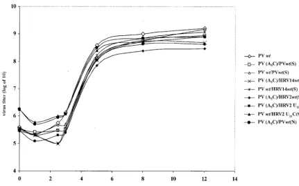
One-step growth curve of dual-crepolioviruses.To examine to what extent rescue by cognate or foreign cres influences virus yield, we tested virus production with genomes carrying a
on November 8, 2019 by guest
http://jvi.asm.org/
lethal A5C mutation in PV-cre(2C) as well as a second cre
(PV-cre, HRV2-cre, HRV14-cre, or HRV2-creU11C) element.
Two locations of the second cre were used for these experi-ments: (i) between the P1 and P2 coding sequences and (ii) between the cloverleaf and the IRES element in the 5⬘NTR. As shown in Fig. 5, the dual-creviruses with a second copy of functional viral cre displayed similar growth kinetics. These results suggest that, in vivo, the uridylylation of VPg might not be the rate-limiting step of poliovirus RNA replication. Note that HRV2-cre, a weaker template for in vitro VPg uridylyla-tion by poliovirus 3Dpol (Fig. 4A, lane 9), still stimulated the
dual-crevirus to amplify by over 2 orders of magnitude at 12 h postinfection (Fig. 5). The results also suggested that the lo-cation of the rescuer cre has little effect on the recovery of virus, as PV (A5C)/PVwt(N) has growth kinetics similar to
those of PV (A5C)/PVwt(S) and both proliferated at a rate
comparable to that for wt PV (Fig. 5).
To exclude the possibility that the insertion of a secondcre
impairs replication of the resulting virus, we also inserted the abovecreelements into the wt poliovirus genome and studied the resulting viruses in one-step growth experiments. As shown in Fig. 5, a dual-crevirus carrying wtcrein the 2C coding region grew just as well as wt poliovirus, regardless of the content of the second cre, and slightly better than the dual-cre viruses carrying the A5C mutation.
The growth phenotypes of the constructs carrying mutations in thecre element could also be due to rapid mutation to wt sequences or second-site suppressor mutations. Sequence analysis of the genomic RNAs recovered after two passages in HeLa cells did not reveal changes in thecresequences. This
result excludes direct reversion, but it does not eliminate sec-ond-site suppressor mutations. This interesting possibility is currently under investigation.
In vitro uridylylation of VPg using PV-creG1and A14
mu-tants and artificialcreRNAs as templates.We were also in-terested in the ability of PV-cre’s to direct VPg uridylylation when either the G1or A14nucleotides were altered. As shown
in Fig. 4C, transversions at these positions caused a strong defect in the protein priming reaction. Mutant PV-cres carry-ing either G1C or G1U or A14C mutations in the core cre
sequence lost more than 90% of VPg uridylylation activity compared to thewtPV-cre(Fig. 4C, compare lane 2 with lanes 4, 6, and 7), whereas PV-creA14U is about 50% as functional
as wt PV-cre(Fig. 4C, compare lanes 8 and 2). On the other hand, transitions are better tolerated at both nucleotide posi-tions: PV-creG1A and PV-creA14G retained 50 and 90% of wt
PV-cre activity for the uridylylation of VPg in vitro, respec-tively (Fig. 4C, compare lane 2 with lanes 5 and 9). As a negative control, mutant PV (SLtm) (Fig. 2) was constructed to carry the debilitating A5C change and two point mutations
in the upper stem region (G4462A and C4465U); these three
mutations are predicted to alter the secondary structure ofcre
drastically. As expected, PV (SLtm) showed only a background level signal for the in vitro uridylylation of VPg (Fig. 4C, lane 3), which is in accordance with previous findings with full-length viral RNA and poliovirus replicons (9, 30). These results suggest a preference for purine at the N1and N14nucleotide
positions in the core sequence of PV-cre(2C), specifically a G1
and an A14.
We also tested our artificial cre, carrying the wt core
se-FIG. 4. In vitro uridylylation of VPg using either a dual-creP/L replicon or mutated PV-creRNAs as the template. Uridylylation reactions were performed as described in Materials and Methods. (A) Dual-crereplicons carrying a lethal (A5C) mutation in the 2CATPasecoding sequence with
a rescuercrefrom HRV14 or HRV2 or a mutant U11C HRV2-cre(predicted by the Zuker program to enlarge the terminal loop of HRV2-creto
resemble that of HRV14-cre). (B) VPg uridylylation using inactivated dual-cre(SLtm; triple mutant) replicons as templates and the rescuer wt PV-creinserted at different locations. (C) Mutant or wtcreRNAs as templates in VPg uridylylation.
on November 8, 2019 by guest
http://jvi.asm.org/
[image:7.603.51.532.74.318.2]quence from PV-cre(2C) and a stem of unrelated nucleotide sequence, in the in vitro VPg uridylylation assay. As shown in Fig. 4C (compare lanes 1 and 2), the artificial creis as func-tional as the wt PV-crefor directing the synthesis of VPg-pU and VPg-pUpU under the experimental conditions, in agree-ment with our data for the dual-crereplicon shown in Fig. 3A (lane 19).
Analysis of full-length poliovirus genomes carrying muta-tions within the loop of PV-cre(2C).Further characterization of the effect of mutations within the core PV-cresequence was carried out by using mutant full-length polioviruses. The growth phenotype and uridylylation activities of these full-length constructs are shown in Fig. 6A and B, respectively. Similar to the wt, C8A, A9G, and C8U A9G mutants developed
complete cytopathic effect (CPE) after 24 h of incubation at 37°C posttransfection. A delay in the appearance of CPE was observed with the A9U mutant, an observation suggesting that
this mutant RNA may be impaired in replication. We sus-pected that the inefficient replication may lead to a reversion or other genetic alterations. This was confirmed by sequence analysis (reverse transcription-PCR) of RNA extracted from the virus that was collected after three passages following the original transfection. It reveals a reversion at the site of mu-tagenesis (U9A) to the original codon (resulting in a change
from Leu118 to His). In addition, we detected a change at
position A7G in the core sequence (synonymous mutation). No
CPE was detected with C10U C11U mutant RNA even after
three additional blind passages in HeLa R19 cells. This result is not unexpected since the substitutions introduced into this double mutant are likely to lead to base pairing with the critical
A5and A6nucleotides in the corecresequence. This is
sup-ported only by computer-aided folding; no biochemical evi-dence has been generated. The C10U C11U mutation also
greatly reduces the yield of VPg-pU and VPg-pUpU products in in vitro uridylylation (Fig. 6B). The results are also consis-tent with the data from dual-crereplicons [P/L (A5C)/PV G3A
C11U(S)] shown in Fig. 3A, where a mutation in the PV-cre
loop which reduces the size of the terminal loop reduced lu-ciferase activity 10-fold relative to that for the P/L carrying wt PV-cre. However, since the double mutation changes the amino acid sequence from H118R119 to H118C119, we cannot
rule out the possibility that this may critically impair 2CATPase
function. The C10U and C11U mutations have not yet been
tested in a dual-creconstruct.
All mutant versions of the full-length RNAs tested above are functional templates in the in vitro VPg uridylylation reaction (Fig. 6B). However, smaller amounts of pU and VPg-pUpU products were produced than were produced with wt PV1 RNA (Fig. 6B, compare lane 1 with lanes 3, 5, 7, 9, and 11). Among them, only one (C10U C11U mutant; 10% of wt
activity in uridylylation; Fig. 6B, compare lane 1 with lane 7) rendered the genome nonviable. Polypeptide 3CDpro
signifi-cantly stimulated uridylylation in all cases, as its absence greatly reduced synthesis of the VPg-pU and VPg-pUpU prod-ucts (Fig. 6B, lanes 2, 4, 6, 8, 10, and 12). The existence of viable mutants containing amino acid changes at His118 in
2CATPase suggests that this His residue is not essential for
2CATPasefunction in the replication of poliovirus (Fig. 6A).
However, whereas the His118Arg change was tolerated, a the
[image:8.603.77.509.70.339.2]His118Leu change produced a replication phenotype that led to
FIG. 5. One-step growth curves for various dual-crepolioviruses. HeLa R19 cells were infected with virus derived from wt and mutant poliovirus cDNA plasmids, and the virus yield was determined at the indicated time points (see Materials and Methods).
on November 8, 2019 by guest
http://jvi.asm.org/
reversion to the original codon and amino acid. Since this reversion is accompanied by a synonymous change in the pre-ceding codon, it is impossible to decide whether the growth phenotype of the A9U mutant resulted from the amino acid
change, from the structure of thecre, or both. Finally, a G1U
mutation in cognate PV-cre(2C) of poliovirus RNA rendered the genome nonviable, even after three blind passages in HeLa cells (data not shown). Taken together, the data support the hypothesis that critical features of thecrerequired for repli-cation are contained in a 14-nt core sequence in the apical portion of the RNA stem-loop.
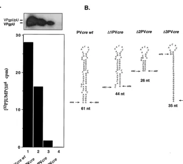
Minimal length of PV-cre(2C) required for uridylylation.
We have performed experiments to analyze the relationship between the length of the stem in PV-cre(2C) and VPg uridy-lylation. Two truncated PV-creRNAs were synthesized in vitro and tested as templates in the in vitro uridylylation assays. As shown in Fig. 7, both RNAs were able to serve as templates, but the yield was reduced to 70 to 80% of that with the wt PV-cre[⌬1 PV-cre(2C)] and to 25% of that with⌬2 PV-cre(2C) (compare lane 1 with lanes 2 and 3). The data indicate that the upper stem and loop of PV-cre(2C) carry the minimal struc-tural determinants for the assembly of a uridylylation-compe-tent complex. On the other hand, an RNA spanning the se-quence 4470 to 4504 [⌬3 PV-cre(2C)] of the poliovirus genome is totally inactive as a template in the reaction (Fig. 7, compare lanes 1 and 4), suggesting that the sequence determinants
contained in this region must be contained within a certain spatial arrangement to be recognized by the proteins of the uridylylation complex.
Proteins binding to the poliovirusoriI [PV-cre(2C)].The function of PV-cre(2C) is likely to involve an RNP complex consisting of more than one protein. As shown previously, PV-cre(2C) serves as an efficient template for 3Dpol-catalyzed
VPg uridylylation only if 3CDproor 3Cpro is present (23, 26)
(see our results above). We therefore used a binding assay to test interactions between viral and possibly cellular polypep-tides and PV-cre(2C).
We first investigated if nonstructural proteins that originated from the P3 region of the polyprotein (3CDpro, 3AB, and
3Dpol) and the cellular protein PCBP2 possess specific affinity
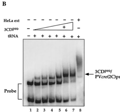
to PV-cre(2C) by gel shift assays, carried out in the presence of a 1,000-fold excess of nonspecific competitor tRNA (10). Pu-rified polypeptide 3AB, known to be a nonspecific RNA-bind-ing protein (25, 38), does not bind to PV-cre(2C) under these conditions (Fig. 8A, compare lane 1 with lane 6). In contrast, 3CDproand its cleavage product 3Cproretarded the mobility of
the labeled probe in the presence of the 1,000-fold excess of tRNA (Fig. 8A, lanes 2 and 4, respectively). Gel shift assays carried out with increasing amounts of 3CDpro-His showed
[image:9.603.105.487.68.342.2]that the amount of RNP complex formed is dependent on the concentration of the protein in the reaction mixture up to about 400 nM 3CDpro(Fig. 8B, lanes 2 to 7, and 9A). This is
FIG. 6. Summary of the effect of nucleotide substitutions within the PV-cre(2C) loop in full-length PV1(M) genome RNA on genome replication and uridylylation. (A) Phenotypic properties of viruses derived from transfection experiments with HeLa R19 cells. Following transfection into HeLa cells the cell lysates were subjected to three additional passages and the viral RNAs were sequenced following reverse transcription-PCR, as described in Materials and Methods. The letters above the nucleotides indicate the deduced amino acid sequence and its location within 2CATPase. Mutations or reversions in nucleotides are in boldface and italics. Nucleotide reversions are shown with lowercase letters.
(B) VPg uridylylation was assayed as described in Materials and Methods with full-length wt or mutant RNAs as templates. Where indicated,
poliovirus 3CDprowas added to the reaction mixtures.
on November 8, 2019 by guest
http://jvi.asm.org/
true regardless of whether the recombinant 3CDprois attached
to a His tag at the C terminus (Fig. 8) or is free of any extra tag sequence (Fig. 9A). The formation of an RNP complex be-tween 3CDproand PV-cre(2C) could be competed by the
ad-dition of a 200-fold excess of nonlabeled PV-cre(2C) RNA (Fig. 8C). Moreover, this RNP complex is similarly susceptible to competition by the addition of an unlabeled plus sense cloverleaf RNA but not by its cRNA sequence (referred to as a minus sense cloverleaf; Fig. 9B). This competition experi-ment supports our claim of specificity since plus strand clover-leaf RNA is known to bind 3CDpro(4, 10, 41). As expected, a
100-fold excess of domain II RNA of the poliovirus IRES, a hairpin containing an AAACCA sequence in the loop region, did not compete in 3CDpro/PV-cre(2C) complex formation
(data not shown).
As 3AB did not bind to PV-cre(2C), it also did not supershift a 3CDpro/PV-cre(2C) complex (data not shown). This is in
contrast to the formation of a 3AB/3CDpro/cloverleaf complex,
which we have shown previously to be essential for plus strand RNA synthesis (10, 38, 39). Similarly, the cellular RNA binding protein PCBP2 can form a complex with 3CDpro and the 5⬘
cloverleaf (7, 21). Just like 3AB, purified PCBP2 did not bind PV-cre(2C) RNA (Fig. 8A, lane 3), nor did it supershift the
3CDpro/PV-cre(2C) complex (data not shown). These results
are consistent with our previous report showing that the addi-tion of purified 3AB or PCBP2 had no effect on the uridylyla-tion of VPg in the presence of 3Dpol, 3CDpro, and PV-cre(2C)
(26). It is likely therefore, that 3AB and PCBP2 do not play a direct role in VPg uridylylation.
The viral RNA polymerase 3Dpoldid not bind PV-cre(2C)
RNA under the conditions of the gel shift assay (Fig. 8A, lane 5) and was also unable to influence the formation of the 3CDpro/PV-cre(2C) complex (Fig. 9A) in filter binding
exper-iments. Notably, 1g of 3Dpol, the amount of protein normally
used in uridylylation experiments, did not affect the dose-re-sponse curve resulting from the binding of increasing amounts of 3CDproto the PV-creprobe (Fig. 9A).
[image:10.603.115.484.71.402.2]Components of an S10 HeLa cell extract formed one or more RNP complexes with PV-cre(2C), even in the presence of competitor tRNA (Fig. 8B, lane 8). Preliminary data suggested that a 50-kDa protein present in HeLa cell extracts is the major component that binds PV-cre(2C) (data not shown). However, early studies have indicated that the addition of an extract of uninfected HeLa cells did not stimulate the uridylylation reac-tion under the condireac-tion of the experiments (26). Currently we
FIG. 7. In vitro uridylylation using shortened PV-cre(2C) RNAs as templates. (A) Effect of truncation on the ability of PV-cre(2C) RNA to serve as the template in VPg uridylylation. The uridylylation of VPg was measured by the standard method described in Materials and Methods. The yield of the reaction (graph) was shown by autoradiography. (B) Schematic drawing of the secondary structure of complete and truncated PV-creRNAs utilized in this experiment. The first and last nucleotides as well as the length of each RNA template are indicated.
on November 8, 2019 by guest
http://jvi.asm.org/
do not know the identity of the cellular protein, nor do we know its function.
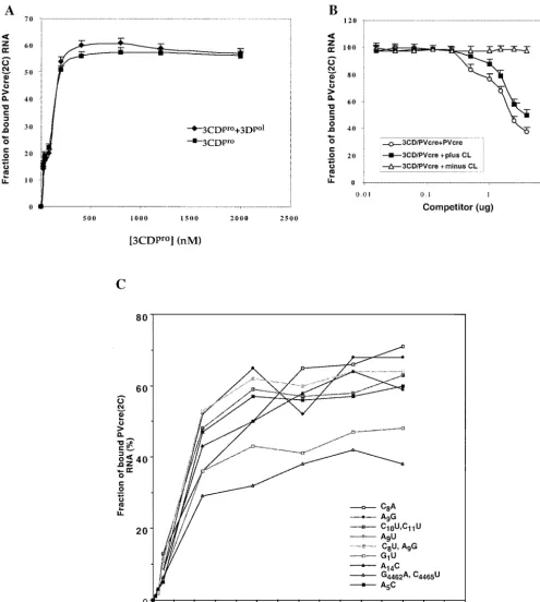
To test whether mutations in the loop or stem of PV-cre(2C) affect the binding to 3CDpro, we performed filter-binding
[image:11.603.272.511.76.298.2]as-says with selected mutant PV-cre RNA probes. As shown in Fig. 9C, use of C8A, A9G, C10U C11U, A9U, C8U A9G, or A5C
loop mutant RNA or A14C stem mutant RNA resulted in the
retention of amounts of 3CDpro on the nitrocellulose
mem-brane similar to that resulting from the use of the wt PV-cre
probe. Interestingly, quantification of the binding of the PV-cre
G1U and G4462A C4465U (mut1 in reference 30) mutant RNAs
revealed an 8- to 15-fold reduction in binding to 3CDpro, as
judged by a decreased amount of RNP complex retained on the nitrocellulose membranes. The diminution in binding of PV-creG1U and G4462A C4465U RNAs by 3CDprois in
agree-ment with our uridylylation studies and the phenotypic prop-erties of either the full-length viral genome (G4462A C4465U)
(30) or a replicon RNA (G1U) (Fig. 3B).
DISCUSSION
Originally, the highly structured 5⬘and 3⬘NTR sequences of entero- and rhinovirus genomes were considered necessary and sufficient for the initiation of RNA replication (1). The discovery of internalcreelements, first described by McKnight and Lemon (18) for HRV14, which are also essential for viral RNA replication, came as a surprise. Similarly surprising was the subsequent observation that these elements can direct 3Dpol-catalyzed uridylylation of VPg, thereby providing the
primer for the initiation of RNA synthesis (8, 26, 30). A sug-gestion that the poliovirus cloverleaf is involved in VPg uridy-lylation was provided in a recent report by Lyons et al. (16). This finding, together with the recent model of the circulariza-tion of the poliovirus genome prior to RNA synthesis (11), suggests that at least three RNA domains (5⬘-terminal clover-leaf,cre, and 3⬘-terminal poly[A]) must interact with viral and cellular polypeptides to orchestrate the initiation of minus
FIG. 8. Relative abilities of poliovirus and HeLa cell proteins to form complexes with PV-cre(2C) RNA. The electrophoretic mobility shift assays were carried out with a32P-labeled PV-cre(2C) probe in the
presence of a 1,000-fold excess of tRNA as described in Materials and Methods. (A) Lane 6, radiolabeled PV-cre (63-nt, 7 nM) probe in binding buffer; lane 1, 3AB (0.8M); lane 2, 3CDpro(His tagged, 0.5
M); lane 3, PCBP (1.1M); lane 4, 3Cpro(1.2M); lane 5, 3Dpol(1
M). (B) Binding curve for PV-cre/3CDprowith increasing amounts of
3CDpro. Lane 1, PV-creprobe in binding buffer; lanes 2 to 7, probe in
binding buffer with increasing amounts (0.05, 0.13, 0.2, 0.4, 0.5, and 0.6 M) of 3CDpro(His tagged); lane 8, PV-creprobe in binding buffer
and HeLa cell extract proteins (15g). (C) Competition for the bind-ing of 3CDproto the radiolabeled PV-cre probe (63 nt, 7 nM) with
unlabeled PV-cre. Binding reactions were performed in the absence (lane 1) or in the presence (lanes 2 to 7) of recombinant 3CDpro(His
tagged). Lanes 3 to 7 contain radiolabeled PV-creprobes in binding buffer, purified 3CDpro(0.5M; His tagged), and increasing
concen-trations (from 10 to 500 M excess) of unlabeled PV-creRNA as the competitor.
on November 8, 2019 by guest
http://jvi.asm.org/
FIG. 9. Filter binding assays to assess the effect of addition of 3Dpolor competitor (mutant PV-creor cloverleaf) RNAs on the binding of
3CDproto PV-cre(2C). (A) Binding reaction between PV-cre(2C) (63 nt, 7 nM) and 3CDpro(0.01 to 0.8M, untagged) performed in the absence
(squares) or in the presence (diamonds) of 1M 3Dpol. 3CDproused in this experiment and that described in the panel B legend is a recombinant
(3Cpro/H40A) protein that does not carry additional tag sequences (23). The filter binding data collected are shown as the means of three
measurements at each data point. (B) Effect of plus and minus strand poliovirus cloverleaf and wt PV-creon the binding of 3CDproto PV-cre.
Competition was performed in the presence of either PV-cre(circles) or plus strand cloverleaf (squares) or minus strand cloverleaf (triangles) RNA competitors. Quantitative analysis of the 3CDpro/PV-crecomplexes was carried out by using a filter-binding assay as described in Materials and
Methods. (C) Comparison of the relative binding affinities of wt and mutant PV-creRNAs for the site within 3CDproin a filter-binding assay as
described in Materials and Methods.
on November 8, 2019 by guest
http://jvi.asm.org/
strand synthesis. The details of these events are not yet under-stood.
We have focused on thecreelements of poliovirus, HRV14, and HRV2 to decipher structural parameters by genetic and biochemical analyses. With the exception of those of foot-and-mouth disease virus (17), allcreelements described so far are located in the ORFs encoding components of polyproteins (8, 9, 15, 18). Genetic analysis requires site-directed mutagenesis of the cognate PV-cre(2C) without destroying 2CATPase
func-tion, a strategy that is severely limited. Therefore, in this study, the cognate cre was inactivated by a single point mutation (A5C) and a secondcrewas inserted to rescue genome
repli-cation. Since McKnight and Lemon (18) and Goodfellow et al. (9) had shown previously that HRV14-creand PV3-cre, respec-tively, could be moved from their original locations, our strat-egy was likely to work. Indeed, the data presented here show that a secondcreelement, even if it was derived from a differ-ent picornavirus, was efficidiffer-ent in rescuing replication. As any genetic alteration at the second cre does not complicate the proper processing and functioning of viral proteins, the
dual-cresystem was suitable for genetic analyses. Remarkably, the location of the secondcrehad no detectable effect on its res-cuing function, even when it mapped to the region between the cloverleaf and the IRES.
Our genetic and biochemical analyses have led to the des-ignation of a core sequence (Fig. 2B) essential forcrefunction. A similar core sequence has been proposed very recently also by Yang et al. (40) based on an analysis of HRV14-cre. Muta-tions within the conserved 14-nt core sequence (Fig. 2) pro-duce phenotypes that suggest that several purines within this core sequence, i.e., G1, A5, A6, and A14, are necessary forcre
function. Interestingly, transition mutations in the loop (C10U
and C11U) that may lead to base pairing with A5 and A6
abolished cre function in uridylylation and rendered the ge-nome nonviable even after four passages. This observation indicates that a degree of structural integrity in the core se-quence is essential for its function. The preferred nucleotide in position 1 of the core is the purine G, which is highly sensitive to base pairing since a mutant carrying an A14C transversion
reduces the luciferase signal dramatically in the dual-cre rep-licon system and nearly abolishescre-dependent uridylylation of VPg.
Although PV-cres with mutation in G1 or A14of the core
sequence produced a reduction increfunction when used as a rescuer in the dual-crereplicon system, the role of G1appears
to be more important than that of A14. Transversions are not
tolerated at the G1position, whereas the A14U mutant is still
active in VPg uridylylation in vitro and in the rescue function in vivo. Initially, we were surprised about the functional activity of the A14U mutant since either a C or a U at nt 14 of the core
sequence could base pair with G1. However, since U14has the
option to base pair with A2and since A2is not essential forcre
function (J. Yin and E. Wimmer, unpublished data), an A2::U14base pairing may not be debilitating. Perhaps the
ob-served conservation of an A at position 14 is solely due to the fact that this A is the least favorable nucleotide to base pair with G1.
Experiments with truncated PV-cre elements (Fig. 7) sup-port our previous finding that imsup-portant structural determi-nants forcrefunction are contained in the upper stem and loop
region.⌬2 PV-cre(2C) RNA, consisting of only 26 nt, can still function as a template for VPg uridylylation although the tem-plate activity is greatly reduced. The artificial cre has even fewer virus-specific nucleotides, yet it is not only an excellent template for uridylylation in vitro but also an efficient rescue element in vivo (Fig. 3 and 4). These results suggest that the sequences contained in the middle and bottom regions of the
crestem might be required only for proper structural presen-tation of the core sequence. This conclusion is in agreement with evidence derived from mutational analyses ofcreelements of poliovirus and HRVs (8, 9, 18, 19, 30, 40).
The flexibility by which the poliovirus genome tolerates dif-ferent locations of the cre element is surprising. Even when placed between two highly structured entities, the cloverleaf and the IRES, nearly wt function was retained. These results suggest that sequences in the vicinity of the cognatecre play little if any role increfunction. Mason et al. (17) have recently shown that thecreelement of foot-and-mouth disease virus, an aphthovirus, is located in the 5⬘-terminal region of its genome and could be moved to the 3⬘NTR. This result, together with the other known locations of creelements in entero-, rhino-, and cardiovirus genomes, supports the notion thatcre- medi-ated uridylylation is location independent.
We have previously reported thatcreRNAs of HRV14 and HRV2 can serve as templates in the uridylylation of poliovirus VPg in vitro in the presence of poliovirus 3Dpoland 3CDpro(8,
26). We now show that these heterologous cre elements are capable also of rescuing RNA synthesis in dual-cregenomes of replicons in vivo. The efficiency of the rescuing ability covaries with the efficiency of in vitro uridylylation: HRV14-creis far superior to HRV2-cre(Fig. 3A). Of the threecres that we have analyzed, that of HRV2 has the smallest loop domain. Whereas this configuration must be optimal for HRV2 repli-cation, it is suboptimal for poliovirus polypeptides such as VPg, 3Dpol, and 3CDprothat must recognize it. It appears that the
poliovirus polypeptides, as well as those of HRV14, prefer a larger loop since a U11C mutation in HRV2-creenhances
sig-nificantly both the rescue function and template function in uridylylation.
Our dual-creviruses showed less-than-expected differences when analyzed in one-step growth curve experiments. Com-pared to the results from in vitro uridylylation assays using dual-cre P/L replicon RNAs, the differences in viral growth kinetics in vivo are far less pronounced. The reason for this unexpected result is unknown. One possible explanation is that in infected cells VPg-pUpU is synthesized in excess of what is needed to produce progeny RNAs. Another possibility is that reversions in the mutated PV-cre(2C) emerge rapidly to ob-scure the original replication phenotype. However, we have sequenced bothcreregions in plaque-purified dual-creviruses isolated after the second passage and have found that the A5C
mutation in the 2CATPaselocus was always retained. However,
in two out of four progeny viruses that were plaque purified after two passages of PV (A5C)/HRV2-wt(S) virus, we found
an identical U11C transition in HRV2-cre, a mutation that
enhances HRV2-crefunction in the context of the poliovirus genome. In any event, we cannot exclude the possibility that some of the isolates after the second passage harbor second-site suppressor mutations.
Nothing has been known about the mechanism of