JOURNAL OFVIROLOGY, May 2003, p. 6055–6061 Vol. 77, No. 10 0022-538X/03/$08.00⫹0 DOI: 10.1128/JVI.77.10.6055–6061.2003
Copyright © 2003, American Society for Microbiology. All Rights Reserved.
NOTES
Amphipathic Helix-Dependent Localization of NS5A Mediates
Hepatitis C Virus RNA Replication
Menashe Elazar,
1Kwang Ho Cheong,
1,2Ping Liu,
1Harry B. Greenberg,
1,2†
Charles M. Rice,
3and Jeffrey S. Glenn
1,2*
Division of Gastroenterology and Hepatology, Stanford University School of Medicine,1and Department of
Microbiology and Immunology, Veterans Administration Medical Center,2Palo Alto, California,
and Center for the Study of Hepatitis C, Rockefeller University,
New York, New York3
Received 31 October 2002/Accepted 7 February 2003
We identified an N-terminal amphipathic helix (AH) in one of hepatitis C virus (HCV)’s nonstructural proteins, NS5A. This AH is necessary and sufficient for membrane localization and is conserved across isolates. Genetically disrupting the AH impairs HCV replication. Moreover, an AH peptide-mimic inhibits the mem-brane association of NS5A in a dose-dependent manner. These results have exciting implications for the HCV life cycle and novel antiviral strategies.
Hepatitis C virus (HCV) is a significant cause of morbidity and mortality, infecting over 100 million people worldwide (1, 9). Despite recent progress, current therapies remain inade-quate for the majority of patients (27, 29, 45). HCV is a pos-itive, single-stranded RNA virus. Its 9.6-kb genome encodes a single ⬃3,000-amino-acid polyprotein which is proteolytically processed by cellular and viral proteinases into structural (components of the mature virus) and nonstructural (NS) (el-ements proposed to help replicate new virions) proteins (2, 6, 33). Like other plus-strand RNA viruses, HCV is thought to replicate its RNA in association with cytoplasmic membranes (4, 8, 12, 25, 34), although how the RNA replication complex is assembled and maintained remains unknown. That NS5A, one of the NS proteins of HCV, may play a key role in mem-brane-associated RNA replication is suggested by its apparent association with host cell membranes (17, 30, 37). NS5A has also been reported to interact with a variety of host cell pro-teins (13, 41, 44) and to determine the response to interferon therapy in some patients (11), although to date its precise role in HCV replication is not clear.
The study of HCV replication has been hampered by the lack of a convenient cell culture system. The recent advent of high-efficiency HCV subgenomic replicons (5), however, now opens the prospect of performing detailed molecular genetic studies. Such replicons, based on the original report by Loh-mann et al. (28), contain all thecisandtranselements required for HCV RNA replication and should allow an analysis of structure-function relationships of engineered HCV mutants.
In the hope of further characterizing the role of NS5A in the
HCV life cycle and identifying potential novel targets for an-tiviral therapy, we have been studying the cell biology of this NS protein. We were particularly interested in its mechanism of membrane association since it has no obvious transmem-brane or endoplasmic reticulum (ER)-targeting domains (32). Here we report the identification of a key mechanism of the membrane association of NS5A and show that disrupting this mechanism abolishes HCV RNA replication.
Disruption of the N-terminal AH of NS5A abolishes mem-brane localization.To study the determinants of NS5A intra-cellular localization, we first expressed NS5A in Huh-7 cells (a liver-derived cell line) by using the vaccinia virus expression system and a vector that encodes most of the HCV NS proteins (16). The cells were fixed with 4% formaldehyde and stained with a monoclonal antibody against NS5A (Virostat, Portland, Maine) and a Texas red-labeled mouse secondary anti-body (15). The staining pattern of NS5A in the transfected cells shows a cytoplasmic localization characteristic of mem-brane-associated proteins (Fig. 1A, middle panel). A perinu-clear punctate vesicular staining suggestive of a Golgi-like in-tracellular distribution pattern was readily observed, as was, occasionally, a reticular chicken wire-like staining pattern, characteristic of the ER. Both patterns have been reported previously when NS5A is expressed either alone or in combi-nation with other HCV NS proteins in a variety of expression systems (18, 21, 30, 37). As mentioned previously, although these findings strongly suggest that NS5A is associated with intracellular membrane compartments, no obvious transmem-brane domains, ER retention domains, or evidence of lipid modifications—features commonly responsible for membrane association of proteins—have been reported.
Inspection of the extreme N-terminal amino acid sequence of NS5A revealed the presence of a potential amphipathic ␣-helix (36) (Fig. 1A, bottom panel). Preliminary deletion analysis suggested that the membrane localization is mediated
* Corresponding author. Mailing address: Division of Gastroenter-ology and HepatGastroenter-ology, Stanford University School of Medicine, CCSR Building, Room 3115, 269 Campus Dr., Palo Alto, CA 94305-5187. Phone: (650) 725-3373. Fax: (650) 723-5488. E-mail: jeffrey.glenn @stanford.edu.
† Present address: Aviron, Inc., Mountain View, CA 94043.
6055
on November 8, 2019 by guest
http://jvi.asm.org/
by the N-terminal portion of NS5A (results not shown; consis-tent with results in references 20 and 39). We hypothesized that the N-terminal amphipathic helix (AH) might be respon-sible for mediating the membrane association of NS5A. As a
[image:2.603.107.483.67.492.2]first test of this hypothesis, we used PCR mutagenesis (14) to construct a mutant of pBRTM/HCV 827–3011, termed 827/ 5A⌬7–28, wherein the predicted AH of NS5A was deleted. This mutation resulted in a dramatic change in the intracellular
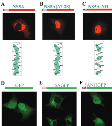
FIG. 1. Disruption of the hydrophobic face of the N-terminal AH of NS5A abolishes the intracellular membrane localization pattern of NS5A. (A) (Top) The region of NS5A found to harbor an AH is shown in gray (above the picture) and in more detail in the bottom panel. (Middle) Huh-7 cells were infected with a vaccinia virus expressing T7 RNA polymerase and then transfected with plasmid pBRTM/HCV 827–3011 containing the HCV NS proteins downstream of a T7 promoter. Expression of NS5A was detected by indirect immunofluorescence using a monoclonal antibody against NS5A and a Texas red-labeled anti-mouse secondary antibody. (Bottom)␣-Helix net diagram of the N terminus of NS5A wherein the cylindrical␣-helical segment is “cut” longitudinally along one face and then “flattened” into the plane of the page. The amino acid sequence of NS5A from amino acids 4 to 27 in the N-terminal to C-terminal direction is shown. Hydrophobic amino acids in the amphipathic helix are shaded in green. Note the long, continuous stretch of such amino acids along one face of the helix, defining its amphipathic nature. (B) Same as for panel A except that plasmid 827/5A⌬7–28, which harbors a mutation of NS5A wherein the AH segment has been deleted (deleted amino acids shown between red brackets in bottom panel), was substituted for pBRTM/HCV 827–3011. (C) Same as for panel A except that plasmid 827/5ANH, wherein mutations which disrupt the hydrophobic nature of the AH (depicted in the bottom panel) were engineered into NS5A, was used for transfection. (D) Cells transfected with plasmid T7-GFP, which expresses wild-type GFP, a non-membrane-associated protein of 236 amino acids (schematically represented in the top panel). It is diffusely distributed throughout the cytoplasm and nucleus. (E) Cells transfected with plasmid T7-5AGFP, which expresses a fusion protein wherein the N-terminal segment of NS5A (containing the first 31 of the 448 amino acids of the full-length protein) which harbors the AH is grafted onto GFP (schematically represented in the top panel). The fusion protein displays an intracellular localization of a membrane-associated protein. (F) Cells transfected with plasmid T7-5ANHGFP, which is identical to T7-5AGFP except that the AH in the NS5A segment has been disrupted with the same mutations as in panel C (schematically represented in the top panel). This mutant displays a localization pattern similar to that of the native GFP.
on November 8, 2019 by guest
http://jvi.asm.org/
distribution of NS5A (Fig. 1B, middle panel). Cytoplasmic membrane localization was completely abolished and replaced predominantly by a nuclear localization pattern.
We interpreted this result as being consistent with activation of a previously described cryptic nuclear localization signal (NLS) located within the C-terminal domain of NS5A (20). The NLS presumably became functional after the loss of the normal cytoplasmic membrane-targeting sequence of NS5A, which apparently lies within the deleted segment. These results are similar to those recently reported with an NS5A mutant harboring a larger N-terminal deletion consisting of the first 45 amino acids (7).
To test the hypothesis that it is indeed the amphipathic nature of the deleted putative helix which mediates intracellu-lar localization of NS5A, the hydrophobic face of the helix was disrupted by site-specific PCR mutagenesis. Our strategy was to introduce three charged amino acids spaced at intervals along the predicted N-terminal␣-helix such that no sustained hydrophobic patch remained (Fig. 1C, bottom panel). The primer 5ANH (5⬘-TCCGGCTCCTGGCTAAGGGACGACT GGGACTGGGAATGCGAGGTGCTGAGCGACGATAA GACC-3⬘) was used to change the coding sequence for isoleu-cine-8, isoleucine-12, and phenylalanine-19 of NS5A to encode aspartate, glutamate, and aspartate, respectively. On expres-sion of this mutant, termed 827/5ANH, in Huh-7 cells, com-plete loss of the characteristic membrane localization pattern of NS5A was observed (Fig. 1C, middle panel). Again, as seen for the mutant lacking the entire helix, nuclear localization was the predominant pattern observed. Interestingly, in both NS5A mutants, a few cells were occasionally noted in which the sharp nuclear localization pattern was replaced by diffuse staining throughout the cell (data not shown). Even then, however, no characteristic localization to cytoplasmic membrane structures was observed. Perhaps these cells represented a different phase of the cell cycle since no attempt had been made to synchro-nize cell division in any of our experiments. We conclude that the predicted N-terminal AH is necessary for the normal cy-toplasmic membrane localization of NS5A.
We next sought to test the hypothesis that the N-terminal AH of NS5A is also sufficient to confer cytoplasmic membrane localization. For this, we first constructed a plasmid, T7GFP, in which the HCV-encoding sequences of pBRTM/HCV 827– 3011 were replaced with the gene for the jellyfish Aequorea
victoria green fluorescent protein (GFP), a
non-membrane-associated protein (43). Next we modified plasmid T7GFP to construct plasmid T75AGFP, in which the first 31 amino acids of NS5A containing the AH are fused in frame to the N terminus of GFP. On expression in Huh-7 cells, GFP displays a diffuse cytoplasmic distribution pattern and also readily en-ters the nucleus (Fig. 1D). Grafting the N-terminal AH of NS5A onto GFP results in a dramatic change in intracellular distribution: the fusion protein can no longer enter the nucleus and appears restricted to cytoplasmic membrane structures (Fig. 1E). A similar finding has been recently reported (7). To determine the critical element within the grafted NS5A seg-ment responsible for conferring the membrane association, we made plasmid T75ANHGFP, in which we introduced into the fusion protein the same mutations as in Fig. 1C, designed to destroy the hydrophobic face of the AH. As shown in Fig. 1F, disrupting the amphipathic nature of the NS5A helix portion of
the fusion protein restored the distribution pattern character-istic of GFP. These results argue for the notion that the AH has an independent function—namely, membrane targeting— as opposed to being simply a part of NS5A crucial for proper folding.
We next wished to complement the above morphological data with biochemical evidence. To determine the effect of disrupting the amphipathic nature of the N-terminal␣-helix of NS5A on the membrane association of NS5A, we performed membrane flotation assays (26) on cells expressing wild-type and AH mutant NS5A. Whole-cell extracts from Huh-7 cells transfected with pBRTM/HCV 827–3011 or 827/5ANH were overlaid with a 5 to 40% OptiPrep (Sigma) step density gradi-ent and subjected to ultracgradi-entrifugation for 4 h at 40,000⫻g
[image:3.603.304.537.69.335.2]in an SW60 rotor. Fractions were collected from the top, and the proteins in each gradient fraction were precipitated with methanol-chloroform and analyzed by Western blotting for NS5A using a monoclonal antibody against NS5A and a horse-radish peroxidase-labeled anti-mouse secondary antibody. In such assays, non-membrane-associated proteins remain at the bottom of the gradient whereas membranes—and associated proteins—“float” toward less dense gradient fractions present
FIG. 2. An intact AH is required for biochemical association of NS5A with membranes. Huh-7 cells were infected with a vaccinia virus expressing T7 RNA polymerase and then transfected with plasmid pBRTM/HCV 827–3011 (A) or 827/5ANH (B), which expresses wild-type or an AH mutant of NS5A, respectively. Cell extracts were ana-lyzed by membrane flotation assays for membrane association of NS5A by overlaying the extracts with an OptiPrep gradient (5 to 40%) and subjecting them to ultracentrifugation. Non-membrane-associated proteins remain at the bottom (right side) of the gradient, whereas membranes and associated proteins float toward the less dense gradi-ent fractions presgradi-ent at the top (left side). Numbers correspond to OptiPrep gradient fractions, which were analyzed by Western blotting using a monoclonal antibody to NS5A as the probe.
VOL. 77, 2003 NOTES 6057
on November 8, 2019 by guest
http://jvi.asm.org/
at the top. As shown in Fig. 2, wild-type NS5A quantitatively floated with the membrane fraction (Fig. 2A) whereas NS5A with a genetically disrupted AH did not (Fig. 2B). Moreover, these Western blots show that this is not simply a function of the mutant NS5A being expressed at a lower level than the wild-type protein. Finally, both NS5B and NS4B have been reported to each be independently targeted to membranes (19, 35) and are thought to interact with NS5A as part of a multi-protein complex (23, 38). However, as shown in Fig. 1C and 2B, neither of these potential interactions appears to be suffi-cient to keep AH-mutated NS5A on the membranes.
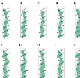
Genetic disruption of the NS5A AH impairs HCV RNA rep-lication.The above experiments identified an essential struc-tural element capable of mediating the membrane association of NS5A when expressed in cultured cells. To assess the pos-sible importance of this element in natural infections of HCV, we examined the predicted amino acid sequences of the N terminus of NS5A contained in isolates obtained from a variety of patients and representing multiple genotypes. As shown in Fig. 3, while there was considerable variability in the specific amino acid sequence of this region among isolates, the amphi-pathic nature of the predicted ␣-helix was preserved. More-over, when a similar analysis was extended to all sequenced isolates currently available in public databases, the same result was observed (7; Y. Yang and J. Glass, personal
communica-tion). This suggests that such a structural motif is indeed im-portant for some essential aspect of the HCV life cycle.
[image:4.603.132.455.66.379.2]The dramatic relocalization of NS5A to the nucleus after disruption of the amphipathic nature of its N-terminal helix and the strict preservation of this motif in all known HCV isolates suggest that disruption of the AH may have significant consequences for HCV RNA replication. To test this hypoth-esis, we constructed high-efficiency second-generation bicis-tronic subgenomic RNA replicons of HCV (5) harboring a neomycin resistance gene along with an HCV polyprotein con-taining NS5A with either a wild-type or mutated N-terminal AH (Fig. 4, top panels). The RNA replication efficiency of these replicons was then assayed on the basis of their ability to establish G418-resistant colonies after transfection of Huh-7 cells. As shown in Fig. 4, while a replicon with a wild-type NS5A N terminus gave rise to numerous colonies, as previ-ously observed (5), disruption of the amphipathic nature of the N-terminal helix of NS5A resulted in a dramatic inhibition of HCV genome replication. It is unlikely that these results are due to an increased cytotoxicity associated with the mutant NS5A, since we observed no decreased transfection efficiency or ability to establish colonies by using plasmids encoding a drug resistance marker along with just the wild-type or mutant NS5A proteins (data not shown). It is difficult to suggest that NS5A simply cannot tolerate mutations, since a much larger
FIG. 3. The AH of NS5A is conserved across HCV isolates. Amino acid sequences of the AH of NS5A obtained from sequenced isolates of HCV from around the world and representing a variety of genotypes are shown. Note that although there are differences in particular amino acids (shown in red) compared to a reference sequence (A), the amphipathic nature of the helix (indicated by the green hydrophobic continuous band) is conserved. (A) Genotype 1a (accession no. AF009606), (B) genotype 1b (accession no. P26663), (C) genotype 2a (accession no. P26660), (D) genotype 2b (accession no. AB030907), (E) genotype 3a-K (accession no. D28917), (F) genotype 3a-NZL (accession no. D17763), (G) genotype 3b (accession no. D49374), (H) genotype 4a (accession no. Y11604), (I) genotype 10a (accession no. D63821), (J) genotype 11a (accession no. D63822).
on November 8, 2019 by guest
http://jvi.asm.org/
alteration (47 deleted amino acids) than our three point mu-tations was tolerated without any effect on RNA replication (5). Nor are these results likely to be due to disruption of a critical RNA structural motif, since multiple mutations appear to be tolerated within the mutated segment of NS5A (including wobble mutations at the amino acid codons mutated in NS5ANH [Fig. 3]). Rather, it is the altered intracellular tar-geting of NS5A that is most clearly associated with the pro-found inhibition of HCV RNA replication.
Because the three mutations completely disrupting the am-phipathic nature of the NS5A AH contained in Bart5X79I resulted in no HCV RNA replication, we hypothesized that a partial disruption of the hydrophobic face of the AH might be associated with an intermediate level of replication. Indeed, while Bart79I with a wild-type AH had a⬃3% G418 transduc-tion efficiency and Bart5X79I gave rise to no colonies, when just one of the mutations illustrated in Fig. 1C (I12E) was introduced into the Bart79I replicon, a G418 transduction ef-ficiency of ⬃0.0003% was observed (data not shown). This 4-log-unit-reduced level of replication is comparable to that observed with the first-generation HCV replicons without adaptive mutations (5, 28). A more complete description of a larger collection of such intermediate mutants will be pre-sented elsewhere.
Most, if not all, plus-strand RNA viruses appear to replicate in association with cytoplasmic membranes (4, 8, 12, 25, 34). The detailed mechanisms of RNA replication complex assem-bly, however, are not known. The AH of NS5A might play a role in localizing, inducing, or maintaining such membrane-associated replication complexes and may be an example of a
more general theme. For example, the NS protein 2C of both poliovirus and hepatitis A virus also contains a membrane-associating AH (24, 42), which, at least for poliovirus, appears to play a role in RNA synthesis (31) and the induction of membrane rearrangements derived from the host ER and Golgi (40) on which replication complexes are localized (3). In this regard, the HCV NS4B protein has also recently been associated with the induction of novel intracellular membrane structures hypothesized to be the site of HCV RNA replication (10). It is interesting that we also have found an AH within NS4B, which will be presented elsewhere.
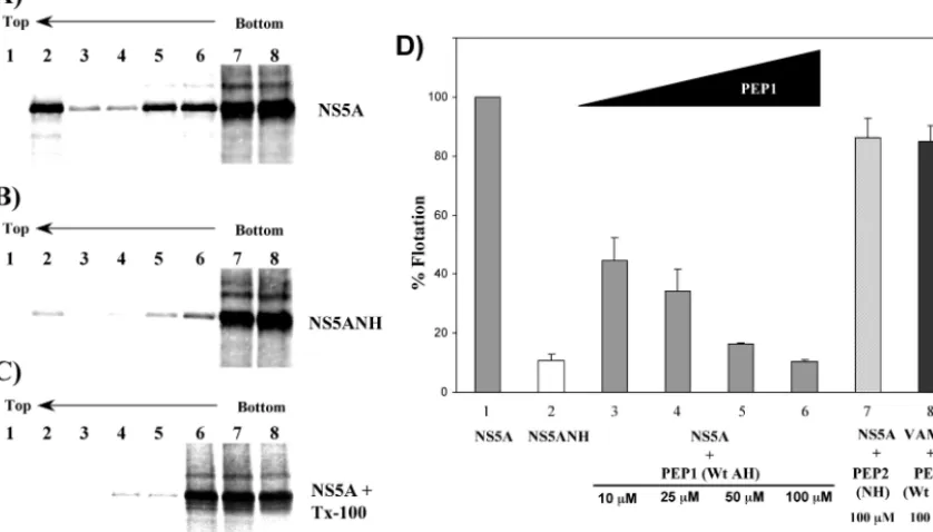
Pharmacologic disruption of NS5A AH-mediated membrane association.Finally, because genetically mutating the ability of the AH to associate with membranes disrupts RNA replication efficiently, we hypothesized that the AH-mediated membrane association of NS5A might also be amenable to pharmacologic disruption and thereby form the basis for a novel approach to anti-HCV therapy. To test this hypothesis, we used a modified version of the membrane flotation assay to evaluate the ability of a first-generation inhibitor to competitively block the asso-ciation of NS5A with host cell membranes. Because NS5A associates with membranes in a posttranslational manner (7), aliquots of in vitro-translated and35S-labeled NS5A proteins
prepared using the TNT reticulocyte lysate kit (Promega, Mad-ison, Wis.) were combined with a membrane fraction derived from Huh-7 cells, with or without synthetic peptides, and over-laid with a 5 to 40% OptiPrep step gradient. Following ultra-centrifugation, the proteins in each gradient fraction were pre-cipitated with methanol-chloroform and analyzed by sodium dodecyl sulfate-polyacrylamide gel electrophoresis. The
per-FIG. 4. Disrupting the amphipathic nature of the 5A helix impairs replication of HCV subgenomic replicons. (Top) Schematic representation of HCV high-efficiency subgenomic replicons used for HCV replication assays. (A) Bart79I, a high-efficiency subgenomic replicon of HCV harboring the neomycin resistance gene (neo) and the HCV NS proteins. (5⬘NTR and 3⬘NTR represent the nontranslated regions at the end of the HCV genomic RNA, which contain presumed recognition sequences for the viral replication machinery. E-IRES, encephalomyocarditis internal ribosome entry site.) (B) The amphipathic helix-disrupting mutations of Fig. 1C were introduced into the Bart79I replicon, yielding the depicted Bart5X79I. (Bottom) The above replicons were electroporated into Huh-7 cells, and neomycin-resistant colonies were selected and stained with crystal violet. Each dot represents a colony of Huh-7 cells that was able to grow in the presence of neomycin due to the presence of efficiently replicating intracellular replicons.
VOL. 77, 2003 NOTES 6059
on November 8, 2019 by guest
http://jvi.asm.org/
[image:5.603.62.519.69.293.2]centage of protein floating to the top of the gradient with the membrane fraction (fraction 2 from the top) was then quanti-fied using a PhosphorImager (Molecular Dynamics, Sunnyvale, Calif.). Similar to the results shown in Fig. 2, wild-type NS5A specifically floated with the low-density gradient membrane fraction (Fig. 5A). In contrast, when NS5A contained the mu-tations (depicted in Fig. 1C) that disrupt the amphipathic na-ture of the N-terminal helix, minimal flotation was observed (Fig. 5B). As expected, if the cell extracts were treated with the detergent Triton X-100 to dissolve the target membranes prior to gradient assembly, no flotation of NS5A was observed (Fig. 5C). These results are in good agreement with the morphologic and biochemical evidence for AH-mediated membrane asso-ciation (Fig. 1 and 2), especially considering the potential lim-itations of this type of in vitro assay (22). Moreover, as shown in Fig. 5D, a synthetic peptide (PEP1) designed to mimic the wild-type AH not only inhibited membrane association of NS5A but also did so in a dose-dependent manner. A control peptide (PEP2) mimicking the mutated AH had no such effect on NS5A membrane association. An additional measure of specificity against NS5A was provided by testing the effect of PEP1 on the membrane association of VAMP2, a control host cell protein whose membrane association is mediated by its own AH in a posttranslational fashion (22). The precise mech-anistic details of how the peptide exerts its inhibition remains
[image:6.603.87.506.80.319.2]to be determined, although a variety of interesting possibilities can be envisaged. For example, the host cell membrane recep-tor for NS5A may be composed of a specific protein—presum-ably different from that used by VAMP2—which can be spe-cifically blocked by PEP1 but not by the control peptide, PEP2. Alternatively, PEP1 may somehow perturb the lipid membrane itself so as to alter efficient binding by NS5A. Finally, PEP1 might directly bind to NS5A and thereby prevent the latter from efficient membrane binding. Experiments are being per-formed to help distinguish between these models. Whatever the precise mechanism, however, the maximal extent of NS5A inhibition achieved pharmacologically is comparable to that obtained by genetic mutation of the AH in NS5A. In addition, unlike current treatments for HCV, the synthetic peptide ap-pears equally effective against NS5A derived from different genotypes (data not shown), including those most refractory to current therapies. This convenient membrane flotation assay has also proven well suited for current efforts focused on study-ing the mechanistic details of the AH membrane-targetstudy-ing domain and ideal for guiding ongoing development of peptido-mimetic compounds designed to resemble key elements, or bind to specific features, of the AH. Such compounds repre-sent an exciting potential addition to current anti-HCV com-bination therapy regimens.
FIG. 5. Pharmacologic disruption of AH-mediated membrane association of NS5A. (A and B) Membrane association of NS5A (containing a wild-type AH) (A) and NS5ANH (NS5A with a genetically mutated AH) (B) proteins was analyzed by in vitro membrane flotation assays. Numbers correspond to OptiPrep gradient (5 to 40%) fractions. Non-membrane-associated proteins remain at the bottom (right side) of the gradient, whereas membranes and associated proteins float toward the less dense gradient fractions present at the top (left side). (C) Same as panel A except that the reaction mixture was treated with the detergent Triton X-100 to dissolve the membranes prior to gradient assembly. (D) Pharmacologic inhibition of NS5A membrane association. Huh-7 cell-derived membranes were treated with the indicated concentration of PEP1 (SG SWLRDVWDWICTVLTDFKTWLQSKLDYKD), a peptide mimicking the wild-type amphipathic helix of NS5A, or PEP2 (SGSWLRDDW DWECTVLTDDKTWLQSKLDYKD), a control peptide mimicking the NS5ANH mutant amphipathic helix, or mock treated with water and incubated with in vitro-translated NS5A, NS5ANH, or VAMP2 before being analyzed by membrane flotation assays. The percentage of NS5A, NS5ANH, or VAMP2 floating to the top of the gradient with the membrane fraction (fraction 2 from the top) under the indicated conditions was quantified using a PhosphorImager and expressed relative to the mock-treated control. Error bars represent standard error of the mean.
on November 8, 2019 by guest
http://jvi.asm.org/
M.E. and K.H.C. contributed equally to this work.
We thank Ellie Ehrenfeld and Karla Kirkegaard for helpful discus-sions and critical reading of the manuscript and David Andrews for providing the VAMP2 clone.
M.E. is the recipient of the ALF Postdoctoral Research Fellowship Award and Stanford University Dean’s Fellowship. J.S.G. is supported by an Amgen/AASLD American Liver Foundation Award and a Bur-roughs Wellcome Fund Career Award. This work was also supported in part by the Eli Lilly-Stanford University HCV initiative, a pilot study award from the Stanford Digestive Disease Center, a Veterans Ad-ministration REAP Award, and an NIH U19 award.
REFERENCES
1. Alter, M. J., D. Kruszon-Moran, O. V. Nainan, G. M. McQuillan, F. Gao, L. A. Moyer, R. A. Kaslow, and H. S. Margolis.1999. The prevalence of hepatitis C virus infection in the United States, 1988 through 1994. N. Engl. J. Med.341:556–562.
2. Bartenschlager, R., and V. Lohmann.2000. Replication of hepatitis C virus. J. Gen. Virol.81:1631–1648.
3. Bienz, K., D. Egger, T. Pfister, and M. Troxler.1992. Structural and func-tional characterization of the poliovirus replication complex. J. Virol.66:
2740–2747.
4. Bienz, K., D. Egger, Y. Rasser, and W. Bossart.1980. Kinetics and location of poliovirus macromolecular synthesis in correlation to virus-induced cyto-pathology. Virology100:390–399.
5. Blight, K. J., A. A. Kolykhalov, and C. M. Rice.2000. Efficient initiation of HCV RNA replication in cell culture. Science290:1972–1974.
6. Branch, A. D.2000. Hepatitis C virus RNA codes for proteins and replicates: does it also trigger the interferon response? Semin. Liver Dis.20:57–68. 7. Brass, V., E. Bieck, R. Montserret, B. Wolk, J. A. Hellings, H. E. Blum, F.
Penin, and D. Moradpour.2002. An amino-terminal amphipathic alpha-helix mediates membrane association of the hepatitis C virus nonstructural protein 5A. J. Biol. Chem.277:8130–8139.
8. Chu, P. W., and E. G. Westaway.1992. Molecular and ultrastructural analysis of heavy membrane fractions associated with the replication of Kunjin virus RNA. Arch. Virol.125:177–191.
9. Di Bisceglie, A. M.2000. Natural history of hepatitis C: its impact on clinical management. Hepatology31:1014–1018.
10. Egger, D., B. Wolk, R. Gosert, L. Bianchi, H. E. Blum, D. Moradpour, and K. Bienz.2002. Expression of hepatitis C virus proteins induces distinct membrane alterations including a candidate viral replication complex. J. Vi-rol.76:5974–5984.
11. Enomoto, N., I. Sakuma, Y. Asahina, M. Kurosaki, T. Murakami, C. Yamamoto, Y. Ogura, N. Izumi, F. Marumo, and C. Sato.1996. Mutations in the nonstructural protein 5A gene and response to interferon in patients with chronic hepatitis C virus 1b infection. N. Engl. J. Med.334:77–81. 12. Froshauer, S., J. Kartenbeck, and A. Helenius.1988. Alphavirus RNA
rep-licase is located on the cytoplasmic surface of endosomes and lysosomes. J. Cell Biol.107:2075–2086.
13. Gale, M. J. J., M. J. Korth, N. M. Tang, S. L. Tan, D. A. Hopkins, T. E. Dever, S. J. Polyak, D. R. Gretch, and M. G. Katze.1997. Evidence that hepatitis C virus resistance to interferon is mediated through repression of the PKR protein kinase by the nonstructural 5A protein. Virology230:217– 227.
14. Glenn, J. S., J. C. Marsters, Jr., and H. B. Greenberg.1998. Use of a prenylation inhibitor as a novel antiviral agent. J. Virol.72:9303–9306. 15. Glenn, J. S., J. M. Taylor, and J. M. White.1990. In vitro-synthesized
hepatitis delta virus RNA initiates genome replication in cultured cells. J. Virol.64:3104–3107.
16. Grakoui, A., C. Wychowski, C. Lin, S. M. Feinstone, and C. M. Rice.1993. Expression and identification of hepatitis C virus polyprotein cleavage prod-ucts. J. Virol.67:1385–1395.
17. Hijikata, M., H. Mizushima, Y. Tanji, Y. Komoda, Y. Hirowatari, T. Akagi, N. Kato, K. Kimura, and K. Shimotohno.1993. Proteolytic processing and membrane association of putative nonstructural proteins of hepatitis C virus. Proc. Natl. Acad. Sci. USA90:10773–10777.
18. Huang, Y., Y. Uchiyama, T. Fujimura, H. Kanamori, T. Doi, A. Takamizawa, T. Hamakubo, and T. Kodama.2001. A human hepatoma cell line expressing hepatitis C virus nonstructural proteins tightly regulated by tetracycline. Biochem. Biophys. Res. Commun.281:732–740.
19. Hugle, T., F. Fehrmann, E. Bieck, M. Kohara, H. G. Krausslich, C. M. Rice, H. E. Blum, and D. Moradpour.2001. The hepatitis C virus nonstructural protein 4B is an integral endoplasmic reticulum membrane protein. Virology
284:70–81.
20. Ide, Y., L. Zhang, M. Chen, G. Inchauspe, C. Bahl, Y. Sasaguri, and R. Padmanabhan.1996. Characterization of the nuclear localization signal and subcellular distribution of hepatitis C virus nonstructural protein NS5A. Gene182:203–211.
21. Kim, J. E., W. K. Song, K. M. Chung, S. H. Back, and S. K. Jang.1999. Subcellular localization of hepatitis C viral proteins in mammalian cells. Arch. Virol.144:329–343.
22. Kim, P. K., C. Hollerbach, W. S. Trimble, B. Leber, and D. W. Andrews.
1999. Identification of the endoplasmic reticulum targeting signal in vesicle-associated membrane proteins. J. Biol. Chem.274:36876–36882.
23. Koch, J. O., and R. Bartenschlager.1999. Modulation of hepatitis C virus NS5A hyperphosphorylation by nonstructural proteins NS3, NS4A, and NS4B. J. Virol.73:7138–7146.
24. Kusov, Y. Y., C. Probst, M. Jecht, P. D. Jost, and V. Gauss-Muller.1998. Membrane association and RNA binding of recombinant hepatitis A virus protein 2C. Arch. Virol.143:931–944.
25. Lazarus, L. H., and R. Barzilai.1974. Association of foot-and-mouth disease virus replicase with RNA template and cytoplasmic membranes. J. Gen. Virol.23:213–218.
26. Lecat, S., P. Verkade, C. Thiele, K. Fiedler, K. Simons, and F. Lafont.2000. Different properties of two isoforms of annexin XIII in MDCK cells. J. Cell Sci.113:2607–2618.
27. Liang, T. J., B. Rehermann, L. B. Seeff, and J. H. Hoofnagle.2000. Patho-genesis, natural history, treatment, and prevention of hepatitis C. Ann. Intern. Med.132:296–305.
28. Lohmann, V., F. Korner, J. Koch, U. Herian, L. Theilmann, and R. Barten-schlager.1999. Replication of subgenomic hepatitis C virus RNAs in a hepatoma cell line. Science285:110–113.
29. McHutchison, J. G., S. C. Gordon, E. R. Schiff, M. L. Shiffman, W. M. Lee, V. K. Rustgi, Z. D. Goodman, M. H. Ling, S. Cort, J. K. Albrecht, and the Hepatitis Interventional Therapy Group.1998. Interferon alfa-2b alone or in combination with ribavirin as initial treatment for chronic hepatitis C. N. Engl. J. Med.339:1485–1492.
30. Moradpour, D., P. Kary, C. M. Rice, and H. E. Blum.1998. Continuous human cell lines inducibly expressing hepatitis C virus structural and non-structural proteins. Hepatology28:192–201.
31. Paul, A. V., A. Molla, and E. Wimmer.1994. Studies of a putative amphi-pathic helix in the N-terminus of poliovirus protein 2C. Virology199:188– 199.
32. Pelham, H. R. B.1995. Sorting and retrieval between the endoplasmic reticulum and Golgi apparatus. Curr. Opin. Cell Biol.7:530–535. 33. Reed, K. E., and C. M. Rice.2000. Overview of hepatitis C virus genome
structure, polyprotein processing, and protein properties. Curr. Top. Micro-biol. Immunol.242:55–84.
34. Rice, C. M.1996.Flaviviridae: the viruses and their replication, p. 931–959.
InB. N. Fields, D. M. Knipe, and P. M. Howley (ed.), Fields virology, 3rd ed. Lippincott-Raven Publications, Philadelphia, Pa.
35. Schmidt-Mende, J., E. Bieck, T. Hugle, F. Penin, C. M. Rice, H. E. Blum, and D. Moradpour.2001. Determinants for membrane association of the hepa-titis C virus RNA-dependent RNA polymerase. J. Biol. Chem.276:44052– 44063.
36. Segrest, J. P., H. De Loof, J. G. Dohlman, C. G. Brouillette, and G. M. Anantharamaiah.1990. Amphipathic helix motif: classes and properties. Proteins8:103–117.
37. Selby, M. J., Q.-L. Choo, K. Berger, G. Kuo, E. Glazer, M. Eckart, C. Lee, D. Chien, C. Kuo, and M. Houghton.1993. Expression, identification and sub-cellular localization of the proteins encoded by the hepatitis C viral genome. J. Gen. Virol.74:1103–1113.
38. Shirota, Y., H. Luo, W. Qin, S. Kaneko, T. Yamashita, K. Kobayashi, and S. Murakami.2002. Hepatitis C virus (HCV) NS5A binds RNA-dependent RNA polymerase (RdRP) NS5B and modulates RNA-dependent RNA poly-merase activity. J. Biol. Chem.277:11149–11155.
39. Song, J., M. Nagano-Fujii, F. Wang, R. Florese, T. Fujita, S. Ishido, and H. Hotta.2000. Nuclear localization and intramolecular cleavage of N-termi-nally deleted NS5A protein of hepatitis C virus. Virus Res.69:109–117. 40. Suhy, D. A., T. H. Giddings, and K. Kirkegaard.2000. Remodeling the
endoplasmic reticulum by poliovirus infection and by individual viral pro-teins: an autophagy-like origin for virus-induced vesicles. J. Virol.74:8953– 8965.
41. Tan, S.-L., and M. G. Katze.2001. How hepatitis C virus counteracts the interferon response: the jury is still out on NS5A. Virology284:1–12. 42. Teterina, N. L., A. E. Gorbalenya, D. Egger, K. Bienz, and E. Ehrenfeld.
1997. Poliovirus 2C protein determinants of membrane binding and rear-rangements in mammalian cells. J. Virol.71:8962–8972.
43. Tsien, R. Y.1998. The green fluorescent protein. Annu. Rev. Biochem.
67:509–544.
44. Tu, H., L. Gao, S. T. Shi, D. R. Taylor, T. Yang, A. K. Mircheff, Y. Wen, A. E. Gorbalenya, S. B. Hwang, and M. M. Lai.1999. Hepatitis C virus RNA polymerase and NS5A complex with a SNARE-like protein. Virology263:
30–41.
45. Zeuzem, S., S. V. Feinman, J. Rasenack, E. J. Heathcote, M. Y. Lai, E. Gane, J. O’Grady, J. Reichen, M. Diago, A. Lin, J. Hoffman, and M. J. Brunda.
2000. Peginterferon alfa-2a in patients with chronic hepatitis C. N. Engl. J. Med.343:1666–1672.
VOL. 77, 2003 NOTES 6061