postmortem brains indicate effects on synaptic function
.
White Rose Research Online URL for this paper:
http://eprints.whiterose.ac.uk/131161/
Version: Published Version
Article:
Martins-de-Souza, D., Guest, P.C., Vanattou-Saifoudine, N. et al. (2 more authors) (2012)
Phosphoproteomic differences in major depressive disorder postmortem brains indicate
effects on synaptic function. European Archives of Psychiatry and Clinical Neuroscience,
262 (8). pp. 657-666. ISSN 0940-1334
https://doi.org/10.1007/s00406-012-0301-3
© The Author(s) 2012. This article is published with open access at Springerlink.com.
[email protected] https://eprints.whiterose.ac.uk/ Reuse
This article is distributed under the terms of the Creative Commons Attribution (CC BY) licence. This licence allows you to distribute, remix, tweak, and build upon the work, even commercially, as long as you credit the authors for the original work. More information and the full terms of the licence here:
https://creativecommons.org/licenses/
Takedown
If you consider content in White Rose Research Online to be in breach of UK law, please notify us by
O R I G I N A L P A P E R
Phosphoproteomic differences in major depressive disorder
postmortem brains indicate effects on synaptic function
Daniel Martins-de-Souza•Paul C. Guest•
Natacha Vanattou-Saifoudine•Hassan Rahmoune•
Sabine Bahn
Received: 16 November 2011 / Accepted: 31 January 2012 / Published online: 21 February 2012 ÓThe Author(s) 2012. This article is published with open access at Springerlink.com
Abstract There is still a lack in the molecular compre-hension of major depressive disorder (MDD) although this condition affects approximately 10% of the world popu-lation. Protein phosphorylation is a posttranslational mod-ification that regulates approximately one-third of the human proteins involved in a range of cellular and bio-logical processes such as cellular signaling. Whereas phosphoproteome studies have been carried out extensively in cancer research, few such investigations have been carried out in studies of psychiatric disorders. Here, we present a comparative phosphoproteome analysis of post-mortemdorsolateral prefrontal cortex tissues from 24 MDD patients and 12 control donors. Tissue extracts were ana-lyzed using liquid chromatography mass spectrometry in a data-independent manner (LC-MSE). Our analyses resulted
in the identification of 5,195 phosphopeptides, corre-sponding to 802 non-redundant proteins. Ninety of these proteins showed differential levels of phosphorylation in tissues from MDD subjects compared to controls, being 20 differentially phosphorylated in at least 2 peptides. The majority of these phosphorylated proteins were associated with synaptic transmission and cellular architecture not only pointing out potential biomarker candidates but mainly shedding light to the comprehension of MDD pathobiology.
Keywords Major depressionPhosphoproteome ProteomePhosphorylationMass spectrometry Postmortem
Introduction
Major depressive disorder (MDD) is characterized by feelings of low mood and self-esteem and by loss of interest or pleasure in activities [12]. The consequences of MDD include negative effects on work and social relationships and associated comorbidities such as substance abuse and anxiety, which results in an enormous financial burden on healthcare services. The combination of direct costs, mor-tality costs arising from depression-related suicides and costs associated with effects on the workplace were esti-mated to be over 80 billion dollars in the United States alone in the year 2000 [14]. MDD is now thought to be a leading cause of disability worldwide and has been hypothesized to be the most incident disease of the twenty-first century [12]. Despite the fact that a number of molecular and image-based studies have been performed, an understanding of the underlying pathophysiology is still lacking, and there are still no robust empirical means of increasing our ability to diagnose such conditions accurately.
Electronic supplementary material The online version of this article (doi:10.1007/s00406-012-0301-3) contains supplementary material, which is available to authorized users.
D. Martins-de-Souza (&)
P. C. Guest N. Vanattou-SaifoudineH. RahmouneS. Bahn Department of Chemical Engineering and Biotechnology, University of Cambridge, Tennis Court Road, Cambridge, Cambridgeshire CB2 1QT, UK
e-mail: [email protected]
D. Martins-de-Souza
Max Planck Institute of Psychiatry, Proteomics and Biomarkers, Munich, Germany
S. Bahn
Department of Neuroscience, Erasmus Medical Centre, Rotterdam, The Netherlands
The Human Proteome Organization (HUPO) emerged from the Human Genome Project as a means of under-standing gene and protein functions that may lead to the understanding of diseases such as MDD and to the iden-tification of diagnostic/prognostic biomarkers [44]. The human genome is now known to contain approximately 35,000 genes [26,58], although the number of proteins is anticipated to be at least one order of the magnitude greater. One reason for this high number is due to the fact that proteins can undergo posttranslational modifications such as phosphorylation [48], which can give rise to mul-tiple forms of the same gene product.
Recent advances have been made in the development and application of large-scale molecular profiling tech-niques such as transcriptomics [43] and proteomics [4,38,
39] in studies of biological tissues from MDD patients. However, there have only been a few studies on differential patterns of protein phosphorylation in MDD [38]. Phos-phorylation controls a diverse range of cellular processes such as cell signaling via switching mechanism of the kinase-mediated addition of a high energy phosphate group to a serine, threonine or tyrosine residue on a protein. The large-scale analyses and quantification of phosphoproteins and/or phosphopeptides using mass spectrometry are known as phosphoproteomics and have been employed mostly in studies of cancer [2,10] and neurodegenerative disorders studies [9, 15]. For instance, the hyperphosph-orylation of TAU is one of the central mediators of Alz-heimer’s disease (AD) pathogenesis. Studies of TAU phosphorylation have proven to be an effective example of how to discover molecular mechanisms and signaling pathways involved in pathogenic processes in the brain. Phosphoproteomics offer us the possibility of investigating in a large-scale manner the functional role of proteins, which is a subject mostly neglected in large-scale proteome studies. Investigation of changes in the phosphorylation states of proteins, which are independent of changes in their total expression, can provide insights about molecular signaling and mechanisms such as neuroplasticity and synaptic transmission.
Here, we have carried out a differential phosphoproteo-mic analysis of postmortem dorsolateral prefrontal cortex (DLPFC) tissue from MDD patients (n=24) compared to matched controls (n=12) using liquid chromatography mass spectrometry in a data-independent mode (LC-MSE). Our interest in the DLPFC arises from the important role that this brain structure plays in MDD pathogenesis [24]. It was of particular importance to determine whether differential phosphorylation is involved in the pathogenesis of MDD and whether such molecules might be used as potential biomarker candidates [32] as a means of developing novel molecular biomarker tests to improve diagnosis and for use as surrogate biomarkers in drug discovery studies.
Methods and materials
Brain tissue samples
Postmortem DLPFC tissues (Brodmann area 9) from 24 MDD patients and 12 matched control subjects were obtained from the Stanley Medical Research Institute brain collection (Bethesda, MD, USA) (Table 1 and Supple-mentary Material 1). Consent was obtained by question-naire-based telephone interview and signed by the interviewer and a witness. The Institutional Review Board at the Uniformed Services University of Health Sciences determined that the procedure was exempt from federal and state regulations governing human research, since speci-mens were obtained from cadavers and anonymized with respect to personal information.
Sample preparation
Brain tissue samples (20 mg) were homogenized individ-ually in 100lL of 7 M urea, 2 M thiourea, 4% CHAPS,
2% ASB-14 and 70 mM DTT [40] using the Sample Grinding Kit (GE Healthcare; Little Chalfont, Bucks, UK). Samples were centrifuged for 10 min at 16,0009g. The supernatants were collected and protein concentrations determined using the Bradford dye-binding assay (Sigma; Poole, Dorsett, UK).
Shotgun LC-MSEproteomics workflow
The following workflow was established previously [37]. Protein samples (15 lg) were subjected to sodium
doce-cylsulfate polyacrylamide gel electrophoresis (SDS-PAGE) for pre-fractionation to enhance phosphoproteome cover-age. Protein bands were visualized using Coomassie blue staining, and each lane containing stained protein bands was sliced to produce 3 horizontal sections. Gel sections were subjected to trypsin digestion in situ and resulting peptide mixtures were lyophilized. The peptides were suspended in 0.1% formic acid and injected (0.5lg) in
duplicate into a nano Ultra Performance Liquid Chroma-tography instrument containing a BEH-130 C18 column (75lm9200 mm) at a flow rate of 0.3lL/min connected
Statistical analyses
Wilcoxon signed-rank test was used to determine signifi-cant differences between the groups under comparison (p\0.05) in case data are not normally distributed. False discovery rate (FDR) was calculated according to Benja-mini and Hochberg [5]. No adjustments were made for multiple comparisons as previously supported [53]. This approach is to avoid the exclusion of possible true positives since proteomic data are not necessarily random but can be physiologically interdependent observations, even though a Q-value threshold of approximately 0.4 and a fold change cut off of 10% have been established.
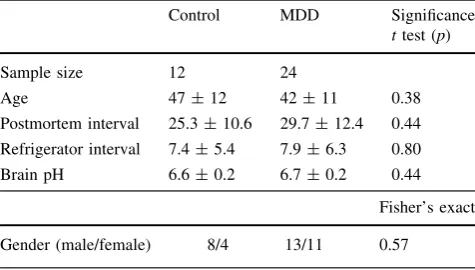
Considering that MDD and controls groups are matched for demographic variables (Table1), results here are unlike to have suffered influence of gender, age, alcohol abuse, smoking, postmortem interval and refrigeration interval. By using principal component analysis (PCA), we could observe that potential interferences of medication are also unlikely (Supplementary Material 1).
Phosphoproteome analyses
Potential phosphorylated molecules were identified auto-matically by PLGS based on the experimentally deter-mined loss of a 80 Da PO3-ion from peptides containing one or more phosphorylation consensus sequences featur-ing serine, threonine or tyrosine residues. Quantification was performed using the ElucidatorÓsystem.
Western blot validation
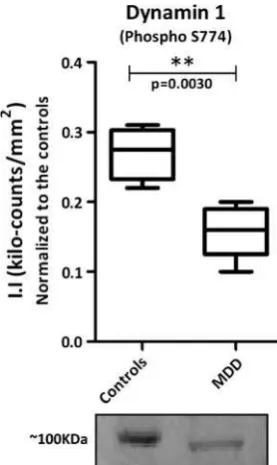
Brain tissue samples were prepared as described above. Samples were arranged in randomized order such that each of the two diagnostic groups (Controls vs. MDD) was represented on each gel. For each sample, 40lg of total
protein was electrophoresed using pre-cast Novex 10–20% Tricine polyacrylamide gels (Invitrogen; Paisley, UK) at
150 V for about 60 min, followed by semidry electropho-retic transfer to Immobilon-FL polyvinyldiphenyl fluoride (PVDF) membranes (Millipore; Watford, UK). The mem-branes were incubated in a 1:1 mixture of Odyssey blocking buffer (Li-COR Biosciences; Cambridge, UK). Membranes were then incubated overnight at 4°C with anti-Dynamin 1 (phospho S774) antibody (ab55324) at 1/500 dilutions (Abcam; Cambridge, UK). The membranes were washed in Tris-buffered saline (TBS) containing 0.1% Tween-20 for 1 h at room temperature (4920 min) and then incubated for 1 h at room temperature with the appropriate IR-dye-conjugated secondary antibodies (1:7,500 for secondary rabbit antibody Li-COR Biosci-ences) in blocking buffer. Immunoreactive protein bands were visualized using the Odyssey Infra-red imaging sys-tem (Li-COR Biosciences) and the integrated intensities (II) of the bands measured. Values which lay outside the mean by more than twice the standard deviation were excluded from the analysis.
Biological classification
Differentially phosphorylated proteins in MDD DLPFC were classified according to their biological pathways and subcellular localization using the Human Protein Reference Database (http://www.hprd.org). For interpreting func-tional significance of differentially phosphorylated pro-teins, the associated SwissProt accession identification codes for each phosphoprotein were uploaded into the Ingenuity Pathways Knowledge Base (IPKB) (http://www.
ingenuity.com), and these were analyzed to identify
potential interactions between these proteins and other proteins in the IPKB and for determining the most signif-icant biological, disease and canonical pathways associated with these proteins (significance determine using Fisher’s exact test).
Results
[image:4.595.51.289.579.714.2]Using our shotgun LC-MSE approach, we could identify 5,195 phosphopeptides in all the 36 analyzed samples corresponding to 802 distinct proteins. Comparing MDD patient samples to controls, significant differences in phosphorylation levels (p\0.05—Wilcoxon signed-rank test) were observed for 116 phosphopeptides, correspond-ing to 90 distinct proteins (Table2). Ten of these proteins (11.1%—Table2in black) were found with differences in protein expression, which impairs the confirmation of their differential phosphorylation. Fifty-three proteins (58.9%) presented phosphorylation differences in one phosphory-lation site in a single peptide, while 17 proteins (18.9%) presented phosphorylation differences in at least 2 Table 1 Demographic information for the samples used in the study
(mean±SD)
Control MDD Significance
ttest (p)
Sample size 12 24
Age 47±12 42±11 0.38
Postmortem interval 25.3±10.6 29.7±12.4 0.44
Refrigerator interval 7.4±5.4 7.9±6.3 0.80
Brain pH 6.6±0.2 6.7±0.2 0.44
Fisher’s exact
Gender (male/female) 8/4 13/11 0.57
Table 2 Differentially phosphorylated peptides identified and their correspondent proteins
phosphorylation sites in a single peptide. More consis-tently, 20 proteins (22.2%) were differentially phosphory-lated in more than 1 peptide (Table2 in gray). For 3 of them—alpha-crystallin B chain (CRYAB), 60 kDa heat shock protein, mitochondrial (HSPD1) and myelin basic protein (MBP)—phosphopeptides presented phosphoryla-tion differences in opposite direcphosphoryla-tions. The presented results are not likely to have suffered neither interference of demographics (Table1) nor medication according to a PCA which did not show clustering of samples (Supple-mentary material 1).
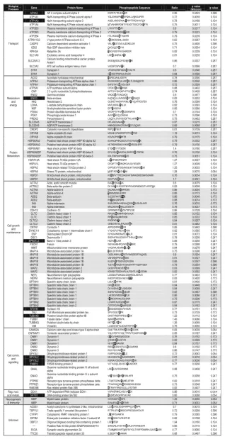
These phosphoproteins were assigned according to their biological processes in order to comprehend the
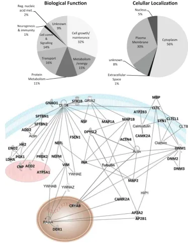
[image:6.595.169.544.232.715.2]biochemical pathways associated with the differential phosphorylation signaling (Fig.1). Seven different bio-logical processes were represented, being ‘‘cell growth and maintenance’’ the most frequent class (Fig.1a). Dif-ferentially phosphorylated proteins were also classified according to their cellular localization (Fig.1b). Although most of them are cytoplasmic (56%), there was a signif-icant coverage of membrane proteins (30%), which are important targets not only for protein signaling, but for potential drug targets. Phosphoproteins were also sub-mitted to a systems biology analyses in IPKB as to be discussed ahead (Fig. 2). Considering the large-scale nature of our analyses, we performed a Western blot
Fig. 1 aBiological function andbcellular localization of the differentially phosphorylated proteins in MDD brains
Fig. 2 Network of proteins interactions among the differentially phosphorylated proteins according to systems biology analyses by ingenuity pathways knowledge base
validation of phosphorylated Dynamin 1, confirming the LC-MSE findings (Fig.3).
Discussion
Despite their obvious importance, there is still a lack about phosphoproteomic studies in psychiatric disorders [38]. It is estimated that protein phosphorylation regulates approximately one-third of the human proteins in a wide range of cellular processes [59]. Moreover, phosphoprote-omic analyses could provide information about phosphor-ylation status, increasing the understanding about the functional aspects of the MDD. Even not using any special preparation for this purpose, we evaluate the phosphopro-teomic differences in MDD compared to controls. We focused our discussion and our illustrations in the 20 pro-teins that showed differences in phosphorylation in at least 2 peptides, since those provide more confidence regarding differential phosphorylation. Nevertheless, the remaining 70 proteins are also important protein targets to be con-sidered, proved by the fact that they fit in a biological and molecular context as further presented.
The identification of differential phosphorylation in sub-units of clathrin (CLTC and CLTCL1), spectrin (SPTBN1 and SPTBN2) and synapsin (SYN1) as well as the identification and validation of dynamin (DNM1) (Fig.3) reinforces the impairment of synaptic transmission in MDD [16]. As rep-resented in Fig.4, our phosphoproteomic findings suggest a
generalized dysregulation of the cytoskeleton signaling, which may compromise cell morphology and synaptic trans-mission. SPTBN1 and SPTBN2 together with other spectrin subunits like spectrin alpha chain (SPTAN1) are responsible for connecting plasma membrane to the actin cytoskeleton, playing roles in cell morphology, organelles organization and transmembrane proteins arrangement [6]. Alpha-actinin-4 (ACTN4) also belongs to the spectrin superfamily playing complementary roles to SPTBN1 and SPTBN2 and has been implicated in central nervous system (CNS) disorders such as schizophrenia and epilepsy [46]. Beta-adducin (ADD2) also plays structural roles in binding and regulating actin and spectrin filaments and contains phosphorylation sites for protein kinase C and calmodulin binding site [7]. Interest-ingly, Add2 KO mice presented differences in expression and phosphorylation levels of alpha- and gamma-adducin as well as impairments in synaptic plasticity, motor coordination, behavioral and learning deficits [51]. In addition, mRNA of sodium potassium transporting subunit alpha-3 (ATP1A3), calcium calmodulin dependent protein kinase type II alpha chain (CAMK2A) and vimentin (VIM) were found differen-tially expressed in MDD DLPFC [57].
Microtubule-associated proteins (MAPs) such as MAP1B and MAP2, which we found differentially phos-phorylated in MDD DLPFC, bind to tubulin subunits for regulating microtubules assembly and stability. These are necessary for several cellular processes in brain tissue such as axonogenesis and dendritogenesis during neurodevel-opment and function [1] as supported by studies in MAP1B null mice [8]. Moreover, MAP2 acts stabilizing and shap-ing dendrites durshap-ing neuron development [13]. Interest-ingly, we found MAP1B less phosphorylated in MDD brain, in line with previous functional studies which sug-gested that a dephosphorylated state of MAP1B may trig-ger cytoskeletal alterations that will impair long-term potentiation leading ultimately to impaired synaptic plas-ticity [62]. In addition, MAP1B null adult mice presented alterations in myelin sheath diameter as well as conduc-tance velocity of peripheral axons in the corpus callosum, which is in line with the differential phosphorylation we found for MBP in MDD DLPFC [8]. Stress-unsusceptible rats from a chronic social defeat stress model presented lower levels of Map1b mRNA in the frontal cortex and hippocampus [20]. Although we did not find significant differences in proteins expression when comparing MDD and control subjects, the reduced gene expression in the social defeat model and differential protein phosphoryla-tion in brain tissue reinforce the role of MAP1B in the impairment of the synaptic transmission in MDD. More-over, the inhibition of GSK-3beta by lithium leads to the loss of phosphorylated MAP-1B and thus to axonal remodeling [30], suggesting MAP1B as a potential drug target.
[image:7.595.99.238.57.290.2]MAP1B is also involved in growth cone and axonal activity [45] so as alpha-internexin (INA) and dihy-dropyrimidinase-related protein 2 (DPYSL2). INA, which is also present in dendrites, is one of the responsible pro-teins to maintain neuronal caliber and is involved in neu-ronal morphogenesis together with neurofilaments L, M (NEFL and NEFM) [11], which we also found differen-tially phosphorylated. DPYSL2, which acts in the regula-tion of axon guidance, vesicle trafficking and synaptic function, has been shown to bind and be modulated by antidepressants and neuroactive molecules [19]. INA and DPYSL2 have been found differentially expressed in schizophrenia brains [34,35,41]. While their phosphory-lation status has not been mentioned on schizophrenia, differences in expression of these proteins have not been related to MDD so far.
Synapsin-1 (SYN1) is a neuronal membrane phospho-protein that anchors to synaptic vesicles which is involved in axogenesis, synaptogenesis and modulation of neuro-transmitter release [56] so as N-ethylmaleimide-sensitive fusion protein (NSF), which is a key synaptic component especially in synaptic transmission SNARE-mediated [60]. SYN1 has been implicated in psychiatric disorders and could be a target for their treatment [61]. Synaptic vesicles
reserved in the axon are converted to ready-to-release vesicles depending on SYN1 phosphorylation. When pre-synaptic membranes depolarize, there is a calcium influx into the axonal nerve, which can involve plasma membrane calcium-transporting ATPase 3 (ATP2B3) that we also found to be differentially phosphorylated. Calcium ions bind to calmodulin, and the complex calcium/calmodulin activates protein kinases that phosphorylate SYN1. In turn, SYN1 dissociates from the vesicle membrane, leading it for release [25,54] (Fig.4). Interestingly, SYN1 interacts with amphiphysin (AMPH) [47], which we found to be differ-entially expressed in MDD brains [36].
When ready-to-release synaptic vesicles reach the axo-nal membrane, they are mechanically involved by dyna-mins like DNM1, DNM2 and DNM3, which forms a spiral around the vesicle forcing their rupture at the expense of GTP hydrolysis [18] (Fig.4). Interestingly, differences in the phsophorylation levels of clathrin heavy chain 1 and 2 (CLTC and CLTCL1) were also observed in MDD DLPFC. Clathrin complex performs a pivotal role in shaping synaptic vesicles and can be recycled after vesicle cycles also playing roles in axon guidance and organization of cellular membrane [29]. The roles of DNM1 and CLTC as well as the pivotal role of NSF can be observed in SPTBN1
INA
DPYSL2
DNM
complex
Synap c vesicle
SYN1
Ca+2 Ca+2
Ca+2
P CALM
ATP2B3 ATP4A
SLC25A5
CLTC & CLTCL1 ACTN4
ADD2
ADD2
ADD2
Neuron
Oligodendrocyte
MPB
MAP1B
MAP2 Dendrites
Dendrites
MAP1B
MAP2 CRYAB
HSPs
HSPs
HSPs
HSPs
INA
DNM
complex ACO2
ENO2 PGK1HK2 LDHA ATP5A1
CNP
DDX1
CAMK2A
Fig. 4 Integrated view of the role players of the synaptic dysfunction in MDD brains. Colored proteins were found differentially phosphorylated between MDD patients versus controls
[image:8.595.115.478.56.374.2]http://stke.sciencemag.org/content/vol2004/issue264/images/ data/re19/DC2/slowtrack2.swf. Differences in the phos-phorylation levels of key proteins such as DNM1 and CLTC can impair significantly synaptic transmission.
Although synaptic transmission is a known mechanism in MDD, revealing exactly which proteins, peptides and amino acids are differentially phosphorylated can lead to potential targets for therapeutic studies, paving way to the discovery of new drugs.
MBP, which is the major structural component of the myelin sheets in the CNS, and alpha-crystallin B (CRYAB) were identified with differential phosphorylation by two peptides, being one more phosphorylated and the other less phosphorylated. MBP gene and protein expression have been found altered consistently in schizophrenia [1,31,33,
42] and multiple sclerosis [55]. In addition, the differential expression of MBP in the entorhinal cortex in schizo-phrenia patients correlates to migrational disturbances of pre-alpha cell clusters leading to deficits in axonal myeli-nation and disturbed connectivity during neurodevelop-ment [49]. CRYAB was also previously found differentially expressed in schizophrenia [41]. Therefore, the differential phosphorylation of MBP and CRYAB protein modification cannot point out a process specific for MDD, but can indicate trait processes of neuropsychiatric disorders. Considering the structural role of these proteins added to the cytoskeleton impairment described above, we here present the possible players of the impaired cytoar-chitecture of MDD brain tissue. Recently, there has been found a correlation between the phosphorylation of CRY-AB with apoptosis in breast cancer cells [27]. In addition, CRYAB acts as molecular chaperone [50], and the differ-ential phosphorylation of other 3 heat shock proteins here found—putative heat shock protein HSP 90-beta-3 (HSP90AB3P), putative heat shock protein HSP 90-alpha A2 (HSP90AA2) and 60 kDa heat shock protein, mito-chondrial (HSPD1)—may suggest impaired cellular envi-ronment. Interestingly, CRYAB has an autokinase activity, and posttranslational modifications decrease their role as chaperone [21].
Energy metabolism pathways have been associated intimately with MDD [3,22,52]. In line with these results, we observed recently that several oxidative phosphoryla-tion enzymes were differentially expressed in MDD [36]. We found differences in the phosphorylation levels of metabolic enzymes such as aconitate hydratase (ACO2), enolase (ENO2 and ENO3), hexokinase-2 (HK2) and L-lactate dehydrogenase A (LDHA).
As for any postmortem brain tissue research, some potential limitations of our study have to be addressed. Factors such as age, gender, postmortem interval and oth-ers may confound global proteomic analyses especially in postmortem studies [23]. However, these factors are
unlikely to have had significant effects on our analyses, considering that the compared groups are matched for demographic variables and these have not shown signifi-cant differences (Table 1). In addition, no segregation of subjects has been observed using principle component analyses for medication effects (Supplementary material 1). The limited sample sizes can also be a drawback in postmortem brain tissue studies, suggesting, therefore, replication in an independent sample cohort. In addition, it is suggested validation in a larger number of samples for overcoming errors from multiple testing. Even with all potential drawbacks, we believe that the analysis of post-mortem tissue from patients while studying brain disorders is indispensable as reported recently [17]. These studies have generated insights for psychiatric studies that have been useful in basic and applied research.
Interestingly, the differences in phosphorylation that we observed in MDD DLPFC brain tissue can be connected in a net of interactions and roles as described in Figs.2and4. The advantage of a hypothesis-free analysis as we per-formed is to give the opportunity to reveal the real players of the studied disorder. Moreover, considering the signal-ing role of phosphorylation, which is indispensable for cellular structure, neuroplasticity and communication, the protein candidates presented here be considered therapeutic targets to be further explored in drug response and drug discovery studies.
Acknowledgments Authors thank sincerely all tissue donors and their families. Their comprehension and consent is essentially important to our research and to the lives of patients. We also thank the Stanley Medical Research Institute for their financial support and material donation. Special thanks to Professor E. Torrey Fuller and Dr. Maree Webster.
Conflict of interest None.
Open Access This article is distributed under the terms of the Creative Commons Attribution License which permits any use, dis-tribution, and reproduction in any medium, provided the original author(s) and the source are credited.
References
1. Al-Bassam J, Ozer RS, Safer D, Halpain S, Milligan RA (2002) MAP2 and tau bind longitudinally along the outer ridges of microtubule protofilaments. J Cell Biol 157:1187–1196 2. Ashman K, Villar EL (2009) Phosphoproteomics and cancer
research. Clin Transl Oncol 11:356–362
3. Baxter LR Jr, Schwartz JM, Phelps ME, Mazziotta JC, Guze BH, Selin CE, Gerner RH, Sumida RM (1989) Reduction of prefrontal cortex glucose metabolism common to three types of depression. Arch Gen Psychiatry 46:243–250
major psychiatric disorders: evidence for disease-associated changes. Proteomics 6:3414–3425
5. Benjamini Y, Hochberg Y (1995) Controlling the false discovery rate: a practical and powerful approach to multiple testing. J R Stat Soc 57:289–300
6. Bennett V, Baines AJ (2001) Spectrin and ankyrin-based path-ways: metazoan inventions for integrating cells into tissues. Physiol Rev 81:1353–1392
7. Bennett V, Gardner K, Steiner JP (1988) Brain adducin: a protein kinase C substrate that may mediate site-directed assembly at the spectrin-actin junction. J Biol Chem 263:5860–5869
8. Bouquet C, Soares S, von Boxberg Y, Ravaille-Veron M, Propst F, Nothias F (2004) Microtubule-associated protein 1B controls directionality of growth cone migration and axonal branching in regeneration of adult dorsal root ganglia neurons. J Neurosci 24:7204–7213
9. Cavallarin N, Vicario M, Negro A (2010) The role of phos-phorylation in synucleinopathies: focus on Parkinson’s disease. CNS Neurol Disord Drug Targets 9:471–481
10. Chong PK, Lee H, Kong JW, Loh MC, Wong CH, Lim YP (2008) Phosphoproteomics, oncogenic signaling and cancer research. Proteomics 8:4370–4382
11. Duprey P, Paulin D (1995) What can be learned from interme-diate filament gene regulation in the mouse embryo. Int J Dev Biol 39:443–457
12. Fava M, Kendler KS (2000) Major depressive disorder. Neuron 28:335–341
13. Goedert M, Crowther RA, Garner CC (1991) Molecular charac-terization of microtubule-associated proteins tau and MAP2. Trends Neurosci 14:193–199
14. Greenberg PE, Kessler RC, Birnbaum HG, Leong SA, Lowe SW, Berglund PA, Corey-Lisle PK (2003) The economic burden of depression in the United States: how did it change between 1990 and 2000? J Clin Psychiatry 64:1465–1475
15. Hanger DP, Seereeram A, Noble W (2009) Mediators of tau phosphorylation in the pathogenesis of Alzheimer’s disease. Expert Rev Neurother 9:1647–1666
16. Harrison PJ (2002) The neuropathology of primary mood disor-der. Brain 125:1428–1449
17. Harrison PJ (2011) Using our brains: the findings, flaws, and future of postmortem studies of psychiatric disorders. Biol Psy-chiatry 69:102–103
18. Henley JR, Cao H, McNiven MA (1999) Participation of dyn-amin in the biogenesis of cytoplasmic vesicles. FASEB J 13(Suppl 2):S243–S247
19. Hensley K, Venkova K, Christov A, Gunning W, Park J (2011) Collapsin response mediator protein-2: an emerging pathologic feature and therapeutic target for neurodisease indications. Mol Neurobiol 43:180–191
20. Kanarik M, Alttoa A, Matrov D, Koiv K, Sharp T, Panksepp J, Harro J (2011) Brain responses to chronic social defeat stress: effects on regional oxidative metabolism as a function of a hedonic trait, and gene expression in susceptible and resilient rats. Eur Neuropsychopharmacol 21:92–107
21. Kantorow M, Piatigorsky J (1994) Alpha-crystallin/small heat shock protein has autokinase activity. Proc Natl Acad Sci USA 91:3112–3116
22. Kennedy SH, Evans KR, Kruger S, Mayberg HS, Meyer JH, McCann S, Arifuzzman AI, Houle S, Vaccarino FJ (2001) Changes in regional brain glucose metabolism measured with positron emission tomography after paroxetine treatment of major depression. Am J Psychiatry 158:899–905
23. Kleinman JE, Law AJ, Lipska BK, Hyde TM, Ellis JK, Harrison PJ, Weinberger DR (2011) Genetic neuropathology of schizo-phrenia: new approaches to an old question and new uses for postmortem human brains. Biol Psychiatry 69:140–145
24. Koenigs M, Grafman J (2009) The functional neuroanatomy of depression: distinct roles for ventromedial and dorsolateral pre-frontal cortex. Behav Brain Res 201:239–243
25. Krueger BK, Forn J, Greengard P (1977) Depolarization-induced phosphorylation of specific proteins, mediated by calcium ion influx, in rat brain synaptosomes. J Biol Chem 252:2764–2773 26. Lander ES, Linton LM, Birren B, Nusbaum C, Zody MC,
Baldwin J, Devon K, Dewar K, Doyle M, FitzHugh W, Funke R, Gage D, Harris K, Heaford A, Howland J, Kann L, Lehoczky J, LeVine R, McEwan P, McKernan K, Meldrim J, Mesirov JP, Miranda C, Morris W, Naylor J, Raymond C, Rosetti M, Santos R, Sheridan A, Sougnez C, Stange-Thomann N, Stojanovic N, Subramanian A, Wyman D, Rogers J, Sulston J, Ainscough R, Beck S, Bentley D, Burton J, Clee C, Carter N, Coulson A, Deadman R, Deloukas P, Dunham A, Dunham I, Durbin R, French L, Grafham D, Gregory S, Hubbard T, Humphray S, Hunt A, Jones M, Lloyd C, McMurray A, Matthews L, Mercer S, Milne S, Mullikin JC, Mungall A, Plumb R, Ross M, Shownkeen R, Sims S, Waterston RH, Wilson RK, Hillier LW, McPherson JD, Marra MA, Mardis ER, Fulton LA, Chinwalla AT, Pepin KH, Gish WR, Chissoe SL, Wendl MC, Delehaunty KD, Miner TL, Delehaunty A, Kramer JB, Cook LL, Fulton RS, Johnson DL, Minx PJ, Clifton SW, Hawkins T, Branscomb E, Predki P, Richardson P, Wenning S, Slezak T, Doggett N, Cheng JF, Olsen A, Lucas S, Elkin C, Uberbacher E, Frazier M et al (2001) Initial sequencing and analysis of the human genome. Nature 409:860–921
27. Launay N, Tarze A, Vicart P, Lilienbaum A (2010) Serine 59 phosphorylation of {alpha}B-crystallin down-regulates its anti-apoptotic function by binding and sequestering Bcl-2 in breast cancer cells. J Biol Chem 285:37324–37332
28. Li GZ, Vissers JP, Silva JC, Golick D, Gorenstein MV, Gerom-anos SJ (2009) Database searching and accounting of multiplexed precursor and product ion spectra from the data independent analysis of simple and complex peptide mixtures. Proteomics 9:1696–1719
29. Lippincott-Schwartz J, Roberts TH, Hirschberg K (2000) Secre-tory protein trafficking and organelle dynamics in living cells. Annu Rev Cell Dev Biol 16:557–589
30. Lucas FR, Goold RG, Gordon-Weeks PR, Salinas PC (1998) Inhibition of GSK-3beta leading to the loss of phosphorylated MAP-1B is an early event in axonal remodelling induced by WNT-7a or lithium. J Cell Sci 111(Pt 10):1351–1361
31. Martins-de-Souza D (2010) Proteome and transcriptome analysis suggests oligodendrocyte dysfunction in schizophrenia. J Psychi-atr Res 44:149–156
32. Martins-de-Souza D (2010) Is the word ‘biomarker’ being properly used by proteomics research in neuroscience? Eur Arch Psychiatry Clin Neurosci 260(7):561–562
33. Martins-De-Souza D, Dias-Neto E, Schmitt A, Falkai P, Gor-manns P, Maccarrone G, Turck CW, Gattaz WF (2010) Proteome analysis of schizophrenia brain tissue. World J Biol Psychiatry 11:110–120
34. Martins-de-Souza D, Gattaz WF, Schmitt A, Maccarrone G, Hunyadi-Gulyas E, Eberlin MN, Souza GH, Marangoni S, Nov-ello JC, Turck CW, Dias-Neto E (2009) Proteomic analysis of dorsolateral prefrontal cortex indicates the involvement of cyto-skeleton, oligodendrocyte, energy metabolism and new potential markers in schizophrenia. J Psychiatr Res 43:978–986
35. Martins-de-Souza D, Gattaz WF, Schmitt A, Novello JC, Ma-rangoni S, Turck CW, Dias-Neto E (2009) Proteome analysis of schizophrenia patients Wernicke’s area reveals an energy metabolism dysregulation. BMC Psychiatry 9:17
36. Martins-de-Souza D, Guest PC, Harris LW, Vanattou-Saifoudine N, Webster MJ, Rahmoune. H., Bahn S (2011) Identification of proteomic signatures associated with depression and psychotic
depression in post-mortem brains from major depression patients. Transl Psychaitry. doi:10.1038/TP.2012.13
37. Martins-de-Souza D, Guest PC, Steeb H, Pietsch S, Rahmoune H, Harris LW, Bahn S (2011) Characterizing the proteome of the human dorsolateral prefrontal cortex by shotgun mass spec-trometry. Proteomics 11:2347–2353
38. Martins-de-Souza D, Guest PC, Vanattou-Saifoudine N, Wes-seling H, Rahmoune H, Bahn S (2011) The need for phospho-proteomic approaches in psychiatric research. J Psychiatr Res 45:1404–1406
39. Martins-de-Souza D, Harris LW, Guest PC, Turck CW, Bahn S (2010) The role of proteomics in depression research. Eur Arch Psychiatry Clin Neurosci 260:499–506
40. Martins-de-Souza D, Menezes de Oliveira B, dos Santos Farias A, Horiuchi RS, Crepaldi Domingues C, de Paula E, Marangoni S, Gattaz WF, Dias-Neto E, Camillo Novello J (2007) The use of ASB-14 in combination with CHAPS is the best for solubilization of human brain proteins for two-dimensional gel electrophoresis. Brief Funct Genomic Proteomic 6:70–75
41. Martins-de-Souza D, Schmitt A, Roder R, Lebar M, Schneider-Axmann T, Falkai P, Turck CW (2010) Sex-specific proteome differences in the anterior cingulate cortex of schizophrenia. J Psychiatr Res 44:989–991
42. Martins de Souza D, Dias-Neto E (2009) RNA Biomarkers in Schizophrenia. In: Turck CW (ed) Biomarkers for psychiatric disorders. Springer, pp 97–127
43. Mehta D, Menke A, Binder EB (2010) Gene expression studies in major depression. Curr Psychiatry Rep 12:135–144
44. Mueller M, Martens L, Apweiler R (2007) Annotating the human proteome: beyond establishing a parts list. Biochim Biophys Acta 1774:175–191
45. Nozumi M, Togano T, Takahashi-Niki K, Lu J, Honda A, Taoka M, Shinkawa T, Koga H, Takeuchi K, Isobe T, Igarashi M (2009) Identification of functional marker proteins in the mammalian growth cone. Proc Natl Acad Sci USA 106:17211–17216 46. Oikonomou KG, Zachou K, Dalekos GN (2011) Alpha-actinin: a
multidisciplinary protein with important role in B-cell driven autoimmunity. Autoimmun Rev 10:389–396
47. Onofri F, Giovedi S, Kao HT, Valtorta F, Bongiorno Borbone L, De Camilli P, Greengard P, Benfenati F (2000) Specificity of the binding of synapsin I to Src homology 3 domains. J Biol Chem 275:29857–29867
48. Ozlu N, Akten B, Timm W, Haseley N, Steen H, Steen JA (2010) Phosphoproteomics. Wiley Interdiscip Rev Syst Biol Med 2:255–276
49. Parlapani E, Schmitt A, Erdmann A, Bernstein HG, Breunig B, Gruber O, Petroianu G, von Wilmsdorff M, Schneider-Axmann T, Honer W, Falkai P (2009) Association between myelin basic protein expression and left entorhinal cortex pre-alpha cell layer disorganization in schizophrenia. Brain Res 1301:126–134 50. Pigaga V, Quinlan RA (2006) Lenticular chaperones suppress the
aggregation of the cataract-causing mutant T5P gamma C-crys-tallin. Exp Cell Res 312:51–62
51. Porro F, Rosato-Siri M, Leone E, Costessi L, Iaconcig A, Ton-giorgi E, Muro AF (2010) beta-adducin (Add2) KO mice show
synaptic plasticity, motor coordination and behavioral deficits accompanied by changes in the expression and phosphorylation levels of the alpha- and gamma-adducin subunits. Genes Brain Behav 9:84–96
52. Rezin GT, Amboni G, Zugno AI, Quevedo J, Streck EL (2009) Mitochondrial dysfunction and psychiatric disorders. Neurochem Res 34:1021–1029
53. Rothman KJ (1990) No adjustments are needed for multiple comparisons. Epidemiology 1:43–46
54. Schulman H, Greengard P (1978) Stimulation of brain membrane protein phosphorylation by calcium and an endogenous heat-stable protein. Nature 271:478–479
55. Sinclair C, Mirakhur M, Kirk J, Farrell M, McQuaid S (2005) Up-regulation of osteopontin and alphaBeta-crystallin in the normal-appearing white matter of multiple sclerosis: an immunohisto-chemical study utilizing tissue microarrays. Neuropathol Appl Neurobiol 31:292–303
56. Sudhof TC (1990) The structure of the human synapsin I gene and protein. J Biol Chem 265:7849–7852
57. Tochigi M, Iwamoto K, Bundo M, Sasaki T, Kato N, Kato T (2008) Gene expression profiling of major depression and suicide in the prefrontal cortex of postmortem brains. Neurosci Res 60:184–191
58. Venter JC, Adams MD, Myers EW, Li PW, Mural RJ, Sutton GG, Smith HO, Yandell M, Evans CA, Holt RA, Gocayne JD, Amanatides P, Ballew RM, Huson DH, Wortman JR, Zhang Q, Kodira CD, Zheng XH, Chen L, Skupski M, Subramanian G, Thomas PD, Zhang J, Gabor Miklos GL, Nelson C, Broder S, Clark AG, Nadeau J, McKusick VA, Zinder N, Levine AJ, Roberts RJ, Simon M, Slayman C, Hunkapiller M, Bolanos R, Delcher A, Dew I, Fasulo D, Flanigan M, Florea L, Halpern A, Hannenhalli S, Kravitz S, Levy S, Mobarry C, Reinert K, Rem-ington K, Abu-Threideh J, Beasley E, Biddick K, Bonazzi V, Brandon R, Cargill M, Chandramouliswaran I, Charlab R, Cha-turvedi K, Deng Z, Di Francesco V, Dunn P, Eilbeck K, Evan-gelista C, Gabrielian AE, Gan W, Ge W, Gong F, Gu Z, Guan P, Heiman TJ, Higgins ME, Ji RR, Ke Z, Ketchum KA, Lai Z, Lei Y, Li Z, Li J, Liang Y, Lin X, Lu F, Merkulov GV, Milshina N, Moore HM, Naik AK, Narayan VA, Neelam B, Nusskern D, Rusch DB, Salzberg S, Shao W, Shue B, Sun J, Wang Z, Wang A, Wang X, Wang J, Wei M, Wides R, Xiao C, Yan C et al (2001) The sequence of the human genome. Science 291:1304–1351
59. Walaas SI, Greengard P (1991) Protein phosphorylation and neuronal function. Pharmacol Rev 43:299–349
60. Wickner W, Schekman R (2008) Membrane fusion. Nat Struct Mol Biol 15:658–664
61. Yamagata Y (2003) New aspects of neurotransmitter release and exocytosis: dynamic and differential regulation of synapsin I phosphorylation by acute neuronal excitation in vivo. J Pharma-col Sci 93:22–29