Mapping of Sequences in Pseudorabies Virus pUL34 That Are
Required for Formation and Function of the Nuclear Egress Complex
Lars Paßvogel, Patricia Trübe, Franziska Schuster, Barbara G. Klupp, Thomas C. Mettenleiter Institute of Molecular Biology, Friedrich-Loeffler-Institut, Greifswald-Insel Riems, Germany
The nuclear egress complex (NEC) is required for efficient translocation of newly synthesized herpesvirus nucleocapsids from the nucleus to the cytosol. It consists of the type II membrane protein pUL34 which interacts with pUL31 at the inner nuclear membrane (INM). To map regions within pUL34 required for nuclear membrane targeting and pUL31 interaction, we con-structed deletion/substitution mutations. Previously, we showed that 50 C-terminal amino acids (aa) of pseudorabies virus (PrV) pUL34, including the transmembrane domain, could be functionally replaced by cellular lamina-associated polypeptide 2(Lap2) sequences. In contrast, replacement of the C-terminal 100 aa abrogated complementation but not pUL31 interac-tion. To further delineate essential sequences within this region, C-terminal pUL34 truncations of 60, 70, 80, 85, and 90 aa fused to Lap2sequences were generated. While truncations up to 85 aa were functional, deletion of the C-terminal 90 aa abrogated function, which indicates that the important region is located between aa 171 and 176. Amino acids 173 to 175 represent RQR, a motif suggested to mediate INM targeting. Mutagenesis to RQG revealed that the mutant protein exhibited pronounced Golgi localization, but a fraction still reached the INM. Deletion mutations in the N-terminal domain of pUL34 demonstrated that absence of the first 4 aa was tolerated, while removal of 9 or more residues resulted in a nonfunctional protein. In addition, mu-tation of three conserved cysteines did not abrogate pUL34 function, whereas alteration of a conserved glutamine/tyrosine se-quence yielded a nonfunctional protein.
H
erpesvirus capsids are assembled and viral genomes are pack-aged in the host cell nucleus while further virion maturation occurs in the cytosol. For transit from the nucleus to the cyto-plasm, herpesvirus nucleocapsids bud at the inner nuclear mem-brane (INM), thereby acquiring a primary envelope which subse-quently fuses with the outer nuclear membrane (ONM) to release the nucleocapsids into the cytosol. This process can be regarded as a vesicle (primary envelope)-mediated transport of cargo (nucleo-capsids) through the nuclear envelope. Although mechanistic de-tails of this process are not yet known, conserved herpesviral pro-teins homologous to herpes simplex virus 1 (HSV-1) pUL31 and pUL34 are required for efficient translocation (reviewed in refer-ences1to3).The pUL34 homologs constitute type II, tail-anchored mem-brane proteins which are efficiently targeted to the nuclear enve-lope. The pUL34 interaction partner pUL31 is diffusely distrib-uted in the nucleoplasm in the absence of pUL34 but relocates to the INM in its presence, where both proteins form the heterodi-meric nuclear egress complex (NEC). This complex recruits cel-lular and viral kinases to phosphorylate the nuclear lamina, a tight network of intermediate filaments underneath the INM, to partly dissolve or soften the lamina, and to permit access of nucleocap-sids to the budding sites (reviewed in references1,3, and4). Ex-pression of only these two viral proteins is sufficient for the for-mation of vesicles from the INM which resemble primary envelopes, indicating that no other viral components are required for membrane bending, fusion, and fission during primary envel-opment (5,6).
How nucleocapsids are targeted to the budding site is unclear. Recently, an interaction between pUL31 and the capsid via pUL25 within the C-capsid specific component (CCSC; renamed capsid vertex-specific component, or CVSC) (7) has been shown for HSV-1 (8), and interactions between pUL31 and pUL25 have been demonstrated by yeast two-hybrid analyses of
alphaherpes-virus homologs (8–10). However, pseudorabies virus (PrV) pUL25 seems not to be required for capsid binding of pUL31 (11). Moreover, HSV-1 and PrV nucleocapsids lacking pUL25 are often found close to the INM, indicating that they are able to dock at putative budding sites although primary envelopment does not ensue (12,13). A direct link of pUL34 to the capsid via the major capsid protein has been suggested based on coprecipitation (14), but this requires confirmation.
The nuclear envelope consists of two lipid bilayers, which are designated the inner and outer nuclear membranes. Both mem-branes are connected at multiple nuclear pores. Nevertheless, they possess unique protein compositions, and at least 100 different integral INM proteins have been identified (15). The ONM is contiguous and shares proteins with the rough endoplasmic retic-ulum (ER) but also contains a specific subset of polypeptides. How the selectivity of ONM and INM protein composition is realized is still not completely clear (16). Three different models are pro-posed to explain how membrane proteins specific for the INM reach their final destinations: (i) diffusion and retention, (ii) ac-tive transport using classical nuclear localization signals (NLS), and (iii) active trafficking via inner nuclear membrane sorting (NMS) motifs (17–21). Recent studies indicate that the SUN do-main (Sad1p and UNC-84 homology dodo-main) containing pro-teins of the INM contains several different localization signals throughout the proteins (16,21,22). Mammalian SUN2 carries
Received3 January 2013 Accepted29 January 2013
Published ahead of print6 February 2013
Address correspondence to Thomas C. Mettenleiter, [email protected].
Copyright © 2013, American Society for Microbiology. All Rights Reserved. doi:10.1128/JVI.00021-13
on November 7, 2019 by guest
http://jvi.asm.org/
three different motifs: a classical NLS, a basic arginine-rich cluster which probably functions as a Golgi retrieval signal, and the SUN domain (22). TheCaenorhabditis eleganshomolog of mammalian SUN, Unc-84, contains four different trafficking signals including two classical NLS, an inner nuclear membrane-targeting signal, and the SUN domain (21). Only when all four signals are simul-taneously deleted does Unc-84 completely fail to localize to the INM and to function, indicating that multiple mechanisms are used to ensure proper targeting.
HSV-1 and PrV pUL34 are targeted to the nuclear envelope independent of other viral proteins although the presence of the alphaherpesvirus-specific kinase pUS3 increases the efficiency (23, 24). While a putative NLS is predicted for HSV-1 pUL34 (PSort [http://www.psort.org/]) (25) and HSV-1 pUL34 lacking its transmembrane domain efficiently localizes to the nucleoplasm (26), a similar motif is absent in PrV pUL34.
To define the regions necessary for pUL34 intracellular target-ing and function, we (27) and others (28,29) constructed chime-ric proteins in which different parts of pUL34 were replaced by corresponding regions of either homologous viral proteins (hu-man cytomegalovirus [HCMV] pUL50 and Epstein-Barr virus [EBV] BFRF1), other viral tail-anchored membrane proteins (HSV-1 pUS9 and pUL56), cellular proteins specific for the inner nuclear membrane (Emerin, Lap2, and lamin B receptor), or ER-resident tail-anchored cellular proteins (Bcl and Vamp). These data indicated that the transmembrane domain can be functionally replaced by various alpha-helical membrane anchors with a length of at least 15 amino acids (aa) (27,28). Deletion of the C-terminal 14 amino acids in HSV-1 pUL34, leaving only 15 hydrophobic amino acids at the C terminus, did not impair func-tion (28), and extension of the lumenal domain by at least 22 additional amino acids had also no detectable effect on correct targeting and complementation, indicating that the C-terminal part serves solely to anchor the protein in the membrane. In PrV, the substituted region could be extended to 50 C-terminal amino acids without functional loss. However, when 100 C-terminal amino acids were replaced by Lap2sequences, the chimeric pro-tein was nonfunctional in complementation of a UL34-deleted PrV mutant (PrV-⌬UL34), although it still recruited pUL31 to the nuclear membrane into speckles indicative of vesicle formation (5,
27). These results localized a region which is required for function of pUL34 beyond pUL31 interaction to a stretch of 50 amino acids between aa 161 and aa 211 of the PrV protein.
The interaction domain of pUL34 homologs with pUL31 has been mapped in several herpesviruses, and important amino acids have been identified in HSV-1 (26,29,30), murine cytomegalovi-rus (MCMV) (31,32), and HCMV (33). In PrV, yeast two-hybrid studies showed that the N-terminal 162 amino acids of pUL34 are sufficient for interaction with pUL31 (34), while in HSV-1 the interaction domain was further pinpointed to aa 137 to 181 (cor-responding to aa 123 to 167 in PrV pUL34). This region mediated interaction with pUL31, necessary for colocalization of pUL31 and pUL34 and essential for function (35). However, sequences in the N-terminal part of HSV-1 pUL34 preceding the proposed in-teraction domain also mediated binding to pUL31 (29). A pUL34 mutant lacking amino acids 91 to 275 still interacted with pUL31 (29), whereas charged cluster mutants located outside the pro-posed interaction domain failed to bind pUL31 (26). In addition, data from MCMV and HCMV homologs also suggest that (nearly) the complete pUL34 N terminus is required for efficient
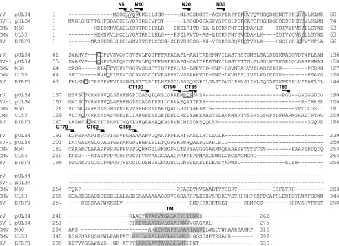
binding to pUL31 homologs (aa 1 to 181 in HCMV) (33). More-over, in HCMV, MCMV, and HSV-1, two amino acids within the conserved N-terminal part were found to be especially important for complex formation, represented by glutamate at position 53 and tyrosine at position 54 in PrV pUL34 (Fig. 1) (26, 31–33), indicating that in all herpesvirus pUL34 homologs, the complete N-terminal part may be involved in pUL31 binding.
To further identify important functional domains and essen-tial amino acids in PrV pUL34, we generated additional chimeric constructs and replaced sequentially either 60, 70, 80, 85, or 90 amino acids of the PrV pUL34 C terminus by Lap2sequences as described previously (27). We show that pUL34 lacking the C-ter-minal 85 amino acids and fused to the Lap2C terminus (pUL34-LapCT85) efficiently complemented the defect of PrV-⌬UL34, while pUL34-LapCT90 failed to, thus indicating that functionally important sequences were deleted. Recruitment of pUL31 was un-impaired, as described previously for pUL34-LapCT100 (27), but nucleocapsids were unable to leave the nucleus. Within this re-gion, amino acid positions 173 to 175 are represented by RQR. The three-residue motif RXR has been shown to be present in the cytoplasmic domains of glycoproteins B in HSV-1 and HCMV as well as in the cellular lamin B receptor. Fused to a reporter protein (CD8), this signal was sufficient for INM localization (36,37). To study the role of this motif in pUL34 localization, it was altered to RQG by site-directed mutagenesis. Moreover, to study require-ments for pUL31 interaction in the N-terminal part of pUL34, N-terminal truncations were generated, and conserved amino ac-ids were mutated and tested for pUL31 recruitment as well as functional complementation.
MATERIALS AND METHODS
Cells and viruses.Rabbit kidney (RK13) or porcine kidney (PSEK) cells were cultivated in Dulbecco’s modified Eagle’s minimum essential me-dium supplemented with 10% or 5% fetal calf serum, respectively. Wild-type PrV Kaplan (PrV-Ka) (38) was grown on RK13 or PSEK cells, whereas PrV-⌬UL34 was propagated on RK13-UL34 cells as described previously (23,39).
Generation of mutant pUL34.Chimeric pUL34-LapCT proteins were generated as described recently (27) using primers listed inTable 1and PrV-Ka DNA and plasmid pEGFP-Lap2(40) as the templates. After fusion, PCR products were cloned into EcoRV-cleaved pcDNA3 (Invitro-gen). Correct amplification and cloning were controlled by restriction enzyme cleavage and sequencing.
N-terminal truncations were introduced by PCR using the primers listed inTable 1on pcDNA-UL34 as the template (39). PCR products were furnished with artificially introduced EcoRI and XhoI cleavage sites for cloning into pcDNA3. Correct amplification and cloning were verified by sequencing.
Point mutations were created by site-directed mutagenesis using an Agilent Technologies QuikChange II XL site-directed mutagenesis kit. pcDNA-UL34 served as the template for primers listed inTable 1. All plasmids were sequenced for correct amplification and cloning (data not shown).
Generation of stable cell lines.To generate cell lines which stably express mutant pUL34, RK13 cells were transfected by calcium phosphate precipitation (41). The transfected cells were selected in medium supple-mented with 0.5 mg/ml G418 (Invitrogen). Resistant cell clones were picked, and pUL34 expression was examined by indirect immunofluores-cence and Western blot analysis with a pUL34-specific polyclonal rabbit serum (39).
Laser scanning confocal microscopy.To determine the localization of the mutated pUL34, RK13 cells were transfected with the different pcDNA-UL34 constructs by calcium phosphate precipitation. For
on November 7, 2019 by guest
http://jvi.asm.org/
calization studies of pUL34 and pUL31, RK13 cells were cotransfected with pcDNA-UL31 (34). At 48 h after transfection, cells were fixed with 3% paraformaldehyde for 20 min, washed with 50 mM NH4Cl, and per-meabilized with 0.5% Triton X-100. Immunostaining for pUL34 and/or pUL31 was performed with polyclonal rabbit anti-pUL31 (34) and poly-clonal rabbit or mouse anti-pUL34 serum (5,39), which was diluted 1:500 in phosphate-buffered saline (PBS), and incubated for 1 h at room tem-perature. Cell culture supernatant of hybridoma cell lines expressing anti-Golgi complex monoclonal antibody P3-b4-3 was used at a dilution of 1:10 (42,43). Bound antibody was detected by Alexa Fluor 488 goat anti-rabbit IgG and Alexa Fluor 555 goat anti-mouse IgG (Invitrogen), which were diluted 1:1,000 in PBS. Fluorescence images were obtained using a confocal laser scanning microscope (LSM510 [Zeiss, Jena, Germany] and Leica SP5 [Wetzlar, Germany]).
Complementation assay.To assay for functional complementation, RK13 cell lines expressing the pUL34 constructs were infected with PrV-Ka or PrV-⌬UL34 at a multiplicity of infection (MOI) of 5 or 3 as indicated in the legends toFig. 3and6. After 1 h at 4°C the inoculum was replaced by prewarmed medium and incubated for an additional hour at 37°C to allow virus entry. Remaining extracellular virus was inactivated by low-pH treatment (44). To determine progeny virus titers, infected cells and supernatants were harvested and titrated on RK13-UL34 cells. The
mean values of at least three independent experiments and the corre-sponding standard deviations were determined.
For determination of plaque size, cells were infected with PrV-Ka or PrV-⌬UL34 under plaque assay conditions and fixed 2 days later. Diam-eters of 30 plaques each were measured microscopically and calculated compared to PrV-Ka on the corresponding cell line, set as 100%. Mean values of three independent assays were plotted.
RESULTS
Localization of functionally important regions in the C-termi-nal part of pUL34.Recently, we showed that deletion of 50 C-ter-minal amino acids of PrV pUL34 including the predicted trans-membrane region and replacement with a corresponding region of the cellular Lap2resulted in a functional chimeric protein (27). However, extension of the deleted region to 100 C-terminal amino acids retaining only amino acids 1 to 161 of pUL34 fused to the C terminus of Lap2with the transmembrane region resulted in lack of complementation of the UL34 deletion mutant despite continuing pUL31 binding (27). To further delineate the func-tionally important sequences between amino acids 161 and 211, truncation mutants of pUL34 were generated lacking 60, 70, 80, or FIG 1Sequence comparison of pUL34 homologs. Deduced amino acid sequences of PrV (accession numberAFI70799), HSV-1 (AER38042), HCMV (AAS48955), MCMV (CAP08099), and EBV (YP_401649) were compared using the constraint-based multiple protein alignment tool (COBALT) (49) (and matched to the alignment shown in reference33). Amino- and carboxy-terminal truncations as well as site-specific mutations introduced into PrV pUL34 and analyzed in this report are indicated. The RXR motif present in PrV pUL34 is boxed; the deduced transmembrane regions (TM) are shaded. A predicted amino-terminal alpha-helix (33) is marked in PrV pUL34.
Functional Domains of PrV pUL34
on November 7, 2019 by guest
http://jvi.asm.org/
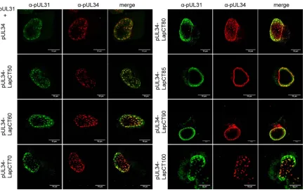
[image:3.585.49.541.65.422.2]90 C-terminal amino acids (Fig. 1) and fused to the same Lap2 sequences used for pUL34-LapCT50 (amino acids 374 to 452 of Lap2). The corresponding chimeras LapCT60, pUL34-LapCT70, pUL34-LapCT80, and pUL34-LapCT90 showed nu-clear rim staining after transfection of the expression plasmids into RK13 cells and relocated coexpressed pUL31 to the nuclear boundary, indicating functional interaction as shown for pUL34-LapCT50 and pUL34-LapCT100 (Fig. 2) (27).
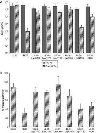
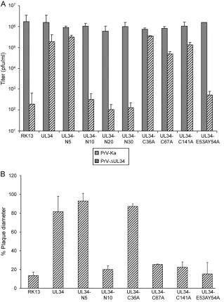
Cell lines stably expressing the different pUL34-Lap2 chime-ric proteins were infected with either PrV-Ka or PrV-⌬UL34 at an MOI of 5 and harvested 30 h postinfection. As shown inFig. 3A, pUL34-LapCT60, pUL34-LapCT70, and pUL34-LapCT80 com-plemented the defect of PrV-⌬UL34 to titers only approximately 5- to 10-fold lower than those of PrV-Ka on the same cells. How-ever, PrV-⌬UL34 titers derived from RK13-UL34-LapCT90 were similar to those from nontransfected RK13 cells (Fig. 3A). Titers of PrV-Ka from the different pUL34-LapCT-expressing cell lines were above 106PFU/ml and similar to titers from nontransfected
RK13 cells, indicating that none of the constructs exerted a signif-icant dominant negative effect on virus replication.
Since these data indicated presence of a region important for pUL34 function independent of the pUL31 interaction between pUL34 amino acids 171 LapCT90) and 181 (pUL34-LapCT80), an additional chimeric protein was constructed lack-ing amino acids 176 to 262 of pUL34, designated pUL34-LapCT85. This protein localized to the nuclear rim, interacted with pUL31 (Fig. 2), and complemented PrV-⌬UL34 to a
signifi-cant extent (Fig. 3A), delineating the functionally important do-main between amino acids 171 and 176 of pUL34.
Besides its role in nuclear egress, pUL34 may also influence direct viral cell-to-cell spread (27,45). Therefore, we tested the efficiency of plaque formation of PrV-⌬UL34 on the different pUL34-Lap2-expressing cell lines. Wild-type-like plaque diam-eters were found on cells expressing pUL34-LapCT80, while plaque size was slightly reduced on UL34-LapCT60, LapCT70, and LapCT85 cells. RK13-UL34-LapCT90 failed to efficiently complement cell-to-cell spread of PrV-⌬UL34, and plaque diameters were only marginally larger than on RK13 cells (Fig. 3B), thereby mirroring the results of one-step growth analyses (Fig. 3A).
These data indicate that pUL34 sequences preceding amino acid 176 (present in pUL34-LapCT85) (Fig. 1) are required for pUL34 function during viral replication. Since amino acids 1 to 162 were shown to be sufficient for pUL31 interaction (27,34), this result points to a function beyond recruitment of pUL31 to the INM.
[image:4.585.42.546.76.375.2]The RQR motif does not specifically target pUL34 to the INM.The pUL34 amino acid sequences present in functional LapCT85 but lacking in noncomplementing pUL34-LapCT90 comprise the tripeptide RQR at amino acids 173 to 175 (Fig. 1). This RXR motif has been shown to be sufficient for tar-geting HCMV and HSV-1 glycoprotein B and lamin B receptor as well as a CD8 reporter protein to the INM (36,37). To study the role of this sequence on PrV pUL34 localization, RQR was altered TABLE 1Primers used in this study
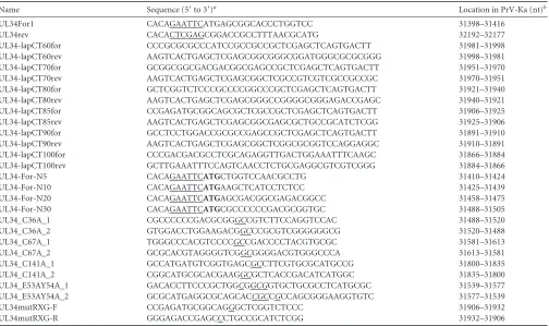
Name Sequence (5=to 3=)a Location in PrV-Ka (nt)b
UL34For1 CACAGAATTCATGAGCGGCACCCTGGTCC 31398–31416
UL34rev CACACTCGAGCGGACCGCCTTTAACGCATG 32192–32177
UL34-lapCT60for CCCGCGCGCCCATCCGCCGCCGCTCGAGCTCAGTGACTT 31981–31998
UL34-lapCT60rev AAGTCACTGAGCTCGAGCGGCGGGCGGATGGGCGCGCGGG 31998–31981
UL34-lapCT70for GCGGCGGCGACGACGGCGAGCCGCTCGAGCTCAGTGACTT 31951–31970
UL34-lapCT70rev AAGTCACTGAGCTCGAGCGGCTCGCCGTCGTCGCCGCCGC 31970–31951
UL34-lapCT80for GCTCGGTCTCCCGCCCCGGCCCGCTCGAGCTCAGTGACTT 31921–31940
UL34-lapCT80rev AAGTCACTGAGCTCGAGCGGGCCGGGGCGGGAGACCGAGC 31940–31921
UL34-lapCT85for CCGAGATGCGGCAGCGCTCGCCGCTCGAGCTCAGTGACTT 31906–31925
UL34-lapCT85rev AAGTCACTGAGCTCGAGCGGCGAGCGCTGCCGCATCTCGG 31925–31906
UL34-lapCT90for GCCTCCTGGACCGCGCCGAGCCGCTCGAGCTCAGTGACTT 31891–31910
UL34-lapCT90rev AAGTCACTGAGCTCGAGCGGCTCGGCGCGGTCCAGGAGGC 31910–31891
UL34-lapCT100for CCCGACGACGCCTCGCAGAGGTTGACTGGAAATTTCAAGC 31866–31884
UL34-lapCT100rev GCTTGAAATTTCCAGTCAACCTCTGCGAGGCGTCGTCGGG 31884–31866
UL34-For-N5 CACAGAATTCATGCTGGTCCAACGCCTG 31410–31424
UL34-For-N10 CACAGAATTCATGAAGCTCATCCTCTCC 31425–31439
UL34-For-N20 CACAGAATTCATGAGCGACGGCGAGACGGCC 31458–31475
UL34-For-N30 CACAGAATTCATGCGCCCCCCGACGCGGTGC 31488–31505
UL34_C36A_1 CGCCCCCCGACGCGGGCCGTCTTCCAGGTCCAC 31488–31520
UL34_C36A_2 GTGGACCTGGAAGACGGCCCGCGTCGGGGGGCG 31520–31488
UL34_C67A_1 TGGGCCCACGTCCCCGCCGACCCCTACGTGCGC 31581–31613
UL34_C67A_2 GCGCACGTAGGGGTCGGCGGGGACGTGGGCCCA 31613–31581
UL34_C141A_1 GCCATGATGTCGGTGAGCGCCTTCGTGCGCATGCCG 31800–31835
UL34_C141A_2 CGGCATGCGCACGAAGGCGCTCACCGACATCATGGC 31835–31800
UL34_E53AY54A_1 GACACCTTCCCGCTGGCGGCGGTGCTGCGCCTCATGCGC 31539–31577
UL34_E53AY54A_2 GCGCATGAGGCGCAGCACCGCCGCCAGCGGGAAGGTGTC 31577–31539
UL34mutRXG-F CCGAGATGCGGCAGGGCTCGGTCTCCC 31906–31932
UL34mutRXG-R GGGAGACCGAGCCCTGCCGCATCTCGG 31932–31906
aRestriction sites introduced for convenient cloning are underlined, artificial start codons are shown in bold, and mismatches for site-directed mutagenesis underlined and in
italics.
bPositions correspond to GenBank accession number BK001744. nt, nucleotide.
on November 7, 2019 by guest
http://jvi.asm.org/
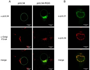
to RQG by site-directed mutagenesis, and cell lines were isolated which stably expressed pUL34RQG. Indirect immunofluores-cence analyses with the rabbit anti-pUL34 serum (39) revealed a perinuclear pattern characteristic for Golgi localization in addi-tion to nuclear rim staining (Fig. 4A). Double fluorescence stain-ing usstain-ing a Golgi complex-specific monoclonal antibody (42,43) verified colocalization in these punctate structures in the cytosol but not at the nuclear rim, indicating that pUL34RQG, but not wild-type pUL34, was present in the Golgi apparatus (Fig. 4A). However, after cotransfection with pcDNA-UL31, nuclear speck-les were still observed in which both proteins colocalized, demon-strating that, despite its prominent Golgi localization, a fraction of pUL34RQG also reached the INM to recruit nucleoplasmic pUL31 (Fig. 4B). These data suggest that the RQR motif is not essential for pUL34 targeting to the nuclear membrane but might act as a Golgi retrieval signal to relocate Golgi compartment-lo-calized molecules back into the ER and from there into the nuclear membrane.
Despite its enhanced Golgi localization, pUL34RQG partially complemented the defect of PrV-⌬UL34 in one-step growth kinetics, with approximately 25-fold reduced titers (Fig. 3A) and reduced plaque diameters (ca. 60% compared to PrV-Ka) (Fig. 3B), correlating with the continuing, but reduced, pres-ence of pUL34RQG at and recruitment of pUL31 to the INM. Delineation of the pUL31 interaction domain.Yeast two-hy-brid studies showed that amino acids 1 to 162 of PrV pUL34 are sufficient for interaction with pUL31 (34), and a pUL34-LapCT chimera comprising only amino acids 1 to 161 of PrV pUL34 was able to recruit pUL31 to the nuclear rim (27). In HSV-1 pUL34, the pUL31 interaction domain was reported at amino acids 137 to
181 (35). However, a PrV pUL34-Lap2chimeric construct in which the N-terminal 50 amino acids of pUL34 were replaced by Lap2sequences, thus retaining the respective homologous re-gion (PrV pUL34 aa 123 to 167), did not mediate recruitment of pUL31 to the INM and failed to complement the defect in
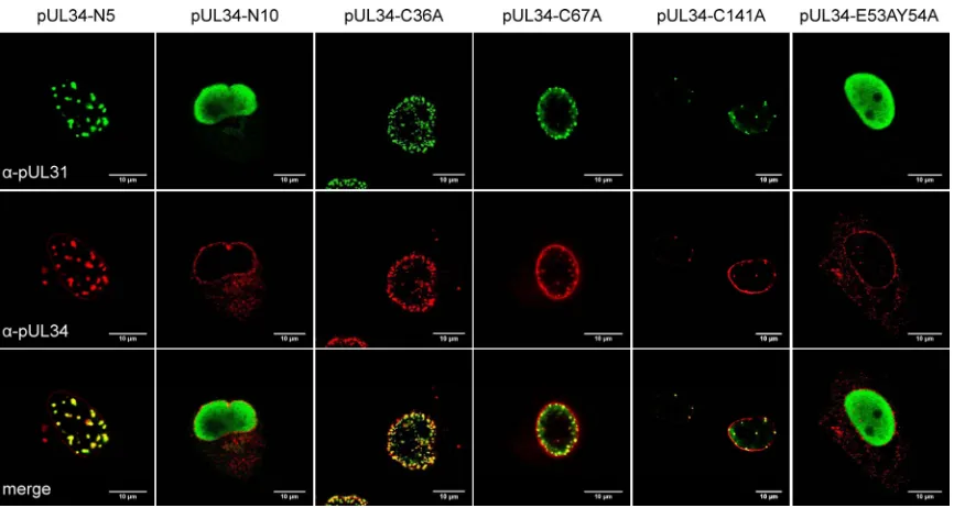
PrV-⌬UL34. Thus, at least in PrV pUL34, this domain is not sufficient for functional interaction (27). Therefore, to further delineate the pUL31 interaction domain, N-terminal truncations of 4, 9, 19, 29, or 39 N-terminal amino acids were created (designated pUL34-N5, -N10, -N20, -N30, and -N40, respectively). For all constructs, an artificial start codon was added by the primers used for ampli-fication (Table 1;Fig. 1). Transfection of the pcDNA3 expression constructs revealed that all proteins localized to the nuclear rim as did native pUL34 (data not shown). However, after cotransfection with pcDNA-UL31, only pUL34-N5 showed the typical punctate nuclear rim pattern, with costaining of both pUL34 and anti-pUL31 sera indicating interaction and vesicle formation (5) (Fig. 5). In contrast, all other N-terminally truncated pUL34 proteins were unable to recruit pUL31 to the INM (exemplarily shown for pUL34-N10) (Fig. 5) since the protein was found diffusely distrib-uted in the nucleoplasm as in cells expressing only pUL31 (34). These results demonstrate that deletion of as few as nine N-termi-nal pUL34 amino acids abolishes pUL31 recruitment to the INM. To test for complementation, cell lines stably expressing pUL34-N5, pUL34-N10, pUL34-N20, and pUL34-N30 were gen-erated and tested after infection with either Ka or
PrV-⌬UL34. Only RK13-UL34-N5 complemented PrV-⌬UL34 to wild-type-like titers and plaque diameters while all the others failed, with values similar to those found on nontransfected RK13 FIG 2Nuclear envelope localization and pUL31 interaction of LapCT chimeras. RK13 cells were cotransfected with expression plasmids for pUL34-LapCT and pUL31 and analyzed by indirect immunofluorescence with monospecific murine anti-pUL34 (red) and rabbit anti-pUL31 (green) sera. Images were recorded with a confocal laser scanning microscope (LSM510 [Zeiss] and SP5 [Leica]). Scale bar, 10m.␣, anti.
Functional Domains of PrV pUL34
on November 7, 2019 by guest
http://jvi.asm.org/
[image:5.585.77.511.64.335.2]cells (Fig. 6). These data further restrict the pUL31 interaction domain in pUL34 to sequences between amino acids 5 and 161.
Conserved amino acids in the N-terminal domain of pUL34. Since truncations in the N terminus, besides deletion of the first 4 amino acids, resulted in nonfunctional proteins, we aimed to identify functionally important amino acids in the conserved N-terminal part of PrV pUL34. Similar studies have been performed for pUL34 homologs of HSV-1 (charge cluster mutants) (26), MCMV (linker insertions) (31), and HCMV (point mutations) (33). We first focused on the conserved cysteine residues. These had not been addressed in other studies but might play an impor-tant role for pUL34 structure. To this end, cysteine residues at
positions 36, 67, and 141 of PrV pUL34 (Fig. 1) were changed to alanines by site-directed mutagenesis. While cysteine at position 36 is strictly conserved also positionally within pUL34 homologs in all herpesvirus subfamilies, the cysteines at positions 67 and 141 are found at similar, though not identical, positions (Fig. 1) (33). Interaction between the mutated pUL34 constructs and pUL31 was tested after cotransfection of the corresponding expression plasmids with pcDNA-UL31 into RK13 cells, followed by coim-munostaining at 2 days posttransfection. As shown inFig. 5, all cysteine mutants still localized to the nuclear rim and recruited pUL31 into nuclear speckles, indicating continuing interaction.
The ability to functionally complement PrV-⌬UL34 was again FIG 3Functional complementation of PrV-⌬UL34 by the pUL34-LapCT chimeras and pUL34RQG. (A) Cell lines stably expressing the indicated proteins were infected with PrV-Ka or PrV-⌬UL34 at an MOI of 5, and viral progeny were harvested at 30 h postinfection and titrated on RK13-UL34 cells. Values represent the means of three independent experiments with the corresponding standard deviations. (B) To test for complementation of direct cell-to-cell spread, cells were infected with PrV-Ka or PrV-⌬UL34 under plaque assay conditions and fixed at 2 days postinfection. Diameters of 30 plaques for each virus and cell line were measured microscopically in three independent experiments. Plaque diameters of PrV-Ka on a given cell line were set as 100%, and diameters of plaques induced by PrV-⌬UL34 were calculated accordingly.
on November 7, 2019 by guest
http://jvi.asm.org/
[image:6.585.136.453.66.508.2]assayed on cell lines stably expressing the mutated pUL34 proteins (Fig. 6A). While RK13-UL34-C36A complemented PrV-⌬UL34 to wild-type-like titers, infectious progeny derived from RK13-UL34-C67A and RK13-UL34-C141A reached lower titers than PrV-Ka on the corresponding cell lines, but titers were still well above those of the nontransgenic control cells. However, plaque diameters formed by PrV-⌬UL34 after infection of RK13-UL34-C67A and RK13-UL34-C141A were similar to those found after infection of nontransfected RK13 cells (Fig. 6B), indicating that these residues are important for a step beyond nuclear egress.
Lastly, we wanted to validate our assay system by mutating amino acids which had already been shown to be important for pUL34 function in other herpesviruses. In the HCMV and HSV-1 pUL34 homologs, amino acids corresponding to glutamate 53 and tyrosine 54 in PrV pUL34 have been shown to be important for pUL31 interaction and protein function (26,33). To test for their role in PrV, both amino acids were simultaneously altered to ala-nines, giving rise to pUL34-E53AY54A (Fig. 1). In transient ex-pression assays, pUL34-E53AY54A localized to the nuclear rim but was unable to relocate pUL31 (Fig. 5), indicating that these two amino acids play an important role for pUL31 interaction also in PrV pUL34. On transgenic cells stably expressing pUL34-E53AY54A, titers and plaque diameters of PrV-⌬UL34 were sim-ilar to those from nontransfected RK13 cells (Fig. 6), demonstrat-ing that these two conserved amino acids of pUL34 play an important role in herpesvirus replication, presumably by direct involvement in interaction with pUL31.
DISCUSSION
Conserved herpesvirus proteins designated pUL34 and pUL31 in PrV and HSV-1 physically interact to form the NEC, which is required for efficient nuclear egress, by mediating primary enve-lope formation, fusion, and fission (reviewed in references1to3). While the N-terminal part is well conserved between the pUL34 homologs throughout the herpesvirus subfamilies, the C-terminal half including the membrane anchor region of these type II mem-brane proteins is more divergent (Fig. 1) (31,33,45). The N-ter-minal domain is required for interaction with and recruitment of pUL31 to the INM as a prerequisite for primary envelopment during nuclear egress (27,29,31,33,34).
In the absence of a crystal structure for the NEC, several mu-tagenesis and/or deletion analyses have been performed to iden-tify regions in either protein required for complex formation and function. Although the results improved our understanding of the NEC, the picture is far from being complete. Thus, in this study we extend previous analyses on PrV pUL34 by creating truncated and site-specifically mutated versions of the protein and testing them for pUL31 interaction and for functional complementation of a pUL34 deletion mutant.
Previously, it has been shown that replacement of the C-termi-nal transmembrane domain of pUL34 by corresponding regions of other cellular INM proteins or of other related or unrelated viral polypeptides was functionally tolerated, whereas deletion of the transmembrane domain resulted in a nonfunctional protein. FIG 4Intracellular localization of pUL34RQG. (A) RK13 cells were transfected with plasmids expressing pUL34 or pUL34RQG, fixed, and costained with a monoclonal antibody against Golgi proteins (P3-b4; red) (42,43) and rabbit anti-pUL34 serum (green). (B) RK13 cells were cotransfected with pcDNA-UL34RQG and pcDNA-UL31, fixed, and stained 2 days later with rabbit anti-pUL31 and mouse anti-pUL34 sera. Bound antibody was visualized after incubation with Alexa Fluor 488 anti-rabbit and Alexa Fluor 555 anti-mouse secondary IgG. Images were taken with a confocal laser scanning microscope (LSM510; Leica SP5). Scale bar, 10m.
Functional Domains of PrV pUL34
on November 7, 2019 by guest
http://jvi.asm.org/
[image:7.585.109.475.66.353.2]Thus, the presence of a membrane anchor in pUL34 but not its viral origin is necessary for function since it can be replaced by a variety of alpha-helical sequences of at least 15 amino acids (27–
29). We showed recently that even the exchange of 50 C-terminal residues of PrV pUL34 with Lap2sequences resulted in a func-tional protein which efficiently mediated nuclear egress and infec-tious virus formation (27). However, extension of the substituted region to 100 amino acids, retaining only amino acids 1 to 161 of the 262 amino acids of PrV pUL34, resulted in a nonfunctional protein although it was still able to recruit pUL31. To further delineate this functionally important region, we sequentially de-leted sequences from the C terminus of PrV pUL34 and fused the truncated proteins with the membrane anchor domain of cellular Lap2(27). We show here that extension of the deletion up to amino acid 177 of PrV pUL34 did not impair pUL31 interaction and NEC function. In contrast, deletion of an additional 5 amino acids resulted in a nonfunctional protein which was, however, still able to recruit pUL31. Thus, most of the less-conserved C-termi-nal region of pUL34 is actually not required for protein function. This suggests that this part of the protein could simply act as a “stalk” exposing the N-terminal pUL31 interaction domain for recruitment of the complex partner. Apparently, heterologous se-quences are able to substitute for authentic pUL34 sese-quences to provide this feature.
The 5 amino acids which differ between functional pUL34-LapCT85 and nonfunctional pUL34-LapCT90 contain the tripep-tide RQR. This motif bears striking resemblance to the RXR se-quence shown to locate glycoproteins B of HSV-1 and HCMV as well as nonviral reporter CD8 to the INM (36,37). To test the influence of this arginine-rich motif on pUL34 localization, we altered its sequence to RQG. After transient as well as stable ex-pression, a fraction of the mutated protein still located to the nu-clear rim and interacted with pUL31. However, a major fraction of
the mutated pUL34 was found to colocalize with a Golgi complex-specific protein in perinuclear structures, indicative of localization to the Golgi apparatus. Thus, after mutation of the RQR sequence, a fraction of pUL34 does not reach the nucleus but may leak into the cellular secretory pathway to the Golgi compartment. This indicates that the RQR motif may function as a Golgi retrieval signal relocating pUL34 from the secretory pathway. Similar re-sults were obtained for the cellular SUN2 protein in which a clus-ter of arginines mediates Golgi retrieval (22).
RXR motifs have been described on ER-resident proteins. They were first identified as ER retention signals for type II membrane proteins (46). Protein kinase A and C phosphorylation sites flank the RXR sequence, suggesting phosphorylation-controlled ER ex-port of RXR-containing proteins as a quality control mechanism (47). Frequently, a dileucine endocytic sorting motif is located in the vicinity (48). In PrV pUL34 the sequence comprising the RQR motif is predicted as a phosphorylation site (http://elm.eu.org/) with a preceding dileucine (both in boldface) sequence (SQTQRLLDRAEMRQRSVSRP) (Fig. 1). Further experimenta-tion is needed to test whether pUL34 is phosphorylated at this position and to elucidate the influence of phosphorylation as well as the importance of the dileucine motif on pUL34 localization and function. Interestingly, a linker insertion within the dileucine sequence in the MCMV homolog was detrimental to protein func-tion (31).
Whereas the C-terminal, less-conserved part of pUL34 shows little sensitivity toward alterations, the well-conserved N terminus is highly sensitive. Only the amino-terminal four amino acids could be deleted without impairing pUL34 function and interac-tion with pUL31. Extending the truncainterac-tion to nine amino-termi-nal amino acids resulted in a nonfunctioamino-termi-nal protein. In this in-stance loss of pUL34 function correlated with loss of pUL31 interaction, which again emphasizes the importance of complex FIG 5Interaction of N-terminally truncated and site-specifically mutated pUL34 with pUL31. To test whether the N-terminally truncated pUL34 and mutated constructs were able to interact with pUL31, plasmids expressing the indicated proteins were cotransfected with pcDNA-UL31. Two days later cells were fixed and stained with pUL34-specific murine serum and pUL31-specific rabbit serum. Bound antibodies were visualized after incubation with the corresponding secondary antibodies under the confocal laser scanning microscope (LSM510; Leica SP5). Scale bar, 10m.
on November 7, 2019 by guest
http://jvi.asm.org/
[image:8.585.77.510.64.295.2]formation for nuclear egress. These data support results of other investigators (29, 31, 33), demonstrating that interaction of pUL34 with pUL31 requires more than the interaction domain mapped by Liang and Baines (35). Most likely, the complete well-conserved amino terminal part of pUL34 is necessary for complex formation.
To test for the importance of specific amino acids in this re-gion, we mutated three conserved cysteine residues to alanines. Whereas Cys36 of PrV pUL34 is positionally identical in pUL34 homologs of other herpesviruses, Cys67 and Cys141 are slightly offset in pUL34 homologs of HCMV, MCMV, and EBV (Fig. 1) (33). All of these mutants still interacted with pUL31, resulting in nuclear rim localization. However, different phenotypes in func-tional complementation were observed. pUL34-C36A efficiently
complemented the replication and cell-to-cell spread defect of PrV-⌬UL34. In contrast, whereas titers found after infection of RK13-UL34-C67A and RK13-UL34-C141A were only slightly re-duced, direct cell-to-cell spread was nearly abrogated, and plaque diameters were similar to those found after infection of nontrans-genic RK13 cells. This unexpected result points to an important function of PrV pUL34 Cys 67 and Cys 141 in a step beyond nuclear egress.
Alteration of the highly conserved dipeptide Glu53/Tyr54 of PrV pUL34 to alanine abolished pUL31 interaction and function of pUL34. These amino acids have already been shown to be re-quired for function of pUL34 homologs in HSV-1 (26) and HCMV (33). Moreover, a linker insertion between them in MCMV pUL34 resulted in a dominant negative protein (32). A FIG 6Complementation of PrV-⌬UL34 by N-terminally truncated and site-specifically mutated pUL34. (A) Cell lines stably expressing N-terminally truncated or site-specifically mutated pUL34 were infected at an MOI of 3 with PrV-Ka or PrV-⌬UL34 and harvested after 24 h. Infectious progeny was titrated on RK13-UL34 cells. Given are mean values of three independent assays with the corresponding standard deviations. (B) To test for complementation of cell-cell spread, cell lines were infected with PrV-Ka or PrV-⌬UL34 under plaque assay conditions. Plaque diameters of 30 plaques each were measured microscopically in three independent assays. Values of PrV-Ka on the corresponding cell line were set at 100%, and diameters of PrV-⌬UL34 plaques were calculated accordingly.
Functional Domains of PrV pUL34
on November 7, 2019 by guest
http://jvi.asm.org/
[image:9.585.135.454.67.497.2]cell line stably expressing HSV-1 pUL34Y68A, which is equivalent to PrV Tyr54, replicated a corresponding pUL34 deletion mutant to approximately 60-fold lower titers than the parental protein and complemented direct viral cell-to-cell spread only very inef-ficiently. In pUL34Y68A-expressing cells the nuclear envelope showed blebs, presumably as the result of disconnecting nuclear envelope from the underlying lamina, pointing to an exaggerated lamina disruption. In addition, trafficking of gE was inhibited in this cell line after infection with the UL34 deletion mutant, but interaction of pUL34Y68A with pUL31 was not tested (45). Col-lectively, these data identify a conserved dipeptide motif in her-pesvirus pUL34 homologs which is highly important for function of the NEC.
In summary, our data show that most of the C-terminal por-tion of pUL34, which shows little to no conservapor-tion between the herpesvirus pUL34 homologs, can be deleted and replaced by het-erologous sequences if a transmembrane anchor is provided. Thus, this portion of the protein may primarily, if not solely, act to anchor pUL34, and, consequently, the NEC, in the INM and pro-vide a stalk for presenting the N-terminal domain for interaction with pUL31. In contrast, the well-conserved N-terminal part of pUL34 is essential for NEC function during virus replication and nuclear egress although conserved cysteine residues, in contrast to a similarly conserved EY motif, are not required. Amino acids 5 to 161 of PrV pUL34 are sufficient for pUL31 binding, while the precise termini of this interaction domain remain to be identified, preferably by elucidation of the crystal structure of the NEC. How-ever, sequences up to amino acid 176 also play a fundamental role for viral replication independent of pUL31 binding. In this region, trafficking signals such as the presumptive Golgi retrieval motif RQR could modulate the intracellular location of pUL34 and, therefore, impact its function in nuclear and extranuclear events of herpesvirus replication.
ACKNOWLEDGMENTS
This study was supported by the Deutsche Forschungsgemeinschaft (DFG Me 854/12-1).
We thank C. Meinke for expert technical assistance.
REFERENCES
1.Mettenleiter TC, Klupp BG, Granzow H.2009. Herpesvirus assembly: an update. Virus Res.143:222–234.
2.Mettenleiter TC, Müller FM, Granzow H, Klupp BG.2013. The way out: what we know and do not know about herpesvirus nuclear egress. Cell Microbiol.15:170 –178.
3.Johnson DC, Baines JD.2011. Herpesviruses remodel host membranes for virus egress. Nat. Rev. Microbiol.9:382–394.
4.Marschall M, Feichtinger S, Milbradt J.2011. Regulatory roles of protein kinases in cytomegalovirus replication. Adv. Virus Res.80:69 –101. 5.Klupp BG, Granzow H, Fuchs W, Keil GM, Finke S, Mettenleiter TC.
2007. Vesicle formation from the nuclear membrane is induced by coex-pression of two conserved herpesvirus proteins. Proc. Natl. Acad. Sci. U. S. A.104:7241–7246.
6.Desai PJ, Pryce EN, Henson BW, Luitweiler EM, Cothran J. 2012. Reconstitution of the Kaposi’s sarcoma-associated herpesvirus nuclear egress complex and formation of nuclear membrane vesicles by coexpres-sion of ORF67 and ORF69 gene products. J. Virol.86:594 –598. 7.Cockrell SK, Huffman JB, Toropova K, Conway JF, Homa FL.2011.
Residues of the UL25 protein of herpes simplex virus that are required for its stable interaction with capsids. J. Virol.85:4875– 4887.
8.Yang K, Baines JD. 2011. Selection of HSV capsids for envelopment involves interaction between capsid surface components pUL31, pUL17, and pUL25. Proc. Natl. Acad. Sci. U. S. A.108:14276 –14281.
9.Fossum E, Friedel CC, Rajagopala SV, Titz B, Baiker A, Schmidt T,
Kraus Stellberger TT, Rutenberg C, Suthram S, Bandyopadhyay S, Rose D, von Brunn A, Uhlmann M, Zeretzke C, Dong YA, Boulet H, Koegl M, Bailer SM, Koszinowski HU, Ideker T, Uetz P, Zimmer R, Haas J.
2009. Evolutionarily conserved herpesviral protein interaction networks. PLoS Pathog.5:e1000570. doi:10.1371/journal.ppat.1000570.
10. Uetz P, Dong YA, Zeretzke C, Atzler C, Baiker A, Berger B, Rajagopala SV, Roupelieva M, Rose D, Fossum E, Haas J.2006. Herpesviral protein networks and their interaction with the human proteome. Science311: 239 –342.
11. Leelawong M, Guo D, Smith GA.2011. A physical link between the pseudorabies virus capsid and the nuclear egress complex. J. Virol.85: 11675–11684.
12. Klupp BG, Granzow H, Keil GM, Mettenleiter TC.2006. The capsid-associated UL25 protein of the alphaherpesvirus pseudorabies virus is nonessential for cleavage and encapsidation of genomic DNA but is re-quired for nuclear egress of capsids. J. Virol.80:6235– 6246.
13. Kuhn J, Leege T, Klupp BG, Granzow H, Fuchs W, Mettenleiter TC.
2008. Partial functional complementation of a pseudorabies virus UL25 deletion mutant by herpes simplex virus type 1 pUL25 indicates overlap-ping functions of alphaherpesvirus pUL25 proteins. J. Virol.82:5725– 5734.
14. Ye GJ, Vaughan KT, Vallee RB, Roizman B.2000. The herpes simplex virus 1 UL34 protein interacts with a cytoplasmic dynein intermediate
chain and targets nuclear membrane. J. Virol.74:1355–1363.
15. Schirmer EC, Gerace L.2005. The nuclear membrane proteome: extend-ing the envelope. Trends Biochem. Sci.30:551–558.
16. Antonin W, Ungricht R, Kutay U.2011. Traversing the NPC along the pore membrane: targeting of membrane proteins to the INM. Nucleus
2:87–91.
17. Braunagel SC, Williamson ST, Ding Q, Wu X, Summers MD.2007. Early sorting of inner nuclear membrane proteins is conserved. Proc. Natl. Acad. Sci. U. S. A.104:9307–9312.
18. Holmer L, Worman HJ.2001. Inner nuclear membrane proteins: func-tions and targeting. Cell. Mol. Life Sci.58:1741–1747.
19. King MC, Lusk CP, Blobel G.2006. Karyopherin-mediated import of integral inner nuclear membrane proteins. Nature442:1003–1007. 20. Lusk CP, Blobel G, King MC.2007. Highway to the inner nuclear
mem-brane: rules for the road. Nat. Rev. Mol. Cell Biol.8:414 – 420.
21. Tapley EC, Ly N, Starr DA.2011. Multiple mechanisms actively target the SUN protein UNC-84 to the inner nuclear membrane. Mol. Biol. Cell
22:1739 –1752.
22. Turgay Y, Ungricht R, Rothballer A, Kiss A, Csucs G, Horvath P, Kutay U.2010. A classical NLS and the SUN domain contribute to the targeting of SUN2 to the inner nuclear membrane. EMBO29:2262–2275. 23. Klupp BG, Granzow H, Mettenleiter TC.2001. Effect of the pseudorabies
virus US3 protein on nuclear membrane localization of the UL34 protein and virus egress from the nucleus. J. Gen. Virol.82:2363–2371. 24. Reynolds AE, Ryckman BJ, Baines JD, Zhou Y, Liang L, Roller RJ.2001.
UL31 and UL34 proteins of herpes simplex virus type 1 form a complex
that accumulates at the nuclear rim and is required for envelopment of nucleocapsids. J. Virol.75:8803– 8817.
25. Horton P, Nakai K.1997. Better prediction of protein cellular localization sites with the k nearest neighbors classifier. Proc. Int. Conf. Intell. Syst. Mol. Biol.5:147–152.
26. Bjerke SL, Cowan JM, Kerr JK, Reynolds AE, Baines JD, Roller RJ.2003. Effects of charged cluster mutations on the function of herpes simplex virus type 1 UL34 protein. J. Virol.77:7601–7610.
27. Schuster F, Klupp BG, Granzow H, Mettenleiter TC.2012. Structural determinants for nuclear envelope localization and function of pseudora-bies virus pUL34. J. Virol.86:2079 –2088.
28. Ott M, Tascher G, Haßdenteufel S, Zimmermann R, Haas J, Bailer SM.
2011. Functional characterization of the essential tail anchor of the herpes simplex virus type 1 nuclear egress protein pUL34. J. Gen. Virol.92:2734 – 2745.
29. Roller RJ, Bjerke SL, Haugo AC, Hanson S.2010. Analysis of a charge cluster mutation of herpes simplex virus type 1 UL34 and its extragenic suppressor suggests a novel interaction between pUL34 and pUL31 that is necessary for membrane curvature around capsids. J. Virol.84:3921– 3934.
30. Roller RJ, Haugo AC, Kopping NJ. 2011. Intragenic and extragenic suppression of a mutation in herpes simplex virus 1 UL34 that affects both nuclear envelope targeting and membrane budding. J. Virol.85:11615– 11625.
on November 7, 2019 by guest
http://jvi.asm.org/
31. Bubeck A, Wagner M, Ruzsics Z, Lotzerich M, Iglesias M, Singh IR, Koszinowski HU.2004. Comprehensive mutational analysis of a herpes-virus gene in the viral genome context reveals a region essential for herpes-virus replication. J. Virol.78:8026 – 8035.
32. Rupp B, Ruzsics Z, Buser C, Adler B, Walther P, Koszinowski UH.
2007. Random screening for dominant-negative mutants of the cytomeg-alovirus nuclear egress protein M50. J. Virol.81:5508 –5517.
33. Milbradt J, Auerochs S, Sevvana M, Müller YA, Sticht H, Marschall M.
2012. Specific residues of a conserved domain in the N terminus of the human cytomegalovirus pUL50 protein determine its intranuclear inter-action with pUL53. J. Biol. Chem.287:24004 –24016.
34. Fuchs W, Klupp BG, Granzow H, Osterrieder N, Mettenleiter TC.2002. The interacting UL31 and UL34 gene products of pseudorabies virus are involved in egress from the host-cell nucleus and represent components of primary enveloped but not mature virions. J. Virol.76:364 –378. 35. Liang L, Baines JD.2005. Identification of an essential domain in the
herpes simplex virus 1 UL34 protein that is necessary and sufficient to interact with UL31 protein. J. Virol.79:3797–3806.
36. Meyer GA, Radsak KD.2000. Identification of a novel signal sequence that targets transmembrane proteins to the nuclear envelope inner mem-brane. J. Biol. Chem.275:3857–3866.
37. Meyer G, Gicklhorn D, Strive T, Radsak K, Eickmann M. 2002. A three-residue signal confers localization of a reporter protein in the inner nuclear membrane. Biochem. Biophys. Res. Commun.291:966 –971. 38. Kaplan AS, Vatter AE.1959. A comparison of herpes simplex and
pseu-dorabies viruses. Virology7:394 – 407.
39. Klupp BG, Granzow H, Mettenleiter TC.2000. Primary envelopment of pseudorabies virus at the nuclear membrane requires the UL34 gene prod-uct. J. Virol.74:10063–10073.
40. Beaudouin J, Gerlich D, Daigle N, Eils R, Ellenberg J.2002. Nuclear envelope breakdown proceeds by microtubule-induced tearing of the lamina. Cell108:83–96.
41. Graham FL, van der Eb AJ. 1973. A new technique for the assay of infectivity of human adenovirus 5 DNA. Virology52:456 – 467. 42. Grossmann A, Weiland F, Weiland E.1989. Autoimmunity induced by
lactate dehydrogenase-elevating virus: monoclonal autoantibodies against Golgi antigens and other subcellular elements. Autoimmunity2:201–211. 43. Klupp BG, Granzow H, Klopfleisch R, Fuchs W, Kopp M, Lenk M, Mettenleiter TC.2005. Functional analysis of the pseudorabies virus UL51 protein. J. Virol.79:3831–3840.
44. Mettenleiter TC.1989. Glycoprotein gIII deletion mutants of pseudora-bies virus are impaired in virus entry. Virology171:623– 625.
45. Haugo AC, Szpara ML, Parsons L, Enquist LW, Roller RJ.2011. Herpes simplex virus type 1 pUL34 plays a critical role in cell-to-cell spread of virus in addition to its role in virus replication. J. Virol.85:7203–7215. 46. Schutze MP, Peterson PA, Jackson MR.1994. An N-terminal
double-arginine motif maintains type II membrane proteins in the endoplasmic reticulum. EMBO J.13:1696 –1705.
47. Scott DB, Blanpied TA, Ehlers MD.2003. Coordinated PKA and PKC phosphorylation suppresses RXR-mediated ER retention and regulates the surface delivery of NMDA receptors. Neuropharmacology45:755– 767.
48. Michelsen K, Yuan H, Schwappach B.2005. Hide and run. Arginine-based endoplasmic-reticulum-sorting motifs in the assembly of hetero-multimeric membrane proteins. EMBO Rep.6:717–722.
49.Papadopoulos JS, Agarwala R. 2007. COBALT: constraint-based alignment tool for multiple protein sequences. Bioinformatics 23: 1073–1079.
Functional Domains of PrV pUL34