Inhibitory Effect on Php4 Function
Philippe Vachon, Alexandre Mercier, Mehdi Jbel, and Simon Labbé
Département de Biochimie, Faculté de Médecine et des Sciences de la Santé, Université de Sherbrooke, Sherbrooke, QC, Canada
When iron is scarce,Schizosaccharomyces pombecells repress transcription of several genes that encode iron-using proteins. Php4 mediates this transcriptional control by specifically interacting with the CCAAT-binding core complex that is composed of Php2, Php3, and Php5. In contrast, when there is sufficient iron, Php4 is inactivated, thus allowing the transcription of many genes that encode iron-requiring proteins. Analysis by bimolecular fluorescence complementation and two-hybrid assays showed that Php4 and the monothiol glutaredoxin Grx4 physically interact with each other. Deletion mapping analysis revealed that the glutaredoxin (GRX) domain of Grx4 associates with Php4 in an iron-dependent manner. Site-directed mutagenesis iden-tified the Cys172 of Grx4 as being required for this iron-dependent association. Subsequent analysis showed that, although the thioredoxin (TRX) domain of Grx4 interacts strongly with Php4, this interaction is insensitive to iron. Fine mapping analysis revealed that the Cys35 of Grx4 is necessary for the association between the TRX domain and Php4. Taken together, the results revealed that whereas the TRX domain interacts constitutively with Php4, the GRX domain-Php4 association is both modulated by iron and required for the inhibition of Php4 activity in response to iron repletion.
I
ron poses a particular dilemma to cells because of its dualprop-erties being an essential metal for cell viability but also being cytotoxic. As a redox-active metal, iron is an important cofactor in a variety of enzymes that are intimately linked to essential cellular functions such as DNA synthesis, oxidative phosphorylation, and
the biosynthesis of metabolites (10,36). Ironically, this same
re-dox property allows iron to undergo Fenton-type chemical reac-tions that result in the production of hydroxyl radicals that can
lead to cell death (8). Therefore, cells have developed homeostatic
mechanisms to acquire adequate, but not excessive, concentra-tions of iron.
In the model organismSchizosaccharomyces pombe, the
GATA-type transcription factor Fep1 binds to chromatin and represses the expression of a number of genes involved in iron acquisition
when the intracellular iron levels are elevated (24,34,35). In
con-trast, when iron levels are low, Fep1 is unable to bind chromatin, which results in transcriptional activation of iron transport genes
(18). Iron-dependent regulators possessing similar functions and
sequences to Fep1 have been identified in several fungi, but not in
Saccharomyces cerevisiae(2,6,7,9,15,20). Recent studies have shown that the monothiol glutaredoxin Grx4 is a binding partner of Fep1 and that it plays a critical role in inhibiting Fep1 function when cells undergo a transition from iron-sufficient to
iron-lim-iting conditions (17,23). Although the mechanism by which Grx4
communicates the low levels of iron to Fep1 remains unclear, deletion mapping analysis revealed that the thioredoxin (TRX) domain of Grx4 interacts strongly and constitutively with the
C-terminal region of Fep1 (17). Further analysis has shown that,
under conditions of iron starvation, the glutaredoxin (GRX) do-main of Grx4 associates with Fep1 through its N terminus. A po-tential mechanism for the Grx4-mediated inhibition of Fep1 func-tion would be that the Fep1-GRX domain associafunc-tion induces an inhibitory conformational change that inactivates the Fep1 DNA-binding domain, blocking its interaction with chromatin and therefore its repressive action on target gene expression.
Under conditions of iron deprivation,S. pombecells produce
the CCAAT-binding subunit Php4 (28). Php4 then assembles
with a heteromeric DNA-binding complex that contains three
other subunits designated Php2, Php3, and Php5 (26). Together,
the Php2/Php3/Php4/Php5 heteromeric complex binds CCAAT
cis-acting elements and represses expression of genes encoding
several iron-dependent proteins, including the iron-responsive
transcriptional repressor Fep1 (29). On the other hand, when iron
is abundant, Php4 fails to act as a repressor, andfep1⫹
tion is therefore derepressed. Fep1 is responsible for the
transcrip-tional repression of thephp4⫹gene, thus creating a reciprocal
regulatory mechanism in which Fep1 and Php4 mutually control each other’s expression as a function of iron availability.
Subsequent studies have revealed the involvement of an addi-tional regulatory mechanism by which Php4 is inactivated by iron
(27). This mechanism does not operate at the transcriptional level;
instead, it operates at the posttranslational level. In cells undergo-ing transition from low to high iron, it has been determined that
Php4 is exported from the nucleus to the cytoplasm (27). This
nuclear-to-cytosolic export of Php4 requires both the exportin
Crm1 and the monothiol glutaredoxin 4 (Grx4) (27).
Consis-tently,grx4⌬mutant cells show a markedly decreased
transcrip-tion of genes encoding iron-dependent proteins as a result of the
action of the constitutively active Php4 (27). The constant
repres-sion of Php4 target gene expresrepres-sion measured in thegrx4⌬mutant
cells has been intrinsically associated with a constitutive Php4
nu-clear retention (27). Subsequently, bimolecular fluorescence
complementation (BiFC) and two-hybrid assays have revealed
that Grx4 is a binding partner of Php4 (27). BiFC experiments
have shown that VN-Php4-Grx4-VC complexes (VN is the Venus amino-terminal fragment, and VC is the Venus carboxyl-terminal
Received22 February 2012 Accepted10 April 2012 Published ahead of print20 April 2012
Address correspondence to Simon Labbé, [email protected].
Copyright © 2012, American Society for Microbiology. All Rights Reserved.
doi:10.1128/EC.00060-12
on September 8, 2020 by guest
http://ec.asm.org/
fragment) display fluorescence signals in the nuclei of iron-defi-cient cells, whereas BiFC signals accumulate in the cytoplasm of
cells exposed to high iron levels (27).
S. pombeGrx4 is a multidomain monothiol glutaredoxin (4). The primary amino acid sequence of Grx4 contains two major regions that are denoted as the thioredoxin (TRX)-like and glu-taredoxin (GRX)-like domains, respectively. The N-terminal
TRX-like domain of Grx4 contains a WAAPC35K sequence that is
reminiscent of the thioredoxin active site motif WCGPCK (4,11).
Mutational analysis of the Cys35 residue located within the TRX domain of Grx4 revealed that it is required for the establishment
of a strong, iron-independent association with Fep1 (17).
Consis-tently, a recent study ofS. cerevisiaesuggested that the TRX
do-main may serve as a docking site for the interacting partners of the
multidomain monothiol glutaredoxins Grx3 and Grx4 (12). In
baker’s yeast, other functions for the TRX domain have been pro-posed, including a role in the targeting of the monothiol
glutare-doxin Grx3 to the nucleus (31), as well as a regulatory role in actin
cytoskeleton remodeling and a role in cellular defense against
ox-idative stress (40). However, whether the TRX domain’s
partici-pation in the repolarization of the actin cytoskeleton involves any protein-protein interactions remains to be determined.
Based on biochemical studies with other multidomain
glutare-doxins orthologous toS. pombeGrx4, it has been shown that the
C-terminal GRX-like domain contains a CGFS active site motif
(11,41). In the case ofS. pombeGrx4, this motif is located within
residues 172 to 175 (172CGFS175). The CGFS-type monothiol
glu-taredoxins can form [2Fe-2S]-bridged homodimers (1, 16,38).
Indeed, the combination of two glutaredoxin molecules (contain-ing one CGFS motif each) provides two Cys ligands with which
they can hold a 2Fe-2S cluster (1,16,38). Within the complex, the
addition of two glutathione molecules provides the other two cluster ligands, resulting in a glutathione-ligated [2Fe-2S] center
that is held within the monothiol glutaredoxin dimer (16,42).
Several studies have pointed out important roles for multidomain monothiol glutaredoxins. Of note, these proteins participate in
mitochondrial and cytosolic Fe-S protein biogenesis (41), they
specifically deliver and transfer Fe-S clusters into proteins and
subcellular compartments (32) and they relay cellular iron status
to several iron-responsive transcription factors (17,21,23,27,33,
39,43).
Based on the previous finding that Grx4 is a Php4-binding partner that is required for its inactivation under conditions of
high levels of iron (27), the mechanism by which Grx4 and Php4
physically interact with each other as a function of iron availability was investigated. Deletion mapping analysis revealed that the TRX domain interacts constitutively with Php4, whereas the GRX do-main associates in an iron-dependent manner with a dodo-main cor-responding to amino acids 188 to 254 of Php4. Further analyses by BiFC assays revealed that the GRX domain is required for iron-mediated inhibition of Php4 activity, as well as for its nuclear export behavior. Taken together, the results reported here reveal that the presence of the GRX domain of Grx4 is critical to the communication of an excess of iron to the Grx4-Php4 complex.
MATERIALS AND METHODS
Yeast strains and growth media.TheS. pombestrains used in this study were all isogenic derivatives of FY435 (h⫹his7-366 leu1-32 ura4-⌬18 ade6-M210) (27) and includedgrx4⌬(h⫹his7-366 leu1-32 ura4-⌬18 ade6-M210 grx4⌬::KANr) andphp4⌬grx4⌬(h⫹his7-366 leu1-32
ura4-⌬18 ade6-M210 php4⌬::loxP grx4⌬::KANr).S. pombecells were cultured
in yeast extract plus supplements (YES) medium that contained 3% glu-cose and 225 mg/liter of adenine, histidine, leucine, uracil, and lysine. Strains used for plasmid integration were grown in synthetic Edinburgh minimal medium (EMM) in which specific amino acids were absent as required for plasmid selection and maintenance.S. pombeliquid cultures were seeded at anA600of 0.5 and then grown to exponential phase. Once
at log phase (A600of⬃0.9), the cells were treated with either 2,2=-dipyridyl
(Dip) (250M) or FeCl3(100M) or were left untreated for 90 min,
unless otherwise indicated.grx4⌬andphp4⌬grx4⌬mutant strains and control strains were grown in culture jars under microaerobic conditions using the BD GazPack EZ system (BD Diagnostic System, Sparks, MD). In the case of two-hybrid experiments, S. cerevisiae strain L40 [MATa
his3⌬200 trp1-901 leu2-3,112 ade2 LYS2::(lexAop)4-HIS3 URA3::
(lexAop)8-lacZ] (47) was grown in a synthetic minimal medium (pH 6.1)
containing 83 mg/liter of histidine, adenine, uracil, and lysine, 2% dex-trose, 50 mM MES [2-(N-morpholino)ethanesulfonic acid], and 0.67% yeast nitrogen base lacking both copper and iron (MP Biomedicals, So-lon, OH).
Plasmids.Thirteen previously described plasmids, VP16-1Php4295,
VP16-1Php4254, VP16-1Php4218, VP16-54Php4254, VP16-152Php4254,
VP16-188Php4254, VP16-219Php4254, LexA-2Grx4244, LexA-2Grx4142
(do-main TRX), LexA-105Grx4244 (domain GRX), LexA-2Grx4142(C35A),
LexA-Grx4(C35A), and LexA-Grx4(C172A) (17,27), were used in this study. The prey plasmids pVP16-54Php4295, pVP16-112Php4295,
pVP16-152Php4295, pVP16-188Php4295, pVP16-219Php4295, and VP16-152Php4254
(C221A; C227A) were created by cloning different truncated versions of thephp4⫹gene into pVP16 (47). The truncated versions of thephp4⫹ gene were generated by PCR using primers which contained BamHI and NotI restriction sites at their ends. After amplification, the purified DNA fragments were digested with these two enzymes and then cloned into the corresponding sites of pVP16 (47) as described previously (27).
TheS. pombe grx4⫹gene was obtained by PCR amplification using primers that contained SalI and Asp718 restriction sites using genomic DNA from strain FY435 as the template. The purified DNA fragment was digested and then cloned into the SalI and Asp718 sites of the pJK-194*promphp4⫹-TAP plasmid (prom stands for promoter), creat-ing pJK-194*promphp4⫹-TAP-grx4⫹. The pJK-194*promphp4⫹- GFP-php4⫹plasmid (GFP stands for green fluorescent protein) has been de-scribed previously (27). The gene encodingGFPwas PCR amplified from the pSF-GP1 plasmid (22) and inserted into the BamHI and SalI restric-tion sites of pGEM-7Zf (Promega, Madison, WI). A SalI-Asp718grx4⫹ gene fragment was generated by PCR and cloned into the plasmid pGEM-7Zf in which theGFPgene had been previously introduced. The resulting pGEM-7Zf⫹GFP-grx4⫹plasmid was subsequently digested with BamHI and XhoI, and the insert was cloned into the corresponding sites of the previously described pSP1-194*promphp4⫹plasmid (27). The resulting construct was named pSP1-194*promphp4⫹-GFP-grx4⫹. To create the pSP1-194*promphp4⫹-GFP-105Grx244plasmid, pSP1-194*promphp4⫹
-GFP-grx4⫹was codigested with SalI and ApaI. A copy of the C-terminal region of Grx4 (the coding sequence corresponding to amino acid resi-dues 105 to 244) was generated by PCR using primers that contained SalI and ApaI sites and was then exchanged with the SalI-ApaI DNA fragment in plasmid pSP1-194*promphp4⫹-GFP-grx4⫹.
A DNA fragment encoding Grx4-VC was isolated from pGEM-grx4⫹-VC(27) using the BamHI and Asp718 restriction enzymes. The purified DNA fragment was inserted into the integrative vector pJK-1200grx4⫹(17) from which the BamHI-Asp718grx4⫹gene fragment had previously been removed, leaving only the SacII-BamHI grx4⫹ promoter segment at the 5= terminal end of the polylinker region (pJK-1200promgrx4⫹). The resulting plasmid was denoted pJK-1200promgrx4⫹Grx4-VC. To generate pJK-1200promgrx4⫹VC-105Grx244,
the first BamHI-SalI DNA restriction fragment containing the coding region of VC was produced by PCR amplification using pFA6a-VC-kanMX6 (45) as the template. A second SalI-Asp718 DNA restriction
Iron Inhibition of Php4 Function
on September 8, 2020 by guest
http://ec.asm.org/
fragment containing the C-terminal region of Grx4 (amino acid residues 105 to 244) was generated by PCR amplification from the pSP1-194*promphp4⫹-GFP-105Grx244plasmid. The pJK-1200promgrx4⫹ VC-105Grx244plasmid was constructed by a three-piece ligation that
simulta-neously introduced the BamHI-SalI PCR-amplified fragment containing VC and the SalI-Asp718 PCR-amplified fragment harboring105Grx244
into the BamHI-Asp718-digested pJK-1200promgrx4⫹vector.
RNA isolation and analysis.Total RNA was extracted using a hot phenol method as described previously (3) and was quantified spectro-photometrically. In the case of the RNase protection assays, 15g of RNA per reaction were used as described previously (28). Riboprobes derived from the plasmids pSKisa1⫹, pSKphp4⫹(28), and pSKgrx4⫹(27) were used to detect theisa1⫹,php4⫹, andgrx4⫹transcripts, respectively. An act1⫹riboprobe derived from the linearized plasmid pSKact1⫹(28) was used to detectact1⫹mRNA as an internal control for normalization dur-ing quantification of the RNase protection products. The riboprobes de-rived from the plasmids pKSlacZ, pKSACT1(25), and pSKVP16(27) were used to determine thelacZ,ACT1, andVP16mRNA levels, respectively.
Two-hybrid analysis.Precultures of each L40 cotransformed strain harboring the indicated bait and prey plasmids were grown to anA600of
0.4 and were then either left untreated or were cultured in the presence of Dip (250M) or FeCl3(100M) for 4 h. Aliquots were withdrawn, and
the-galactosidase activity was assayed usingo-nitrophenyl--D
-galacto-pyranoside as the substrate.-Galactosidase activity levels were measured within the linear response range and were expressed in standard Miller units (30). The values reported here are the averages of triplicates of three independent cotransformants. In addition to-galactosidase assays, a riboprobe derived from the plasmid pKSlacZwas used to monitor steady-state levels of thelacZmRNA derived from the integrated (lexAop)8-lacZ
reporter construct in the L40 strain. Furthermore, a riboprobe detecting the actin (ACT1) mRNA levels fromS. cerevisiaewas used as an internal control. For Western blotting experiments, total cell lysates were prepared as described previously (49). After electrophoresis on 9% sodium dodecyl sulfate-polyacrylamide gels, protein samples were analyzed by immuno-blotting. Antibodies used for protein detection were monoclonal antibod-ies anti-LexA 2-12, which is directed against the LexA DNA-binding do-main, and anti-VP16 1-21, which is directed against the VP16 activation domain (Santa Cruz Biotechnology, Santa Cruz, CA). A monoclonal anti-3-phosphoglycerate kinase (anti-PGK) antibody (Molecular Probes, Eu-gene, OR) was used to detect PGK and served as an internal control.
Bimolecular fluorescence complementation assay.Analysis by BiFC assays was performed as described previously (27). Fluorescence and differential interference contrast images of the cells were obtained using an Eclipse E800 epifluorescence microscope (Nikon, Melville, NY) equipped with an ORCA ER digital cooled camera (Hamamatsu, Bridgewater, NJ). BiFC signals were visualized using a magnification of ⫻1,000 with a transmission window of 465 to 495 nm, whereas the chromosomal material (as marked by Hoechst 33342 staining) was detected with a window of 340 to 380 nm. The cell fields shown in this study represent a minimum of five independent experiments. The merged images were obtained using the Simple PCI software, version 5.3.0.1102 (Compix, Sewickly, PA).
RESULTS
Php4 interacts with both the N- and C-terminal regions of Grx4.
Previously it has been shown that full-length Php4 and Grx4
phys-ically interact with each other (27). More specifically, the minimal
region of Php4 (residues 152 to 254) necessary for interaction with
Grx4 was elucidated (27); however, the amino acid regions of
Grx4 that are required for its association with Php4 remain un-known. To gain insight on the regions of Grx4 that interact with Php4, an N-terminal segment (residues 2 to 142) and a C-terminal segment (residues 105 to 244) of Grx4 were initially studied. No-tably, the N-terminal 142-residue region of Grx4 includes a thi-oredoxin (TRX)-like domain, whereas its C-terminal region
(amino acids 105 to 244) contains a glutaredoxin (GRX)-like
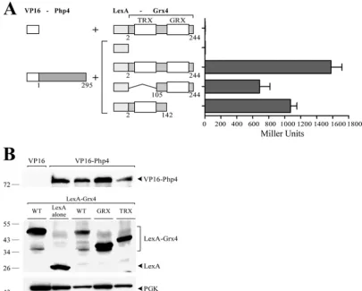
do-main. -Galactosidase assays of LexA-2Grx4142 and
LexA-105Grx4244 coexpressed with VP16-Php4 revealed high activity
levels (1,072⫾70 and 686⫾118 Miller units, respectively).
Al-though these levels of-galactosidase activity were lower by 32%
and 57% compared to that of the full-length LexA-2Grx4244
pro-tein (1,578⫾119 Miller units), a clear and elevated
transactiva-tion of the reporter gene expression was observed, revealing that
both N- and C-terminal regions of Grx4 interact with Php4 (Fig.
1A). For negative controls, the VP16 transactivation domain
with-out the Php4 protein or the LexA DNA-binding domain was
as-sayed without the Grx4 protein. In both cases,-galactosidase
activity was absent, resulting in background values typical of pairs
of noninteracting proteins (Fig. 1A). Immunoblot analyses of
pro-tein extracts using anti-LexA and anti-VP16 antibodies clearly in-dicated that the fusion proteins were expressed in the
cotrans-formed cells (Fig. 1B). Although we consistently detected LexA
polypeptide alone, full-length VP16-Php4 protein, LexA-Grx4 protein, and its truncated derivatives, we were unable to detect the VP16 polypeptide alone. This result may be due to its low
pre-dicted molecular mass (⬃8 kDa). On the basis of these data, we
concluded that the TRX- and GRX-containing regions of Grx4 both interact with Php4.
Whereas the association between the TRX domain and Php4 is constitutive, the GRX domain interacts in an iron-dependent manner with Php4.As we have previously shown using a two-hybrid approach, the interaction between the full-length LexA-Grx4 and VP16-Php4 fusion proteins is not modulated by the
cellular iron status (27). Indeed, in the present study, the
full-length LexA-Grx4 and VP16-Php4 chimeric proteins interacted
with each other, producing a constitutive steady-state level oflacZ
mRNA as assayed by RNase protection experiments under both
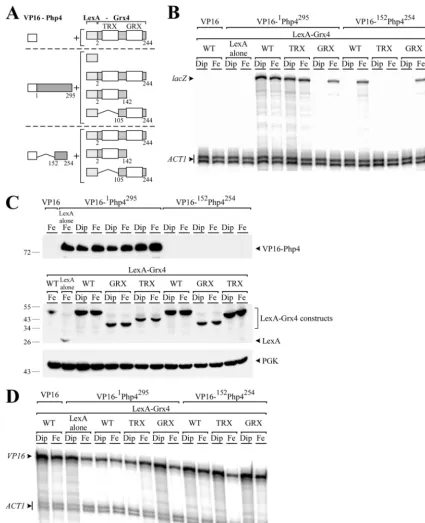
low and high iron concentrations (Fig. 2AandB). Similarly, when
the GRX domain of Grx4 was deleted (LexA-2Grx4142), leaving
only the TRX-containing region, and the resulting construct was
then tested for interaction with the full-length VP16-1Php4295
protein, the levels oflacZmRNA were constitutive and
unrespon-sive to cellular iron status (Fig. 2AandB). Surprisingly, when the
TRX domain was removed (LexA-105Grx4244, leaving only the
GRX-containing region) and the resulting construct was then
tested for association with VP16-1Php4295, thelacZmRNA was
exclusively detected in the presence of iron and not under
iron-limiting conditions (Fig. 2AandB). In this study, iron was limited
by the addition of the permeant iron chelator 2,2=-dipyridyl (Dip)
at a concentration of 250M. In contrast, iron-treated cultures
were supplemented with 100M FeCl3.
Based on these data and the fact that we had previously deter-mined that the Php4 domain corresponding to amino acids 152 to 254 constituted the minimal module sufficient for interaction
with Grx4 (27), the region on Grx4 that was required for
interac-tion with the Php4 domain corresponding to amino acids 152 to 254 was investigated. Initially, the possibility of interaction
be-tween VP16-152Php4254with the N-terminal 2 to 142 residues of
Grx4 (including the TRX domain) was examined. This first
com-bination showed an absence oflacZtranscript, irrespective of iron
status (Fig. 2AandB). We then tested whether the C-terminal 105
to 244 residues of Grx4 (including the GRX domain) was involved in the interaction with amino acids 152 to 254 of Php4. In these
experiments, LexA-105Grx4244 clearly activatedlacZmRNA
ex-pression under iron-replete conditions, but not under
on September 8, 2020 by guest
http://ec.asm.org/
ing conditions. These results revealed an interaction between the GRX domain of Grx4 and the amino acid region consisting of residues 152 to 254 of Php4 that was iron dependent. We consistently found that the interaction between the full-length
LexA-Grx4 and VP16-152Php4254occurred only in response to
excess iron and not under conditions of iron deficiency (Fig. 2A
andB). These results suggested that the iron-dependent
inter-action between these two proteins took place through the GRX domain of Grx4. With the exception of the VP16 polypeptide
alone and the short VP16-152Php4254protein, all of the fusion
proteins tested for two-hybrid interactions were expressed as
confirmed by immunoblot analyses (Fig. 2C). Given this
situ-ation, the mRNA levels of VP16 alone, VP16-1Php4295, and
VP16-152Php4254 were assayed by RNase protection
experi-ments. The results showed that constructs containingVP16
alone or fused with full-length or truncatedphp4, were all
ex-pressed, with transcripts being detected in the case of each prey
construct (Fig. 2D). We therefore concluded, on the basis of
this data that iron fosters the interaction between the GRX domain of Grx4 and the C-terminal 152 to 254 amino acid residues of Php4, whereas the TRX domain establishes a con-stitutive and iron-independent association with Php4.
Cys35 of Grx4 is required for interaction between Php4 and the TRX domain, whereas Cys172 is necessary for the iron-de-pendent association between the GRX domain and the C-termi-nal region of Php4.To further investigate the interaction between both the TRX and GRX domains of Grx4 with Php4, two-hybrid
experiments usingLexA-grx4fusion alleles that contained point
mutations in two highly conserved cysteine residues were per-formed. The N-terminal TRX domain of Grx4 contains a WAAP
C35K sequence that is reminiscent of the thioredoxin active site
motif WCGPCK (11). The C-terminal GRX-like domain of Grx4
contains the highly conserved residues C172GFS that are required
for monothiol glutaredoxin cellular functions (41). Initially, the
LexA-Grx4(C35A) mutant construct was coexpressed with either
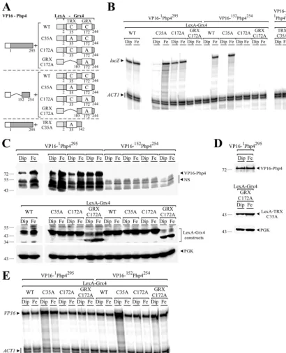
VP16-1Php4295or VP16-152Php4254. Under low-iron conditions,
no significant levels oflacZmRNA were detected, indicating that
the interactions between LexA-Grx4(C35A) and VP16-1Php4295
or LexA-Grx4(C35A) and VP16-152Php4254were either not
signif-icant or were absent (Fig. 3AandB). In contrast, when these two
cotransformants were incubated in the presence of iron, lacZ
mRNA was readily detected, revealing the presence of
iron-depen-dent interactions between LexA-Grx4(C35A) and VP16-1Php4295
or LexA-Grx4(C35A) and VP16-152Php4254 (Fig. 3A and B).
FIG 1The N- and C-terminal regions of Grx4 interact with Php4. (A) The VP16-Php4 fusion protein was coexpressed with the full-length LexA-Grx4 protein or its truncated derivatives. The amino acid sequences of the Php4 and Grx4 proteins are numbered relative to their initiator codons, respectively. Each set of constructs was coexpressed in theS. cerevisiaestrain L40 under basal conditions. As a measure of protein-protein interactions, liquid-galactosidase assays were performed, and results are shown as the means plus standard deviations (error bars) of triplicate determinations. (B) Cell lysates from aliquots of the cultures containing the constructs shown in panel A were analyzed by immunoblotting using anti-VP16, anti-LexA, or anti-phosphoglycerate kinase (anti-PGK) (as an internal control) antibodies. The positions of protein standards (molecular masses in kilodaltons) are indicated to the left of the panel. WT, wild type.
Iron Inhibition of Php4 Function
on September 8, 2020 by guest
http://ec.asm.org/
FIG 2Two domains of Grx4 are involved in the association with Php4, but only the GRX domain interacts in an iron-dependent manner. (A) Schematic illustrations of chimeric VP16-Php4 and LexA-Grx4 molecules that were used as prey and bait, respectively. The N-terminal 142 amino acid residues of Grx4 encompass its TRX domain, whereas residues 105 to 244 of Grx4 contain the GRX domain. The amino acid sequence numbers refer to the positions relative to the first amino acid of each protein. (B) Each set of constructs was coexpressed in theS. cerevisiaestrain L40 grown to anA600of 0.4 and then treated with Dip (250M) or FeCl3(100M) for 3 h. After total RNA extraction, thelacZandACT1steady-state mRNA levels were analyzed by RNase protection assays. Results shown are representative of three independent experiments. (C) Cell lysates from aliquots of the cultures containing the constructs shown in panel B were analyzed by immunoblotting using anti-VP16, anti-LexA, or anti-PGK (as an internal control) antibodies. The positions of the molecular mass standards are indicated on the left. (D) Aliquots of the cultures containing the constructs in panel B were also examined by RNase protection assays for the steady-state levels of theVP16transcript. Actin (ACT1) mRNA levels were probed as an internal control.
on September 8, 2020 by guest
http://ec.asm.org/
When the GRX domain was mutated [LexA-Grx4(C172A)] and
then tested for interaction with the full-length VP16-1Php4295
fu-sion protein, high levels oflacZmRNA were detected under both
iron-limiting and iron-replete conditions (Fig. 3AandB). In
con-trast, two-hybrid assays of LexA-2Grx4142(C35A) coexpressed
with VP16-1Php4295 or LexA-Grx4(C172A) coexpressed with
VP16-152Php4254showed nolacZmRNA under either
iron-limit-ing or iron-replete conditions (Fig. 3AandB). On the basis of
FIG 3Cys35 of Grx4 is necessary for the Php4-TRX domain interaction, whereas the Grx4 Cys172 is required for the iron-dependent Php4-GRX domain association. (A) Schematic illustrations of the VP16-Php4 and LexA-Grx4 fusion proteins and their mutant derivatives. The point mutations of the Grx4 Cys35 and Cys172 are indicated with an A (instead of the wild-type [WT] C residues). The amino acid sequence numbers refer to the positions relative to the first amino acid of each protein. (B) Cotransformed cells were grown to mid-logarithmic phase and then incubated in the presence of Dip (250M) or FeCl3(Fe) (100M) for 3 h. Total RNA was isolated and analyzed by RNase protection assays to measure the steady-state levels of thelacZtranscript. Actin (ACT1) mRNA levels were probed as an internal control. The results shown are representative of three independent experiments. (C and D) Whole-cell extracts were prepared from aliquots of the cultures containing the constructs shown in panel B and were analyzed by immunoblotting using either anti-VP16, anti-LexA, or anti-PGK (as an internal control) antibodies. (E) Total RNA isolated from aliquots of the cultures containing the constructs shown in panel C were also examined by RNase protection assays for the steady-state levels of theVP16transcript. Actin (ACT1) mRNA levels were probed as an internal control.
Iron Inhibition of Php4 Function
on September 8, 2020 by guest
http://ec.asm.org/
these data, we concluded that Cys35 of Grx4 was necessary for interaction between Php4 and the TRX domain of Grx4, whereas Cys172 of Grx4 was required for Grx4 interaction with the C-ter-minal 152 to 254 residues of Php4. Furthermore, the results showed that interaction was modulated by iron in the absence of the Cys35 of Grx4. To further investigate the requirement for Cys172, a truncated version of the N-terminal end of Grx4 containing only residues 105 to 244 of the GRX domain was
constructed. When the Cys172 ¡ Ala mutation
[LexA-105
Grx4244(C172A)] was tested for interaction with either
VP16-1Php4295 or VP16-152Php4254 by two-hybrid analysis, no lacZ
mRNA was detected by RNase protection assays (Fig. 3AandB).
Given these results and those obtained above (Fig. 2), we
con-cluded that the Cys172 residue located within the GRX domain of Grx4 is absolutely required because its exchange abrogates the
interaction between the LexA-105Grx4244and VP16-1Php4295or
LexA-105Grx4244and VP16-152Php4254in response to iron.
West-ern blot analyses of protein extracts using LexA and anti-VP16 antibodies showed that the fusion proteins were expressed
in the cotransformed cells, independently of the iron levels (Fig.
3CandD). Since we were unable to detect the VP16-152Php4254
fusion protein (presumably due to its low molecular weight), we
ascertained the mRNA levels of theVP16-php4⫹fusion alleles by
RNase protection assays (Fig. 3E). Results showed thatVP16
-1php4295 and VP16-152php4254 fusion alleles were clearly
ex-pressed, with transcripts detected in the case of each prey
con-struct (Fig. 3E).
Minimal C-terminal region of Php4 required for interaction with the GRX domain of Grx4.The full-length Grx4 protein has previously been shown to interact with the C-terminal 152 to 254
residues of Php4 (27). To gain additional insight into the Php4
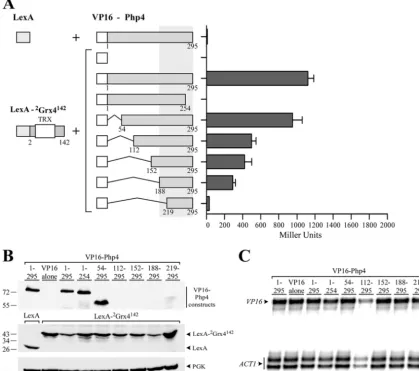
domain that is responsible for interaction with the GRX domain of Grx4, seven chimeric proteins were generated using different segments of the Php4 protein. These segments comprised amino
acid residues 1 to 295 (VP16-1Php4295), 1 to 254 (VP16-1Php4254),
1 to 218 (VP16-1Php4218), 152 to 254 (VP16-152Php4254), 188 to
254 (VP16-188Php4254), 219 to 254 (VP16-219Php4254), and 152 to
254 [VP16-152Php4254(C221A; C227A)], in which both Cys221
and Cys227 were mutated to alanine residues. -Galactosidase
assays of VP16-1Php4295, VP16-1Php4254, VP16-152Php4254, or
VP16-188Php4254coexpressed with LexA-105Grx4244showed high
levels of activity (637⫾56, 1,772⫾42, 1,366⫾15, and 973⫾12
Miller units, respectively) (Fig. 4A). Coexpression of
VP16-1
Php4218, VP16-219Php4254, or VP16-152Php4254(C221A; C227A)
with LexA-105Grx4244resulted in dramatically reduced levels of
-galactosidase activity (19⫾1, 7⫾1, and 21⫾4 Miller units,
respectively) that were similar to the values obtained for pairs of
noninteracting proteins (Fig. 4A). Furthermore, when Cys221 or
Cys227 was individually replaced with alanine, the mutant Php4 protein failed to interact with Grx4 (data not shown). All fusion proteins tested for two-hybrid interactions were detected by immunoblot analyses except for the chimeric
VP16-152Php4254, VP16-188Php4254, VP16-219Php4254, and
VP16-152
Php4254(C221A; C227A) proteins (Fig. 4B). As previously
per-formed in the case of the very short versions of VP16-Php4
fusions, the levels ofVP16-152Php4254, VP16-188Php4254, VP16
-219Php4254, and VP16-152Php4254(C221A; C227A) mRNA were
monitored to confirm that they were produced in the two-hybrid
experiments (Fig. 4C). On the basis of the results of the analysis of
the reporter gene expression, we concluded that the C-terminal
region of Php4 from residues 188 to 254 is sufficient for the inter-action between Php4 and the GRX domain of Grx4. Furthermore, the results showed that both Cys221 and Cys227 in Php4 are re-quired for Php4-GRX domain association.
Regions of Php4 that interact with the TRX domain of Grx4.
In additional two-hybrid assays, the LexA-2Grx4142fusion protein
that contained only the TRX domain was used as bait. Results showed that the construct in which the last 41 amino acids of Php4
were deleted had no-galactosidase activity when coexpressed
with LexA-2Grx4142(Fig. 5A). Deletions of amino acids on the
N-terminal side in VP16-54Php4295, VP16-112Php4295,
VP16-152
Php4295, and VP16-188Php4295showed successively less
-ga-lactosidase activity (15%, 56%, 62%, and 74%, respectively)
com-pared to VP16-1Php4295. Further deletion to amino acid 219
drastically abolished almost all of the-galactosidase activity (by
97%) compared to the level observed with the full-length
VP16-1Php4295protein (Fig. 5A). The LexA protein alone and
LexA-2
Grx4142fusion proteins were detected by immunoblotting using
an anti-LexA antibody (Fig. 5B). Although the long VP16-Php4
fusions (VP16-1Php4295, VP16-1Php4254, and VP16-54Php4295)
were consistently detected by immunoblotting (Fig. 5B), the short
fusion products (VP16-112Php4295, VP16-152Php4295,
VP16-188Php4295, and VP16-219Php4295) were not consistently
de-tected by immunoblotting (Fig. 5B). To ensure that these
fu-sion proteins were expressed in the cotransformed cells, RNase protection analyses were performed so as to verify the expres-sion levels of these VP16-Php4 constructs. Results showed that
the transcripts were detected in each case (Fig. 5C). Taken
to-gether, the two-hybrid interaction assays revealed that the last 41 amino acid residues of the C terminus of Php4 and the first 218 N-terminal amino acids of Php4 constitute two important regions that are required for optimal interaction between Php4 and the TRX domain of Grx4.
The GRX domain is sufficient for the iron-mediated inhibi-tion of Php4 funcinhibi-tion.Given the fact that two-hybrid assays showed that the GRX domain strongly associates with Php4 in an iron-dependent manner, we further investigated the effect of this domain on Php4 function. These experiments were in keeping with the fact of the importance of the Php4-dependent regulation
of two target genes,isa1⫹andsdh4⫹. Theisa1⫹gene encodes an
iron-using protein implicated in the iron-sulfur cluster assembly
paths (48), whereassdh4⫹encodes an iron-dependent
mitochon-drial membrane anchor subunit of the succinate dehydrogenase
(28). Integrative plasmids harboring thegrx4⫹,TAP-grx4⫹, and
GFP-grx4⫹alleles were expressed under the control of thegrx4⫹
promoter. Similarly, theGFP-GRXandGFP-GRX C172Aalleles,
which expressed only the GRX domain (wild type and mutated,
respectively), were integrated into agrx4⌬mutant strain and were
also expressed under thegrx4⫹promoter. To assess the effects of
the expression of the different Grx4 fusion proteins on Php4 func-tion, we carried out RNase protection experiments examining the relative transcriptional profiles of two Php4-regulated target
genes,isa1⫹andsdh4⫹(Fig. 6). Whengrx4⌬cells expressing the
GFP-GRX(GRX domain alone) allele were grown under
condi-tions of iron deficiency, isa1⫹andsdh4⫹gene expression was
repressed (⬃5.5- and⬃6.2-fold, respectively) compared to the
transcript levels detected from either control (untreated) or
iron-treated cells. As shown inFig. 6,grx4⌬cells in which wild-type
grx4⫹or functionalTAP-grx4⫹andGFP-grx4⫹alleles were
rein-tegrated regained their capacity to downregulateisa1⫹orsdh4⫹
on September 8, 2020 by guest
http://ec.asm.org/
gene expression in response to iron starvation. In the case of these
transformedgrx4⌬cells,isa1⫹andsdh4⫹transcript levels were
readily detected under both standard (untreated) and iron-replete
conditions. In fact,isa1⫹andsdh4⫹transcript levels were more
abundant (⬃9- to 16-fold) than those of mRNAs observed under
iron-limiting conditions. Under iron-replete conditions,grx4⌬
cells expressingGFP-GRXexhibited a less pronounced
derepres-sion of theisa1⫹andsdh4⫹transcript levels thangrx4⌬cells
ex-pressing thegrx4⫹,TAP-grx4⫹orGFP-grx4⫹alleles did (Fig. 6
and data not shown). This result may be due to the fact that the GRX domain alone (without the TRX domain) is less competent in inhibiting Php4 in response to iron. When an integrative
plas-mid expressing theGFP-GRX C172Aallele was transformed in
grx4⌬ cells, no sdh4⫹ transcript was observed, revealing that
Cys172 was required for the GRX domain-mediated inhibition of
Php4 function in response to iron (Fig. 6C andD). Taken
to-gether, the results revealed that the GRX domain of Grx4 appears to be sufficient to inactivate a large proportion of the Php4 pro-teins in response to iron.
The GRX domain and Php4 associate with each other in the presence of iron.The ability of the GRX domain to interact with
Php4 in anS. cerevisiaetwo-hybrid system led us to investigate
whether such interaction could be detected inS. pombe. To
ad-dress this point, we used a BiFC approach in fission yeast in which case Venus amino-terminal fragment (VN) and Venus carboxyl-terminal fragment (VC) were fused to the N-carboxyl-terminal portions of
FIG 4The GRX domain of Grx4 interacts with residues 188 to 254 of the C-terminal region of Php4. (A) The LexA-105Grx4244fusion protein was coexpressed with the full-length VP16-Php4 or its truncated derivatives. The amino acid sequences of the Grx4 and Php4 proteins are numbered relative to their initiator codons. Each set of constructs was coexpressed in theS. cerevisiaestrain L40 in the presence of iron (10M FeCl3). Protein-protein interactions were detected by-galactosidase assays, and the results are indicated in Miller units. The values are means plus standard deviations (error bars) of three independent experiments. (B) Whole-cell extracts were prepared from aliquots of the cultures containing the constructs described above for panel A and were analyzed by immunoblotting using either an anti-VP16 or anti-LexA antibody. As an internal control, total extract preparations were probed with an anti-phosphoglycerate kinase (anti-PGK) antibody. (C) Total RNA from aliquots of the cultures containing the constructs shown in panel A was isolated and then used in RNase protection experiments. A representative experiment was used to detect steady-statelacZandACT1mRNA levels.
Iron Inhibition of Php4 Function
on September 8, 2020 by guest
http://ec.asm.org/
Php4 and the GRX domain, respectively. Integrative plasmids
ex-pressing the tagged (VN-php4⫹andVC-GRX) coding sequences
were cotransformed intogrx4⌬php4⌬mutant cells which were
grown to mid-logarithmic phase. At this stage of growth, cells were incubated for 2 h in the presence of the iron chelator Dip
(250M) or exogenous iron (100M). Microscopic analysis of
iron-starved cells did not exhibit a BiFC signal. Under iron-limit-ing conditions, VN-Php4 and VC-GRX did not reconstitute a functional fluorophore, revealing that they were not interacting
with each other (Fig. 7A). In contrast, when the cells were
incubated in the presence of exogenous iron, VN-Php4 and VC-GRX produced a BiFC signal that appeared in the cyto-plasm and was, to a great extent, excluded from the nucleus (Fig. 7A). Furthermore, when integrative plasmids coexpress-ingVN-php4⫹andVC-GRX C172Aalleles were examined by
BiFC inS. pombe, no BiFC signal was observed irrespective of
the cellular iron status (Fig. 7Aand data not shown).
Consis-tent with the results obtained by two-hybrid analysis, the
inter-action between the GRX domain and Php4 occurred in an
iron-dependent manner when assayed by BiFC inS. pombeand was
dependent on the presence of the Cys172 residue. As previously
reported (27), the VN-tagged Php4 and VC-tagged Grx4
pro-teins used in this study (as positive controls) produced a BiFC signal in the nucleus under low-iron conditions, whereas the BiFC signal was mainly detected in the cytoplasm of the cells
under iron-replete conditions (Fig. 7A). To ensure that the
VN- and VC-tagged versions of the Php4 and Grx4 protein derivatives were expressed, total RNA was prepared from the cultures analyzed by BiFC under both iron starvation and iron-replete conditions and was analyzed by RNase protection
as-says (Fig. 7BandC). Although the transcripts for each
con-struct were not found at the same steady-state levels, thephp4⫹
andgrx4⫹mRNAs were clearly detected under both iron
star-vation and excess iron. Together, these results indicated that both the GRX domain and iron are required for relocalization of the Grx4-Php4 complex from the nucleus to the cytoplasm.
FIG 5The TRX domain of Grx4 interacts with two distinct regions of Php4. (A) The TRX domain of Grx4 was coexpressed with a series of VP16-Php4 truncations in theS. cerevisiaeL40 strain. Protein-protein interactions were detected by-galactosidase assays, and the results are indicated in Miller units. The values are means plus standard deviations (error bars) of triplicate experiments. Amino acids are numbered relative to the first amino acid of either the Grx4 or Php4 protein. (B) Total cell extract preparations from aliquots of the cultures used in the assays described above for panel A were analyzed by immunoblotting with anti-VP16, anti-LexA, or anti-PGK antibodies. (C) Aliquots of the cultures used in the assays described above for panel A were also examined by RNase protection assays for steady-stateVP16andACT1mRNA levels.
on September 8, 2020 by guest
http://ec.asm.org/
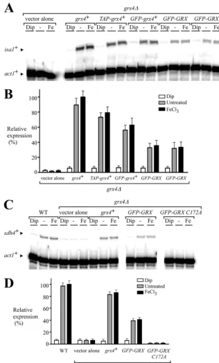
FIG 6The GRX domain inhibits Php4 activity in response to iron. (A) Cells harboring agrx4⌬deletion were transformed with an empty integrative vector or with thegrx4⫹,TAP-grx4⫹,GFP-grx4⫹, orGFP-GRXallele. For theGFP-GRXallele, results obtained with two independent transformants are shown. Mid-logarithmic-phase cultures were left untreated (⫺) or were treated with Dip (250M) or FeCl3(Fe) (100M) for 90 min. Total RNA was prepared from each sample and analyzed by RNase protection assays. The steady-state levels of theisa1⫹andact1⫹mRNAs are indicated by the black arrowheads. (B) Graphic representation of the quantification of the results of three independent RNase protection assays, including the experiment shown in panel A. The histogram values represent the averages plus standard deviations (error bars) of triplicate determinations. (C) Strain AMY34 (grx4⌬) was transformed with an empty integrative vector or with thegrx4⫹,GFP-GRX, orGFP-GRX C172Aallele. Mid-logarithmic-phase cultures of isogenic strains FY435 (WT) and AMY34 (grx4⌬) were left untreated (⫺) or were treated with Dip (250M) or FeCl3(Fe) (100M) for 90 min. Total RNA was prepared from each sample and analyzed by RNase protection assays. The steady-state levels of thesdh4⫹andact1⫹mRNAs are indicated by the black arrowheads. (D) The histogram values represent the averages plus standard deviations of triplicate determinations.
Iron Inhibition of Php4 Function
on September 8, 2020 by guest
http://ec.asm.org/
DISCUSSION
InS. pombe, thephp4⫹gene encodes a subunit of the CCAAT-binding protein complex which includes three other subunits that
are denoted Php2, Php3, and Php5 (26). The genes encoding
Php2, Php3, and Php5 are constitutively expressed, whereas
tran-scripts ofphp4⫹are induced under conditions of iron starvation
and repressed under iron-replete conditions (28). Under low-iron
conditions, Php4 acts as a negative regulatory subunit of the CCAAT-binding factor and fosters the repression of several genes encoding iron-using proteins. It has previously been shown that
the deletion of thegrx4⫹gene makes Php4 constitutively active,
suggesting that Grx4 plays a critical role in inhibiting Php4
func-tion (27). Further analysis by BiFC assays showed that Grx4 is a
binding partner of Php4 (27). On the basis of these findings, we
sought to determine the mechanism by which Grx4 and Php4 interact with each other as a function of iron availability. The results presented here indicate that both the TRX and GRX do-mains of Grx4 interact with Php4. Although the TRX domain interacted strongly and constitutively with Php4, the GRX domain associated in an iron-dependent manner with Php4. These results
were different than those reported in the case of theS. cerevisiae
monothiol glutaredoxins Grx3 and Grx4 with respect to their
as-sociations with Aft1 (39). In this case, two-hybrid experiments
showed that both the GRX and TRX domains of Grx3 and Grx4
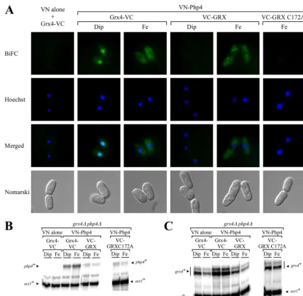
FIG 7Php4 and the GRX domain interact in an iron-dependent manner in livingS. pombecells. (A) A mutant strain disrupted in thegrx4⫹andphp4⫹genes (grx4⌬php4⌬) was transformed with a vector alone (VN alone) and withgrx4⫹-VCorVN-php4⫹andgrx4⫹-VCorVN-php4⫹andVC-GRXorVN-php4⫹and VC-GRX C172A. Cells expressing the indicated alleles were grown to mid-logarithmic phase and then treated with 250M Dip or 100M FeCl3(Fe) for 2 h. After treatment, the cells were visualized by fluorescence microscopy using BiFC and Hoechst stain. The merged images are shown in the last row of color panels. Nomarski optics was used to monitor cell morphology. For simplicity, the images of cells cotransformed with VN alone andgrx4⫹-VCorVN-php4⫹and VC-GRX C172Awere taken from iron-replete cells because the fluorescent images from the iron-deficient cells were identical. Aliquots of the cultures described above for panel A were analyzed by RNase protection assays. Steady-state mRNA levels ofphp4⫹andact1⫹were probed in panel B, while those ofgrx4⫹andact1⫹ were analyzed in panel C. Each RNase protection experiment in panels B and C was assayed three independent times.
on September 8, 2020 by guest
http://ec.asm.org/
interacted with Aft1, exhibiting similar levels of-galactosidase activity, with no specification with respect to the interactions (be-tween these polypeptides) as a function of iron availability. Al-though the nature of the differences between these respective ob-servations is unclear, it is possible that the composition and length of the GRX and TRX domains may be contributing factors that would explain the differences between the results reported here
and those published by others (39). Alternatively, the differences
between the results of the two-hybrid studies may be due to the
fact that Php4 (S. pombe) and Aft1 (S. cerevisiae) do not share
significant amino acid sequence identity with each other (only 4.3%). It is possible that these two proteins use distinct mecha-nisms or partners in their interactions with monothiol glutare-doxins. Recently, it has been determined that the last 16 amino
acid residues ofS. cerevisiaeGrx4 serve as a specific binding region
for the transcription factor Aft1 (12). When this region was
sub-stituted for the last 14 amino acid residues of S. pombeGrx4
(which represents the C-terminal end of the protein), the
interac-tion between the chimeric Grx4 and Aft1 was lost (12). This lack of
interaction was consistent with the fact that there is only limited identity (⬃31%) between the C-terminal ends of the Grx4
pro-teins fromS. pombeandS. cerevisiae.
InS. cerevisiae, the monothiol glutaredoxins Grx3 and Grx4 can transport Fe/S clusters to diverse locations and then
subse-quently sort them to different enzymes (32). It has been
demon-strated that both the TRX and GRX domains of Grx3 and Grx4 are
required for this function (12). Although a similar function forS.
pombeGrx4 has not yet been ascertained, this protein may also participate in intracellular Fe/S cluster delivery. A second role for
S. cerevisiaeGrx3 and Grx4 is to communicate the presence of iron to Aft1. This condition leads to Aft1 inactivation and its
subse-quent export from the nucleus to the cytoplasm (5,21,37,46).
Deletion mapping analysis has revealed that neither the TRX do-main alone nor the GRX dodo-main alone can mediate the
iron-dependent inhibition of Aft1 (12).S. cerevisiaeGrx4 must contain
the two domains, TRX and GRX, to inactivate Aft1 function. The
results presented here are different from those reported for S.
cerevisiaeGrx4. In the case ofS. pombeGrx4, the expression of the GRX domain alone was sufficient to mediate a significant iron inhibition of Php4 function. This observation was supported by
two experimental results. First,grx4⌬cells in which either a
full-lengthgrx4⫹allele or theGRXdomain coding sequence was
rein-tegrated regained the capacity to upregulateisa1⫹gene expression
in response to iron (Fig. 6). In contrast, the deletion of thegrx4⫹
gene resulted in constitutively active Php4, thereby maintaining
the target geneisa1⫹in a constant state of repression (Fig. 6) (27).
Second, in cells harboring aphp4⌬grx4⌬double deletion,
coex-pression of VN-Php4 and VC-GRX domain produced BiFC sig-nals in the cytoplasm of iron-replete cells, indicating that the GRX domain can associate with Php4 and that it contributes to its in-activation via its relocalization from the nucleus to the cytoplasm.
Therefore, based on results reported here, it would appear thatS.
pombeGrx4 functions as an iron sensor for Php4. Furthermore, the presence of the GRX domain is sufficient for iron inhibition of Php4 function. Interestingly, the results also showed that the GRX domain was sufficient to allow recognition of the Php4-Grx4
complex by the exportin Crm1 (27) since the Php4-GRX complex
was exported in response to iron.
Yeast two-hybrid analysis showed that the TRX domain of Grx4 binds Php4 in a constitutive manner. Two Php4 regions were involved in the Php4-TRX domain association. The first region was relatively short and encompassed the last 40 C-terminal resi-dues of Php4 (resiresi-dues 255 to 295), while the second region was larger and involved amino acid residues 55 to 218 of Php4. These data strongly suggest that the TRX domain functions as a docking domain for the association between Grx4 and Php4. Although the precise amino acid residues responsible for the interaction be-tween the TRX domain and Php4 must await finer mapping
anal-ysis, the Cys35residue located within the TRX domain was found
to be required for this interaction.
Previously it was shown that Grx4 is required for inhibition of Php4 function in response to iron. The question thus arises con-cerning the mechanism of this inhibition. As opposed to the TRX domain, the GRX domain of Grx4 interacted in an iron-depen-dent manner with Php4. Under high-iron conditions, the GRX domain associated with Php4 through a minimal domain encom-passing amino acid residues 188 to 254 of Php4. This minimal C-terminal region of Php4 contains two Cys residues located at positions 221 and 227. Interestingly, these two Cys residues are highly conserved in other Php4-like proteins, including HapX fromAspergillus nidulans, Hap43 fromCandida albicans, HapX fromAspergillus fumigatus, and HapX fromCryptococcus
neofor-FIG 8A proposed model for the interaction between the monothiol glutaredoxin Grx4 and Php4. In the absence of iron (⫺Fe), Php4 forms a complex with Php2, Php3, Php5, and Grx4. Because only the TRX domain interacts with Php4, this leaves the repression domain of Php4 available to repress target gene expression. When cells undergo a transition from iron-limiting to iron-sufficient conditions, the GRX domain interacts with Php4, causing an inhibitory effect on Php4 function. The inactivation of Php4 leads to its release from the Php2/Php3/Php5 complex and its subsequent export from the nucleus to the cytoplasm (27). This lack of Php4 then allows the CCAAT-binding Php2/Php3/Php5 heterotrimer complex to activate gene expression. Php4-TRX and Php4-GRX domain associa-tions require the presence of Cys35 and Cys172 of Grx4, respectively.
Iron Inhibition of Php4 Function
on September 8, 2020 by guest
http://ec.asm.org/
mans (13, 14, 19, 44). Substitution of these two Cys residues
(Cys221and Cys227) by alanine residues abolished the association
between the LexA-105Grx4244and VP16-152Php4254fusion
pro-teins (Fig. 4). Similarly, substitution of Cys172to alanine in the
GRX domain of Grx4 disrupted the interaction between this do-main and Php4. One model that could explain these observations would be that the GRX domain forms a [2Fe-2S] cluster with
Php4. This putative [2Fe-2S] cluster is coordinated by Cys172
lo-cated in the CGFS motif of Grx4, Cys221and Cys227of Php4, and
the cysteine residue of one molecule of glutathione (GSH). At this time, however, the model remains speculative, and the mecha-nism by which Grx4 communicates the presence of iron to Php4 needs further investigation. Experiments are under way to inves-tigate this mechanism. It is important to mention that, in support of the model, a number of studies have revealed that CGFS-type monothiol glutaredoxins can act as scaffolds for the delivery of
[2Fe-2S] clusters to acceptor proteins (1,16,38). Under
condi-tions of excess iron, Grx4 would acquire an [2Fe-2S] cluster that would in turn trigger the interaction between the GRX domain and the C-terminal region (residues 188 to 254) of Php4. This interaction between the GRX domain and Php4 would induce an inhibitory conformational change that would disrupt the Php4/ Php2/Php3/Php5 heteromeric complex, leading to Php4 release
and inactivation (Fig. 8). The Grx4-mediated inactivation of Php4
would lead to its recruitment by the exportin Crm1 and then to its
subsequent export from the nucleus to the cytoplasm (27).
Con-versely, under conditions of iron starvation, the GRX domain would dissociate from the C-terminal portion (residues 188 to 254) of Php4, resulting in the ability of Php4 to bind the Php2/ Php3/Php5 heterotrimeric complex and thereby repressing the
transcription of its target genes (Fig. 8). Because the TRX domain
of Grx4 is constitutively associated with Php4, other cellular com-ponents may be required to communicate the cellular iron levels to the GRX domain of Grx4. These components may participate in signaling and/or delivery of a putative [2Fe-2S] cluster that would be inserted into the GRX domain of Grx4 to inactivate Php4. Fur-ther studies are needed to decipher the detailed mechanism by which Php4 is inactivated by the monothiol glutaredoxin Grx4.
ACKNOWLEDGMENTS
We are grateful to Gilles Dupuis and William Home for critically reading the manuscript and for their valuable comments. We are indebted to Won-Ki Huh (Institute of Microbiology, Seoul National University) for the gift of pFA6a-VN173-KANMX6 and pFA6a-VC155-KANMX6 plasmids.
P.V. and M.J. were recipients of studentships from the Faculté de Médecine et des Sciences de la Santé of the Université de Sherbrooke and the Fonds Québecois de la Recherche sur la Nature et les Technologies (FQRNT), respectively. This study was supported by Natural Sciences and Engineering Research Council of Canada grants MOP-238238-2010-15 and MOP-396029-2010-DAS to S.L.
REFERENCES
1.Bandyopadhyay S, et al.2008. Chloroplast monothiol glutaredoxins as scaffold proteins for the assembly and delivery of [2Fe-2S] clusters. EMBO J.27:1122–1133.
2.Chao LY, Marletta MA, Rine J.2008. Sre1, an iron-modulated GATA DNA-binding protein of iron-uptake genes in the fungal pathogen Histo-plasma capsulatum.Biochemistry47:7274 –7283.
3.Chen D, et al.2003. Global transcriptional responses of fission yeast to environmental stress. Mol. Biol. Cell14:214 –229.
4.Chung WH, Kim KD, Roe JH.2005. Localization and function of three monothiol glutaredoxins inSchizosaccharomyces pombe.Biochem. Bio-phys. Res. Commun.330:604 – 610.
5.Ehrensberger KM, Bird AJ.2011. Hammering out details: regulating metal levels in eukaryotes. Trends Biochem. Sci.36:524 –531.
6.Haas H.2003. Molecular genetics of fungal siderophore biosynthesis and uptake: the role of siderophores in iron uptake and storage. Appl. Micro-biol. Biotechnol.62:316 –330.
7.Haas H, Eisendle M, Turgeon BG.2008. Siderophores in fungal physi-ology and virulence. Annu. Rev. Phytopathol.46:149 –187.
8. Halliwell B, Gutteridge JM. 1992. Biologically relevant metal ion-dependent hydroxyl radical generation. An update. FEBS Lett.307:108 – 112.
9.Harrison KA, Marzluf GA.2002. Characterization of DNA binding and the cysteine rich region of SRE, a GATA factor inNeurospora crassa in-volved in siderophore synthesis. Biochemistry41:15288 –15295. 10. Hentze MW, Muckenthaler MU, Galy B, Camaschella C.2010. Two to
tango: regulation of mammalian iron metabolism. Cell142:24 –38. 11. Herrero E, de la Torre-Ruiz MA. 2007. Monothiol glutaredoxins: a
common domain for multiple functions. Cell. Mol. Life Sci.64:1518 – 1530.
12. Hoffmann B, et al.2011. The multidomain thioredoxin-monothiol glu-taredoxins represent a distinct functional group. Antioxid. Redox Signal.
15:19 –30.
13. Hortschansky P, et al.2007. Interaction of HapX with the CCAAT-binding complex-a novel mechanism of gene regulation by iron. EMBO J.
26:3157–3168.
14. Hsu PC, Yang CY, Lan CY.2011.Candida albicansHap43 is a repressor induced under low-iron conditions and is essential for iron-responsive transcriptional regulation and virulence. Eukaryot. Cell10:207–225. 15. Hwang LH, Seth E, Gilmore SA, Sil A. 2012. SRE1 regulates
iron-dependent and -iniron-dependent pathways in the fungal pathogen Histo-plasma capsulatum.Eukaryot. Cell11:16 –25.
16. Iwema T, et al.2009. Structural basis for delivery of the intact [Fe2S2] cluster by monothiol glutaredoxin. Biochemistry48:6041– 6043. 17. Jbel M, Mercier A, Labbé S. 2011. Grx4 monothiol glutaredoxin is
required for iron limitation-dependent inhibition of Fep1. Eukaryot. Cell
10:629 – 645.
18. Jbel M, Mercier A, Pelletier B, Beaudoin J, Labbé S.2009. Iron activates in vivoDNA binding ofSchizosaccharomyces pombetranscription factor Fep1 through its amino-terminal region. Eukaryot. Cell8:649 – 664. 19. Jung WH, et al.2010. HapX positively and negatively regulates the
tran-scriptional response to iron deprivation inCryptococcus neoformans.PLoS Pathog.6:e1001209. doi:10.1371/journal.ppat.1001209.
20. Jung WH, Sham A, White R, Kronstad JW.2006. Iron regulation of the major virulence factors in the AIDS-associated pathogenCryptococcus neoformans.PLoS Biol.4:e410. doi:10.1371/journal.pbio.0040410. 21. Kaplan CD, Kaplan J.2009. Iron acquisition and transcriptional
regula-tion. Chem. Rev.109:4536 – 4552.
22. Kim J, Hirsch JP.1998. A nucleolar protein that affects mating efficiency inSaccharomyces cerevisiaeby altering the morphological response to pheromone. Genetics149:795– 805.
23. Kim KD, Kim HJ, Lee KC, Roe JH. 2011. Multi-domain CGFS-type glutaredoxin Grx4 regulates iron homeostasis via direct interaction with a repressor Fep1 in fission yeast. Biochem. Biophys. Res. Commun.408: 609 – 614.
24. Labbé S, Pelletier B, Mercier A.2007. Iron homeostasis in the fission yeastSchizosaccharomyces pombe.Biometals20:523–537.
25. Labbé S, Zhu Z, Thiele DJ.1997. Copper-specific transcriptional repres-sion of yeast genes encoding critical components in the copper transport pathway. J. Biol. Chem.272:15951–15958.
26. McNabb DS, Tseng KA, Guarente L.1997. TheSaccharomyces cerevisiae Hap5p homolog from fission yeast reveals two conserved domains that are essential for assembly of heterotetrameric CCAAT-binding factor. Mol. Cell. Biol.17:7008 –7018.
27. Mercier A, Labbé S.2009. Both Php4 function and subcellular localiza-tion are regulated by iron via a multistep mechanism involving the glu-taredoxin Grx4 and the exportin Crm1. J. Biol. Chem.284:20249 –20262. 28. Mercier A, Pelletier B, Labbé S.2006. A transcription factor cascade involving Fep1 and the CCAAT-binding factor Php4 regulates gene ex-pression in response to iron deficiency in the fission yeast Schizosaccharo-myces pombe.Eukaryot. Cell5:1866 –1881.
29. Mercier A, Watt S, Bähler J, Labbé S.2008. Key function for the CCAAT-binding factor Php4 to regulate gene expression in response to iron defi-ciency in fission yeast. Eukaryot. Cell7:493–508.
on September 8, 2020 by guest
http://ec.asm.org/
30. Miller JH.1972. Experiments in molecular genetics. Cold Spring Harbor Laboratory Press, Cold Spring Harbor, NY.
31. Molina MM, Belli G, de la Torre MA, Rodriguez-Manzaneque MT, Herrero E.2004. Nuclear monothiol glutaredoxins ofSaccharomyces cerevisiaecan function as mitochondrial glutaredoxins. J. Biol. Chem.279: 51923–51930.
32. Muhlenhoff U, et al.2010. Cytosolic monothiol glutaredoxins function in intracellular iron sensing and trafficking via their bound iron-sulfur cluster. Cell Metab.12:373–385.
33. Ojeda L, et al.2006. Role of glutaredoxin-3 and glutaredoxin-4 in the iron regulation of the Aft1 transcriptional activator inSaccharomyces cerevisiae. J. Biol. Chem.281:17661–17669.
34. Pelletier B, Beaudoin J, Mukai Y, Labbé S.2002. Fep1, an iron sensor regulating iron transporter gene expression inSchizosaccharomyces pombe.J. Biol. Chem.277:22950 –22958.
35. Pelletier B, Trott A, Morano KA, Labbé S.2005. Functional character-ization of the iron-regulatory transcription factor Fep1 from Schizosaccha-romyces pombe.J. Biol. Chem.280:25146 –25161.
36. Philpott CC, Leidgens S, Frey AG.27 January 2012. Metabolic remod-eling in iron-deficient fungi. Biochim. Biophys. Acta doi:10.1016/ j.bbamcr.2012.01.012.
37. Philpott CC, Protchenko O.2008. Response to iron deprivation in Sac-charomyces cerevisiae.Eukaryot. Cell7:20 –27.
38. Picciocchi A, Saguez C, Boussac A, Cassier-Chauvat C, Chauvat F.
2007. CGFS-type monothiol glutaredoxins from theCyanobacterium syn-echocystisPCC6803 and other evolutionary distant model organisms pos-sess a glutathione-ligated [2Fe-2S] cluster. Biochemistry46:15018 –15026. 39. Pujol-Carrion N, Belli G, Herrero E, Nogues A, de la Torre-Ruiz MA.
2006. Glutaredoxins Grx3 and Grx4 regulate nuclear localisation of Aft1 and the oxidative stress response inSaccharomyces cerevisiae.J. Cell Sci.
119:4554 – 4564.
40. Pujol-Carrion N, de la Torre-Ruiz MA.2010. Glutaredoxins Grx4 and Grx3 ofSaccharomyces cerevisiaeplay a role in actin dynamics through their Trx domains, which contributes to oxidative stress resistance. Appl. Environ. Microbiol.76:7826 –7835.
41. Rouhier N, Couturier J, Johnson MK, Jacquot JP.2010. Glutaredoxins: roles in iron homeostasis. Trends Biochem. Sci.35:43–52.
42. Rouhier N, et al.2007. Functional, structural, and spectroscopic charac-terization of a glutathione-ligated [2Fe-2S] cluster in poplar glutaredoxin C1. Proc. Natl. Acad. Sci. U. S. A.104:7379 –7384.
43. Rutherford JC, et al.2005. Activation of the iron regulon by the yeast Aft1/Aft2 transcription factors depends on mitochondrial but not cytoso-lic iron-sulfur protein biogenesis. J. Biol. Chem.280:10135–10140. 44. Schrettl M, et al.2010. HapX-mediated adaption to iron starvation is
crucial for virulence ofAspergillus fumigatus.PLoS Pathog.6:e1001124. doi:10.1371/journal.ppat.1001124.
45. Sung MK, Huh WK.2007. Bimolecular fluorescence complementation analysis system for in vivo detection of protein-protein interaction in Sac-charomyces cerevisiae.Yeast24:767–775.
46. Ueta R, Fujiwara N, Iwai K, Yamaguchi-Iwai Y. 2007. Mechanism underlying the iron-dependent nuclear export of the iron-responsive transcription factor Aft1p inSaccharomyces cerevisiae.Mol. Biol. Cell18: 2980 –2990.
47. Vojtek AB, Cooper JA, Hollenberg SM.1997. Searching for interacting proteins with the two-hybrid system II, p 29 – 42.InBartel P, Fields S (ed), The yeast two-hybrid system: a practical approach. Oxford University Press, New York, NY.
48. Wu G, et al.2002. Iron-sulfur cluster biosynthesis: characterization of Schizosaccharomyces pombeIsa1. J. Biol. Inorg. Chem.7:526 –532. 49. Znaidi S, Pelletier B, Mukai Y, Labbé S.2004. TheSchizosaccharomyces
pombecorepressor Tup11 interacts with the iron-responsive transcription factor Fep1. J. Biol. Chem.279:9462–9474.
Iron Inhibition of Php4 Function