Reveals a Conserved Mode of Receptor Recognition
Guangwen Lu,aNa Zhang,aJianxun Qi,aYan Li,aZhujun Chen,aChunfu Zheng,bGeorge F Gao,a,c,d,eJinghua Yana
CAS Key Laboratory of Pathogenic Microbiology and Immunology, Institute of Microbiology, Chinese Academy of Sciences, Beijing, Chinaa; Soochow University, Institutes of Biology and Medical Sciences, Suzhou, Chinab; Laboratory of Protein Engineering and Vaccines, Tianjin Institute of Industrial Biotechnology, Chinese Academy of Sciences, Tianjin, Chinac; Research Network of Immunity and Health (RNIH), Beijing Institutes of Life Science, Chinese Academy of Sciences, Beijing, Chinad; Office of Director-General, Chinese Center for Disease Control and Prevention (China CDC), Beijing, Chinae
ABSTRACT
Herpes simplex virus 1 (HSV-1) and HSV-2 are among the most prevalent human pathogens. Both viruses can recognize, via the surface envelope glycoprotein D (gD), human nectin-1 as a functional receptor. Previous studies have successfully elucidated the molecular basis of the binding between HSV-1 gD and nectin-1 by cocrystallography. Despite a high sequence identity between HSV-1 and HSV-2 gDs, the atomic intermolecule details for the HSV-2-gD/nectin-1 interaction remain elusive. Here, we report the crystal structures of both the unbound and the nectin-1-bound HSV-2 gDs. The free-gD structure expectedly comprises an IgV-like core and the surface-exposed terminal extensions as observed in its HSV-1 counterpart but lacks traceable electron den-sities for a large portion of the terminal elements. These terminal residues were clearly traced in the complex structure as a defin-itive loop in the N terminus and an␣-helix in the C terminus, thereby showing a conserved nectin-1-binding mode as reported for HSV-1 gD. The interface residues in nectin-1 were further mutated and tested for the gD interaction by surface plasmon reso-nance. The resultant binding patterns were similar for HSV-1 and HSV-2 gDs, further supporting a homologous receptor-bind-ing basis by the two viruses for nectin-1. These data, together with a cell-based fusion assay showreceptor-bind-ing a cross-inhibition of the gD/nectin-1-mediated cell-cell fusion by soluble HSV-1 and HSV-2 gDs, provided solid structural and functional evidence that HSV-1 and HSV-2 recognize nectin-1 via the same binding mode. Finally, we also demonstrated that nectin-1 I80 is an important residue involved in gD interaction.
IMPORTANCE
Despite intensified studies, a detailed picture of the molecular features in the HSV-2-gD/nectin-1 interaction remains unavail-able. Previous work focused on HSV-1 gD, which folds into an IgV-like core with large terminal extensions and utilizes the ex-tension elements to engage nectin-1. Here, we report the crystal structures of HSV-2 gD in both the free and the nectin-1-bound forms. The atomic intermolecule details for HSV-2-gD/nectin-1 interaction are clearly presented. The observed binding mode is identical to that reported for its HSV-1 counterpart. This structural observation was further supported by our comparative func-tional assays showing that nectin-1 mutations similarly affect the ligand-receptor interaction of both virus gDs. Taken together, we provide comprehensive structural and functional data demonstrating a conserved receptor-binding mode between HSV-1 and HSV-2 for nectin-1. Our results also indicate that the tropism difference between the two viruses likely arises from aspects other than the gD/nectin-1 binding features.
H
erpes simplex virus (HSV) belongs to the Alphaherpesvirus subfamily in the Herpesviridaefamily (1). Two HSV types have been identified thus far, including HSV-1 and HSV-2, both of which can infect humans. The two viruses can infect neurons and thereby cause neurological diseases. They can also establish latency in sensory neurons and result in the infected patient being a lifelong virus carrier (2). Although HSV infections are usually asymptomatic, intermittent reactivation of the latent viruses can lead to severe diseases and sometimes be life-threatening in im-munocompromised individuals (1). The asymptomatic nature of HSV infection is also considered to facilitate the virus spread in the population (3). HSV-1 primarily causes oral and ocular lesions and is also related to genital infections, though less frequently (4,5). HSV-2 causes mainly genital tissue damages (5,6).
HSV infection starts with the virus entry into susceptible host cells (1, 7). This entry process is a complicated set of multiple events that involve orchestrated interplays between the glycopro-teins embedded in the viral envelope and the host surface mole-cules and receptors (7–9). The current thesis indicates the
involve-ment of at least 5 (of 11) virus envelope glycoproteins in HSV entry, including glycoproteins B, C, D, H, and L (gB, gC, gD, gH, and gL) (7–9). The initial attachment of the virus particles on the cell surface could be mediated by gB or gC, which can recognize sugar moieties like heparan sulfate proteoglycan (HSPG) (10,11). This is normally followed by a specific interaction between gD and its canonical receptors (12,13). The final entry step of membrane fusion, then, occurs at either the plasma (14) or the endosome (15) membrane, mediated by an activated core fusion machinery
Received1 July 2014 Accepted8 September 2014
Published ahead of print17 September 2014
Editor:R. M. Longnecker
Address correspondence to Jinghua Yan, [email protected].
G.L. and N.Z. contributed equally to this work.
Copyright © 2014, American Society for Microbiology. All Rights Reserved.
doi:10.1128/JVI.01906-14
on November 7, 2019 by guest
http://jvi.asm.org/
composed of gB and gH/gL (16–19). In this cascade of events, the gD-receptor interaction is suggested as a key factor for successful virus entry. In addition to tethering virus particles onto cells ex-pressing specific gD receptors, it is also believed that gD binding to receptor would displace its C-terminal profusogenic domain (PFD), thereby activating the gB/gH/gL fusion executor (20–22). Furthermore, gD is also an important contributing factor to a broad cell-type tropism of HSV by engaging diversified cellular receptors (23). Thus far, a series of host molecules have been re-ported to be recognized by HSV gD. These include herpesvirus entry mediator (HVEM) (24), nectin-1 (25), nectin-2 (26), and 3-O-sulfonated-heparan sulfate (3-O-S-HS) (27), of which nec-tin-1 plays an indispensable role in the infection and pathogenesis of both HSV-1 and HSV-2 (28–30).
Nectin-1 belongs to the Ca2⫹-independent cell adhesion
mol-ecule family and functions via homo- or heterodimerization in the formation of adherent junctions (31). The molecule also serves as a functional receptor for both HSV-1 and HSV-2 (12) and is shown to play a more determinant role than other gD receptors (e.g., HVEM) in HSV-2 infection (28,29). With a truncated gD molecule (spanning residues 1 to 285), which according to previ-ous reports (32–34) shows a much higher affinity for receptors than the longer gD form (spanning residues 1 to 306), the molec-ular basis of the binding between HSV-1 gD and nectin-1 has been successfully elucidated via costructures (35,36). It was revealed that the gD molecule contains a V-set immunoglobulin-like (IgV-like) core that is wrapped by large terminal extensions and utilizes the extensive N- and C-terminal extension elements to engage nectin-1 (35,36). This forms a striking contrast to the binding mode of HSV-1 gD with HVEM, which shows the utilization of an N-terminal hairpin in gD for receptor binding (37). Nevertheless, either way of receptor engagement by HVEM or nectin-1 should displace a “prelocated” C-terminal loop in gD as observed in a previous dimeric structure (38). This characteristic structure is consistent with earlier functional analyses showing that the expo-sure of the gD PFD is a key step in triggering fusion (20,21). The current structural investigations, however, are almost exclusively based on the HSV-1 gD protein. Inspired by the high sequence identity (⬃85%) between HSV-1 and HSV-2 gDs, there has been functional evidence showing that the two viral proteins exhibit similar affinities for nectin-1 (39) and are interchangeable in virus infection (40) and entry inhibition (41). In addition, a recent study reported a complex structure between HSV-2 gD and a hu-man monoclonal antibody, E317, which reveals quite similar structures for HSV-1 and HSV-2 gD proteins (42). These results point to a homologous nectin-1-binding mode of the two viruses. Nevertheless, the molecular basis on the HSV-2 gD recognition by nectin-1 remains elusive, despite an indispensable role of nectin-1 in HSV-2 infection.
In this study, we present the atomic intermolecule details for HSV-2-gD/nectin-1 interaction via the crystal structures of HSV-2 gD in both the free and the receptor-bound forms. We further conduct a thorough mutational analysis characterizing the binding properties of nectin-1 with both HSV-1 and HSV-2 gDs. A conserved receptor binding mode between the two viruses for nectin-1 was solidly confirmed. In addition, we also demonstrated that nectin-1 I80 acts as an important interface residue for gD interaction, in addition to previously identified key amino acids, including N77, M85, and F129 (36,43,44).
MATERIALS AND METHODS
Preparation of the proteins.The soluble proteins of HSV-1 gD and nec-tin-1 were prepared as previously described (35), and HSV-2 gD (the gene of HSV-2 gD was a kind gift from Peng Tao at Guangzhou Institutes of Biomedicine and Health, Chinese Academy of Sciences) was prepared similarly to HSV-1 gD. In brief, the HSV-1 (strain KOS) and HSV-2 (strain 333) gD ectodomain proteins (residues 1 to 285) were expressed in Hi5 cells using recombinant baculoviruses. The GP67 signal sequence was engineered to the N terminus of gD to facilitate protein secretion, and a C-terminal hexa-His tag was used for affinity chromatography purifica-tion. The pooled gD proteins from the Ni-nitrilotriacetic acid (Ni-NTA) column were further purified by gel filtration using a Superdex 200 (GE) column.
The nectin-1 ectodomain protein (residues 30 to 335) was obtained by refolding of theEscherichia coli-expressed inclusion bodies, which were first added into a buffer consisting of 100 mM Tris (pH 9.0), 400 mM
L-arginine, 2 mM EDTA, 4 M urea, 5 mM reduced glutathione, and 1 mM
oxidized glutathione and then sequentially dialyzed against water and a buffer containing 10 mM Tris (pH 9.0), and 10 mM NaCl. The refolded protein was then concentrated and loaded onto a Superdex 200 column for further purification.
Comparative mutagenesis study by surface plasmon resonance (SPR).A QuikChange site-directed mutagenesis kit was used to obtain the nectin-1-expressing plasmids with the indicated mutations. The mutant proteins were prepared using the same refolding and purification proce-dure as described for the wild-type (WT) nectin-1. All the purified pro-teins were then exchanged to a buffer containing 10 mM HEPES (pH 8.0), 150 mM NaCl, 3 mM EDTA, and 0.005% Tween 20, concentrated to approximately 4 mg/ml, and stored at 4°C for further use.
The binding assay was carried out with a BIAcore 3000 system (GE Healthcare). The gD protein (HSV-1 or HSV-2) was immobilized on CM5 chips at⬃750 response units using a standard amine coupling method and was then tested for the binding by 1M the individual nec-tin-1 proteins (WT or mutant) at room temperature. The amount of bound protein in response units was recorded for comparison.
Kinetic study.The kinetic analysis was performed by SPR using the BIAcore 3000 system. Gradient concentrations of HSV-1 or HSV-2 gD protein were flowed at 30l/min over nectin-1 (WT or mutant), immo-bilized at⬃750 response units, and tested for binding at 25°C. The run-ning buffer is composed of 10 mM HEPES (pH 8.0), 150 mM NaCl, 3 mM EDTA, and 0.005% Tween 20. In each run, 10 mM NaOH was used to regenerate the chip. The binding data were fitted using the 1:1 Langmuir binding model or the steady-state affinity model for dissociation constant (Kd) calculations. The calculated kinetic parameters are summarized in Table 1.
Crystallization and data collection and processing.The crystalliza-tion and diffraccrystalliza-tion data colleccrystalliza-tion for free HSV-2 gD have been described previously (45). To obtain HSV-2 gD/nectin-1 complex crystals, the gD and nectin-1 proteins were mixed at a 3:1 molar ratio and then used to screen the Hampton Research kits via the vapor diffusion sitting drop method. The conditions were then optimized. Diffractable crystals were finally obtained by mixing 1l of the protein preparation (3 mg/ml) with 1l reservoir solution consisting of 0.2 M sodium chloride, 0.1 M sodium cacodylate (pH 6.0), and 8% (wt/vol) polyethylene glycol (PEG) 8000, followed by incubation at 18°C for about 2 weeks.
The diffraction data of the complex crystal were collected at beamline BL17U of the Shanghai Synchrotron Radiation Facility (SSRF) using the synchrotron radiation at 100 K. The cryoprotectant solution is composed of 15% (vol/vol) glycerol and 85% (vol/vol) reservoir solution. Data were then processed with HKL2000 (46) for indexing, integration, and scaling.
Structure determination. Via molecular replacement using the PHASER (47) program in the CCP4 suite (48), the free HSV-2 gD and the nectin-1-bound complex structures were solved with a free HSV-1 gD structure (PDB code1L2G, chain A) and an HSV-1-gD/nectin-1 complex structure (PDB code3U82) as the search models, respectively. The built
on November 7, 2019 by guest
http://jvi.asm.org/
structure models were further refined using REFMAC (49) and Phenix (50) and manually adjusted in Coot (51) for several cycles. In each cycle, the stereochemistry of the model was monitored by Procheck (52). The final data processing and structure refinement statistics are summarized inTable 2.
Cell fusion blocking assay.The fusion mediated by HSV gB/gD/ gH/gL and the receptors has been validated in various cell types (21,53). In this study, we set up a cell-based fusion system using CHO-K1 cells as previously reported (53). In brief, the genes of HSV-1 gB, gD, gH, and gL, HSV-2 gD, and human nectin-1 were cloned into the pCDNA4.0-myc-his vector to yield the respective plasmids for protein expression in mamma-lian cells. The T7 polymerase and T7 luciferase expression plasmids were constructed previously in our lab. The expressing plasmids for HSV-1 gB, gH, gL, HSV-1 or HSV-2 gD, and the T7 luciferase and the plasmids for nectin-1 and the T7 polymerase were separately cotransfected into CHO-K1 cells using Lipo2000 (Invitrogen) according to the manufactur-er’s instructions. Twelve hours posttransfection, the nectin-1-expressing cells were first incubated for 2 h with gradient concentrations (0.1, 0.5, and 2.5M) of soluble gD proteins of HSV-1 or HSV-2 or hNUDT16 (an irrelevant protein that was prepared as previously described [54]) and then mixed with the cells expressing gB/gD/gH/gL and further incubated for 6 h for cell fusion. The luciferase activity was tested using a luciferase assay system kit bought from Promega.
The virus entry and entry inhibition assays.A genetically modified HSV-1 virus with a luciferase gene has been described previously (55). For the virus entry assay, the WT nectin-1 construct (as used in the cell fusion assay) and the mutant constructs with the indicated mutations (obtained by site-directed mutagenesis) were used to transfect CHO-K1 cells using Lipo2000. The transfection efficiency was characterized by flow cytom-etry. The cells in a 48-well plate were then incubated with 100l of virus solution (1⫻106PFU) for 2 h. After washing and a 4-hour incubation in fresh medium, the cells were harvested, lysed, and tested for luciferase activity. The entry inhibition assay follows the same procedure as the one described for the entry assay, except that the virus was incubated with the WT or mutant nectin-1 proteins (final concentration, 100g/ml) for 1 h before it was inoculated into the nectin-1-expressing cells.
Protein structure accession numbers.The coordinates and the re-lated structural factors determined in this study have been deposited into the Protein Data Bank with the PDB codes of4MYVfor the free HSV-2 gD structure and4MYWfor the HSV-2-gD/nectin-1 complex structure.
RESULTS
[image:3.585.42.543.77.155.2]The structure of free HSV-2 gD.We previously managed to col-lect a 1.8-Å resolution data set from HSV-2 gD crystals (45). The solved structure, with anRworkof 0.189 and anRfreeof 0.220 (Table 2), includes two gD molecules per asymmetric unit. For each mol-ecule, clear electron densities could be traced for residues V37 to R184 and L195 to R222. These amino acids are arranged into a
TABLE 1Kinetic statistics of the SPR assaya
Binding pair Kon(M⫺1s⫺1) Koff(s⫺1) Kd(M) 2/RUmaxc Model
HSV-1 gD to nectin-1/WT 5.7⫻105 7.11⫻10⫺3 1.25⫻10⫺8 0.894/24.2 Langmuir
HSV-1 gD to nectin-1/I80A —b — 1⫻10⫺6 5.44/55.5 Steady-state affinity
HSV-1 gD to nectin-1/tetra-TQEN 1.13⫻106 2.57⫻10⫺3 2.27⫻10⫺9 2.28/46.3 Langmuir
HSV-2 gD to nectin-1/WT 5.45⫻105 1.04⫻10⫺2 1.91⫻10⫺8 0.42/24.6 Langmuir
HSV-2 gD to nectin-1/I80A — — 1.47⫻10⫺6 2.99/46 Steady-state affinity
HSV-2 gD to nectin-1/tetra-TQEN 8.25⫻105 6.44⫻10⫺3 7.81⫻10⫺9 1.8/47 Langmuir
aThe slow-on/slow-off kinetic data were analyzed by the 1:1 Langmuir binding model. The values of association rate (K
on), dissociation rate (Koff), and the subsequent dissociation
constant (Kd) were calculated and are listed. b—, fast-on/fast-off kinetics for which theK
ons andKoffs cannot be calculated; the steady-state model was used for theKddetermination. c
The goodness of fit between the experimental data and the calculated model was characterized by the chi-square (2
) value, which is less than 1/10 of the maximum response unit (RUmax).
TABLE 2Data collection and refinement statistics
gD gD⫹nectin-1
Data collection
Space group P21 P22121
Cell dimensions
a,b,c(Å) 63.6, 55.4, 65.3 53.3, 170.9, 192.3 ␣,,␥(°) 90.0, 96.3, 90.0 90.0, 90.0, 90.0
Wavelength (Å) 0.97916 0.97939
Resolution (Å)a 50.0–1.8 (1.86–1.80) 50.0–3.2 (3.31–3.20) RsysorRmerge
a,b 0.085 (0.297) 0.091 (0.492)
I/Ia 15.9 (2.71) 15.7 (2.88)
Completeness (%)a 96.8 (81.2) 99.7 (99.5)
Redundancya 3.9 (2.8) 4.1 (3.8)
Total no. of reflections 158118 122980
No. of unique reflections 40614 30129
Refinement
Resolution (Å) 36.3–1.80 38.9–3.19
Rwork/Rfreec 0.189/0.220 0.196/0.247
No. of atoms
Protein 2834 8516
Ligand/ion
Water 407 57
B factors
Protein 26.0 82.2
Ligand/ion
Water 29.9 56.0
Root mean square deviations
Bond lengths (Å) 0.008 0.005
Angles (°) 1.315 0.924
Ramachandran plotd
Most favored region (%) 87.0 83.0
Additionally allowed region (%)
10.7 15.2
Generously allowed region (%)
2.3 1.8
Disallowed region (%) 0 0
a
Values for the outmost resolution shell are given in parentheses.
bR
merge⫽ ⌺i⌺hkl|Ii⫺ ⬍I⬎|/⌺i⌺hklIi, whereIiis the observed intensity and⬍I⬎is the
average intensity from multiple measurements.
cR
work⫽ ⌺| |Fo|⫺|Fc| |/⌺| Fo|, whereFoandFcare the structure-factor amplitudes
from the data and the model, respectively.Rfreeis theRfactor for a subset (5%) of
reflections that was selected prior to refinement calculations and was not included in the refinement.
dRamachandran plots were generated by using the program PROCHECK.
on November 7, 2019 by guest
http://jvi.asm.org/
[image:3.585.298.543.273.653.2]nine-stranded (A=, B, C, C=, C⬙, D, E, F, and G) IgV-like core in the center and several surface components of␣-helices (␣1,␣1=,␣2) and loops wrapping the center core on the exterior (Fig. 1A). Overall, this observed structure is very similar to the previously reported gD structures. For example, when superimposed, the free gD structure obtained in this study shows a root mean square deviation (rmsd) of⬃0.78 Å over 158 equivalent C␣atoms to a free HSV-1 gD structure (PDB code1L2G), an rmsd of⬃0.85 Å over 160 C␣atoms to an HSV-1 gD structure binding with HVEM (PDB code1JMA), an rmsd of⬃0.77 Å over 156 C␣atoms to an HSV-1 gD structure binding with nectin-1 (PDB code3U82), and an rmsd of⬃0.71 Å over 155 C␣atoms to the recently reported HSV-2 gD structure in the antibody-binding form (PDB code
3W9E).
Despite a conserved fold as revealed by the low rmsd values, our HSV-2 gD structure exhibits an unexpected feature in the external extension region relative to the aforementioned gD struc-tures. A large portion of the external elements, including those covering residues P23 to R36, A185 to P194, and F223 to A239, lacks traceable electron densities in our structure. The equivalent
amino acids, however, are consistently shown in the reference structures to form a long loop and a short strand (str2) in the N terminus, an intervening G-␣2 loop, and a long helix (␣3) in the C terminus (Fig. 1B). Further conformational difference was ob-served in the IgV-like core for the loops connecting strands C⬙and D (the C⬙-D interloop) and strands E and F (the E-F interloop) (Fig. 1C). It is interesting that this E-F interloop also exhibits different conformations for the two molecules in the HSV-2 gD asymmetric unit, with one protomer (chain A) aligning similarly but the other protomer (chain B) orienting distantly from the reference gD structures (Fig. 1C).
The complex structure of HSV-2 gD bound to nectin-1.To elucidate the molecular basis of the nectin-1 recognition by HSV-2 gD, complex crystals were prepared and the structure was successfully solved at 3.2-Å resolution. The constructed model, with anRworkof 0.196 and anRfreeof 0.247 (Table 2), contains two 1:1 binding complexes of the HSV-2 gD to nectin-1 in the asym-metric unit. For each complex, gD residues P23 to P254 and nec-tin-1 amino acids V33 to E335 are successfully traced in the elec-tron density map. On the whole, the gD molecule shows a typical
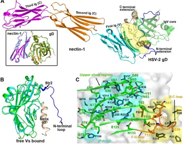
FIG 1Structure of the unbound HSV-2 gD. (A) Ribbon representation of the overall structure. The two molecules (chains A and B) present in the asymmetric unit are shown. The IgV-like core is in green, while the N- and C-terminal extensions are highlighted in blue and orange, respectively. The secondary elements are labeled as in HSV-1 gD (37). The loop linking residues R184 and L195 is invisible and is therefore indicated as a dotted curve. The terminal residues are marked. (B, C) Comparison of the free HSV-2 gD structure (green for chain A and blue for chain B) with previously reported gD structures, including those of free HSV-1 gD (red), HSV-1 gD with HVEM (cyan), HSV-1 gD with nectin-1 (orange), and HSV-2 gD with an antibody (magenta). The colored arrows mark, in the individual structures, the positions of the structure termini and the conformations of the loops referred to in the text. Those elements that are invisible in our structure but are clearly traced in the reference structures are circled and labeled. (B) Superimposition of the structures highlighting the external extension region (shown in ribbon). (C) Superimposition of the structures highlighting the IgV-like core.
on November 7, 2019 by guest
http://jvi.asm.org/
[image:4.585.138.449.63.407.2]structure of an IgV core wrapped by extensive terminal exten-sions, and nectin-1 folds into an overall rod-like structure with tandemly arranged V-C-C Ig-like domains. As expected, HSV-2 gD binds to the membrane-distal IgV-like domain of nectin-1 (Fig. 2A). In comparison to the free HSV-2 gD structure, many of the untraceable terminal residues are unambiguously observed in the complex structure, presenting as an extended loop in the N terminus and an␣-helix (␣3) in the C terminus to engage the receptor (Fig. 2B).
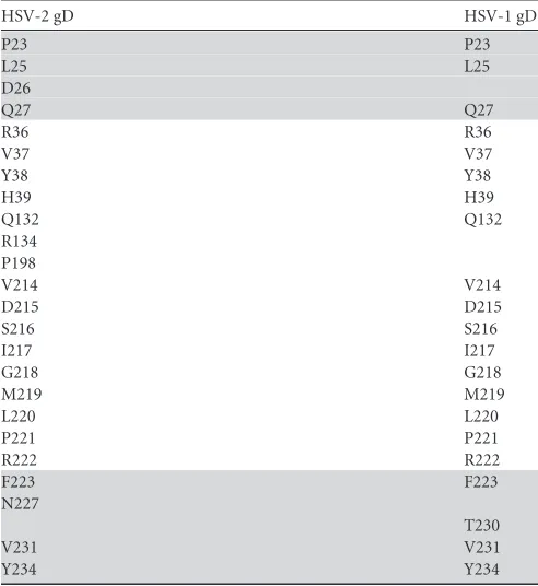
The overall binding mode between HSV-2 gD and nectin-1 is very similar to that observed in a previously reported HSV-1-gD/ nectin-1 complex structure (Fig. 2A). The binding relies on the surface components in the terminal extensions of HSV-2 gD en-gaging the rear (CC=C⬙FG) sheet of the nectin-1 IgV domain (Fig. 2A). A high complementarity in both the shape and the hydro-phobicity character is observed between the two molecules at this interface, bearing extended surface areas of about 938.4 Å2and 946.7 Å2in gD and nectin-1, respectively. We further detailed the interface amino acids with a 4.5-Å resolution cutoff to character-ize the receptor/ligand footprints in these two interacting mole-cules. A total of 24 residues are revealed in HSV-2 gD and are observed to sterically congregate into two main surface patches (patch1 and patch2), as reported for HSV-1 gD (35). The patch1 residues, including P23, L25 to Q27, F223, N227, V231, and Y234,
locate in the distal N-terminal loop and the␣3 helix. While the remaining interface residues of R36 to H39, Q132, R134, P198, and V214 to R222 form the much more extended surface patch2 (Fig. 2C). As expected, essentially the same patches of amino acids are also shown to be responsible for the HSV-1-gD/nect1 in-teraction (Table 3).
As to the receptor, the identified gD footprint in nectin-1 is composed of 26 amino acids, including S59, K61, T63, Q64, T66, Q68, G73 to N77, I80 to N82, M85 to S88, L90, E125, A127 to T131, and N133. Five residues (S59, K61, and F129 to T131) are observed to locate in the B-C and F-G loops, constituting a bottom loop region to contact mainly patch1 of HSV-2 gD. While an upper sheet (the CC=C⬙FG sheet) region, with the remaining in-terface residues, interacts in a much more extended manner with HSV-2 gD patch2 (Fig. 2C). This observed amino acid interaction pattern is the same for the two HSV-2-gD/nectin-1 complexes of the asymmetric unit and is also very similar to that observed in the HSV-1-gD/nectin-1 complex structure (35).
Comparative mutagenesis study on nectin-1 binding of HSV-1 and HSV-2 gDs.Based on the complex structures, HSV-1 and HSV-2 gDs contact essentially the same series of rear-sheet residues in nectin-1 for recognition. We therefore further sought functional evidence of a conserved nectin-1-binding mode be-tween the two viral gD proteins. With an alanine-scanning
mu-FIG 2Structure of the HSV-2-gD/nectin-1 complex. (A) Ribbon representation of the overall structure. The gD molecule (green for the IgV-like core, blue for the N-terminal extension, and orange for the C-terminal extension) binds to the first IgV-like domain of nectin-1 (colored cyan, orange, and magenta for the first, second, and third Ig-like domains, respectively). The binding interface involves the nectin-1 CC=C⬙FG rear sheet and the gD terminal extensions, which are circled and labeled. A superimposition comparison of a previously reported complex structure between HSV-1-gD (orange) and nectin-1 (magenta) with the bound structure of HSV-2-gD (green) with nectin-1 (cyan) is highlighted in the black box. (B) Structural comparison of the HSV-2 gD structures in the free (cyan) and the nectin-1 bound (green) forms. The fixed conformations of the terminal elements (highlighted in blue for the N terminus and in orange for the C terminus) in the bound structure are labeled. (C) Detailed view of the binding interface between HSV-2 gD (shown in surface) and nectin-1 (shown in ribbon). The interface residues of gD can be roughly categorized to congregate into surface patches 1 (yellow) and 2 (cyan), which contact mainly the B-C/F-G loop region (orange) and the upper sheet region (green) in nectin-1, respectively. The amino acids involved are shown in sticks and labeled.
on November 7, 2019 by guest
http://jvi.asm.org/
[image:5.585.111.475.70.355.2]tagenesis in nectin-1, its interaction with both HSV-1 and HSV-2 gDs were characterized and compared by surface plasmon reso-nance (SPR). A total of 13 amino acids were selected (based on the complex structure solved in this study and our previously solved HSV-1-gD/nectin-1 structure) and mutated either individually or simultaneously. The mutant proteins were then prepared and pu-rified as described for the wild-type protein and were further tested for the binding to gD (HSV-1 and HSV-2) proteins immo-bilized at⬃750 response units. The binding capacity was thereby characterized as the amount of nectin-1 (WT or mutant), in re-sponse units, that is capable of binding to gD when 1M individ-ual nectin-1 proteins flow through the chip.
Overall, the resultant binding patterns are very similar for HSV-1 and HSV-2 gDs (Fig. 3A). A series of residues, including K61, T63, Q64, Q76, N82, L90, E125, P130, and N133, do not significantly affect the gD/nectin-1 interaction. Nonetheless, dra-matically decreased gD binding is observed for nectin-1 mutants with substitutions for residues N77, I80, M85, and F129, which exhibit about one-half (or less) of the binding response units of the WT nectin-1. This result is also consistent with previous stud-ies reporting the functional indispensability of nectin-1 N77, M85, and F129 in the receptor recognition by HSV-1 gD (36,43,
44). It is also noteworthy that the tetramutant (simultaneously replacing T63, Q64, E125, and N133 with alanine) nectin-1, which should be disrupted for the receptor homodimerization based on
a previous report (56), shows an increased binding capacity to both gD proteins (Fig. 3A).
We further mapped the nectin-1 residues N77, I80, M85, and F129 onto the binding interface in our complex structure. F129, which locates in the F-G loop, inserts its side chain into a highly hydrophobic pocket formed by F223, V231, and the apolar carbon atoms of Q27 in HSV-2 gD (Fig. 3B). In the upper sheet region, where the remaining three amino acids reside, I80 and M85 are shown to extensively contact HSV-2 gD Y38, Q131, and P221 via strong hydrophobic interactions, while N77 could contribute to the ligand binding by providing H-bond interactions via both the side chain and main-chain nitrogens with gD D215 and M219 (Fig. 3C). For the other interface residues, their role in gD recog-nition seems to be restricted to a limited number of van der Waals contacts, explaining their individual dispensability.
[image:6.585.40.286.87.355.2]Cross-inhibition of the gD/nectin-1-mediated cell fusion by soluble HSV-1 and HSV-2 gDs.The conserved nectin-1-binding
TABLE 3Comparison between HSV-1 and HSV-2 gDs for the patch residues locating at the nectin-1 binding interfacea
HSV-2 gD HSV-1 gD
P23 P23
L25 L25
D26
Q27 Q27
R36 R36
V37 V37
Y38 Y38
H39 H39
Q132 Q132
R134 P198
V214 V214
D215 D215
S216 S216
I217 I217
G218 G218
M219 M219
L220 L220
P221 P221
R222 R222
F223 F223
N227
T230
V231 V231
Y234 Y234
a
Based on the HSV-2-gD/nectin-1 complex structure solved in this study and a previously reported structure of HSV-1 gD bound to nectin-1 (PDB code3U82), the interface residues within a distance of 4.5 Å from the nectin-1 receptor were selected and aligned in a pairwise manner. The two steric patches composed by these amino acids, as referred to in the text, are patch1 (gray background) and patch2 (white background). Clearly shown is that HSV-1 and HSV-2 gDs utilize essentially the same interface residues for nectin-1 recognition.
FIG 3Homologous receptor binding between HSV-1 and HSV-2 gDs to nec-tin-1. (A) A comparative mutagenesis study characterizing the gD/nectin-1 interaction. The gD proteins of HSV-1 (upper panel) and HSV-2 (lower panel) were immobilized on CM5 chips at⬃750 response units and were then tested for binding by 1M the indicated nectin-1 (WT or mutant) proteins. The amount of bound protein in response units was recorded and presented as a percentage of that of the WT nectin-1 using histograms. Clearly shown is that the interface residue mutations in nectin-1 affect the gD binding similarly for HSV-1 and HSV-2. (B, C) Structural delineation of the role that N77, I80, M85, and F129 play in gD recognition. The binding interface between HSV-2 gD (green) and nectin-1 (cyan) is highlighted for the intermolecule contacts contributed by these four residues. The 2|Fo|-|Fc| electron density map was contoured at 1.0. Dashed lines indicate H bonds. (B) Magnified view of the interface highlighting nectin-1 F129. (C) View of the interface highlighting nectin-1 N77, I80, and M85.
on November 7, 2019 by guest
http://jvi.asm.org/
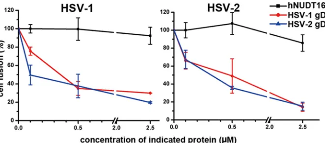
[image:6.585.298.542.282.587.2]mode between HSV-1 and HSV-2 gDs was further confirmed by a cell fusion-based mutual inhibition assay. The gD (HSV-1 or HSV-2) protein, along with the virus fusion executor of gB/gH/gL, and the nectin-1 receptor are separately expressed in CHO-K1 cells. Consistent with previous studies (53), remarkable cell fusion was observed when the nectin-1-expressing cells were mixed with the cells expressing virus gB, gH, and gL of HSV-1 and gD of either HSV-1 or HSV-2. In each case, the fusion is specifically inhibited by the soluble gD protein of its original virus type in a dose-de-pendent manner. In addition, fusion inhibition patterns similar to those observed in the self-inhibiting tests were recorded when the heterogeneous gD proteins were added to the system (Fig. 4). These observations are consistent with previous reports (41,57,
58) and demonstrate a very similar mode of action of HSV-1 and HSV-2 gDs in nectin-1-mediated cell-cell fusion.
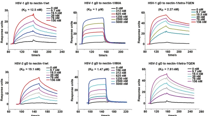
Nectin-1 I80 is an important gD interaction residue.Based on our comparative mutagenesis study, nectin-1 I80 is a newly identified residue that affects the gD binding, in addition to the previously reported amino acids N77, M85, and F129 (36,43,44). We therefore further quantitatively analyzed the contribution of this amino acid to the receptor/ligand engagement. The dissocia-tion constants (Kds) of nectin-1/I80A bound to HSV-1 and HSV-2
gDs were determined to be 1M and 1.47M, respectively. These values are about 2 orders of magnitude lower than those observed for the WT nectin-1 protein (12.5 nM for HSV-1 gD and 19.1 nM for HSV-2 gD), demonstrating that residue I80 indeed plays an important role in the nect1 engagement of gD. We also in-cluded the tetra-TQEN mutant protein in the kinetic assay, be-cause these four residues could disrupt nectin-1 homodimeriza-tion (56) but did not interfere with the gD/nectin-1 interaction individually (Fig. 3A). The calculated binding affinities, withKds
of 2.27 nM for HSV-1 gD and 7.81 nM for HSV-2 gD, increased approximately 2- to 5-fold in comparison to WT nectin-1 (Fig. 5
andTable 1).
We further investigated the role of nectin-1 I80 in the virus cell entry with an infection assay using a genetically modified HSV-1 virus containing a luciferase gene (55). CHO-K1 cells were tran-siently transfected with the nectin-1 (WT or mutant)-expressing plasmids. With similar expression levels (Fig. 6A), mutations of N77A, M85A, and F129A were shown to dramatically impair the
cell entry efficiencies of HSV-1 (Fig. 6B), which is consistent with previous studies (36,43,44). An approximate 2.5-fold decrease in the viral invasion was observed for the I80A mutant in compari-son to that of the WT protein, demonstrating its functional im-portance in nectin-1-mediated virus entry. The tetra-TQEN mu-tant, however, is as capable of mediating virus entry as WT nectin-1 (Fig. 6B). Reciprocally, we further tested in an infection-blocking assay the capability of nectin-1/I80A to inhibit HSV-1 entry into cells by competing with the cell surface nectin-1. Con-sistent with the result of the infection assay, the nectin-1/I80A mutant exhibits compromised blocking activity against the viral invasion of HSV-1 (Fig. 6C). These results demonstrate that nec-tin-1 I80 is an important interface residue involved in the gD binding.
DISCUSSION
As with other enveloped viruses, the initiation of virus infection by HSV requires, as a prerequisite, the specific interactions between viral surface glycoproteins and the host cellular receptors (7–9). Although multiple virus envelope proteins other than gD have been shown to be capable of recognizing host molecules (e.g., both gB and gC can engage HSPG [10, 11] and gB also recognizes PILR␣[59] and NMHC-IIA [60]), the gD-receptor interaction is considered a key step bridging the attachment and the fusion pro-cesses. Of the identified gD receptors, nectin-1 seems to play a more important role in mediating the infection by HSV-2, at least inin vivomurine models (28,29). In comparison to what was seen in the WT or the HVEM knockout mice, an attenuated disease caused by HSV-2 and diminished virus spread were observed in the nectin-1 knockout mice (28). In addition, it was demonstrated that nectin-1, but not HVEM, plays an indispensable role in the HSV-2 infection of the central nervous system by direct cranial inoculation (29). It is therefore an interesting issue to elucidate the molecular basis of the binding between HSV-2 gD and nectin-1. A previous study reported the structure of HSV-2 gD with a neutral-izing antibody, E317 (42). The antibody was shown to exert the neutralization activity by disrupting the gD-receptor interactions. The gD/E317 complex structure, therefore, provided the first structural glimpse on the HSV-2-gD/nectin-1-binding basis. In this study, we report a free HSV-2 gD structure and a complex
FIG 4Similar action modes of HSV-1 and HSV-2 gDs in the nectin-1-mediated cell-cell fusion. By comparing the luciferase activities, the cell fusion in the presence of gradient concentrations (0.1, 0.5, and 2.5M) of either HSV-1 or HSV-2 gD or an irrelevant hNUDT16 protein was plotted as the percentage of that without soluble gDs or hNUDT16. The dotted profiles are shown. (Left) HSV-1-gD-mediated cell fusion; (right), HSV-2-gD-mediated cell fusion. All the results are expressed as means⫾standard deviations (SD) from three independent experiments.
on November 7, 2019 by guest
http://jvi.asm.org/
[image:7.585.128.459.66.212.2]structure of HSV-2 gD bound with nectin-1. By comparing with the previously reported HSV-1-gD/nectin-1 structures (35,36), we demonstrated at the atomic level that gDs of the two virus species engage essentially the same patch of nectin-1 residues for receptor recognition. This homologous binding mode was further supported (i) by our comparative mutagenesis study showing that the individual mutation of the interface residues in nectin-1 af-fects the ligand-receptor interaction similarly for HSV-1 and HSV-2 gDs and (ii) by our fusion inhibition assay showing that soluble gD proteins of the two viruses could mutually inhibit the gD/nectin-1-mediated cell fusion. These comprehensive struc-tural and functional data, together with the previously reported functional evidence (39–41,57,58), demonstrate a conserved
re-ceptor-binding mode between HSV-1 and HSV-2 for nectin-1 and imply that the tropism difference between the two viruses likely arises from other aspects than the gD/nectin-1-binding fea-tures.
Compared to the HSV-1 gD (free or bound) (35–37) or the bound HSV-2 gD structures (42), the unbound HSV-2 gD struc-ture lacks traceable electron densities for a large portion of the terminal extensions at both the N and C termini. This could be either a result of cleavage—removal of the terminal extensions by accidentally contaminated proteases during crystallization— or an indicative clue of the terminal flexibility. It should be noted that in the thus-far-available complex structures of gD with a receptor or an antibody (35–37,42), the bound molecules invariably bind
FIG 5SPR assay characterizing the binding kinetics of gD (HSV-1 and HSV-2) to the indicated nectin-1 proteins (WT or mutant). The profiles of gD at gradient concentrations flowing through individual nectin-1 proteins immobilized at⬃750 response units (RUs) were recorded and are shown. The calculated dissoci-ation constants (Kds) are labeled.
FIG 6Effects of nectin-1 mutations on HSV-1 entry. (A) Flow-cytometric assay characterizing the expression level of each nectin-1 (WT or mutant) protein. The percentages of the positive transfectants (among 10,000 CHO-K1 cells) are shown. (B) Assay characterizing the cell entry efficiency of HSV-1 via WT or mutant nectin-1. CHO-K1 cells expressing the individual nectin-1 proteins are infected with HSV-1 viruses as specified in Materials and Methods. The histogram shows the virus entry efficiencies of HSV-1 using the indicated nectin-1 mutants in comparison to that using WT nectin-1. (C) Quantification of HSV-1 entry into CHO-K1 cells expressing WT nectin-1 in the presence of soluble nectin-1 proteins of WT and mutant forms. The virus entry with bovine serum albumin (BSA) as an irrelevant-protein control is presented as 100%, and those with the indicated nectin-1 proteins are shown as percentages of that of BSA. All the results are expressed as means⫾SD from three independent experiments. Clearly shown is that nectin-1 mutants (N77A, I80A, M85A, and F129A) with compromised gD-binding capabilities exhibit decreased capacities in inhibiting HSV-1 entry.
on November 7, 2019 by guest
http://jvi.asm.org/
[image:8.585.75.510.64.306.2] [image:8.585.114.475.532.646.2]to the gD patches that map to these terminal extension elements. In the free HSV-1 gD structure (37), two gD protomers are packed against each other in the crystal via an interface covering the same extension patch. This raises the possibility that a bound partner (a receptor, an antibody, or another gD molecule) stabilizes the oth-erwise-flexible terminal extension residues of gD into definitive structures. Accordingly, the portion of the terminal extensions were solvent exposed and invisible in the free HSV-2 gD structure rather than being buried with fixed folds as observed in the afore-mentioned gD structures. Our structure therefore further sup-ports the notion that the moveable character of gD N- and C-ter-minal segments would suffice for receptor binding, as documented for HSV-1 gD (61). In support of this view, several studies also reported the replacement of the gD N-terminal loop by uPA, interleukin-13 (IL-13), and the HER2 antibody, the chimeric gDs derived from which are functionally active in mediating virus entry into cells expressing their respective cog-nate receptors (62–64).
We also conducted a thorough mutational analysis for those structurally defined interface residues in nectin-1. In consistency with previous studies (36,43,44), individual substitution of N77, M85, and F129 by alanine dramatically impairs the interaction between gD (HSV-1 or HSV-2) and 1. In addition, nectin-1/I80 also plays an important role in the gD/nectin-1 engagement, which was further demonstrated by our kinetic analysis and the virus entry assay. These four amino acids interact extensively with gD and thereby contribute dramatically to the binding via strong H bond and/or hydrophobic interactions. The remaining inter-face residues, in contrast, do not have a significant role individu-ally. Accordingly, a majority of these amino acids are indeed ob-served to provide only limited van der Waals contacts in the complex structures and thereby are likely functioning to modulate gD recognition in combination. This is, to our knowledge, the first comprehensive alanine-scanning mutagenesis study on nectin-1 for its interaction properties with gD, which represents a useful supplement to the current structural observations. It is also note-worthy that the I80A mutation leads the kinetic profile of nectin-1 binding to gD to a fast-on/fast-off mode, forming a contrast to the kinetic pattern between WT nectin-1 and gD, which is slow-on/ slow-off. We believe that the residue substitution and its resultant change of hydrophobic van der Waals contacts caused the kinetic shift. Similar phenomena of alteredKonand/orKoffrates resulting from mutations of interface residues have also been reported in other protein-protein interactions (65,66).
Nectin-1 can form homodimers in solution (35,56). Based on the solved structures, gD binds to the dimer interface of nectin-1 and would compete against its dimerization (35). In this study, we found that simultaneous mutation of T63, Q64, E125, and N133 to alanine leads to an increased binding affinity between nectin-1 and gD. Although these four residues contact HSV gD, we showed that the individual residues contribute little to the gD/nectin-1 interaction (Fig. 3A). In contrast, they have important roles in nectin-1 homobinding by providing H-bond and salt bridge con-tacts (56). The increased affinity should therefore be a result of residue substitutions that impaired nectin-1 homodimerization but hardly affected its interaction with gD. This is a functional evidence for a competitive relationship between gD/nectin-1 het-erocomplex formation and the nectin-1 dimerization. The result also reminds us of a variant residue usage between gD and the other nectin-1 molecule to engage the same nectin-1 receptor.
This would facilitate the design of a nectin-1 mimic antiviral drug, which can bind to the viral gD protein with high affinity but show low or no specificity for nectin-1. These are interesting issues that are worth pursuing in the future.
ACKNOWLEDGMENTS
This work was supported by the 973 project of China Ministry of Science and Technology (MOST; grant no. 2010CB530104).
Assistance by the staff at the Shanghai Synchrotron Radiation Facility (SSRF-beamline 17U) is acknowledged. We thank Yanfang Zhang and Zheng Fan for their sophisticated technical support.
G.F.G. is the leading principal investigator of the Innovative Research Group of the National Natural Science Foundation of China (NSFC; grant no. 81321063).
REFERENCES
1.Roizman B, Knipe DM, Whitley JR.2007. Herpes simplex viruses, p 2503–2602.InKnipe DM, Howley PM (ed), Fields virology, 5th ed. Lip-pincott Williams & Wilkins, Philadelphia, PA.
2.McLennan JL, Darby G.1980. Herpes simplex virus latency: the cellular location of virus in dorsal root ganglia and the fate of the infected cell following virus activation. J. Gen. Virol.51:233–243.http://dx.doi.org/10 .1099/0022-1317-51-2-233.
3.Looker KJ, Garnett GP, Schmid GP.2008. An estimate of the global prevalence and incidence of herpes simplex virus type 2 infection. Bull. World Health Organ.86:805A– 812A.http://dx.doi.org/10.2471/BLT.07 .046128.
4.Liesegang TJ.2001. Herpes simplex virus epidemiology and ocular impor-tance. Cornea20:1–13.http://dx.doi.org/10.1097/00003226-200101000-00001. 5.Gupta R, Warren T, Wald A.2007. Genital herpes. Lancet370:2127–
2137.http://dx.doi.org/10.1016/S0140-6736(07)61908-4.
6.Brugha R, Keersmaekers K, Renton A, Meheus A.1997. Genital herpes infection: a review. Int. J. Epidemiol.26:698 –709.http://dx.doi.org/10 .1093/ije/26.4.698.
7.Heldwein EE, Krummenacher C.2008. Entry of herpesviruses into mam-malian cells. Cell. Mol. Life Sci.65:1653–1668.http://dx.doi.org/10.1007 /s00018-008-7570-z.
8.Connolly SA, Jackson JO, Jardetzky TS, Longnecker R.2011. Fusing structure and function: a structural view of the herpesvirus entry ma-chinery. Nat. Rev. Microbiol. 9:369 –381. http://dx.doi.org/10.1038 /nrmicro2548.
9.Spear PG.2004. Herpes simplex virus: receptors and ligands for cell entry. Cell. Microbiol.6:401– 410.http://dx.doi.org/10.1111/j.1462-5822.2004 .00389.x.
10. Laquerre S, Argnani R, Anderson DB, Zucchini S, Manservigi R, Glo-rioso JC.1998. Heparan sulfate proteoglycan binding by herpes simplex virus type 1 glycoproteins B and C, which differ in their contributions to virus attachment, penetration, and cell-to-cell spread. J. Virol.72:6119 – 6130.
11. Svennerholm B, Jeansson S, Vahlne A, Lycke E.1991. Involvement of glycoprotein C (gC) in adsorption of herpes simplex virus type 1 (HSV-1) to the cell. Arch. Virol.120:273–279.http://dx.doi.org/10.1007/BF01310482. 12. Spear PG, Eisenberg RJ, Cohen GH.2000. Three classes of cell surface
receptors for alphaherpesvirus entry. Virology275:1– 8.http://dx.doi.org /10.1006/viro.2000.0529.
13. Spear PG, Longnecker R.2003. Herpesvirus entry: an update. J. Virol. 77:10179 –10185.http://dx.doi.org/10.1128/JVI.77.19.10179-10185.2003. 14. Koyama AH, Uchida T.1987. The mode of entry of herpes-simplex virus type-1 into Vero cells. Microbiol. Immunol.31:123–130.http://dx.doi.org /10.1111/j.1348-0421.1987.tb03075.x.
15. Nicola AV, Straus SE.2004. Cellular and viral requirements for rapid endocytic entry of herpes simplex virus. J. Virol.78:7508 –7517.http://dx .doi.org/10.1128/JVI.78.14.7508-7517.2004.
16. Turner A, Bruun B, Minson T, Browne H.1998. Glycoproteins gB, gD, and gHgL of herpes simplex virus type 1 are necessary and sufficient to mediate membrane fusion in a Cos cell transfection system. J. Virol.72: 873– 875.
17. Chowdary TK, Cairns TM, Atanasiu D, Cohen GH, Eisenberg RJ,
Heldwein EE.2010. Crystal structure of the conserved herpesvirus fusion regulator complex gH-gL. Nat. Struct. Mol. Biol.17:882– 888.http://dx .doi.org/10.1038/nsmb.1837.
on November 7, 2019 by guest
http://jvi.asm.org/
18. Heldwein EE, Lou H, Bender FC, Cohen GH, Eisenberg RJ, Harrison SC.2006. Crystal structure of glycoprotein B from herpes simplex virus 1. Science313:217–220.http://dx.doi.org/10.1126/science.1126548. 19. Cairns TM, Whitbeck JC, Lou H, Heldwein EE, Chowdary TK,
Eisen-berg RJ, Cohen GH.2011. Capturing the herpes simplex virus core fusion complex (gB-gH/gL) in an acidic environment. J. Virol.85:6175– 6184. http://dx.doi.org/10.1128/JVI.00119-11.
20. Fusco D, Forghieri C, Campadelli-Fiume G. 2005. The pro-fusion domain of herpes simplex virus glycoprotein D (gD) interacts with the gD N terminus and is displaced by soluble forms of viral receptors. Proc. Natl. Acad. Sci. U. S. A.102:9323–9328. http://dx.doi.org/10 .1073/pnas.0503907102.
21. Cocchi F, Fusco D, Menotti L, Gianni T, Eisenberg RJ, Cohen GH, Campadelli-Fiume G.2004. The soluble ectodomain of herpes simplex virus gD contains a membrane-proximal pro-fusion domain and suffices to mediate virus entry. Proc. Natl. Acad. Sci. U. S. A.101:7445–7450.http: //dx.doi.org/10.1073/pnas.0401883101.
22. Gianni T, Amasio M, Campadelli-Fiume G.2009. Herpes simplex virus gD forms distinct complexes with fusion executors gB and gH/gL in part through the C-terminal profusion domain. J. Biol. Chem.284:17370 – 17382.http://dx.doi.org/10.1074/jbc.M109.005728.
23. Karasneh GA, Shukla D.2011. Herpes simplex virus infects most cell types in vitro: clues to its success. Virol. J.8:481.http://dx.doi.org/10.1186 /1743-422X-8-481.
24. Montgomery RI, Warner MS, Lum BJ, Spear PG.1996. Herpes simplex virus-1 entry into cells mediated by a novel member of the TNF/NGF receptor family. Cell 87:427– 436. http://dx.doi.org/10.1016/S0092 -8674(00)81363-X.
25. Geraghty RJ, Krummenacher C, Cohen GH, Eisenberg RJ, Spear PG. 1998. Entry of alphaherpesviruses mediated by poliovirus receptor-related protein 1 and poliovirus receptor. Science280:1618 –1620.http://dx.doi .org/10.1126/science.280.5369.1618.
26. Warner MS, Geraghty RJ, Martinez WM, Montgomery RI, Whitbeck JC, Xu RL, Eisenberg RJ, Cohen GH, Spear PG.1998. A cell surface protein with herpesvirus entry activity (HveB) confers susceptibility to infection by mutants of herpes simplex virus type 1, herpes simplex virus type 2, and pseudorabies virus. Virology246:179 –189.http://dx.doi.org /10.1006/viro.1998.9218.
27. Shukla D, Liu J, Blaiklock P, Shworak NW, Bai XM, Esko JD, Cohen GH, Eisenberg RJ, Rosenberg RD, Spear PG. 1999. A novel role for 3-O-sulfated heparan sulfate in herpes simplex virus 1 entry. Cell99:13– 22.http://dx.doi.org/10.1016/S0092-8674(00)80058-6.
28. Taylor JM, Lin E, Susmarski N, Yoon M, Zago A, Ware CF, Pfeffer K, Miyoshi J, Takai Y, Spear PG. 2007. Alternative entry receptors for herpes simplex virus and their roles in disease. Cell Host Microbe2:19 –28. http://dx.doi.org/10.1016/j.chom.2007.06.005.
29. Kopp SJ, Banisadr G, Glajch K, Maurer UE, Grunewald K, Miller RJ, Osten P, Spear PG.2009. Infection of neurons and encephalitis after intracranial inoculation of herpes simplex virus requires the entry recep-tor nectin-1. Proc. Natl. Acad. Sci. U. S. A.106:17916 –17920.http://dx .doi.org/10.1073/pnas.0908892106.
30. Karaba AH, Kopp SJ, Longnecker R.2011. Herpesvirus entry mediator and nectin-1 mediate herpes simplex virus 1 infection of the murine cor-nea. J. Virol.85:10041–10047.http://dx.doi.org/10.1128/JVI.05445-11. 31. Takai Y, Ikeda W, Ogita H, Rikitake Y.2008. The immunoglobulin-like
cell adhesion molecule nectin and its associated protein afadin. Annu. Rev. Cell Dev. Biol.24:309 –342.http://dx.doi.org/10.1146/annurev.cellbio.24 .110707.175339.
32. Rux AH, Willis SH, Nicola AV, Hou W, Peng C, Lou H, Cohen GH, Eisenberg RJ.1998. Functional region IV of glycoprotein D from herpes simplex virus modulates glycoprotein binding to the herpesvirus entry mediator. J. Virol.72:7091–7098.
33. Whitbeck JC, Peng C, Lou H, Xu R, Willis SH, Ponce de Leon M, Peng T, Nicola AV, Montgomery RI, Warner MS, Soulika AM, Spruce LA, Moore WT, Lambris JD, Spear PG, Cohen GH, Eisenberg RJ.1997. Glycoprotein D of herpes simplex virus (HSV) binds directly to HVEM, a member of the tumor necrosis factor receptor superfamily and a mediator of HSV entry. J. Virol.71:6083– 6093.
34. Willis SH, Rux AH, Peng C, Whitbeck JC, Nicola AV, Lou H, Hou W, Salvador L, Eisenberg RJ, Cohen GH.1998. Examination of the kinetics of herpes simplex virus glycoprotein D binding to the herpesvirus entry mediator, using surface plasmon resonance. J. Virol.72:5937–5947. 35. Zhang N, Yan J, Lu G, Guo Z, Fan Z, Wang J, Shi Y, Qi J, Gao GF.
2011. Binding of herpes simplex virus glycoprotein D to nectin-1 ex-ploits host cell adhesion. Nat. Commun.2:577.http://dx.doi.org/10 .1038/ncomms1571.
36. Di Giovine P, Settembre EC, Bhargava AK, Luftig MA, Lou H, Cohen GH, Eisenberg RJ, Krummenacher C, Carfi A.2011. Structure of herpes simplex virus glycoprotein D bound to the human receptor nectin-1. PLoS Pathog.7:e1002277.http://dx.doi.org/10.1371/journal.ppat.1002277. 37. Carfi A, Willis SH, Whitbeck JC, Krummenacher C, Cohen GH,
Eisen-berg RJ, Wiley DC.2001. Herpes simplex virus glycoprotein D bound to the human receptor HveA. Mol. Cell8:169 –179.http://dx.doi.org/10 .1016/S1097-2765(01)00298-2.
38. Krummenacher C, Supekar VM, Whitbeck JC, Lazear E, Connolly SA, Eisenberg RJ, Cohen GH, Wiley DC, Carfi A.2005. Structure of unli-ganded HSV gD reveals a mechanism for receptor-mediated activation of virus entry. EMBO J.24:4144 – 4153.http://dx.doi.org/10.1038/sj.emboj .7600875.
39. Krummenacher C, Nicola AV, Whitbeck JC, Lou H, Hou WF, Lambris JD, Geraghty RJ, Spear PG, Cohen GH, Eisenberg RJ.1998. Herpes simplex virus glycoprotein D can bind to poliovirus receptor-related pro-tein 1 or herpesvirus entry mediator, two structurally unrelated mediators of virus entry. J. Virol.72:7064 –7074.
40. Muggeridge MI, Wilcox WC, Cohen GH, Eisenberg RJ.1990. Identifi-cation of a site on herpsimplex virus type-1 glycoprotein-D that is es-sential for infectivity. J. Virol.64:3617–3626.
41. Nicola AV, Peng CR, Lou H, Cohen GH, Eisenberg RJ.1997. Antigenic structure of soluble herpes simplex virus (HSV) glycoprotein D correlates with inhibition of HSV infection. J. Virol.71:2940 –2946.
42. Lee CC, Lin LL, Chan WE, Ko TP, Lai JS, Wang AH.2013. Structural basis for the antibody neutralization of herpes simplex virus. Acta Crys-tallogr. D Biol. CrysCrys-tallogr. 69:1935–1945. http://dx.doi.org/10.1107 /S0907444913016776.
43. Martinez WM, Spear PG.2002. Amino acid substitutions in the V do-main of nectin-1 (HveC) that impair entry activity for herpes simplex virus types 1 and 2 but not for pseudorabies virus or bovine herpesvirus 1. J. Virol. 76:7255–7262. http://dx.doi.org/10.1128/JVI.76.14.7255-7262 .2002.
44. Struyf F, Martinez WM, Spear PG.2002. Mutations in the N-terminal domains of nectin-1 and nectin-2 reveal differences in requirements for entry of various alphaherpesviruses and for nectin-nectin interac-tions. J. Virol. 76:12940 –12950.http://dx.doi.org/10.1128/JVI.76.24 .12940-12950.2002.
45. Chen Z, Lu G, Qi J, Xu X, Zhang N, Yan J, Wang R.2011. X-ray diffraction analysis of glycoprotein D from herpes simplex virus type 2. Sheng Wu Gong Cheng Xue Bao27:1499 –1506.
46. Otwinowski Z, Minor W.1997. Processing of X-ray diffraction data collected in oscillation mode. Methods Enzymol.276:307–326.http://dx .doi.org/10.1016/S0076-6879(97)76066-X.
47. Read RJ.2001. Pushing the boundaries of molecular replacement with maximum likelihood. Acta Crystallogr. D Biol. Crystallogr.57:1373–1382. http://dx.doi.org/10.1107/S0907444901012471.
48. Collaborative Computing Project Number 4.1994. The CCP4 suite: programs for protein crystallography. Acta Crystallogr. D Biol. Crystal-logr.50:760 –763.http://dx.doi.org/10.1107/S0907444994003112. 49. Murshudov GN, Vagin AA, Dodson EJ.1997. Refinement of
macromo-lecular structures by the maximum-likelihood method. Acta Crystallogr. D53:240 –255.http://dx.doi.org/10.1107/S0907444996012255. 50. Adams PD, Afonine PV, Bunkoczi G, Chen VB, Davis IW, Echols N,
Headd JJ, Hung LW, Kapral GJ, Grosse-Kunstleve RW, McCoy AJ, Mo-riarty NW, Oeffner R, Read RJ, Richardson DC, Richardson JS, Terwilliger TC, Zwart PH.2010. PHENIX: a comprehensive Python-based system for macromolecular structure solution. Acta Crystallogr. D Biol. Crystallogr.66: 213–221.http://dx.doi.org/10.1107/S0907444909052925.
51. Emsley P, Cowtan K.2004. Coot: model-building tools for molecular graphics. Acta Crystallogr. D Biol. Crystallogr.60:2126 –2132.http://dx .doi.org/10.1107/S0907444904019158.
52. Laskowski RA, Macarthur MW, Moss DS, Thornton JM. 1993. Pro-check—a program to check the stereochemical quality of protein structures. J. Appl. Crystallogr.26:283–291.http://dx.doi.org/10.1107/S0021889892009944. 53. Pertel PE, Fridberg A, Parish ML, Spear PG.2001. Cell fusion induced by herpes simplex virus glycoproteins gB, gD, and gH-gL requires a gD receptor but not necessarily heparan sulfate. Virology279:313–324.http: //dx.doi.org/10.1006/viro.2000.0713.
54. Lu G, Zhang J, Li Y, Li Z, Zhang N, Xu X, Wang T, Guan Z, Gao GF,
on November 7, 2019 by guest
http://jvi.asm.org/
Yan J.2011. hNUDT16: a universal decapping enzyme for small nucleolar RNA and cytoplasmic mRNA. Protein Cell2:64 –73.http://dx.doi.org/10 .1007/s13238-011-1009-2.
55. Li Y, Wang S, Zhu H, Zheng CF.2011. Cloning of the herpes simplex virus type 1 genome as a novel luciferase-tagged infectious bacterial arti-ficial chromosome. Arch. Virol. 156:2267–2272. http://dx.doi.org/10 .1007/s00705-011-1094-9.
56. Narita H, Yamamoto Y, Suzuki M, Miyazaki N, Yoshida A, Kawai K, Iwasaki K, Nakagawa A, Takai Y, Sakisaka T.2011. Crystal structure of the cis-dimer of nectin-1: implications for the architecture of cell-cell junctions. J. Biol. Chem.286:12659 –12669.http://dx.doi.org/10.1074/jbc .M110.197368.
57. Nicola AV, Willis SH, Naidoo NN, Eisenberg RJ, Cohen GH. 1996. Structure-function analysis of soluble forms of herpes simplex virus gly-coprotein D. J. Virol.70:3815–3822.
58. Johnson DC, Burke RL, Gregory T. 1990. Soluble forms of herpes-simplex virus glycoprotein-D bind to a limited number of cell-surface receptors and inhibit virus entry into cells. J. Virol.64:2569 –2576. 59. Satoh T, Arii J, Suenaga T, Wang J, Kogure A, Uehori J, Arase N,
Shiratori I, Tanaka S, Kawaguchi Y, Spear PG, Lanier LL, Arase H. 2008. PILRalpha is a herpes simplex virus-1 entry coreceptor that associ-ates with glycoprotein B. Cell132:935–944.http://dx.doi.org/10.1016/j .cell.2008.01.043.
60. Arii J, Goto H, Suenaga T, Oyama M, Kozuka-Hata H, Imai T, Minowa A, Akashi H, Arase H, Kawaoka Y, Kawaguchi Y.2010. Non-muscle
myosin IIA is a functional entry receptor for herpes simplex virus-1. Na-ture467:859 – 862.http://dx.doi.org/10.1038/nature09420.
61. Rey FA.2006. Molecular gymnastics at the herpesvirus surface. EMBO Rep.7:1000 –1005.http://dx.doi.org/10.1038/sj.embor.7400807. 62. Zhou G, Roizman B.2007. Separation of receptor-binding and
profuso-genic domains of glycoprotein D of herpes simplex virus 1 into distinct interacting proteins. Proc. Natl. Acad. Sci. U. S. A.104:4142– 4146.http: //dx.doi.org/10.1073/pnas.0611565104.
63. Zhou G, Ye GJ, Debinski W, Roizman B. 2002. Engineered herpes simplex virus 1 is dependent on IL13Ralpha 2 receptor for cell entry and independent of glycoprotein D receptor interaction. Proc. Natl. Acad. Sci. U. S. A.99:15124 –15129.http://dx.doi.org/10.1073/pnas.232588699. 64. Menotti L, Cerretani A, Hengel H, Campadelli-Fiume G.2008.
Con-struction of a fully retargeted herpes simplex virus 1 recombinant capable of entering cells solely via human epidermal growth factor receptor 2. J. Virol.82:10153–10161.http://dx.doi.org/10.1128/JVI.01133-08. 65. Weihofen WA, Liu J, Reutter W, Saenger W, Fan H.2004. Crystal
structure of CD26/dipeptidyl-peptidase IV in complex with adenosine deaminase reveals a highly amphiphilic interface. J. Biol. Chem.279: 43330 – 43335.http://dx.doi.org/10.1074/jbc.M405001200.
66. Wang Q, Qi J, Yuan Y, Xuan Y, Han P, Wan Y, Ji W, Li Y, Wu Y, Wang J, Iwamoto A, Woo PCY, Yuen KY, Yan J, Lu G, Gao GF.2014. Bat origins of MERS-CoV supported by Bat Coronavirus HKU4 usage of hu-man receptor CD26. Cell Host Microbe16:328 –337.http://dx.doi.org/10 .1016/j.chom.2014.08.009.