Determining the E
ff
ective Density and Stabilizer Layer Thickness of
Sterically Stabilized Nanoparticles
Bernice Akpinar,
†Lee A. Fielding,
*
,†,‡Victoria J. Cunningham,
†Yin Ning,
†Oleksandr O. Mykhaylyk,
†Patrick W. Fowler,
†and Steven P. Armes
*
,††
Department of Chemistry, University of She
ffi
eld, Brook Hill, She
ffi
eld, South Yorkshire S3 7HF, U.K.
‡School of Materials, The University of Manchester, Oxford Road, Manchester, M13 9PL, U.K.
*
S Supporting InformationABSTRACT:
A series of model sterically stabilized diblock
copolymer nanoparticles has been designed to aid the
development of analytical protocols in order to determine
two key parameters: the e
ff
ective particle density and the steric
stabilizer layer thickness. The former parameter is essential for
high resolution particle size analysis based on analytical
(ultra)centrifugation techniques (e.g., disk centrifuge
photo-sedimentometry, DCP), whereas the latter parameter is of
fundamental importance in determining the e
ff
ectiveness of
steric stabilization as a colloid stability mechanism. The
diblock copolymer nanoparticles were prepared via
polymer-ization-induced self-assembly (PISA) using RAFT aqueous
emulsion polymerization: this approach a
ff
ords relatively narrow particle size distributions and enables the mean particle
diameter and the stabilizer layer thickness to be adjusted independently via systematic variation of the mean degree of
polymerization of the hydrophobic and hydrophilic blocks, respectively. The hydrophobic core-forming block was
poly(2,2,2-tri
fl
uoroethyl methacrylate) [PTFEMA], which was selected for its relatively high density. The hydrophilic stabilizer block was
poly(glycerol monomethacrylate) [PGMA], which is a well-known non-ionic polymer that remains water-soluble over a wide
range of temperatures. Four series of PGMA
x−
PTFEMA
ynanoparticles were prepared (
x
= 28, 43, 63, and 98,
y
= 100
−
1400)
and characterized via transmission electron microscopy (TEM), dynamic light scattering (DLS), and small-angle X-ray scattering
(SAXS). It was found that the degree of polymerization of both the PGMA stabilizer and core-forming PTFEMA had a strong
in
fl
uence on the mean particle diameter, which ranged from 20 to 250 nm. Furthermore, SAXS was used to determine radii of
gyration of 1.46 to 2.69 nm for the solvated PGMA stabilizer blocks. Thus, the mean e
ff
ective density of these sterically stabilized
particles was calculated and determined to lie between 1.19 g cm
−3for the smaller particles and 1.41 g cm
−3for the larger
particles; these values are signi
fi
cantly lower than the solid-state density of PTFEMA (1.47 g cm
−3). Since analytical
centrifugation requires the density
dif ference
between the particles and the aqueous phase, determining the e
ff
ective particle
density is clearly vital for obtaining reliable particle size distributions. Furthermore, selected DCP data were recalculated by
taking into account the inherent density
distribution
superimposed on the particle size distribution. Consequently, the true
particle size distributions were found to be somewhat narrower than those calculated using an erroneous single density value,
with smaller particles being particularly sensitive to this artifact.
■
INTRODUCTION
Steric stabilization is widely recognized to be the most
important mechanism for achieving long-term colloidal
stability.
1,2Unlike charge stabilization,
3it confers
thermody-namic stability at relatively high solids, is tolerant of added salt
in aqueous formulations,
4and can be designed for a wide range
of media, including both polar solvents
5−11and non-polar
solvents
12−21as well as more exotic solvents such as
supercritical carbon dioxide
22−27or ionic liquids.
28,29In view
of these many advantages, steric stabilization is now used on an
industrial scale across a wide range of commercial sectors.
Examples include the manufacture of copolymer latex
paints,
12,30ceramic dispersions,
31−35ink formulations,
36and
antiwear additives for engine oils.
37−39Steric stabilization is also
known to be a highly e
ff
ective mechanism for preventing the
biofouling of surfaces
40−45and is important in determining the
interfacial adsorption of particles
46as well as the emulsion type
for Pickering emulsi
fi
ers.
47The
ef fective particle density
and the
stabilizer layer thickness
are key parameters for sterically stabilized particles. Knowledge
of the former parameter is vital for high resolution particle size
analysis based on analytical (ultra)centrifugation.
48−50This is
because the density di
ff
erence between the particles and the
continuous phase is one of three primary variables, along with
Received: May 12, 2016
Revised: June 27, 2016
Published: July 7, 2016
Article
pubs.acs.org/Macromolecules License, which permits unrestricted use, distribution and reproduction in any medium,
the particle size and colloidal stability, that determine the rate
of sedimentation (and hence the degree of particle
fractiona-tion). The latter parameter is of fundamental interest and is
directly related to the observed colloidal stability, since it
precisely determines the interparticle separation distance at
which the steric repulsive term becomes important.
2In
principle, small-angle neutron scattering (SANS) can be used
to determine the segment density pro
fi
le of stabilizer chains
normal to the particle surface and hence the mean stabilizer
layer thickness. However, this sophisticated technique usually
requires deuterated polymers for the contrast variation
approach that yields the highest-quality data, but unfortunately
such polymers are typically not available for most commercial
systems of interest. Similarly, small-angle X-ray scattering
(SAXS) can be used to determine stabilizer layer thicknesses.
For example, Ballau
ff
and co-workers have used SAXS to
determine the stabilizer thickness for poly(ethylene
oxide)-stabilized polystyrene (PEO
−
PS) latexes with core diameters
ranging between 70 and 146 nm.
51,52However, the problem of
e
ff
ective particle density was not considered. Moreover, this
PEO
−
PS system is ill-suited to addressing this question
because the density di
ff
erence between the PS core and water
(
∼
0.05 g cm
−3) is simply too small.
According to the well-established mechanism of steric
stabilization, colloidal stability is achieved by creating a
relatively thick dense surface layer of polymer chains.
2,30,53In
a good solvent for the stabilizer, interpenetration of such chains
is unfavorable on both entropic and enthalpic grounds. This
leads to a strong interparticle repulsive term that o
ff
sets the
ever-present van der Waals attractive forces and ensures
long-term colloidal stability. In principle, the stabilizer chains can be
either chemically grafted
4,21,24or merely physically adsorbed on
the surface of the colloidal particles.
16−18A third scenario arises
for amphiphilic diblock copolymer nanoparticles, such as those
prepared by polymerization-induced self-assembly (PISA) using
techniques such as reversible addition
−
fragmentation chain
transfer (RAFT) dispersion or emulsion polymerization.
20,54−70In such cases the solvophilic block comprises the stabilizer
chains, while the solvophobic block forms the particle core.
In the present work, we have exploited RAFT aqueous
emulsion polymerization to prepare a series of
near-monodisperse sterically stabilized diblock copolymer
nano-particles via PISA. The hydrophilic stabilizer block was chosen
to be a well-known non-ionic water-soluble polymer, namely
poly(glycerol monomethacrylate) [PGMA], while
poly(2,2,2-tri
fl
uoroethyl methacrylate) [PTFEMA] was selected as the
hydrophobic core-forming block, mainly because of its
relatively high solid-state density (1.47 g cm
−3, see
Figure 1
).
This model system was designed to enable the determination of
the e
ff
ective particle density (
ρ
particle) and stabilizer shell
thickness (
T
shell) for sterically stabilized diblock copolymer
nanoparticles. Initially, the nanoparticle size and morphology
was assessed using transmission electron microscopy (TEM)
and dynamic light scattering (DLS). SAXS was then utilized to
determine the volume-average diameter, aggregation number
(
N
agg), and
T
shellfor selected nanoparticles. The latter data were
then used to calculate an e
ff
ective particle density (
ρ
particle),
which enabled high resolution particle size analysis for this
model system via disk centrifuge photosedimentometry (DCP).
Finally, it is demonstrated that DCP size distributions can be
corrected for the superimposed density distribution that is an
intrinsic feature of such core
−
shell nanoparticles.
■
EXPERIMENTAL SECTION
Materials. Glycerol monomethacrylate (GMA) was donated by GEO Specialty Chemicals (Hythe, UK) and used without further purification. 2,2,2-Trifluoroethyl methacrylate (TFEMA) and 4,4′ -azobis(4-cyanopentanoic acid) (ACVA; 99%) were purchased from Sigma-Aldrich UK and were used as received. 2-Cyano-2-propyl dithiobenzoate (CPDB) was purchased from STREM Chemicals Ltd. (Cambridge, UK) and was used as received. d6-Acetone and d4 -methanol were purchased from Goss Scientific Instruments Ltd. (Cheshire, UK). All other solvents were purchased from Fisher Scientific (Loughborough, UK) and used as received. Deionized water was used for all experiments.
Synthesis of PGMAxMacro-CTA via RAFT Solution Polymer-ization.A typical protocol for the synthesis of PGMA63is as follows. CPDB RAFT agent (1.650 g, 7.454 mmol), GMA (78.144 g, 488 mmol), and ACVA (0.379 g, 1.352 mmol; CPDB/ACVA molar ratio = 5.0) were weighed into a 500 mL round-bottomflask and degassed with nitrogen for 15 min. Ethanol (148 mL) was deoxygenated separately with nitrogen for 30 min prior to addition to the other reagents. The reaction solution was stirred and degassed in an ice bath for a further 30 min before placing in an oil bath at 70 °C. The polymerization was allowed to proceed for 150 min (GMA monomer conversion = 68% as judged by1H NMR). The crude homopolymer was collected by precipitation into a 10-fold excess of dichloromethane from methanol. This cleanup protocol was repeated twice to afford a pure PGMA macro-CTA (53.14 g, <1% residual monomer). The mean degree of polymerization (DP) was calculated to be 63 as judged by1H NMR. DMF GPC analysis indicated anM
nof 15 000 g mol−1 and anMw/Mnof 1.19 (vs a series of near-monodisperse poly(methyl methacrylate) (PMMA) calibration standards). Other PGMA macro-CTAs with differing mean degrees of polymerization (28, 43, and 98) were prepared using a similar protocol simply by varying the monomer/CPDB molar ratio.
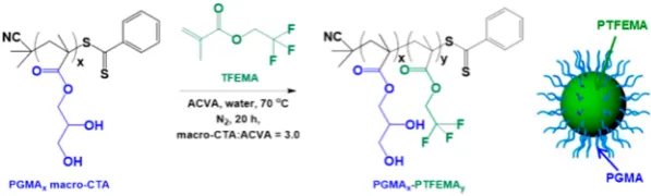
RAFT Aqueous Emulsion Polymerization of PGMAx− PTFEMAy. A typical protocol for the synthesis of PGMA63− PTFEMA400 diblock copolymer nanoparticles was as follows: PGMA63 macro-CTA (0.140 g), ACVA (0.600 mg, 2.14 μmol; macro-CTA/ACVA molar ratio = 3.0), and water (4.58 g, 10% w/w) were weighed into a 14 mL sample vial, sealed with a rubber septum, and degassed with nitrogen for 30 min. TFEMA [3.20 mL, 22.6 mmol, target degree of polymerization (DP) = 400], which had been deoxygenated separately with nitrogen for 15 min, was then added to the solution under nitrogen and immersed in an oil bath set at 70°C. The reaction solution was stirred for 20 h to ensure complete TFEMA monomer conversion, and the polymerization was quenched by exposure to air. 19F NMR spectroscopy analysis of the copolymer dissolved in d6-acetone indicated less than 1% residual TFEMA monomer. Four series of PGMAx−PTFEMAy diblock copolymer
nanoparticle dispersions were prepared by utilizing the PGMAx
[image:2.625.371.520.67.178.2]macro-CTAs described above and varying the degree of polymerization of the PTFEMA block (y) from 100 to 1400.
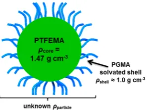
Figure 1.Schematic representation of a sterically stabilized PGMAx− PTFEMAy diblock copolymer nanoparticle. The effective particle
density (ρparticle) in aqueous solution will depend on the radius and density of the PTFEMA core (ρcore) and the thickness (Tshell) and density of the solvated stabilizer shell (ρshell).
1H NMR Spectroscopy.All 1H NMR spectra were recorded at 400 MHz ind6-acetone ord4-methanol using a Bruker Avance-400 spectrometer with 64 scans averaged per spectrum.
19
F NMR Spectroscopy.All19F NMR spectra were recorded at 377 MHz ind6-acetone using either a Bruker Avance-400 spectrometer or Bruker Avance-500 spectrometer with 128 scans averaged per spectrum.
Gel Permeation Chromatography (GPC). The molecular weights and polydispersities of the PGMA macro-CTAs and selected PGMAx−PTFEMAy diblock copolymers were determined by DMF
GPC operating at 60 °C. The setup comprised two Polymer Laboratories PL gel 5μm Mixed C columns connected in series to a Varian 390 LC multidetector suite (refractive index and ultraviolet detector) and a Varian 290 LC pump injection module. The GPC eluent was HPLC-grade DMF containing 10 mmol of LiBr with aflow rate of 1.0 mL min−1. DMSO was used as aflow rate marker, and six near-monodisperse PMMA standards (Mp = 625−489 000 g mol−1) were used for calibration. Chromatograms were analyzed using Varian Cirrus GPC software (version 3.3).
Helium Pycnometry. The solid-state density of PTFEMA homopolymer was determined using a Micromeritics AccuPyc 1330 helium pycnometer operating at 20°C.
Transmission Electron Microscopy (TEM). Copper/palladium TEM grids (Agar Scientific, UK) were coated in-house with a thinfilm of amorphous carbon. The grids were then subjected to a glow discharge for 30 s to create a hydrophilic surface. Each aqueous diblock copolymer dispersion (0.20% w/w, 10.0 μL) was adsorbed onto a freshly treated grid for 1 min and then blotted with filter paper to remove excess solution. To stain the deposited nanoparticles, uranyl formate (9.0μL of a 0.75% w/w aqueous solution) was placed on the sample-loaded grid for 20 s and then carefully blotted to remove excess stain. The grids were then dried using a vacuum hose. Imaging was performed using a Philips CM100 instrument operating at 100 kV and equipped with a Gatan 1 k CCD camera.
Dynamic Light Scattering (DLS). Hydrodynamic particle diameters were obtained using a Malvern Zetasizer NanoZS instrument, equipped with a 4 mW He−Ne solid-state laser operating at 633 nm. Backscattered light was detected at 173°, and the mean particle diameter was calculated from the quadratic fitting of the correlation function using the Stokes−Einstein equation. Highly dilute aqueous dispersions were analyzed using disposable plastic cuvettes after equilibrating at 25°C for 30 s; all measurements were performed in triplicate and averaged values reported.
Small-Angle X-ray Scattering (SAXS). Small-angle X-ray scattering patterns were acquired at a synchrotron source (Diamond Light Source, station I22, Didcot, UK) using monochromatic X-ray radiation and a 2D Pilatus 2M pixel detector (wavelength,λ= 1.0 Å, camera length = 10 m, which gives aqrange from 0.002 to 0.2 Å−1, whereq= 4πsinθ/λis the length of the scattering vector andθis half of the scattering angle). A polycarbonate capillary cell of 2 mm diameter was used as a sample holder for dilute (1.0% w/w) aqueous dispersions of the PGMAx−PTFEMAy nanoparticles. 2D scattering
data were reduced to 1D patterns using Dawn software developed at the Diamond Light Source. Further data processing (background subtraction and calibration to absolute intensity) and analysis were performed using Irena SAS macros for Igor Pro.71
SAXS patterns were also acquired for the four PGMAxmacro-CTAs
and selected nanoparticles using a Bruker AXS Nanostar instrument equipped with a 2D HiSTAR multiwired gas detector, modified with a Xenocs microfocus Genix 3D X-ray source (Cu Kα radiation), a collimator composed of motorized scatterless slits (Xenocs, France), and camera length of 1.46 m. SAXS patterns were recorded over a scattering vector range of 0.008 Å−1<q< 0.16 Å−1, using thin-walled 2 mm glass capillaries. Scattering data were reduced using Irena Nika macros for Igor Pro and analyzed using Irena SAS macros.71
Disk Centrifuge Photosedimentometry (DCP).A CPS Instru-ments model DC24000 disk centrifuge photosedimentometer was used to obtain weight-average particle size distributions. This instrument employed a 405 nm diode sensor for particle detection via turbidimetry (i.e., change in absorbance) near the disk periphery at the maximum centrifugation rate of 24 000 rpm. After reaching this speed, a density gradient was generatedin situbyfilling the empty disc with an aqueous sucrose spinfluid (14.4 mL). Measurements were conducted using a 2−8% w/w aqueous sucrose gradient as the spin fluid, with n-dodecane (0.50 mL) being added to prevent water evaporation and hence extend the gradient lifetime. The instrument was calibrated by injecting 100μL of either 239 or 263 nm near-monodisperse poly(vinyl chloride) (PVC) latex particles (CPS Instruments, Seagate Lane, Stuart, FL), followed by injection of 100 μL of PGMAx−PTFEMAy diblock copolymer nanoparticles in the
form of a 1−5% w/w aqueous dispersion.
■
RESULTS AND DISCUSSION
Copolymer Synthesis.
Four PGMA
xmacro-CTAs were
synthesized via RAFT solution polymerization in ethanol at 70
°
C. These homopolymers had mean degrees of polymerization
of 28, 43, 63, and 98 with DMF GPC analysis indicating narrow
polydispersities (
M
w/
M
n< 1.15) in each case. Chain extension
of these PGMA
xmacro-CTAs using the water-insoluble
TFEMA monomer (aqueous solubility = 0.40 g dm
−3at 20
°
C) via RAFT aqueous emulsion polymerization yielded four
series of PGMA
x−
PTFEMA
y(denoted as G
x-F
yfor brevity)
diblock copolymers (
Figure 2
). As expected,
in situ
self-assembly led to the formation of well-de
fi
ned spherical
nanoparticles with PTFEMA cores and PGMA stabilizer shells.
A series of diblock copolymers were prepared by varying the
target DP of the core-forming PTFEMA block. In principle,
systematic variation of the mean DP of the PTFEMA block
enables the nanoparticle size to be tuned.
64Similarly, varying
the DP of the PGMA stabilizer block allows the stabilizer layer
thickness to be adjusted, as desired.
Each polymerization proceeded to high conversion, as judged
by both
1H and
19F NMR spectroscopy (see
Table 1
). The
19F
NMR spectrum for TFEMA monomer comprises a sharp triplet
at
−
74.5 ppm; the corresponding PTFEMA exhibits a relatively
broad signal at
−
73.9 ppm (see spectra A and C in
Supporting
Information
Figure S1). Comparison of these two integrated
[image:3.625.161.460.68.158.2]signals provides a sensitive method for calculating the
monomer conversion, since
19F is 100% abundant. Moreover,
Figure 2.PISA synthesis of PGMAx−PTFEMAydiblock copolymers via RAFT aqueous emulsion polymerization of TFEMA using a PGMAxunlike
1H NMR spectra,
19F NMR spectra do not su
ff
er from
overlapping signals arising from other species (see spectrum B
in
Figure S1
).
For GPC analysis of diblock copolymers using a refractive
index (RI) detector, there is an implicit assumption that the
two blocks have comparable refractive indices. However, in this
case the RI of the PTFEMA block is 1.42,
72which is close to
that of the DMF eluent (1.43)
73and signi
fi
cantly lower than
that of most non-
fl
uorinated methacrylic polymers (RI = 1.49
−
1.59). Thus, the RI detector necessarily underestimates the
relative signal intensity due to the semi-
fl
uorinated block, which
in turn exaggerates the apparent contamination of the diblock
copolymer by the macro-CTA.
74Indeed, DMF GPC analysis of
the dissolved diblock copolymer chains using an RI detector
indicated a prominent low molecular weight shoulder, which
would normally suggest poor blocking e
ffi
ciency for the
PGMA
x(see graph A in
Figure S2
). However, this shoulder
was substantially suppressed when using a UV GPC detector at
305 nm (which corresponds to the
λ
maxfor the thiocarbonyl
chain-end chromophore). Thus, in reality, relatively high
blocking e
ffi
ciencies were achieved during the synthesis of
these diblock copolymer nanoparticles via RAFT aqueous
emulsion polymerization (see graph B in
Figure S2
).
Initial Particle Characterization.
In all cases the diblock
copolymer nanoparticle dispersions prepared at 20% w/w
solids were free-
fl
owing, which suggested that spherical
particles were obtained, rather than higher order morphologies
such as worms.
68,75DLS studies were conducted on dilute
dispersions of the G
x-F
ynanoparticles (summarized in
Table 1
).
The intensity-average particle diameter increased
monotoni-cally as the PTFEMA target DP was increased (see
Figure 3
).
DLS polydispersity indices were relatively low (typically <0.10)
in each case, indicating relatively narrow size distributions for
G
x-F
ynanoparticles prepared using all four PGMA
x [image:4.625.63.303.130.448.2]macro-CTAs. However, using longer macro-CTAs invariably produced
smaller nanoparticles when targeting a given PTFEMA DP (see
Figure 3
B).
TEM studies con
fi
rmed that only spherical morphologies
were obtained, regardless of the G
x-F
ydiblock composition that
was targeted (see
Figure 4
and
Figure S3
). This kinetically
trapped morphology has also been reported for the synthesis of
many other diblock copolymer nanoparticles via RAFT aqueous
emulsion polymerization.
54,58,64,76However, it is noted that
amphiphilic PTFEMA-based diblock copolymers can form the
full range of copolymer morphologies (i.e., spheres, worms, and
vesicles) when prepared via RAFT
dispersion
polymerization
conducted in
ethanol
.
74Given that such a striking di
ff
erence is
observed for the same core-forming block for syntheses
performed at the same polymerization temperature (70
°
C),
it seems likely that insu
ffi
cient
solvation
of the growing
core-forming chains prevents reorganization to so-called higher
order morphologies during RAFT aqueous emulsion
polymer-Table 1. Summary of TFEMA Conversion and Mean
Intensity-Average (DLS) and Number-Average (TEM)
Diameters Obtained for PGMA
x−
PTFEMA
yDiblock
Copolymer Nanoparticles Prepared via RAFT Aqueous
Emulsion Polymerization
conversion (%) particle diameter (nm)
targeted sample compositiona
1H
NMR
19F
NMR DLS TEMb
G28-F100 >99 >99 42±14 33±3
G28-F200 >99 >99 77±22 63±7
G28-F300 >99 >99 104±20 81±8
G28-F400 99 99 136±20 113±14
G28-F500 98 99 169±36 146±18
G43-F400 99 99 87±18 61±7
G43-F600 99 99 130±21 105±9
G43-F800 99 >99 189±22 144±12
G43-F1000 99 >99 246±9 174±18
G63-F123 >99 >99 34±16 23±3
G63-F184 >99 >99 46±13 32±4
G63-F246 >99 >99 53±13 35±5
G63-F369 >99 99 71±20 42±6
G63-F400 99 99 73±19 63±7
G63-F430 99 99 84±26 56±8
G63-F492 98 >99 91±13 62±10
G63-F615 99 99 110±13 89±9
G63-F737 97 98 127±16 88±12
G63-F983 99 99 156±30 104±11
G63-F1106 99 99 170±25 140±13
G63-F1230 91 92 188±20 164±17
G98-F400 99 99 61±18 49±8
G98-F600 99 99 88±18 58±10
G98-F800 99 99 106±14 79±10
G98-F1000 >99 >99 132±22 98±17
G98-F1400 92 94 161±24 129±19
aThis was assumed to be equal to the actual composition on account
of the high monomer conversions, with the exception of G63-F737, G63 -F1230, and G98-F1400. The actual diblock compositions of these samples were estimated to be G63-F719, G63-F1125, and G98-F1302, respectively.
[image:4.625.325.562.186.454.2]bAt least 100 particles were counted in each case.
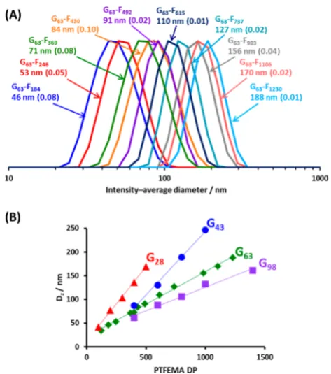
Figure 3.(a) DLS intensity-based size distributions obtained for G63 -Fyparticles prepared at 20% w/w solids via RAFT aqueous emulsion
polymerization of TFEMA at 70°C. (b) Linear correlation between the DLS intensity-average particle diameter and the mean degree of polymerization (DP) of the PTFEMA core-forming block.
ization. Thus, our hypothesis is that the relatively low solubility
of TFEMA monomer in water (as opposed to ethanol) leads to
reduced solvation of the growing PTFEMA chains during PISA.
Taking into account the e
ff
ect of polydispersity and the steric
stabilizer layer thickness, the mean number-average particle
diameters calculated from TEM studies were in fairly good
agreement with DLS studies (see
Table 1
). Again, it was
observed that, for a given PGMA DP, increasing the target
PTFEMA DP produced progressively larger nanoparticles.
Core
−
Shell Particle Density.
The density of core
−
shell
particles,
ρ
particle, can be described by the relationship
ρ ρ ρ
ρ ρ
= +
= + + −
+
V V
V
R R T R
R T
[( ) ]
( )
particle
core core shell shell
particle
core core
3
shell core shell
3 core
3
core shell 3 (1)
where
ρ
coreand
V
corerepresent the density and volume of the
core component,
ρ
shelland
V
shellrepresent the density and
volume of the shell component, and
V
particleis the overall
volume of the particle.
For sterically stabilized nanoparticles comprising a
solvent-free PTFEMA core with
ρ
core= 1.47 g cm
−3and a highly
hydrated PGMA shell such that
ρ
shell≈
1.00 g cm
−3,
eq 1
was
used to calculate
ρ
particleas a function of the core radius (
R
core)
for various (assumed) shell thicknesses
T
shell(see
Figure 5
).
For
R
core≤
100 nm with a
T
shellof between 2.5 and 15 nm,
ρ
particleis strongly dependent on
R
core. However, for particles
with a su
ffi
ciently large
R
corewith respect to
T
shell, there is a
plateau region for which
ρ
particleis no longer strongly dependent
on
R
core. It is also evident that the shell thickness has a strong
in
fl
uence on the particle density, especially when the core
radius is relatively small (
R
core≤
100 nm). Finally, it is noted
that RAFT aqueous emulsion polymerization provides
convenient access to a wide range of well-de
fi
ned nanoparticles
for which
R
core≤
110 nm.
In order to calculate the actual range of e
ff
ective particle
densities for the G
x-F
yparticles discussed herein, it is important
to obtain experimental values of
V
coreand
V
shell(and hence
R
coreand
T
shell). In principle, this information can be obtained by
determining the di
ff
erence between the intensity-average
hydrodynamic diameter reported by DLS for the hydrated
nanoparticles in solution and the number-average diameter
calculated from TEM analysis of the dried nanoparticles.
However, in practice, this approach is unsatisfactory because
DLS and TEM are biased toward di
ff
erent moments of the
particle size distribution. Thus, SAXS, which is a much more
statistically robust and rigorous technique, was used in order to
determine the required structural information for these G
x-F
yparticles.
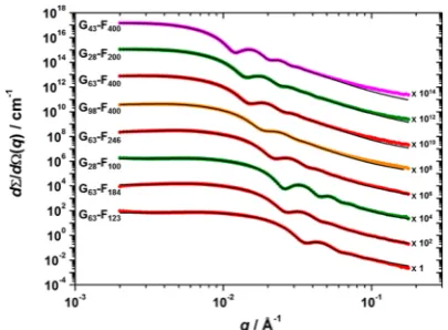
Small-Angle X-ray Scattering.
SAXS patterns were
recorded for 1.0% w/w dispersions of the G
x-F
ycopolymer
nanoparticles.
Figure S4
shows the radially integrated patterns
expressed as the scattering intensity vs the scattering vector,
q
.
In all cases, the gradient of the scattering patterns at low
q
(Guinier region) is approximately zero, supporting the spherical
particle morphology observed by TEM studies (
Figure 4
). The
semi-
fl
uorinated PTFEMA core-forming block has a relatively
high scattering length density (
ξ
PTFEMA= 12.76
×
10
10cm
−2)
compared to the highly hydrated PGMA shell (
ξ
PGMA= 11.94
×
10
10cm
−2,
ξ
water= 9.42
×
10
10cm
−2), so the X-ray scattering is
dominated by the former component. The position of the
fi
rst
minimum in each pattern associated with the particle form
factor is inversely proportional to particle radius; as expected,
this feature shifts to lower
q
for larger particles (higher
PTFEMA DPs). It is also noteworthy that in most cases three
or four minima are observed. This indicates relatively narrow
particle size distributions and suggests that the
q
range chosen
is appropriate for characterizing these nanoparticles.
The scattering intensity resulting from the PGMA chain/
water shells at high
q
is relatively weak in comparison to the
PTFEMA cores. Furthermore, when
fi
tting scattering data it is
important to minimize the number of adjustable parameters in
any given model.
77Thus, the radius of gyration (
R
g) for each of
the four PGMA
xhomopolymers dissolved in aqueous solution
was determined by SAXS before modeling the scattering
patterns obtained for the G
x-F
ydiblock copolymer
nano-particles.
To determine
R
gexperimentally, a 1.0% w/w aqueous
solutions of each PGMA
xhomopolymer was analyzed using a
Gaussian coil model (see
Supporting Information
section C).
78The two
fi
tting parameters used for this model are
R
gand
ν
; the
latter corresponds to the excluded volume fraction governed by
the polymer
−
solvent interaction. Scattering patterns and
models are shown in
Figure 6
. As expected, the normalized
scattering intensity depends on the chain length, with the
longest PGMA
x(
x
= 98) producing the greatest normalized
[image:5.625.80.281.69.201.2]scattering intensity. In each case
ν
was
fi
xed at 0.50, which
corresponds to theta solvent conditions. Prediction of the
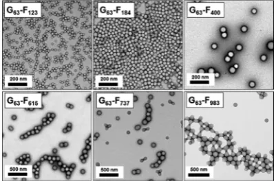
Figure 4. Representative TEM images recorded for G63-Fy diblock [image:5.625.64.297.587.688.2]copolymer nanoparticles prepared by RAFT aqueous emulsion polymerization of TFEMA using a PGMA63 macro-CTA at 20% w/ w solids. A well-defined spherical morphology is observed in each case, with larger particles being obtained when targeting longer core-forming PTFEMA blocks (for a given PGMA block DP).
Figure 5. Relationship between particle density (ρparticle) and core radius (Rcore) for Gx-Fy diblock copolymer nanoparticles of constant
shell thickness (Tshell). The particle density was calculated assuming a PGMA stabilizer shell density of 1.00 g cm−3, a PTFEMA core density of 1.47 g cm−3, and afixed PGMA shell thickness of 2.5, 5.0, 10, or 15 nm.
scattering intensity at low
q
is summarized in section C of the
Supporting Information
. Calculated values correlate well with
the experimental data and support highly hydrated polymer
chains in dilute aqueous solution (see
Table S1
).
R
gvalues of
1.46, 1.75, 2.23, and 2.69 nm were obtained for the four
PGMA
xhomopolymers (where
x
equals 28, 43, 63, or 98,
respectively). Theoretical values of
R
gwere also estimated from
total chain contour lengths and Kuhn length. The contour
length,
L
PGMA, for the PGMA
xblock, is approximately given by
L
PGMA= number of GMA units
×
0.255 nm, where 0.255 nm is
the projected contour length per monomer repeat unit (as
de
fi
ned by two carbon bonds in an
all-trans
conformation). A
Kuhn length of 1.53 nm corresponds to the literature value for
poly(methyl methacrylate).
79Consequently, it follows that
R
g=
(
L
PGMA×
1.53/6)
1/2.
79This approach gave theoretical
R
gvalues
of 1.35, 1.67, 2.03, and 2.53 nm for the four PGMA
homopolymers comprising 28, 43, 63, and 98 GMA monomer
units, respectively. These calculated values are in relatively good
agreement with the experimental values (see
Table S1
).
However, the experimentally determined
R
gvalues are
preferred as no assumptions regarding contour or Kuhn
lengths are required.
In order to model the scattering data obtained for G
x-F
ynanoparticles, the PGMA shell thickness was taken to be equal
to 2
R
g, and the former parameter was assumed to remain
constant for a given PGMA DP, regardless of the PTFEMA DP.
Furthermore, preliminary modeling indicated a mean value for
the solvation of the PTFEMA core (
x
sol) of approximately 0.05,
or just 5% solvent within the PTFEMA cores. This seems
reasonable given the highly hydrophobic character of this block
(its solvent interaction parameter,
χ
H2O, is approximately
7.30).
80Using the aforementioned
R
gand
x
solvalues and a
least-squares
fi
t, a spherical micelle model
78was used to
fi
t
SAXS patterns obtained for a subset of diblock copolymer
nanoparticles comprising a variable PGMA stabilizer DP and a
core-forming PTFEMA DP of up to 400, for which
R
core≤
37
nm (
Figure 7
). A detailed description of the model and
fi
tting
parameters used to analyze these SAXS patterns is given in the
Supporting Information
(see section C and Table S2). It should
be noted that an appropriate structure factor had to be included
in the model in order to obtain reasonably good
fi
ts to the data.
For relatively small nanoparticles (PTFEMA DP
≤
400, or
R
core≤
37 nm), the spherical micelle model produced good
data
fi
ts over the whole
q
range (
Figure 7
). However, for larger
nanoparticles, a systematic deviation between experimental
scattering patterns, and the corresponding data
fi
ts were
observed at high
q
(see
Figure S4
). Despite this technical
problem, the SAXS results obtained for
R
corewere fully
consistent with DLS data shown in
Table 1
. Inspecting
Figure
5
, it is clear that the greatest change in e
ff
ective particle density
occurs for small nanoparticles (
R
core≤
100 nm), and it is
emphasized that the SAXS data
fi
ts are robust in this regime.
Notwithstanding the less satisfactory data
fi
ts obtained for the
larger nanoparticles, SAXS enables
R
coreto be determined with
reasonable accuracy (see following section).
For a
fi
xed PTFEMA DP, both the nanoparticle core radius
and the overall nanoparticle diameter increase when using
shorter PGMA stabilizer blocks. This can be explained by
considering the number of copolymer chains per nanoparticle,
N
agg, which is calculated using the equation
π
= − × ×
N x R
V
(1 ) 4
3
agg sol
core3
chain (2)
where
V
chainis the volume occupied by PTFEMA in a single
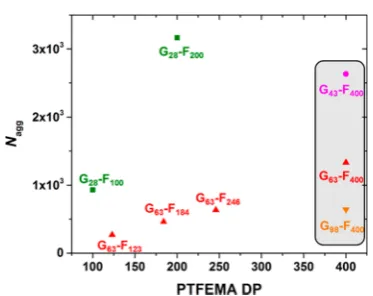
copolymer chain. As the PGMA DP increases for the G
x-F
400nanoparticles,
N
aggis reduced from approximately 2600 to 600
(see gray box in
Figure 8
). This is because longer stabilizer
blocks occupy a larger interfacial area between the nanoparticle
core and shell.
81Moreover, there is a reduction in the
R
core/
L
PTFEMAratio, which provides a measure of the degree of chain
coiling within the core (here
L
PTFEMAis estimated from the
trans
C
−
C bond length assuming a fully stretched chain).
R
core/
L
PTFEMAis reduced from 0.36 for nanoparticles stabilized using
G
43to 0.29 and 0.23 for G
63and G
98, respectively. This suggests
that for longer PGMA stabilizer chains, which produce
nanoparticles with lower aggregation numbers, the hydrophobic
PTFEMA chains are more compact within the (smaller)
nanoparticle cores.
[image:6.625.70.292.64.230.2]For a given PGMA DP,
N
aggincreases as the DP of PTFEMA
becomes larger (
Figure 7
). This observation correlates well
with the monotonic increase in intensity-average diameter
indicated by DLS studies; hence, particle growth is a
Figure 6.Small-angle X-ray scattering patterns recorded for 1.0% w/waqueous solutions of PGMAx homopolymer chains. Solid lines
[image:6.625.343.546.67.216.2]representfits to the data using a Gaussian coil model (see Table S1 and section C in theSupporting Information). TheRgvalues obtained from this model using ν = 0.5 are given, and the numbers in parentheses refer to the theoretical values.
Figure 7. Selected small-angle X-ray scattering patterns (colored circles) recorded for 1.0% w/w aqueous solutions of Gx-Fy
nanoparticles at 20 °C. Solid black lines represent fits to the data using a spherical micelle model.78
consequence of both the greater PTFEMA chain length and a
larger number of copolymer chains per particle.
Calculation and Implications of E
ff
ective Particle
Density.
For the data sets shown in
Figure S4
, the SAXS
patterns between
q
= 0.005 Å
−1and
q
= 0.05 Å
−1were used to
calculate the mean core radius,
R
core, for each nanoparticle
dispersion using the spherical micelle model. Combining this
information with the
R
gdata obtained by SAXS analysis of the
corresponding PGMA stabilizer chains in aqueous solution
enabled the e
ff
ective particle density,
ρ
particle, to be determined
using
eq 1
(see
Figure 9
).
In this analysis,
ρ
corewas taken to be 1.45 g cm
−3(i.e., 5%
solvent is assumed within the nanoparticle cores, as indicated
from SAXS data
fi
ts) and
ρ
shellwas assumed to be 1.00 g cm
−3(we estimate that the volume fraction of the PGMA chains
within the stabilizer shell does not exceed 0.01). Thus, it is clear
that the e
ff
ective particle density,
ρ
particle, of these sterically
stabilized G
x-F
ynanoparticles depends markedly on the precise
x
and
y
values and varies from 1.19 g cm
−3(for G
63-F
123) up to
approximately 1.41 g cm
−3as the core radius approaches 80
nm.
It is also noteworthy that over the same core size range the
G
28series (
T
shell= 2.92 nm) have e
ff
ective particle densities
which are consistently higher than particles stabilized by G
98(
T
shell= 5.38 nm). Such drastic changes in e
ff
ective density over
a relatively narrow range of particle compositions and diameters
can have important implications when conducting particle size
analyses using certain commercial instruments.
For example, DCP is a widely used, high-resolution particle
sizing technique that has been used to characterize a wide range
of colloidal particles including copolymer latexes,
82−87viruses,
88−91colloidal nanocomposites,
50,92−100protein-coated
particles,
101and various inorganic nanoparticles.
35,102−109DCP
is based on the principle of centrifugal sedimentation: particles
are radially fractionated within a rotating disk according to their
size and relative density; i.e., for particles with uniform density,
large particles sediment more quickly than small particles. For
calculating accurate particle size distributions using DCP, the
e
ff
ective particle density is an essential input parameter.
Accordingly, weight-average particle size distributions were
determined by DCP for the G
x-F
ynanoparticles discussed
herein (see
Figure S5
). The e
ff
ective particle densities used to
determine these particle size distributions were calculated from
SAXS analysis (see
Figure 9
). In most cases, these size
distributions are relatively narrow and the trend in
mean-particle diameter agrees well with the DLS, TEM, and SAXS
diameters. In addition, there is no evidence of
fl
occulation in
these particle size distributions; Balmer and co-workers have
recently shown that DCP is very sensitive to such incipient
aggregation.
95 [image:7.625.87.274.62.210.2]In order to illustrate the importance of using an accurate
particle density for DCP analysis,
Figure 10
shows an example
of a particle size distribution determined for G
63-F
184nanoparticles using the solid-state density of PTFEMA (1.47
g cm
−3, blue line). When compared to the particle size
Figure 8.Relationship between aggregation number (Nagg) andcore-forming block DP for selected Gx-Fy nanoparticles prepared using
PGMA28(■), PGMA43(●), PGMA63(▲), and PGMA98(▼) macro-CTAs.
Figure 9. Effective particle densities (ρparticle) calculated for Gx-Fy
nanoparticles using structural parameters derived from SAXS analysis. The weak solvation of the core-forming PTFEMA block indicated by SAXS was taken into account (effectiveρcore= 1.45 g cm−3) andρshell for the highly hydrated shell was taken to be that of water (1.00 g
cm−3). Figure 10.Weight-average particle size distributions determined by disk centrifuge photosedimentometry (DCP) for G63-F184 nano-particles. The blue curve shows theerroneoussize distribution obtained for G63-F184nanoparticles when an upper limit density of 1.47 g cm−3 (which corresponds to the solid-state density of dry PTFEMA homopolymer) is used for DCP analysis. The black curve shows the
corrected particle size distribution obtained when a single effective
particle density is used (1.23 g cm−3). The red curve is thetrueparticle size distribution recalculated to account for thedensity distributionthat is superimposed on the particle size distribution (see Table 2). However, the latter refinement becomes negligible for relatively large Gx-Fynanoparticles (seeFigure S6).
[image:7.625.64.298.400.591.2] [image:7.625.328.556.474.628.2]distribution determined using a corrected e
ff
ective particle
density (black line), it is clear that the former erroneous size
distribution substantially underestimates the mean diameter of
the G
63-F
184nanoparticles.
At this point it is perhaps worth noting that analytical
centrifugation techniques have previously been employed to
determine e
ff
ective particle densities for various nanoparticles
by applying Stokes
’
law to determine particle velocities in media
of di
ff
ering densities.
91,107However, in the present case this
approach would not account for the change in density of the
stabilizer shell, since this largely comprises the spin
fl
uid (or
continuous phase). As a consequence,
ρ
particleis not constant
and depends on the spin
fl
uid density. Thus, we
fi
nd it more
appropriate to use the calculated e
ff
ective densities based on
SAXS analysis rather than relying on the former techniques.
An inherent assumption made during DCP analysis is that all
particles are of equal density. In reality, the stabilizer shell
thickness is essentially constant, but there is some variation in
the nanoparticle core diameter, as con
fi
rmed by the TEM
images shown in
Figure 4
. Consequently, larger particles
possess slightly higher densities and so a density distribution is
imposed on the particle size distribution. Interestingly, this
density distribution for sterically stabilized nanoparticles
(comprising low density shells and high density cores) is
complementary
to that previously reported by Fielding et al. for
polystyrene/silica nanocomposite particles (i.e., high density
shells and low density cores).
50In this earlier study, it was
shown that a mathematical method could be employed to
correct for this density distribution, which enabled the raw
DCP data to be reanalyzed in order to calculate true particle
size distributions. Furthermore, these recalculated particle size
distributions were both broader than those reported using a
single density and also more consistent with particle size
distributions reported using other sizing techniques.
Accordingly, a similar approach to that described previously
50was used herein to correct for the density distribution in the
case of a core
−
shell particle morphology in which the
high-density PTFEMA core is of variable diameter and the
low-density PGMA stabilizer shell is of
fi
xed thickness (see
Supporting Information
section D). Speci
fi
cally, absorbance
versus time raw data sets obtained from DCP measurements
were analyzed assuming a
“
best guess
”
particle density (
ρ
) to
calculate an apparent diameter at the time of detection (
D
t).
The resulting
D
tversus time data sets were then reanalyzed
using a model that relates
D
tto the true particle diameter (
D
p)
according to the following equation:
ρ ρ
Δp( /D Tp shell) =4( − )(r+1) = Δ( /D Tt ) 2
particle fluid
2
0 shell 2
(3)
Here
Δ
pis the di
ff
erence between the density of the core
−
shell
particle (
ρ
particle) and that of the spin
fl
uid (
ρ
fluid), and the
density di
ff
erence,
Δ
0, is
ρ
−
ρ
fluid. For particles with a uniform
shell thickness (
T
shell) and a given core radius (
R
core),
ρ
particlecan be given by simplifying
eq 1
as follows:
ρ = ρ + ρ + −
+
r r r
r
[( 1) ]
( 1)
particle core
3 shell
3 3
3
(4)
where
ρ
coreand
ρ
shellare the densities of the core and shell,
respectively, and
r
is the dimensionless variable
=
r Rcore/Tshell (5)
Substituting
eq 4
into
eq 3
yields a cubic equation (see
Supporting Information
eq D5) that can be solved to give a
physically realistic
D
pvalue for every
D
tcalculated during the
original DCP measurement. This model is actually less complex
than that reported previously because it leads to a
cubic
equation, rather than the
quintic
equation derived earlier.
50The
additional complexity of the earlier model arises from the need
to account for the particulate nature of the shell.
50A
FORTRAN77 program (see section E in the
Supporting
Information
) was written in order to solve the cubic equation
(
eq D5
) for its single real positive root and hence recalculate
the true weight-average particle size distributions for a given set
of G
x-F
ydata obtained by DCP.
Figure 10
shows the DCP data for the G
63-F
184particles (for
which SAXS indicates an
R
coreof 16 nm, see entry 1 in
Table 2
).
As discussed above, the DCP trace obtained when using a
particle density of 1.47 g cm
−3(blue line) clearly undersizes
these nanoparticles when compared to the corresponding
TEM, DLS, and SAXS data. A more realistic particle size
distribution is reported when using an appropriate e
ff
ective
particle density of 1.23 g cm
−3(black line). The red trace shows
the particle size distribution obtained when the data has been
recalculated to account for the superimposed density
distribution. As expected, the recalculated distribution is
narrower
than that determined using a single-value e
ff
ective
particle density. However, this e
ff
ect is only signi
fi
cant for
smaller nanoparticles, where the volume fraction of the
hydrated PGMA stabilizer layer is relatively high, leading to a
more pronounced variation in the particle density (
Figure 9
).
Figure S6
shows that as the nanoparticle mean diameter
increases, the recalculation becomes less signi
fi
cant, and
Table
2
summarizes the di
ff
erences in the reported weight-average
diameters along with SAXS data and particle densities for
comparison (i.e., a subset of those shown in graph C of
Figure
S5
). In principle, this correction will also be negligible for
highly monodisperse particles, since there is minimal variation
in the nanoparticle core volume in this case.
The above technical solution to the problem of a
superimposed density distribution for core
−
shell particles
comprising high-density cores and low-density shells has been
formulated for a model system of sterically stabilized diblock
copolymer nanoparticles. However, the approach is generic and
hence is expected to be useful for various colloidal dispersions
reported in the literature, including sterically stabilized gold
Table 2. Summary of E
ff
ective Particle Densities and Particle
Diameters Determined by Both SAXS and DCP for
PGMA63
−
PTFEMA
yDiblock Copolymer Nanoparticles
Prepared via RAFT Aqueous Emulsion Polymerization
aparticle diameter (nm)
diblock copolymer composition
effective particle density,ρparticle
(g cm−3)
SAXS (2Rcore+
4Rg)
DCP using ρparticle
DCP using ρparticle
distribution
G63-F184 1.23 41±4 45±6 43±4
G63-F430 1.32 72±8 72±8 72±7
G63-F615 1.35 101±10 101±12 101±11
G63-F1106 1.39 157±12 146±16 147±14 aThe DCP particle diameters were determined using both a single effective particle density (ρparticle) and also by superimposing an
nanoparticles
108−111and sterically stabilized magnetite
sols,
112−116both of which are used for biomedical applications.
■
CONCLUSIONS
Four series of PGMA
x−
PTFEMA
ydiblock copolymers were
prepared using RAFT aqueous emulsion polymerization. Very
high conversions (typically >99%) were achieved, as judged by
19F NMR spectroscopy analysis. These diblock copolymers
exhibited narrow, unimodal molecular weight distributions as
judged by UV GPC analysis. Self-assembly in solution is driven
by the
in situ
growth of the highly hydrophobic PTFEMA
block, yielding sterically stabilized spherical nanoparticles with
relatively narrow size distributions, as con
fi
rmed by TEM
studies. Judicious variation of the PGMA
x−
PTFEMA
ydiblock
composition allowed the mean nanoparticle diameter to be
controlled over a relatively wide range, from
∼
30 to
∼
250 nm.
For a
fi
xed DP of the hydrophilic PGMA stabilizer, a
monotonic increase in particle diameter was observed on
increasing the DP of the core-forming PTFEMA block. On the
other hand, a substantial reduction in particle diameter was
observed for PGMA
x−
PTFEMA
400nanoparticles on increasing
the PGMA stabilizer DP (or
x
). SAXS analysis indicated a
corresponding smaller mean number of copolymer chains per
spherical nanoparticle,
N
agg.
The radius of gyration,
R
g, of the PGMA
xprecursor chains in
aqueous solution was calculated theoretically and also
determined experimentally via SAXS. The latter value was
subsequently used as a
fi
xed parameter (along with
x
sol) when
modeling SAXS patterns recorded for PGMA
x−
PTFEMA
ydiblock copolymer nanoparticles in aqueous solution. This
approach enabled calculation of e
ff
ective particle densities for
these model sterically stabilized nanoparticles, which is an
essential parameter for reliable particle size analysis via
analytical centrifugation. As expected, a signi
fi
cant increase in
e
ff
ective particle density was observed as the mole fraction of
the high-density PTFEMA core component was increased. This
model system was designed to enable the determination of the
e
ff
ective particle density and stabilizer layer thickness for
sterically stabilized diblock copolymer nanoparticles. SAXS was
then utilized to determine the volume-average diameter,
N
agg,
and stabilizer shell thickness. These structural parameters were
used to calculate an e
ff
ective particle density, which enabled
high resolution particle size analysis to be conducted for this
model system via disk centrifuge photosedimentometry. Finally,
the resulting particle size distributions were corrected for the
superimposed density distribution that is an intrinsic feature of
such core
−
shell nanoparticles. This led to narrower size
distributions, and this correction is expected to be applicable
to other colloidal dispersions reported in the literature.
■
ASSOCIATED CONTENT
*
S Supporting InformationThe Supporting Information is available free of charge on the
ACS Publications website
at DOI:
10.1021/acs.macro-mol.6b00987
.
19
F NMR spectra of TFEMA monomer and PTFEMA
homopolymer; gel permeation chromatograms of
PGMA
63homopolymer and PGMA
63−
PTFEMA
yco-polymers; TEM images of PGMA
x−
PTFEMA
yparticles;
small-angle X-ray scattering patterns,
fi
tting parameters
and model descriptions for PGMA
xhomopolymers and
PGMA
x−
PTFEMA
yparticles; particle size distributions
determined by disk centrifuge photosedimentometry;
derivation of equation and FORTRAN77 program used
to correct disk centrifuge data (
)
■
AUTHOR INFORMATION
Corresponding Authors
*
lee.
fi
[email protected]
(L.A.F.).
*
s.p.armes@she
ffi
eld.ac.uk
(S.P.A.).
Present AddressB.A.: Department of Chemistry, Imperial College London,
South Kensington Campus, London SW7 2AZ, UK.
Notes
The authors declare no competing
fi
nancial interest.
■
ACKNOWLEDGMENTS
S.P.A. is the recipient of a
fi
ve-year ERC Advanced Investigator
grant (PISA 320372). EPSRC is thanked for a Platform grant
(EP/J007846/1). L.A.F. thanks EPSRC for postdoctoral
support (EP/J018589/1). We are grateful to Diamond Light
Source for providing synchrotron beam-time (SM10237) and
thank the personnel of I22 for their assistance.
■
REFERENCES
(1) Napper, D. H. Steric stabilization.J. Colloid Interface Sci.1977,58
(2), 390−407.
(2) Napper, D. H. Polymeric Stabilization of Colloidal Dispersions; Academic Press: London, 1983.
(3) Hunter, R. J.Foundations of Colloid Science; Oxford University Press: Oxford, 2000.
(4) Lascelles, S. F.; Malet, F.; Mayada, R.; Billingham, N. C.; Armes, S. P. Latex syntheses using novel tertiary amine methacrylate-based macromonomers prepared by oxyanionic polymerization. Macro-molecules1999,32(8), 2462−2471.
(5) Almog, Y.; Reich, S.; Levy, M. Monodisperse polymeric spheres in the micron size range by a single step process.Br. Polym. J.1982,14
(4), 131−136.
(6) Ober, C. K.; Lok, K. P.; Hair, M. L. Monodispersed, micron-sized polystyrene particles by dispersion polymerization.J. Polym. Sci., Polym. Lett. Ed.1985,23(2), 103−108.
(7) Tseng, C. M.; Lu, Y. Y.; Elaasser, M. S.; Vanderhoff, J. W. Uniform polymer particles by dispersion polymerization in alcohol.J. Polym. Sci., Part A: Polym. Chem.1986,24(11), 2995−3007.
(8) Paine, A. J.; Luymes, W.; McNulty, J. Dispersion polymerization of styrene in polar-solvents 0.6. Influence of reaction parameters on particle-size and molecular-weight in poly(n-vinylpyrrolidone)-stabi-lized reactions.Macromolecules1990,23(12), 3104−3109.
(9) Shen, S.; Sudol, E. D.; Elaasser, M. S. Control of particle-size in dispersion polymerization of methyl-methacrylate.J. Polym. Sci., Part A: Polym. Chem.1993,31(6), 1393−1402.
(10) Baines, F. L.; Dionisio, S.; Billingham, N. C.; Armes, S. P. Use of block copolymer stabilizers for the dispersion polymerization of styrene in alcoholic media.Macromolecules1996,29(9), 3096−3102. (11) Ali, A. M. I.; Pareek, P.; Sewell, L.; Schmid, A.; Fujii, S.; Armes, S. P.; Shirley, I. M. Synthesis of poly(2-hydroxypropyl methacrylate) latex particles via aqueous dispersion polymerization.Soft Matter2007,
3(8), 1003−1013.
(12) Barrett, K. E. J.; Thomas, H. R. Kinetics of dispersion polymerization of soluble monomers.I. Methyl methacrylate.J. Polym. Sci., Part A-1: Polym. Chem.1969,7(9PA1), 2621−2650.
(13) Dawkins, J. V.; Taylor, G. Non-aqueous poly(methyl methacrylate) dispersions - radical dispersion polymerization in the presence of ab block copolymers of polystyrene and poly(dimethyl siloxane).Polymer1979,20(5), 599−604.
methacrylate) lattices in nonaqueous media.Colloids Surf. 1986,17
(1), 67−78.
(15) Stejskal, J.; Kratochvil, P.; Konak, C. Structural parameters of spherical-particles prepared by dispersion polymerization of methyl-methacrylate.Polymer1991,32(13), 2435−2442.
(16) Jenkins, A. D.; Maxfield, D.; Dossantos, C. G.; Walton, D. R. M.; Stejskal, J.; Kratochvil, P. Enolate-initiated dispersion polymerization.
Makromol. Chem., Rapid Commun.1992,13(1), 61−63.
(17) Awan, M. A.; Dimonie, V. L.; ElAasser, M. S. Anionic dispersion polymerization of styrene 0.2. Mechanism of particle formation. J. Polym. Sci., Part A: Polym. Chem.1996,34(13), 2651−2664.
(18) Awan, M. A.; Dimonie, V. L.; ElAasser, M. S. Anionic dispersion polymerization of styrene 0.1. Investigation of parameters for preparation of uniform micron-size polystyrene particles with narrow molecular weight distribution. J. Polym. Sci., Part A: Polym. Chem. 1996,34(13), 2633−2649.
(19) Richez, A. P.; Yow, H. N.; Biggs, S.; Cayre, O. J. Dispersion polymerization in non-polar solvent: Evolution toward emerging applications.Prog. Polym. Sci.2013,38(6), 897−931.
(20) Fielding, L. A.; Derry, M. J.; Ladmiral, V.; Rosselgong, J.; Rodrigues, A. M.; Ratcliffe, L. P. D.; Sugihara, S.; Armes, S. P. RAFT dispersion polymerization in non-polar solvents: facile production of block copolymer spheres, worms and vesicles in n-alkanes.Chemical Science2013,4(5), 2081−2087.
(21) Richez, A. P.; Farrand, L.; Goulding, M.; Wilson, J. H.; Lawson, S.; Biggs, S.; Cayre, O. J. Dispersion Polymerization of Poly-(dimethylsiloxane)-Stabilized Polymer Particles from Radical Dis-persion Polymerization in Nonpolar Solvent: Influence of Stabilizer Properties and Monomer Type.Langmuir2014,30(5), 1220−1228. (22) DeSimone, J. M.; Maury, E. E.; Menceloglu, Y. Z.; McClain, J. B.; Romack, T. J.; Combes, J. R. Dispersion polymerizations in supercritical carbon-dioxide.Science1994,265(5170), 356−359.
(23) Hsiao, Y. L.; Maury, E. E.; DeSimone, J. M.; Mawson, S.; Johnston, K. P. Dispersion polymerization of methyl-methacrylate stabilized with poly(1,1-dihydroperfluorooctyl acrylate) in supercritical carbon-dioxide.Macromolecules1995,28(24), 8159−8166.
(24) Shaffer, K. A.; Jones, T. A.; Canelas, D. A.; DeSimone, J. M.; Wilkinson, S. P. Dispersion polymerizations in carbon dioxide using siloxane-based stabilizers.Macromolecules1996,29(7), 2704−2706.
(25) Giles, M. R.; Griffiths, R. M. T.; Aguiar-Ricardo, A.; Silva, M.; Howdle, S. M. Fluorinated graft stabilizers for polymerization in supercritical carbon dioxide: The effect of stabilizer architecture.
Macromolecules2001,34(1), 20−25.
(26) Wang, W. X.; Griffiths, R. M. T.; Naylor, A.; Giles, M. R.; Irvine, D. J.; Howdle, S. M. Preparation of cross-linked microparticles of poly(glycidyl methacrylate) by dispersion polymerization of glycidyl methacrylate using a PDMS macromonomer as stabilizer in super-critical carbon dioxide.Polymer2002,43(25), 6653−6659.
(27) Lee, H.; Terry, E.; Zong, M.; Arrowsmith, N.; Perrier, S.; Thurecht, K. J.; Howdle, S. M. Successful dispersion polymerization in supercritical CO2 using polyvinylalkylate hydrocarbon surfactants synthesized and anchored via RAFT.J. Am. Chem. Soc.2008,130(37), 12242−12243.
(28) Minami, H.; Yoshida, K.; Okubo, M. Preparation of polystyrene particles by dispersion polymerization in an ionic liquid. Macromol. Rapid Commun.2008,29(7), 567−572.
(29) Minami, H.; Kimura, A.; Kinoshita, K.; Okubo, M. Preparation of poly(acrylic acid) particles by dispersion polymerization in an ionic liquid.Langmuir2010,26(9), 6303−6307.
(30) Barrett, K. E. J. Dispersion Polymerization in Organic Media; Wiley: 1974.
(31) Cesarano, J.; Aksay, I. A. Processing of highly concentrated aqueous alpha-alumina suspensions stabilized with poly-electrolytes.J. Am. Ceram. Soc.1988,71(12), 1062−1067.
(32) Allami, H. S.; Billingham, N. C.; Calvert, P. D. Controlled structure methacrylic copolymers as dispersants for ceramics processing.Chem. Mater.1992,4(6), 1200−1207.
(33) Chen, Z. C.; Ring, T. A.; Lemaitre, J. Stabilization and processing of aqueous BaTiO3 suspension with polyacrylic-acid.J. Am. Ceram. Soc.1992,75(12), 3201−3208.
(34) Bhattacharjee, S.; Paria, M. K.; Maiti, H. S. Polyvinyl butyral as a dispersant for barium-titanate in a nonaqueous suspension.J. Mater. Sci.1993,28(23), 6490−6495.
(35) Vamvakaki, M.; Billingham, N. C.; Armes, S. P.; Watts, J. F.; Greaves, S. J. Controlled structure copolymers for the dispersion of highperformance ceramics in aqueous media.J. Mater. Chem.2001,11
(10), 2437−2444.
(36) Magdassi, S.; Bassa, A.; Vinetsky, Y.; Kamyshny, A. Silver nanoparticles as pigments for water-based ink-jet inks.Chem. Mater. 2003,15(11), 2208−2217.
(37) Won, Y. Y.; Meeker, S. P.; Trappe, V.; Weitz, D. A.; Diggs, N. Z.; Emert, J. I. Effect of temperature on carbon-black agglomeration in hydrocarbon liquid with adsorbed dispersant.Langmuir2005,21(3), 924−932.
(38) Growney, D. J.; Mykhaylyk, O. O.; Armes, S. P. Micellization and adsorption behavior of a near-monodisperse polystyrene-based diblock copolymer in nonpolar media.Langmuir2014,30(21), 6047− 6056.
(39) Growney, D. J.; Mykhaylyk, O. O.; Derouineau, T.; Fielding, L. A.; Smith, A. J.; Aragrag, N.; Lamb, G. D.; Armes, S. P. Star diblock copolymer concentration dictates the degree of dispersion of carbon black particles in nonpolar media: Bridging flocculation versus steric stabilization.Macromolecules2015,48(11), 3691−3704.
(40) Stolnik, S.; Dunn, S. E.; Garnett, M. C.; Davies, M. C.; Coombes, A. G. A.; Taylor, D. C.; Irving, M. P.; Purkiss, S. C.; Tadros, T. F.; Davis, S. S.; Illum, L. Surface modification of poly(lactide-co-glycolide) nanospheres by biodegradable poly(lactide)-poly(ethylene glycol) copolymers.Pharm. Res.1994,11(12), 1800−1808.
(41) Feng, W.; Zhu, S. P.; Ishihara, K.; Brash, J. L. Adsorption of fibrinogen and lysozyme on silicon grafted with poly(2-methacryloy-loxyethyl phosphorylcholine) via surface-initiated atom transfer radical polymerization.Langmuir2005,21(13), 5980−5987.
(42) Feng, W.; Brash, J. L.; Zhu, S. P. Non-biofouling materials prepared by atom transfer radical polymerization grafting of 2-methacryloloxyethyl phosphorylcholine: Separate effects of graft density and chain length on protein repulsion.Biomaterials2006,27
(6), 847−855.
(43) Jiang, S. Y.; Cao, Z. Q. Ultralow-fouling, functionalizable, and hydrolyzable zwitterionic materials and their derivatives for biological applications.Adv. Mater.2010,22(9), 920−932.
(44) Zhang, L.; Cao, Z.; Bai, T.; Carr, L.; Ella-Menye, J.-R.; Irvin, C.; Ratner, B. D.; Jiang, S. Zwitterionic hydrogels implanted in mice resist the foreign-body reaction.Nat. Biotechnol.2013,31(6), 553−556.
(45) Alswieleh, A. M.; Cheng, N.; Canton, I.; Ustbas, B.; Xue, X.; Ladmiral, V.; Xia, S.; Ducker, R. E.; El Zubir, O.; Cartron, M. L.; Hunter, C. N.; Leggett, G. J.; Armes, S. P. Zwitterionic poly(amino acid methacrylate) brushes.J. Am. Chem. Soc.2014,136(26), 9404− 9413.
(46) Reed, K. M.; Borovicka, J.; Horozov, T. S.; Paunov, V. N.; Thompson, K. L.; Walsh, A.; Armes, S. P. Adsorption of sterically stabilized latex particles at liquid surfaces: Effects of steric stabilizer surface coverage, particle size, and chain length on particle wettability.
Langmuir2012,28(18), 7291−7298.
(47) Amalvy, J. I.; Unali, G. F.; Li, Y.; Granger-Bevan, S.; Armes, S. P.; Binks, B. P.; Rodrigues, J. A.; Whitby, C. P. Synthesis of sterically stabilized polystyrene latex particles using cationic block copolymers and macromonomers and their application as stimulus-responsive particulate emulsifiers for oil-in-water emulsions.Langmuir2004,20
(11), 4345−4354.
(48) Detloff, T.; Sobisch, T.; Lerche, D. Particle size distribution by space or time dependent extinction profiles obtained by analytical centrifugation (concentrated systems).Powder Technol.2007,174(1− 2), 50−55.
(49) Planken, K. L.; Cölfen, H. Analytical ultracentrifugation of colloids.Nanoscale2010,2(10), 1849−1869.