i
A Dissertation on
“A STUDY ON EVALUATION OF PLASMA D-DIMER AS AN INITIAL DIAGNOSTIC BIOMARKER IN
CEREBRAL VENOUS THROMBOSIS” Dissertation submitted to
THE TAMILNADU Dr.M.G.R. MEDICAL UNIVERSITY CHENNAI – 600 032
With partial fulfillment of the regulations For the award of the degree of
M.D. GENERAL MEDICINE BRANCH – I
Department of General Medicine Coimbatore Medical College Hospital
iii
CERTIFICATE
This is to certify that the dissertation “A STUDY ON EVALUATION OF PLASMA D-DIMER AS AN INITIAL DIAGNOSTIC BIOMARKER IN CEREBRAL VENOUS THROMBOSIS” is a bonafide research work done by Dr.M.Ananthi Post graduate in M.D. General Medicine under my direct guidance and supervision to my satisfaction , in partial fulfillment of the requirements for the degree of M.D. General Medicine.
Date : Professor & Unit chief M-6
Date : Professor & Head Of The Department Department of General Medicine
vi
DECLARATION
I hereby declare that this dissertation entitled “A STUDY ON EVALUATION OF PLASMA D-DIMER AS AN INITIAL DIAGNOSTIC BIOMARKER IN CEREBRAL VENOUS THROMBOSIS” is a bonafide and genuine research work carried out by me under the guidance of Prof. Dr. Isaac Christian Moses M.D., Department of Medicine ,Coimbatore medical college, Coimbatore .
Date :
viii
ACKNOWLEDGEMENT
With great reverence, first of all ,I would like to express my deep sense of gratitude to my guide and teacher, Prof. DR.ISAAC CHRISTIAN MOSES M.D., Professor of Medicine, Coimbatore Medical College, Coimbatore .He has been more than a guide, a good mentor and a source of constant inspiration throughout my course. I also thank him for his able guidance, suggestion and supervision throughout the making of this dissertation.
I would consider it my privilege to extend my respect and gratitude to Prof.DR.KUMAR NATARAJAN M.D., Professor and Head of the Department of Medicine, Coimbatore Medical College, Coimbatore, for all the encouragement and guidance throughout the course and for this dissertation .
I also take this opportunity to thank Prof.Dr.S.Chandrasekaran M.D., Prof. Dr.M.Raveendran M.D., Prof.Dr.N.Sundar M.D., for their moral support and guidance.
ix
I would also like to express my thanks to THE DEAN, Prof.DR.S.REVWATHY M.D.,DGO.,DNB., for her able guidance and encouragement throughout, I am also thankful to the administration and other allied departments of the Coimbatore Medical College for their support in helping me carrying out this study.
I also express my heartfelt sincere thanks to all my fellow postgraduates, for their unconditional support and guidance, and for just being there. No words can express the support extended by my family in all these years; thank you.
My biggest gratitude will always be towards my patients, without whose cooperation this study would have been incomplete. I thank the Almighty for his abundant blessings.
Date :
xiii
ABBREVIATIONS
CVT – Cerebral Venous Thrombosis TB – Tuberculosis
HIV – Human Immuno Deficiency Virus DIC – Disseminated Intravascular Coagulation PNH – Paroxysmal Nocturnal Hemoglobinuria APLA – Anti Phospholipid Antibody Syndrome OCP – Oral Contraceptive Pills
SLE – Systemic Lupus Erythematosis IBD – Inflammatory Bowel Disease SSS – Superior Sagittal Sinus
MRI – Magnetic Resonance Imaging MRV _ Magnetic Resonance Venography CT – Computed tomography
xv
TABLE OF CONTENTS
S.No Title Page no.
1. INTRODUCTION 1
2. AIMS AND OBJECTIVES 3
3. REVIEW OF LITERATURE 4
4. METHODOLOGY 59
5. OBSERVATION AND RESULTS 62
6. DISCUSSION 86
7. SUMMARY 98
8. CONCLUSION 100
9. BIBLIOGRAPHY 101
10. ANNEXURES
i. Proforma 111
ii. Consent Form 115
iii.Key to Master chart 118
xvii
LIST OF TABLES
S .No Table Page no
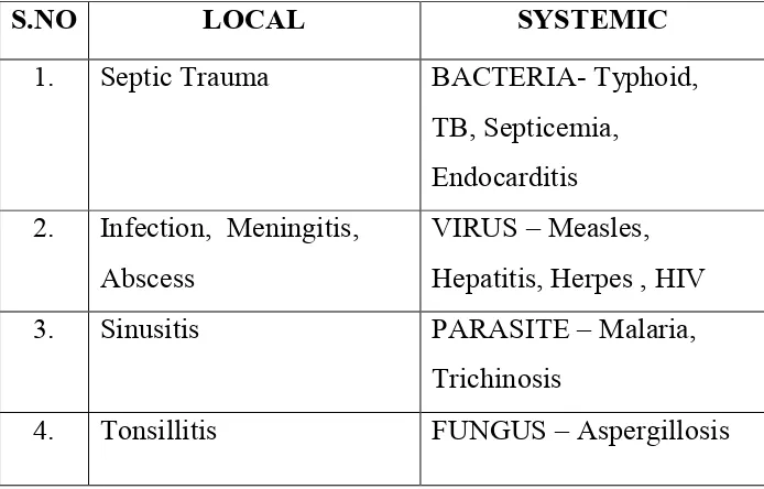
1 Septic Dural Sinus Thrombosis 20
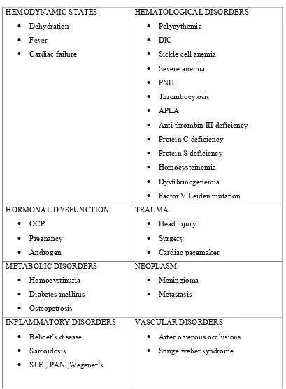
2 Non Septic Dural Sinus Thrombosis 21
3 Genetic Causes Of CVT 23
4 Difference Between CT and MRI/MRV 47 5 Sex Distribution Among Study Population 62 6 Age Distribution Among Study Population 63
7 Clinical Profile of Headache 64
8 Clinical Profile of Convulsions 65
9 Clinical Profile of Vomiting 66
10 Clinical Profile of Loss of Consciousness 67 11 Clinical Profile of Cranial Nerve Involvement 68
12 Profile of Fever 69
13 Spectrum of Papilloedema 70
14 Profile of Focal Deficits 71
15 Sinuses Involved 72
16 D Dimer Distribution 73
17 Spectrum of Double Stranded DNA 74
18 Spectrum of APLA 75
19 Hyperhomocysteinemia In CVT 76
20 Neuro Imaging Studies – CT Brain 77
21 Onset Range 78
xviii
25 Correlation of D Dimer With CT Brain 82 26 Correlation of D Dimer and Onset of Symptoms 83 27 Correlation of D Dimer With MRI/MRV 84 28 Sex Distribution Compared to Other Studies 86 29 Age Distribution Compared to Other Studies 87 30 Comparing Headache with Other Studies 88 31 Comparing Seizures With Other Studies 89 32 Comparing Sensorium With Other Studies 89 33 Comparing Papilloedema With Other Studies 90 34 Comparing Focal Deficits With Other Studies 91 35 Comparing Cranial Nerve Palsy With Other Studies 91 36 Comparing Sinus Involvement in CVT 92 37 Comparing CT Findings With Other Studies 93
38 Comparing APLA With CVT 94
39 Comparing D Dimer and CVT 94
xx
LIST OF GRAPHS
S.No Graphs Page No
1 Frequency of Clinical Manifestations 34 2 Sex Distribution Among the Study Population 62 3 Age Wise Distribution of Cases 63
4 Distribution of Headache 64
5 Distribution of Convulsions 65
6 Clinical Profile of Vomiting 66 7 Clinical Profile of Loss of Consciousness 67
8 Cranial Nerve Involvement 68
9 Profile of Fever 69
10 Spectrum of Papilloedema 70
11 Spectrum of Focal Deficits 71
12 Sinuses Involved 72
13 D Dimer Distribution 73
14 Distribution of ds DNA 74
15 Spectrum of APLA 75
16 Hyperhomocysteinemia in CVT 76
17 CT Brain Abnormality 77
18 Onset Range 78
xxii
LIST OF FIGURES
S.NO FIGURES Page No
1 Anatomy of Cerebral Venous Sinuses 7 2 Anatomy of Superior Sagittal Sinus 8
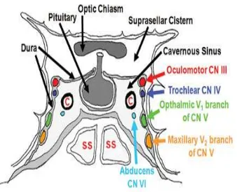
3 Anatomy of Cavernous Sinus 11
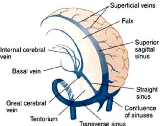
4 Anatomy of cerebral veins 15
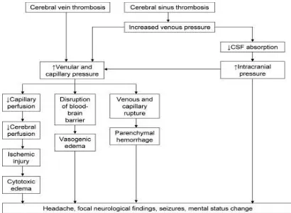
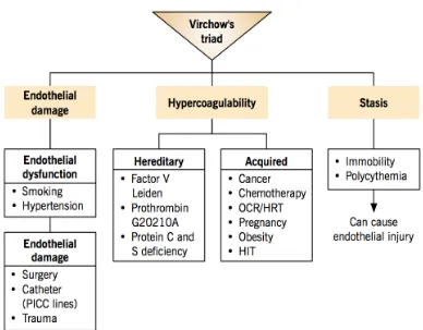
5 Pathogenesis of Thrombosis-Virchow’s Triad 16
6 Pathogenesis of CVT 17
7 Pathology and Etiology 18
8 Pathology of Hemorrhagic Infarct 19
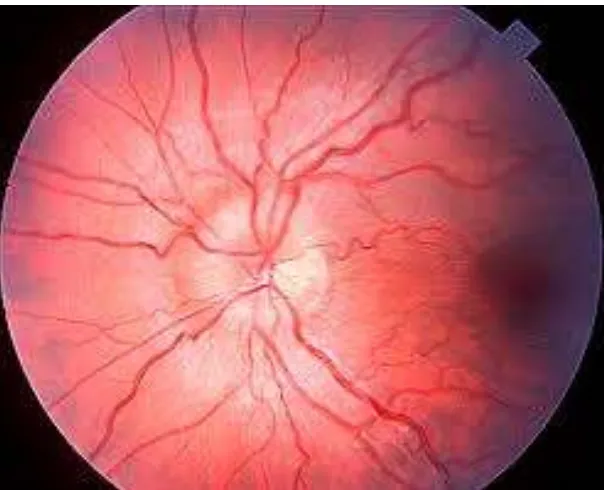
9 Fundus Picture of Papilloedema 33
10 Patterns of Presentation of CVT 36 11 Sinuses and their Clinical Features 37 12 CT Brain Showing Hemorrhagic Infarct 38 13 CT Brain Showing dense Triangle Sign 39 14 CT Brain Demonstrating Empty Delta Sign 40 15 CT Brain Showing Massive Hemorrhagic Infarct 42 16 MRI Brain Showing Transverse Sinus Thrombosis 44
17 MRI – Sigmoid Sinus Thrombosis 45
18 MRV of Left Transverse Sinus Thrombosis 47 19 MRV showing superior sagittal sinus thrombosis 48 20 CT Venography demonstrating the sinuses involved in
CVT
49
21 D Dimer Formation 50
22 D Dimer and CVT Correlation 52
xxiv
ABSTRACT
BACKGROUND :Cerebral venous thrombosis (CVT) is a life threatening condition with varied clinical presentations. It is mainly a diagnosis by means of radiological tool. CT Brain is the initial investigation of choice. It may be normal in 30 – 40 % of the population .MRI with MRV is the gold standard for the diagnosis of CVT. D Dimer is a product of fibrinolysis which is usually used as an exclusion marker of venous thrombosis. Our study aims to analyse the usefulness of D Dimer as an initial diagnostic marker in CVT.
OBJECTIVES:
1. To estimate the level of plasma D-DIMER and its significance in diagnosing Cerebral Venous Thrombosis .
2. To correlate the relationship between plasma D-DIMER and MRI BRAIN in diagnosing Cerebral Venous Thrombosis.
3. To evaluate the diagnostic value of negative D-DIMER in ruling out Cerebral Venous Thrombosis.
METHODOLOGY :
xxv
drawn ,plasma separated and sent to laboratory. D Dimer tests were measured by ELISA test. A value of more than 500 ng/ml were considered positive. All the tests were entered in a data collection sheet in an Excel format and analysed using SPSS software.
RESULTS :
Out of the 50 patients, 28 were females, and 28 were above 35 yrs. The common presenting symptom is headache (96%), followed by seizures, altered sensorium, with papilledema (60%). The most common sinus involved is superior sagittal sinus (70%) followed by sigmoid and transverse sinus. CT Brain was normal in 50 % of the patients. Inherited thrombophilias contribute to 23 %.D Dimer was positive in 78 % of the study population. D Dimer and MRV has a highly significant correlation (0.034). D Dimer and onset of symptoms have a very high significant value of 0.000.
CONCLUSION :
D Dimer can be used as an initial diagnostic bio marker in CVT as there is a significant association with MRI .If patients present very early D Dimer is a highly reliable investigation. Further studies needed for the supportive evidence.
2
INTRODUCTION
Cerebral venous thrombosis (CVT) is a common cerebrovascular
accident due to a multitude of causes. The diagnosis is often missed
because of its varied clinical presentation. The etiological factors are even
more heterogenous making cerebral venous thrombosis a unique clinical
entity .The disease can occur de nova as the first manifestation, or can
overlap on another co existing clinical problem. The occurrence of
thrombus depends upon the site, duration, extent and the rapidity with
which the thrombus occurs. The presentation of CVT is variable in the
form of convulsions, increased intra cranial tension, meningeal infection
or hemorrhage.
Although CVT has its varied presentation, headache is the most
common symptom.1 The diagnosis requires a high index of suspicion. CT Scan may show direct and indirect signs. At times CT may be normal.
MRI with MRV is the diagnostic investigation of choice. The early
diagnosis of this condition will prevent the mortality and morbidity of the
society as it is treatable condition.
Plasma D-Dimer (fibrin degradation product) may be a good
investigation for the diagnosis of CVT. D Dimer has a very high negative
3
pulmonary embolism. Several studies been done for analyzing the value
of D- Dimer in excluding cerebral venous thrombosis. This study aims to
evaluate the usefulness of plasma D-Dimer as an initial diagnostic
5
OBJECTIVES
1. To estimate the level of plasma D-DIMER and its significance in
diagnosing Cerebral Venous Thrombosis .
2. To correlate the relationship between plasma D-DIMER and MRI
BRAIN in diagnosing Cerebral Venous Thrombosis.
3. To evaluate the diagnostic value of negative D-DIMER in ruling out
7
REVIEW OF LITERATURE
HISTORY :
The syndrome of cerebral venous thrombosis was recognized in the
early part of 18th century. Ribes was the first to demonstrate the evidence of cerebral venous thrombosis in 1825.2 John Abercombie of Scotland described this entity in a 24 yr female in 1828.3 Autopsy showed features suggestive of ischemic and hemorrhagic infarct with thrombosed and
sclerosed veins. With the advent of three dimensional flow imaging
modalities, the prevalence of CVT is more common than reported
previously and by early intervention morbidity and mortality has been
reduced drastically.
EPIDEMIOLOGY :
Cerebral venous sinus thrombosis is rare. The incidence in adults is
around 3- 4 cases per million. The major occurrence is in 3 rd decade, out
of which females is around 75 %.4 The use of oral contraceptive pills is the main reason for the disparity between the difference between males
and females. It is more common in the middle east countries, because of
the presence of Behcet’s disease. The incidence of CVT in the west is 3-4
8
ANATOMY OF CEREBRAL VENOUS SYSTEM :6,7
Cerebral venous circulation exhibits following anatomical
characteristics
• Cerebral veins have specific feature that it lacks neither any valves
nor tunica muscularis .The blood flow occurs in various directions
because of the absence of valves. The dilatation of veins is mainly
because of the tunica muscularis.
• There are several inter communications between the cerebral veins,
and because of this only the recovery is either complete or with
minimal residual disability
• Venous sinuses are enclosed between the two layers of duramater.
This is the reason why cerebral veins are not compressed during
raised intra cranial pressure
• There are several emissary veins draining into cerebral veins either
directly or via venous lacunae. The emissary veins include from the
scalp, face, paranasal sinuses, ears, diploic veins and meningeal
veins. This is the reason behind the presence of CVT which occurs
as a complication of infective pathology in catchment areas.
• The superior sagittal sinus receives blood from superficial cortical
veins. The flow is in the opposite direction as that of the sinus thus
9
presence of fibrous septations at the inferior angle of the sinus.
This is why superior sagittal sinus is the most common sinus
involved in CVT .
• Arachnoid villi drain CSF into the superior sagittal sinus .The
occurrence of thrombosis in this sinus causes obstruction of the
CSF flow leading to intracranial hypertension and papilledema .
• Deep cortical veins forms a venous circle around midbrain, like the
arterial circle of willis.
• The cerebral veins drain into the dural sinuses, which then drain
into the two internal jugular veins .
DURAL VENOUS SINUSES:
Cerebral venous system can be classified into two major groups :
1. Superficial system – sagittal sinuses, cortical veins draining
superficial surfaces of both cerebral hemispheres.
2. Deep system – lateral sinus, straight sinus, sigmoid sinus along
10
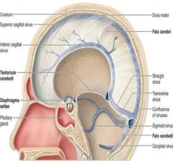
[image:23.612.146.493.64.386.2]
Figure 1: Anatomy of Cerebral Venous Sinuses
I. SUPERIOR SAGITTAL SINUS (SSS) :
o The superior sagittal sinus occupies the upper, attached
margin of the falx cerebri. It is triangular in cross section.
o It begins anteriorly at the crista galli by the union of the tiny
meningeal veins. It communicates with the veins of the
frontal sinus and occasionally with the veins of the nose,
through the foramen caecum.
o It ends near the internal occipital protruberance by turning
to one side, usually the right, and continuous with the right
11
o The junction of all these sinuses is called confluence of
sinuses.
TRIBUTARIES:
Superior cerebral veins
Parietal emissary veins
Venous lacunae
Thrombosis of the SSS may be caused by spread of infection from
the nose, scalp and diploe. This gives rise to rise in intracranial
tension, delirium, convulsions, paraplegia of UMN type.
12
II. INFERIOR SAGITTAL SINUS :
• The inferior sagittal sinus , a channel lies in the posterior
two thirds of the lower, concave margin of the falx cerebri.
• It forms the straight sinus by joining with the great cerebral
veins.
III. STRAIGHT SINUS :
• The straight sinus lies in the median plane within the
junction of falx cerebri and the tentorium cerebelli.
• The inferior sagittal sinus and the great cerebral vein joins to
form the straight sinus. It ends at the internal occipital
protruberance .It continues as the transverse sinus .
• At the termination of the great cerebral veins into the sinus,
there exists a ball valve mechanism , formed by a sinusoidal
plexus of blood vessels ,which regulates the secretion of
CSF.
IV. TRANSVERSE SINUS :
• They are large sinuses..
• The right sinus usually bigger than the left situated in the
posterior part of the attached margin of the tentorium
13
• The superior saggital sinus continues as the right transverse
sinus.
• The straight sinus continues as the left transverse sinus.
• Each sinus extends from the internal occipital protruberance
to the postero inferior angle of the parietal bone at the base
of the mastoid process where it bends and becomes the
sigmoid sinus.
V. SIGMOID SINUS :
• The transverse sinus continues as the sigmoid sinus.
• It is S shaped. It extends from the parietal bone to the
posterior part of the jugular foramen .
• It grooves the mastoid part of the temporal bone .
VI. CAVERNOUS SINUS :
• Each cavernous sinus is a large venous space situated in the
middle cranial fossa .
• It lies on either side of the sphenoid bone.
• Its interior is divided into a number of spaces or caverns by
14
STRUCTURES IN THE LATERAL WALL OF CAVERNOUS
SINUS FROM ABOVE DOWNWARDS:
• Oculomotor nerve
• Trochlear nerve
• Ophthalmic nerve
• Maxillary nerve
STRUCTURES PASSING THROUGH THE CENTRE OF THE
SINUS :
• Internal carotid artery
• Abducent nerve
[image:27.612.146.484.407.681.2]
15 VII. OTHER SINUSES :
• Occipital sinus
• Spheno parietal sinus
• Superior petrosal sinus
• Inferior petrosal sinus
• Basilar plexus of veins
• Petro squamous sinus
• Anterior and posterior intercavernous sinus
GROUPS OF VEINS :
• EXTERNAL CEREBRAL VEINS :
1. SUPERIOR CEREBRAL VEINS :
• These are 6 – 1 2 in number.
• The superolateral surface of the brain is drained by this vein.
• They terminate in the superior sagittal sinus.
2. SUPERFICIAL MIDDLE CEREBRAL VEIN :
• This drains the area around the posterior ramus of lateral
sulcus.
• It terminates in the cavernous sinus.
• It communicates with the superior sagittal , transverse
16
3. DEEP MIDDLE CEREBRAL VEIN :
• The area of insula is drained by this vein.
• It ends in the basal vein.
4. INFERIOR CEREBRAL VEINS :
• They are divided into 2 veins , orbital and tentorial veins.
• The orbital veins terminate in the superior cerebral veins or
in the posterior sagittal sinus.
• The tentorial sinus terminate in the cavernous or any other
surrounding sinus.
5. ANTERIOR CEREBRAL VEINS :
• The corpus callosum and the medial surface of cerebral
hemisphere is drained by these veins and they ends in the
basal vein .
• INTERNAL CEREBRAL VEINS :
• There is one vein on each side .
• It is formed by the union of the thalamostriate and chordal
17
• The right and left veins run posteriorly parallel to each other
in the tela choroidea and unite together to form the great
cerebral vein below the splenium of the corpus callosum .
• TERMINAL VEINS :
1. GREAT CEREBRAL VEIN :
• This is a single median vein. The two internal cerebral veins joined
to form the great cerebral vein. It terminates in the straight sinus.
2. BASAL VEIN :
• There is one vein on each side . It is formed at the anterior
perforated substance by the union of the deep middle cerebral
veins, and the striate veins. It runs posteriorly and terminates
by joining the great cerebral vein
• Ultimately all veins empty into the various cranial venous
Figure 4 : Anatomy of cerebral veins
PATHOGENESIS:
The most commonly involved
sinus, lateral sinus (transverse sinus , sigmoid si
sinus. The least frequently affected sinus
of Gallen. The smaller cortical veins may be affected by thrombus even
without involving the major sinuses .Th
misdiagnosis in computed tomography and magnetic resonance imaging .
The occlusion of the sinuses may be due to the
partial thrombus or by the complete occlusion produced by the extrinsic
compression. The thrombus may extend to the cerebral cortical
18
: Anatomy of cerebral veins
most commonly involved sinuses are the superior
lateral sinus (transverse sinus , sigmoid sinus ) and cavernous
requently affected sinus are straight sinus and the vein
The smaller cortical veins may be affected by thrombus even
without involving the major sinuses .This is one reason for the
misdiagnosis in computed tomography and magnetic resonance imaging .
The occlusion of the sinuses may be due to the formation of
partial thrombus or by the complete occlusion produced by the extrinsic
he thrombus may extend to the cerebral cortical
are the superior saggital
nus ) and cavernous
are straight sinus and the vein
The smaller cortical veins may be affected by thrombus even
is is one reason for the
misdiagnosis in computed tomography and magnetic resonance imaging .
formation of
partial thrombus or by the complete occlusion produced by the extrinsic
19
once the thrombus is blocked by the thrombus. The complete occlusion
of the sinus by the thrombus results in cortical venous infarct .8
Figure 5 : Pathogenesis of Thrombosis – Virchow’s Triad 8,
The occurrence of intracranial hypertension is most common in
venous thrombosis.1 The main pathology is the development of intracellular swelling which occurs due to ischemia produced by the
occlusion. This ischemia damages the cellular membrane pumps which
are energy dependent. The disruption in the blood brain barrier and the
leakage of plasma into the interstitial space causes vasogenic edema . If
the underlying cause is treated promptly it is reversible. There will be
defect in the absorption of CSF leading to increased intracranial tension
20
venous sinuses leads to impaired absorption of CSF and consequently
[image:33.612.126.536.135.436.2]increased intracranial hypertension
Figure 6: Pathogenesis of CVT
Various theories have been postulated for the pathogenesis of cerebral
venous thrombosis. The main factors incriminated are
1. Infective theory
2. Embolism
3. Local endothelial damage
Figure
PATHOLOGY :
The pathological findings are determined by
• Underlying disease pathology
• Nature of sinus/cerebral vein involved
• Interval between the onset and pathological examination
21
Figure 7: Pathology and Etiology
The pathological findings are determined by
Underlying disease pathology
Nature of sinus/cerebral vein involved
22
Cortical vein thrombosis usually presents as a cord like swelling
with minimal or absent hemorrhagic infarction of the brain. This has
been explained based on the intercommunications between various
cortical veins and sinuses. Cortical veins are also swollen and may
rupture at some place giving rise to hemorrhagic infarction and even
intracerebral hemorrhage .Microscopy shows typical changes of
hemorrhage, but specific features appears to be profuse leukocytic
invasion because of patent arteries allowing inflow of inflammatory
cells. 9
Figure 8: Pathology of Hemorrhagic Infarct
AETIOLOGY :
Venous thrombosis anywhere in the body results from increased
activity of mechanism of hemostasis or decreased activity of one or
more anti thrombotic mechanisms or a combination of both. The liquid
23
anti thrombin III , heparin sulphate, thrombomodulin , protein C & S,
fibrinolytic system, normal flow of blood and the absence of
hypercoagulable state. Several causes has been documented as the cause
of CVT. Even with elaborate investigations 20 – 25 % of causes no
etiology has been found.10
[image:36.612.149.496.329.551.2]CAUSES OF CVT :
TABLE 1 :SEPTIC DURAL SINUS THROMBOSIS
S.NO LOCAL SYSTEMIC
1. Septic Trauma BACTERIA- Typhoid,
TB, Septicemia,
Endocarditis
2. Infection, Meningitis,
Abscess
VIRUS – Measles,
Hepatitis, Herpes , HIV
3. Sinusitis PARASITE – Malaria,
Trichinosis
24
TABLE 2 : NON SEPTIC DURAL SINUS THROMBOSIS :
HEMODYNAMIC STATES
• Dehydration
• Fever
• Cardiac failure
HEMATOLOGICAL DISORDERS
• Polycythemia
• DIC
• Sickle cell anemia
• Severe anemia
• PNH
• Thrombocytosis
• APLA
• Anti thrombin III deficiency
• Protein C deficiency
• Protein S deficiency
• Homocysteinemia
• Dysfibrinogenemia
• Factor V Leiden mutation HORMONAL DYSFUNCTION
• OCP
• Pregnancy
• Androgen
TRAUMA
• Head injury
• Surgery
• Cardiac pacemaker METABOLIC DISORDERS
• Homocystinuria
• Diabetes mellitus
• Osteopetrosis
NEOPLASM
• Meningioma
• Metastasis INFLAMMATORY DISORDERS
• Behcet’s disease
• Sarcoidosis
• SLE , PAN ,Wegener’s
VASCULAR DISORDERS
• Arterio venous occlusions
25
I. PROTHROMBOTIC CAUSES OF CVT :
Thrombophilia is a constellation of symptoms with predisposition to
form clots inappropriately. Now ,it has been recognized that congenital
thrombophilia and prothrombotic states are the most common non
infective causes of CVT.11
The predisposition to form clots arise because of an association between
genetic and acquired factors.12 Inherited thrombophilias account for 22.4 %12 of CVT cases .
In patients with age < 45 yrs ,with a family history of venous thrombosis
and who has no apparent risk factors presented with recurrent CVT,
inherited thrombophilia should be thought of .The risk of CVT is more in
patients with inherited thrombophilia with the presence of other risk
factors for CVT .13 The mechanism underlying venous thrombosis include decreased neutralization of thrombi in patients with anti thrombin
deficiency and reduction in the control of thrombin generation in patients
with Factor V Leiden mutation, prothrombin gene mutation and protein
26
II. TABLE 3 :GENETIC CAUSES OF CVT :14
Disorder Gene Inheritance General population With CVT (%) Activated Protein C Resistance Factor v Leiden mutation
AR 2- 15 % 5- 20
Prothrombin
20210
Prothrombin AR 0.1 1- 5
Protein C
deficiency
Protein C
gene
AR 0.2 – 0.4 3- 6
Protein S
deficiency
Protein S
gene
AR 0.03 – 0.13 1-5
Factor VIII elevation Von Willebrand factor deficiency
AR 10 25
Antithrombin III
deficiency
Anti
thrombin III
AR RARE 3--8
Plasminogen
deficiency
Plasminogen
activator 1
AR
The common hereditary thrombophilic states predisposing to CVT are
27
(a) FACTOR V LEIDEN MUTATION :
There is a defect in the Factor V gene , with less susceptibility to
inactivation by activated protein C .15 Factor V a is sequentially cleaved at Arg 506 and Arg679 by activated protein c is demonstrated in 1995.16 They demonstrated that the peptide bond cleavage at Arg506 facilitates
the exposure of the subsequent cleavage sites at Arg306 and Arg679. The
inactivation of factor VIII a by APC is mediated by the Factor V.17 The transition results in the replacement of arginine by glutamine. This is
called as Factor V leiden ,also known as factor v Q506 or Arg 506
Gln.Factor V is a variant of a normal gene.The activated protein C will
not be helpful in cleaving the Factor V .This leads to the production of
increased Factor V within the prothrombinase complex ,producing a
hypercoagulable state and increased thrombin generation.18
(b)PROTHROMBIN GENE MUTATION :
This mutation was first described in 1996 by Poort . Thrombin
is the end product of coagulation cascade which is formed by
prothrombin. Prothrombin has 3 main activities related to coagulation
which includes procoagulant,anticoagulant and anti fibrinolytic
activities.Because of these varied properties, a disorder involving the
28 (c)PROTEIN C DEFICIENCY :
It is an autosomal dominant condition. It is most commonly
associated with familial venous thrombosis.The chromosome 2 is the
gene for protein C , which is related to the factor IX gene .20The heterozygous protein C deficiency is of two types.(Type I and Type II). 21
TYPE I Deficiency :
• It is more common.
• Most of them are heterozygous .
• They have a reduced plasma protein C concentration
• Majority of the mutations are missense and non sense
mutations.
TYPE II DEFICIENCY :
• They have normal plasma Protein C antigen levels with
decreased functional activity.18
The presence of protein C deficiency confers a high risk of
venous thrombosis. More than 60 % of patients had recurrent venous
thrombosis and about 40 % have features of pulmonary embolism.22
(d)PROTEIN S DEFICIENCY :
It was originally discovered and purified in Seattle, leading to
29
PROS 1 and PROS 2. They have been mapped to the chromosome 3.23 Based on total protein S antigen concentration, free protein S
concentration and protein S functional activity, three phenotypes of
protein S deficiency have been documented.
TYPE I PROTEIN S DEFICIENCY :
• This is the classical type .
• This is associated with decreased level of total S antigen,
marked decrease in free protein S antigen and protein S
functional activity.24
TYPE II PROTEIN S DEFICIENCY :
• There is normal total and free protein S levels, but protein S
functional activity is reduced .25
TYPE III PROTEIN S DEFICIENCY :
• This is characterized by normal protein S antigen,decreased
free protein S and its functional activity .26
The clinical manifestations are similar to those seen with protein
C and antithrombin deficiency. The probability of acquiring thrombosis
is increased 9 times higher compared to controls.
(e)DYSFIBRINOGENEMIA :
The dysfibrinogenemia occurs because of the defect in the
30
remains normal.27 The activation of plasminogen with fibrin is mediated by fibrin, and this is reduced with the abnormal type known
as fibrinogen Dusard. It has an autosomal dominant pattern of
inheritance. Most of the patients are asymptomatic. Some of the
patients present with features of bleeding diathesis, thrombophilia or a
combination of both.18The occurrence of venous thrombosis in congenital dysfibrinogenemia is estimated to be 0.8%.
(f)ELEVATED FACTOR VIII:
The risk of venous thrombosis is increased in patients with
increased levels of factor VIII.28,29,30 This accounts for about 20 % of patients with venous thrombosis. In addition, normally 10 % of patients
had increased factor VIII level. The mechanism of thrombosis in
condition is not clear. It has been postulated as an independent risk factor
for recurrent venous thrombosis. False positive results can occur in
pregnancy, inflammation, trauma and renal diseases. However in patients
with thrombosis the elevated levels will persist for a longer duration of
time. This test should be taken as a part of routine examination in patients
with suspected thrombophilia , as this has an increased risk of venous
31 (g)HYPERHOMOCYSTEINEMIA:
Hyperhomocysteinemia can occur both as a congenital or as
an acquired abnormality. Homocysteine is an amino acid which is a by
product of methionine. Homocysteine is metabolized in the body by
transsulfuration and remethylation. The cysteine is produced by the
trans sulfuration of homocysteine and the reaction is catalysed by
cystathinonie beta synthase. The cofactor needed in this reaction is
pyridoxal phosphate. Methionine is produced by the remethylation of
homocysteine and is catalysed by methionine synthase. Vitamin B12 is a
cofactor for methionine synthase. Elevated levels of plasma
homocysteine can thus occur because of enzymatic defect or due to
nutritional deficiencies .The most common mutation associated with
genetic hyperhomocysteinemia results from the production of
thermolabile variant of methylene tetrahydrofolate reductase with
reduced enzymatic activity. This is the most specific and strong risk
factor for CVT.32,33,34
(h) ANTI PHOSPHOLIPID ANTIBODY SYNROME (APLA ):
Antiphospholipid antibodies are acquired serum immunoglobulins.
The anticardiolipin antibodies and lupus anticoagulant are the most
frequently studied antibodies. It is an important cause of systemic arterial
32
an increased risk of CVT at an early age group and they have an more
extensive involvement of sinuses35 and they carry a more frequent risk of thrombosis. This entity has been identified as an important cause of
acquired thrombophilic defect.36
PREGNANCY AND CVT :
• Pregnancy and postpartum period are the important period in
occurrence of CVT .
• Pregnancy itself is a hypercoagulable state because of the
changes in the blood composition
• Obesity , caesarean section , immobilization is also a
contributing factor for CVT in pregnancy.
• Hyperhomocysteinemia is an associated co factor in
pregnancy because of malnutrition and folic acid deficiency.
• Frequency of peri/postpartum CVT is 12/100000 deliveries
CONTRIBUTORS OF THROMBOSIS IN PREGNANCY
• Release of thromboplastin from placenta into circulation
• Ischemia during delivery of fetus
• Reactive thrombocytosis due to hemorrhage
33 CLINICAL FEATURES:
• The clinical presentation of CVT is varied, depending upon
the site, extent and the rate of thrombosis.
• There is a slight female preponderance .37
• Usual onset is in 3 rd or 4 th decade38
• The superior sagittal sinus39 and lateral sinus are more commonly involved than the deep venous system.
• If the symptoms are restricted to dural sinuses it will manifest
with features suggestive of raised intracranial hypertension
• If the cortical veins are involved , focal deficits and seizures
may be the presenting features .40
FACTORS DETERMINING THE CLINICAL PROFILE OF CVT :
• Underlying sinus/venous system involved
• Mode of onset – acute,subacute or chronic
• Time interval between onset and presentation
• Nature of primary disease giving rise to CVT41
SYMPTOMS AND SIGNS : There are varied mechanisms for the
development for signs and symptoms of CVT .
1. Venous obstruction, ischemia ,infarction or hemorrhage and the
34
2. The compressive effect of a mass
3. Venous obstruction leading to sudden increased intra cranial
tension
4. Hyperexcitability with neuronal dysfunction due to the
underlying etiology responsible for a thrombophilic state.
i. HEADACHE :
Headache is the most common presenting symptom .It is the
manifestation in 70 – 80 %42 of the patients. It may present as a very acute, severe presentation43 like thunderclap headache or it may present as subacute or chronic. Usually it is not decreased by taking rest .It
increase gradually over a few days. It may be associated with nausea,
vomiting and other features of raised intra cranial hypertension .It mainly
occurs because of leakage of blood to the surface which stimulates pain
sensitive fibres in the dura or due to rise of intracranial pressure.
ii. FEVER :
It occurs in 15 – 60 % of cases. This is often present with septic
CVT or those secondary to systemic infection 44.Superior saggital sinus thrombosis in infants and children may present only with fever and
35 iii. SEIZURES :
Seizures are more common in patients presenting with focal
neurological deficits. 40 % of patients present with seizures of which it is
focal in 50 % of the cases 45. Seizures may be focal, multifocal or generalized .It occurs commonly due to irritation of the cortex because of
the hemorrhagic venous infarct. Seizures are the most common
manifestation in venous infarct than in arterial stroke .
iv. FOCAL DEFICITS AND ALTERED SENSORIUM :
Focal signs vary with extent and site of thrombosis. They may be motor
or sensory, unilateral or bilateral .It occurs in approximately 60 % of
patients. Thrombosis of the deep venous system produces features of
delirium, amnesia, mutism. If the infarct is big enough to cause
compressive symptoms patient will be in coma because of herniation.
Altered sensorium is usually due to raised intracranial pressure and is
often preceded by headache and severe focal deficits.
v. CRANIAL NERVE SYNDROMES :
They are most commonly seen with lateral venous sinus thrombosis.
These include features suggestive of 6,7,8 cranial nerve involvement . If
36
patient have features suggestive of lower cranial nerve involvement
involving IX , X, XI ,XII .
vi. PAPILLOEDEMA :
The incidence of papilloedema is variable and depends on the etiology,
rate and site of venous thrombosis. It is less common in puerperal CVT.
Severe papilloedema can cause transient as well as permanent visual loss.
The underlying mechanism is due to an increase in venous pressure
secondary to thrombosis of the of the dural venous sinuses, which alters
the pressure gradient resulting in poor CSF absorption through the
[image:49.612.171.474.392.637.2]granulations 47
37 vii.OTHER PRESENTATIONS :
These are uncommon and include signs of meningeal irritation ,
cortical blindness, akinetic mutism , dystonia, attacks of migraine with
aura ,psychiatric disturbances , etc..44,47
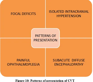
38 PATTERNS OF PRESENTATION :
There are 4 main clinical presentations of CVT
1. FOCAL DEFICITS / PARTIAL SEIZURES :
It is characterized by seizures, headache, altered sensorium, focal
deficits. It may also manifest as isolated seizures.
2. ISOLATED INTRACRANIAL HYPERTENSION :
This comprises headache , nausea,vomiting , papilledema, nerve palsy.
3. SUBACUTE DIFFUSE ENCEPHALOPATHY :
This is characterized by a decreased level of consciousness without
ICT features.
4. PAINFUL OPHTHALMOPLEGIA :This is caused by lesions of the
3 ,4 ,6 cranial nerves, chemosis and proptosis in patients with
39
FOCAL DEFICITS
ISOLATED INTRACRANIAL
HYPERTENSION
PAINFUL
OPHTHALMOPLEGIA
SUBACUTE DIFFUSE
ENCEPHALOPATHY
PATTERNS OF
PRESENTATION
[image:52.612.139.519.118.453.2]
Figure 10: Patterns of presentation of CVT
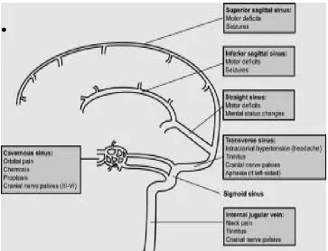
RELATIONSHIP BETWEEN CLINICAL MANIFESTATIONS
AND THE SITE OF OCCLUSION :
• Occlusion of cortical veins – motor/ sensory deficits, seizures or
both
• Occlusion of sagittal sinus - motor deficits, seizures
• Occlusion of lateral sinus – isolated intracranial hypertension
40
• Occlusion of deep cerebral veins – severe coma, delirium ,
bilateral motor deficits
• Occlusion of cavernous sinus – orbital pain , chemosis, proptosis ,
occulomotor palsies 49
[image:53.612.146.509.197.475.2]•
Figure 11: sinuses and their clinical features
INVESTIGATIONS :
NEURO IMAGING OF THE THROMBOSED VESSEL :
The diagnosis of CVT is mainly based on imaging modalities.
It usually shows non specific lesions . Moreover , neuroimaging may be
normal in 25 – 30 % of the patients with CVT . CVT is identified by the
41
occluded vessel. The current gold standard test for the diagnosis of CVT
is MRI with MRV50. The neuroimaging methods commonly used to describe CVT are described below
(A) COMPUTED TOMOGRAPHY (CT BRAIN ) :
Conventional CT is the best non invasive investigation of choice for
identifying CVT. CT may be normal in 25 – 30 % of the patients with
isolated intracranial hypertension50 .However CT BRAIN is the initial investigation in the emergency room not only to find out CVT but also
to rule out other causes which may mimic CVT .The diagnostic value of
CT may be increased, when it is combined with helical CT venography .51
The infarction in the non arterial distribution with hemorrhage implies
the possibility of CVT.
42
The CT findings of CVT may be as follows
• CORD SIGN : This is seen in plain CT scans. It represents the
visualization of a thrombosed cortical veins spontaneously without
any contrast. It is not very reliable as it can be also seen in internal
cerebral vein thrombosis.
• DENSE TRIANGLE SIGN : It represents spontaneous superior
sagittal sinus obscuration by freshly coagulated blood. It is present
in only less than 2 % of cases.
43
• EMPTY DELTA SIGN : It is seen in contrast enhanced CT
and it was first described by Buonanno and colleagues. It
reflects the contrast between the opacified and the non
opacification of the clot inside the sinus. The most common
direct sign in CVT is empty delta sign and it is demonstrated in
approximately 28- 72 % % of the cases.
44 ABSENCE OF EMPTY DELTA SIGN :
• When posterior third of superior saggital sinus is not
involved in thrombosis.
• CT scan is done either early or very late after the onset of
symptoms.
INDIRECT SIGNS OF CEREBRAL VENOUS THROMBOSIS :
• Approximately 20 % of cases will demonstrate intense contrast
enhancement of the falx and tentorium. Superior saggital sinus
thrombosis is usually demonstrated by tentorial enhancement.
There may be dilated transcerebral medullary veins, implying a
major venous stasis, indicating a large SSS thrombosis.
• cerebral edema is characterized by white matter hypodensity
without any enhancement..It is seen in upto 75 % of patients.
• Hemorrhagic venous infarct – in 10 – 50 % of the patients .There
is an associated sub arachnoid hemorrhage or a subdural hematoma
in rare instances. It can only present as the only sign of CVT.
• Non hemorrhagic venous infarct – the incidence is almost equal to
that of hemorrhagic venous infarct. They may be seen in the
45
Figure 15: CT Brain showing massive hemorrhagic infarct
NORMAL CT SCAN - CVT :
In 20 – 30 % of patients with CVT ,CT may be normal. In patients
with isolated intracranial hypertension it may be normal in 50 % of cases
53
. The main aim of CT is rule out the other conditions mimicking CVT.
Very few CT shows the classical domain of CVT . MRI /MRV is the
46
(B) MAGNETIC RESONANCE IMAGING :
MRI with MRV is the sole diagnostic investigation of choice for
CVT39 .The clot can be directly visualized in the sinus with the help of MRI .The clot has a varied picture depending upon the duration of
thrombosis. The thrombosed vessel appears isointense on T1 MRI and
hypointense on T2 image in the very early stages of formation of
thrombus. This phenomenon may not be visualized in MRI. This
difficulty can be overcome by MRV. The flow void becomes absent and
the thrombus becomes hyperintense, first on T1 and later on T2 MRI ,
which is mainly due to the conversion of oxyhemoglobin to
methemoglobin within the thrombus .It starts about 5 days after and lasts
nearly for 35 days. The occlusion will be removed or there may be
recanalisation of the vessels after a certain period, during that period even
MRI is not diagnostic .The abnormalities may persist for more than 6
47
Figure 16: MRI Brain showing transverse sinus thrombosis
MRI is also useful in assessing other associated features apart
from CVT , which includes cerebral edema or infarct . However false
48
DWI - LIMITATIONS
False positive False negative
• Slow flow of blood without
thrombus
• Acute thrombus – early stage
• Isolated cortial vein
thrombosis
• Partial recanalisation
Diffusion weighted imaging – Advantages :
• Direct visualization of clots
• Heterogenous signal intensity
• No diffusion abnormality
49
(C) MAGNETIC RESONANCE VENOGRAPHY :
MRV is now the best diagnostic modality of choice at present .There
are three methods , which are as follows
• Two dimensional time of flight (TOF )
• Three dimensional time of flight
• Phase contrast
The most commonly used method is two dimensional
flight..The slices will be 1.5 and 3mm.The absence of flow in the
veins is the typical appearance in CVT in MRV indicating
complete occlusion of the veins.
LIMITATIONS :
• Difficult to diagnose partial thrombosis
• Difficult to diagnose hypoplasia and thrombosis54
• Difficulty in detecting cortical vein and cavernous sinus
thrombosis
ADVANTAGE :
• Easily repeatable
50
TABLE 4 :DIFFERENCE BETWEEN CT AND MRI/ MRV
S.no Characteristics CT brain MRI/ MRV
1. Visualisation Major venous sinuses Superficial & deep
venous system
2. Time Not time consuming Time consuming
3. Radiation hazard Present Absent
4. With mechanical
devices
Can be used Contraindicated
5. Contrast reactions Present Absent
6. Availability Easy Difficult
[image:63.612.122.549.114.385.2]7. Accuracy Good Excellent
51
Figure 19: MRV Showing superior sagittal sinus thrombosis
(D) CT HELICAL VENOGRAPHY :
CT venography can be used as an alternative tool for MRI.39 CT venography more or less resembles CT angiography. There will be a
little delay after giving contrast in order to visualize the venous system
clearly .The non ionic contrast is injected intravenously at a rate of 3
ml/sec. From the images obtained bone images are deleted. After that
images are reconstructed by maximum intensity projection. The images
of this is closely related to MRI images. Compared to MRV they have
less artifacts.
52
Figure 20:CT Venography demonstrating the sinuses involved in
CVT
ADVANTAGES :
• 3D CT – reduces the mis interpretation of normal anatomic
findings which alike CVT
DISADVANTAGES :
53 (E) D- DIMER :
D- Dimer is a fibrin degradation product .It is a product of
fibrinolysis. It contains two cross linked D fragments of the fibrin
proteins and so it is called . The value of the measurement helps to
predict the presence of thrombosis. The negative D Dimer test always
rules out the possibility of thromboembolism .55,56 The positive test implies the presence of thrombus ,but it does not rules out other possible
causes of elevation..
[image:66.612.163.477.333.624.2]
Figure 21 : D Dimer Formation
54
In the normal human circulation, D Dimers are usually not
present. It is elevated only when the coagulation cascade is activated .The
D Dimer assay depends on the interaction between the monoclonal
antibody and the D Dimer fragment.
D-dimer levels are not always uniform. They have a peak value
during the acute phase of thrombosis. As time goes on the levels of D
Dimer starts decreasing57 .So in patients presenting with subacute or chronic symptoms , there will be not much elevation in the level of D
Dimer. The level of D Dimer correlate with the clot burden. If there is a
lesser clot burden then the levels wont be much high. At present a variety
of techniques are employed for determining D D imer tests.
A study conducted in 2004, proves that D Dimer levels was
abnormal in 34 of 35 patients., giving it a sensitivity of 97.1%, a negative
predictive value of 99.6%, a specificity of 91.2%, and a positive
predictive value of 55.7%58,59. This study is followed by another study,
which proves that even with clear cut thrombosis , 10 % of patients had
normal values of D Dimer. The patients who had only features of isolated
headache without any other symptoms had normal values of D
Dimer. The D DIMER levels above 500 ng/ml is60 considered to be
positive for CVT. The method used for estimation is latex slide
55
[image:68.612.125.479.86.365.2]
Figure 22: D Dimer and CVT Correlation
TREATMENT :
The treatment of CVT is based on assessment of individual cases as
the spectrum of presentation of CVT is varied .Anticoagulants with low
molecular weight heparin or dose adjusted intravenous heparin are used
as first line therapy. It is still a question whether to use anticoagulants in
56
to Martinelli et al ,it is better to delay the initiation of anticoagulation till
the hemorrhage is stabilized.
SYMPTOMATIC TREATMENT :
• The most common cause for CVT is dehydration .In order to treat
the cause of dehydration use of IV fluids is mandatory.
• Anticonvulsants for the patients with seizures. There is no clear
data for the use of prophylactic use of anti convulsants in seizures.
It is continued for a minimum period of 1- 2 yrs and then
withdrawn gradually62. The withdrawl of anticonvulsants should be based on that the patient had no recurrent seizures , has a normal
EEG and no neurological sequelae.
• Measures to reduce increased intracranial hypertension .The
measures includes steroids, mannitol, glycerol, dextran,
acetazolamide, lumbar puncture, shunting or even surgical
decompression .60
• Heparin therapy
• There is no role of steroids in CVT63
• Decompressive hemi craniectomy may be life saving in patients
57 ETIOLOGIC TREATMENT :
The main line of management in CVT is the treatment of the
underlying etiology behind CVT . The use of wide spectrum of antibiotics
for treatment of septic CVT and surgical debridement for primary site of
infection. Malignancies, collagen vascular disorder and hematologic
disorders need specific treatment.
ANTITHROMBOTIC TREATMENT :
AIMS :
• Recanalisation of occluded sinus
• Prevent progression of thrombus
• Treatment of prothrombotic state
• Prevention of recurrence
The optimal duration of heparin therapy is uncertain. Once
symptomatic improvement occurs, heparin is replaced with oral
anticoagulation. The INR should be maintained within 2-3.The treatment
should be continued for 6 months if there is no specific prothrombotic
cause identified . When there is increased risk of further thrombosis like
that of inherited thrombophilias , treatment should be continued for a long
58 THROMBOLYTIC THERAPY :
If patients deteriorate inspite of adequate anticoagulation,
thrombolysis may be considered if facilities are available. 65 Intravenous Urokinase was used in the early 1970’s for thrombolysis . Now r TPA is
the most commonly used agent .Both these agents carry the risk of
hemorrhage. There is no clear cut clinical evidence to show that these
agents have a better clinical outcome .Angio – jet rheolytic
thrombectomy is a novel technique. In this method a vacuum is created
by means of bernoullie effect .This vacuum is used for aspirating the
thrombus. It can be used for even hemorrhagic CVT, as it has an added
advantage of lack of hemorrhagic complications.62
59
Figure 23 : Algorithm for management of CVT Clinical Suspicion of CVT
MRI with MRV
CVT (Confirmed)
No evidence of CVT consider other differential
diagnosis
Initiate anticoagulation (IV heparin or SC LMWH)
Neurological Improvement Neurological deterioration
Continue oral
anticoagulation for 3 to 12 months or lifelong according to the underlying etiology
Severe mass effect or ICH on
repeated imaging
No or mild mass effect on repeated imaging
May consider decompressive hemicraniectomy (lifesaving
procedure )
60 PROGNOSIS :
SHORT TERM OUTCOME :
The cause of death in CVT are
1. Large hemorrhagic infarct
2. Complications like sepsis, uncontrolled seizures, pulmonary
embolism
3. Carcinoma, septicemia, leukemia and PNH
FACTORS SUGGESTING BAD PROGNOSIS :
Rapid progression of thrombus
Patient’s age
Infection
Focal symptoms and coma
Hemorrhagic infarct
Empty delta sign on CT scan
Type of veins involved
FUNCTIONAL RECOVERY :
61 LONG TERM OUTCOME :
During the initial episode of CVT ,if patient had seizures ,the
possibility of having residual epilepsy is around 10 – 30 %.
If the cause of CVT is a hypercoagulable state, there is a possibility
of recurrence of venous thrombosis. But because of long term
63
METHODOLOGY
SOURCE OF STUDY : Patients diagnosed to have Cerebral
venous thrombosis at Coimbatore
Medical college.
DESIGN OF STUDY : Cross sectional Study
PERIOD OF STUDY : AUGUST 2013 – JULY 2014
SAMPLE SIZE : 50
INCLUSION CRITERIA:
• Patients who are diagnosed to have cerebral venous thrombosis by
means of MRI and MRV in Coimbatore medical college within 30
days of symptoms onset, not started on anticoagulation.
• Age above 18 yrs.
EXCLUSION CRITERIA:
• Pregnancy
• Puerperium upto 4 weeks
• Arterial stroke within past 3 months
• Pulmonary embolism or Deep vein thrombosis
• Head injury within past 3 months
64
• Intra cerebral mass lesion
METHODOLOGY:
The patients diagnosed to have cerebral venous thrombosis
confirmed with MRI BRAIN during the study period were included.
After getting informed written consent,careful medical history was
obtained including clinical features, precipitating risk factors, family
history .All patients were subjected to meticulous general and
neurological examination .Patients were subjected to MRI BRAIN with
Venography on a 1.5 Tesla machine.
After confirming the diagnosis of CVT by MRI and MRV ,blood
samples were drawn ,plasma was separated, frozen at -80 C and sent to
the laboratory. A basic hematological work up including complete blood
counts, peripheral blood smear, ESR, PT/APTT were done. D-Dimer tests
were measured by using a conventional ELISA Test. A value of more
than 5oo ng/ml were considered positive tests. In order to find out the
etiology of CVT , dsDNA, APLA, serum homocysteine was done .
STATISTICAL ANALYSIS :
All the dates were entered in a data collection sheet in an Excel
format and analysed using SPSS Software. Numerical values were
65
are reported using number and percentages. probability value (p) value
OBSERVATION AND RESULTS
During the 12 month study period , a total of 50 patients were
studied for finding out the usefulness of D DIMER as an initial diagnostic
bio marker in CVT.
SEX DISTRIBUTION :
Table 5: Sex distribution among
SEX
Female
Male
Total
GRAPH 2–
Among the 50 patients , 28 were females ( 56%) and 22 were males
(44%). The ratio of female: male was 1.3:1.
67
OBSERVATION AND RESULTS
During the 12 month study period , a total of 50 patients were
studied for finding out the usefulness of D DIMER as an initial diagnostic
SEX DISTRIBUTION :
: Sex distribution among the study population
Frequency Percent (%)
Female 28 56.0
22 44.0
50 100.0
– Sex Distribution among the population
Among the 50 patients , 28 were females ( 56%) and 22 were males
(44%). The ratio of female: male was 1.3:1.
56% 44%
Gender Distribution
Female Male
During the 12 month study period , a total of 50 patients were
studied for finding out the usefulness of D DIMER as an initial diagnostic
the study population
Sex Distribution among the population
AGE DISTRIBUTION:
Table 6: Age distribution among the study population
AGE GROUP
Above 35 yrs
Upto 35 yrs
Total
Graph
The age of the patients comes under 2 categories . The majority of them
are above 35 yrs ( 56 %) .The patients below 35 yrs are around 44 % .
SOCIOECONOMIC STATUS :
low socioeconomic status (64.4%) , the rest belonge
socioeconomic status.
68 AGE DISTRIBUTION:
: Age distribution among the study population
AGE GROUP Frequency Percentage (%)
Above 35 yrs 28 56.0
yrs 22 44.0
50 100.0
Graph 3 :Age wise distribution of cases
The age of the patients comes under 2 categories . The majority of them
are above 35 yrs ( 56 %) .The patients below 35 yrs are around 44 % .
SOCIOECONOMIC STATUS : Majority of the cases were within the
low socioeconomic status (64.4%) , the rest belonged to the middle
socioeconomic status.
Age >35 -56% Age <35
-44%
Age range
: Age distribution among the study population
Percentage (%)
The age of the patients comes under 2 categories . The majority of them
are above 35 yrs ( 56 %) .The patients below 35 yrs are around 44 % .
Majority of the cases were within the
SYMPTOMATOLOGY AND SIGNS:
Table
HEADACHE
NO
YES
TOTAL
Graph
Among the 50 patients , 48 patients ( 96 %)
Remaining 2 patients ( 4 % ) didn’t have headache 0
10 20 30 40 50
[image:80.612.179.467.343.557.2]69 SYMPTOMATOLOGY AND SIGNS:
Table 7: Clinical Profile of Headache
HEADACHE FREQUENCY %
2 4.0
YES 48 96.0
TOTAL 50 100.0
Graph 4 : Distribution of Headache
Among the 50 patients , 48 patients ( 96 %) were having headache .
s ( 4 % ) didn’t have headache. NO YES
HEADACHE
Table
Graph
Among the 50 patients , 33 patients (66 %) didn’t have convulsions ,and
the remaining 17 patients (34 %) had convulsions 0
5 10 15 20 25 30 35
CONVULSIONS
NO
YES
TOTAL
70
Table 8: Clinical Profile of Convulsions
Graph 5: Distribution of Convulsions
Among the 50 patients , 33 patients (66 %) didn’t have convulsions ,and
the remaining 17 patients (34 %) had convulsions NO YES
CONVULSION
CONVULSIONS FREQUENCY %
33 66.0
17 34.0
TOTAL 50 100.0
Table
VOMITING
NO
YES
TOTAL
Graph
In our study, 31 patients ( 62
patients(38%) did not have vomiting. 0
NO YES
[image:82.612.175.468.342.600.2]71
Table 9: Clinical Profile of Vomiting
VOMITING FREQUENCY %
19 38.0
31 62.00
50 100.0
Graph 6: Clinical Profile of Vomiting
, 31 patients ( 62 %) of CVT had vomiting and 19
not have vomiting.
5 10 15 20 25 30 35
VOMITING
Table 10: Clinical Profile of Loss of Consciousness
LOC
NO
YES
TOTAL
Graph 7: Clinical Profile of Loss of Consciousness
The patients that presented with loss of consciousness in our study are 13
( 26 %) , and the remaining 37 patients ( 74 ) did not have loss of
consciousness . 0 5 10 15 20 25 30 35 40
LOSS OF CONSCIOUSNESS
72
: Clinical Profile of Loss of Consciousness
LOC FREQUENCY %
NO 37 74.0
YES 13 26.0
TOTAL 50 100.0
: Clinical Profile of Loss of Consciousness
The patients that presented with loss of consciousness in our study are 13
( 26 %) , and the remaining 37 patients ( 74 ) did not have loss of NO YES
LOSS OF CONSCIOUSNESS
: Clinical Profile of Loss of Consciousness
: Clinical Profile of Loss of Consciousness
The patients that presented with loss of consciousness in our study are 13
Table 11: Clinical Profile o CRANIAL NERVE NO YES TOTAL
Graph 8: Graph Showing Cranial Nerve
Among the 50 patients , 11 patients (22%) had cranial nerve involvement
, and the remaining 39 (78%)
0 NO YES
CRANIAL NERVE INVOLVEMENT
73
Clinical Profile of Cranial Nerveinvolvement
CRANIAL NERVE FREQUENCY %
NO 39 78.0
YES 11 22.0