Copyright
© 1994, American Society forMicrobiology
Processing
in
the Hepatitis C Virus E2-NS2 Region:
Identification
of p7 and Two
Distinct
E2-Specific
Products
with Different C Termini
CHAO
LIN,'
BRE1T D.LINDENBACH,'
BELA M.PRAGAT,'
DAVID W. McCOURT,2 ANDCHARLES M.RICE"*
Departmentof Molecular
Microbiology'
and Howard Hughes MedicalInstitute,2 Washington University Schoolof Medicine, St. Louis, Missouri 63110-1093Received 14 February 1994/Accepted 9 May 1994
The hepatitis C virus (HCV) H strain polyprotein is cleaved to produce at least nine distinct products: NH2-C-E1-E2-NS2-NS3-NS4A-NS4B-NS5A-NS5B-COOH. In this report, a series ofC-terminal truncations and fusion with a humanc-mycepitope tag allowedidentification of a tenthHCV-encoded cleavage product,p7, which is located between theE2andNS2 proteins. As determined by N-terminal sequence analysis, p7 begins with position 747 of the HCVHstrain polyprotein. p7 is preceded by a hydrophobic sequence at the Cterminus of E2which may direct its translocation into the endoplasmic reticulum, allowing cleavage at theE2/p7 site by hostsignal peptidase. This hypothesis is supported by the observation that cleavage at theE2/p7andp7/NS2
sites in cell-free translation studies was dependent upon the addition of microsomal membranes. However, unliketypical cotranslational signal peptidase cleavages, pulse-chase experiments indicate that cleavage at the
E2/p7site is incomplete, leading to the production of two E2-specific species, E2 and E2-p7. Possible rolesof p7and E2-p7intheHCV life cycle are discussed.
Hepatitis C viruses (HCV) have recently been recognized as agents of the parentally transmitted form of non-A, non-B hepatitis(10, 40). HCV causes the vast majority of transfusion-associated cases of hepatitis and a significant proportion of community-acquired hepatitis worldwide (reviewed in refer-ences 34 and 36). HCV infection results in various clinical outcomes,including acute hepatitis, chronic hepatitis,
cirrho-sis,andestablishment ofanasymptomatic carrierstatewhich maypersist for life (reviewed in reference 34). Chronic infec-tionsarefrequent, perhapsuniversal,and have been associated withincreased incidence of hepatocellular carcinoma (13, 62). The genome structures of several HCV isolates have been elucidated(9, 11, 14, 30, 38, 39, 52, 54, 55, 66, 67),indicating the existence of several genotypes(6, 65).This group ofclosely relatedenveloped positive-strand viruses isnowclassifiedas a separate genusin theflavivirus family(22),whichincludestwo other genera, the flaviviruses (7) and pestiviruses (12). The HCV genome RNA is approximately 9.4 kb in length and containsahighlyconserved 5'noncoding region (29)followed
byalong openreadingframe encodingapolyproteinof3,010 to 3,033 amino acids.
As established by cell-free translation and cell culture ex-pression studies (2, 16, 25-27, 31-33, 47, 64, 68), the HCV
polyprotein is processed by cellular and viral proteinases to produce the putative viral structural and nonstructural (NS) proteins. The order and nomenclature of thecleavageproducts are as follows: NH2-C-E1-E2-NS2-NS3-NS4A-NS4B-NS5A-NSSB-COOH. C, abasic protein, is believed to be the viral capsid protein; El and E2 are probable virion envelope
glycoproteins;NS2throughNS5BareputativeNSproteins,at least some of which may be the functional equivalents of homologous proteins encoded by flaviviruses and pestiviruses
*Correspondingauthor.Mailingaddress:Department of Molecular
Microbiology, Washington University,School ofMedicine,Box8230, 660 S.EuclidAve., St. Louis,MO 63110-1093. Fax: (314) 362-1232. Electronic mail address: [email protected].
(reviewed in references 36 and 50). Previous studies indicate that the host signal peptidase of the endoplasmic reticulum (ER) catalyzes cleavages in the putative structural region
(C/El, E1/E2, and perhaps E2/NS2) (31), whereas an HCV-encoded serineproteinase located in the N-terminal one-third of the NS3proteinisresponsible for four cleavages in the NS region(3/4A, 4A/4B,4B/5A,and5A/5B)(2, 16, 25, 32, 47,68).
Cleavageatthe2/3 site is mediated byasecond HCV-encoded proteinase which encompasses the NS2 region and the NS3 serine proteinase domain(26, 32).
Although the locations of most HCV-encoded proteins and the corresponding cleavage sites have been determined, the region between structural and NS coding sequences is still
poorlydefined. In apreviousreport,weobservedat leasttwo distinct forms of N-deglycosylated E2-specific proteins and
suggestedthatanadditionalcleavageproductmightbe present between the E2 and NS2proteins (27).Inthisstudy,wehave identified this protein, called p7,by expression ofa series of
C-terminallytruncatedpolyproteinsandbyfusionto ahuman c-myc epitope tag, which allowed isolation of the cleavage product and N-terminal sequence determination. p7 mapped betweenE2 andNS2. The presence ofhydrophobicpotential
signal/anchor sequences preceding the E2/p7 and p7/NS2
cleavage sites and the results of cell-free translation analyses indicate that host signal peptidase maycatalyzeboth of these
cleavages. However, cleavage at theE2/p7site is incomplete,
leading to the production of two stable E2-specific proteins with different Ctermini, E2andE2-p7.
MATERIALS ANDMETHODS
Plasmid constructions. Standard recombinant DNA tech-niques (63) were used for construction of the
expression
plasmids described below. Constructs for expression of
polyproteinswithserialC-terminal truncationsintheE2-NS2
regionwereproducedfrompTM3/HCV1-966,which encodes
5063
on November 9, 2019 by guest
http://jvi.asm.org/
TABLE 1. HCV expressionconstructswith C-terminal truncations in the E2-NS2 region
Construct' HCVsiteb VectorsiteC C-terminal
* ~~~~~~~~~~~~~~~~residues'
pTM3/HCV1-836 Eco47III (2848) Stul 1
pTM3/HCV1-786 BsrBI(2695) StuI 0
pTM3IHCV1-762 EamllO5I(2624) Stul 1
pTM3/HCV1-729 DrdI(2523) StuI 3
pTM3/HCV1-700 ScaI (2440) StuI 1
pTM3/HCV1-660 SstI(2322) Stul 1
a Numbers after HCVrefer to theportionof the HCVpolyproteinencodedby
each construct.Flankingresidues present in thepolyproteinsare notincluded.
For allofthe constructs, threeadditional N-terminalresidues(Met-Cys-Thr)are
predictedtobe presentpriortothe Met residueinitiatingthe HCV-H polypro-tein(27).
bRestriction sites in the HCV cDNA used for the plasmid constructs.
Nucleotidenumbersgiveninparenthesesrefer to thepositionsof these sites in thefull-lengthHCV-H sequence(14), assumingthat the 5'noncoding regionsof HCV-Hand HCV-1(11,29)arethe samelength.Restriction sites in boldface indicate thatprotrudingends were treatedwithT4 DNApolymerasepriorto
ligationtoproduceblunt ends.
cRestriction sitesinplasthidvectorsused forcloning.
dNumber of predicted non-HCV C-terminal residues prior to the first
terminationcodon.
HCVHstrain (HCV-H)amino acid residues 1 to966,bythe
subcloning strategies summarized in Table 1.
Construction ofpSINrepS/HCV171-379-mycandpSINrep5/
HCV370-802-myc will be described in detail elsewhere (44). Briefly, a derivative of the Sindbis virus replicon expression
construct pSINrepS (3), called pSINrepS/c-myc, was assem-bled; it contained, in the polycloning site region, asequence encodinganXbaIrecognitionsitefollowedbyanMluIsite,the coding sequence of the c-mycepitoperecognized by monoclo-nal antibody (MAb) Mycl-9E10 (EQKLISEEDL) (18, 19),
and aUAAtermination codon. Selected regionsof the HCV coding region (as indicated bythe HCV polyprotein residue
numbers)were amplifiedby PCRtocontaina5'XbaIsite,an AUG startcodon, anda3'Mlulsite, allowing in-frame fusion with the c-mycepitope tag. PCR productsweredigested with
XbaI andMluI andcloned into pSINrep5/c-myc.
pTM3/HCV364-802-mycwasconstructedby subcloning the
SacI-XhoI fragment (847 bp) of pSINrep5/HCV370-802-myc
and the NcoI-SacI fragment (893 bp) of pTM3/HCV1-1488 (27) intopTM3(51),which had been digestedwith NcoI and
XhoI. pTM3/HCV364-1207 was produced by subcloning the NcoI-NheI fragment (1,681 bp) of pTM3/HCV1-1488 into pTM3/HCV827-1207(26), which had been digested withNcoI
and NheI.
Cell cultures. The BHK-21 and CV-1 cell lines were ob-tained from the American Type Culture Collection, and the BSC-40 cell line (5) was obtained from D. Hruby (Oregon
State University). Cell monolayers were grown in Eagle's minimal essential medium (MEM) supplemented with2 mM
L-glutamine, nonessential amino acids, penicillin, streptomy-cin, and 10% fetal bovineserum(FBS). TheA16 subcloneof the humanhepatoma HepG2 cell line, generously provided by Alan Schwartz (Washington University, St. Louis, Mo.), was maintained in Dulbecco's modified Eagle medium supple-mentedwithpenicillin, streptomycin, and 10% FBS.
Generation and growth of viruses. A vaccinia virus-T7 expression plasmid containing the entire HCV-H open reading frame, pBRTM/HCV1-3011, has been described previously (27). The corresponding vaccinia virus-HCV recombinant,
vHCV1-3011,
was generated by marker rescue on CV-1 cells(46) and identified by the gpt selection method (20).
Recom-binant viruseswereplaquepurifiedthree times under selective conditions prior to growth of large-scale stocks. Stocks of vHCV1-3011, vHCV1-1488 (27),and vTF7-3, avaccinia virus recombinant expressing the T7 DNA-dependent RNA poly-merase (24),were grownin BSC-40 monolayers andpartially
purified (37),andtiters of infectious progenyweredetermined byplaqueassayonBSC-40 cells (37).
SINrep5/HCV-mycrecombinantviruseswere generated by using 5'-capped SP6 RNA transcripts from
pSINrep5/HCV-myc andpDH(26S)5'SIN helper templates essentially as de-scribed previously (3). The titers of these recombinantswere determinedby an infection assayusingsecondary cultures of chickenembryofibroblasts(3, 23). Briefly,cellswereseeded in 35-mm-diameter tissue culture wells toproduce subconfluent monolayers and were infected with serial dilutions of the
SINrepS/HCV-mycrecombinants(56).Thetiter, expressedin infectiousunits,wasapproximated bydeterminingthe fraction of cellsshowingcytopathiceffectsby24hpostinfection.
Transient expression using the vaccinia virus-T7 hybrid system. For expression assays utilizing vaccinia virus-HCV recombinants, monolayers of BHK-21 or HepG2-A16 cells were infected with vTF7-3 alone or in combination with vHCV1-1488orvHCV1-3011. Themultiplicityof infection for each recombinant was 10 PFU per cell (as determined on
BSC-40 monolayers). After adsorption for 60 min at room temperature, the inoculum was removed and replaced with MEM containing 2% FBS. For pulse-chase experiments, monolayerswerewashedoncewithprewarmedMEMlacking
methionineat3hpostinfectionand thenincubated in thesame medium for 20 minat 37°C.Cellswerelabeledbyincubation for 20 minat37°CwithMEMlackingmethionine and
supple-mentedwith 100 jiCi of
35S-protein
labeling mixture (NEN)per ml. For chase experiments, the labeling mixture was
replacedwithMEMcontaining2%FBS, 1.5 mg of methionine perml,and 100 ,ug ofcycloheximideper mlandincubated for the indicatedperiodsat37°C.Forsteady-statelabeling,
mono-layerswere washed oncewith prewarmedMEM lacking me-thionineat3 hpostinfectionand then labeledbyincubationfor 4 h at37°C withMEMcontaining 1/40 the normal concentra-tion of methionine, 2% FBS, and 25
RCi
of35S-protein
labeling mixture (NEN) per ml. Expression assays of trans-fectedplasmidconstructs utilizedsubconfluentmonolayersof BHK-21 cells in 35-mm-diameter dishes which had beenpreviously infected with vTF7-3 as described above. After removal of theinoculum,cellsweretransfectedat37°Cwitha mixture consisting of 1 ,ug of plasmid DNA and 10 jig of
Lipofectin (Bethesda Research Laboratories) in 0.5 ml of MEM.After2h,the transfection mixturewasremoved and the cellswerelabeled asdescribed above.
Transient expressionofHCV proteinswith
SINrep5/HCV-mycrecombinants. BHK-21 cellswereinfected withSINrepS/ HCV-myc recombinants at a multiplicity of infection of 5 infectious units per cell. After60 minat 37°C, the inoculum wasremoved andreplacedwithMEMcontaining 2% FBS.At 3 hpostinfection, monolayerswerelabeledasdescribedabove. Celllysis, immunoprecipitation,andproteinanalysis.After
labeling, cell monolayers were washed with
phosphate-buff-ered saline andlysedwith asolution of 0.5% sodiumdodecyl
sulfate(SDS),50 mMTris-Cl (pH 7.4), 1mMEDTA, and 20 ,ugofphenylmethylsulfonyl fluoride perml (0.3
ml/106
cells),andcellularDNA wasshearedby repeatedpassage througha
27.5-gauge needle. If the lysateswere not used immediately, aliquotswerestored frozenat-70°C.Sampleswereheated for 10 min at 70°C prior to dilution in the immunoprecipitation buffer containing Triton X-100 and carrier bovine serum albumin (59) andwereclarified by centrifugation at 16,000 x
on November 9, 2019 by guest
http://jvi.asm.org/
gfor15min. Portionsofeachlysatewereincubated either with
theindicated rabbitpolyclonalantiserum(usuallyS ,lI)orwith
mouse MAb (usually 1 ,ll of ascitic fluid) followed by rabbit
antiserum raised against whole mouse immunoglobulin G
(usually 14
[ig)
(Sigma).Immunecomplexeswerecollected byusing Staphylococcus aureus Cowan I (Calbiochem) as
de-scribed previously (59).
Immunoprecipitatesweresolubilized and analyzed by
SDS-polyacrylamide gel electrophoresis (PAGE) (41). After treat-mentforfluorographywithEnhance(Dupont), gelsweredried
andexposedat-70°Cwithprefogged (43) X-rayfilm(Kodak). 14C-methylated molecular mass marker proteins were
pur-chased fromAmersham.
In a previous report, endoglycosidase F (endo F) was
utilized for characterization of the deglycosylated forms of HCV glycoproteins (27). However, this material was
unsuit-able for N-terminal sequence analysis, possibly because of
proteolysisorN-terminal modificationduring overnight endo
F digestions. In this study, we used peptide N-glycosidase F
(PNGaseF), which cleaves between the innermost acetylgly-coamine and asparaginyl residues of high-mannose, hybrid, and complex oligosaccharides from N-linked glycoproteins. PNGaseF digestionswerecarried outessentially accordingto the manufacturer's instructions (New England Biolabs). Briefly, immunoprecipitates were resuspended in 50 mM
so-dium phosphate (pH 7.5) containing 0.5% SDS and 1% 2-mercaptoethanol andwere heated for 10 minat 99°C. The clarified supernatants wereadjusted to 50 mMsodium phos-phate (pH 7.5) containing 1% Nonidet P-40, 10 mM EDTA, and100,ugofphenylmethylsulfonyl fluorideperml andwere
incubated for 60 min at 37°C in the presence or absence of
PNGaseF. Usually, 1,000U(NewEngland Biolabs)ofPNGaseF
wassufficientforcomplete digestionofsamplesfrom 1.5x 10⁵ cells.Inthepresenceof 1%NonidetP-40,upto0.3%SDS had
nosignificant inhibitoryeffectonPNGaseFdigestion. Digested
samples were mixed with an equal volume of 2x Laemmli
samplebuffer (41)and analyzed by SDS-PAGE as described above.
N-terminal sequenceanalysis.HepG2-A16 cellswere
coin-fectedwithvHCV1-1488 and vTF7-3asdescribed above. At 3
h postinfection, monolayers were washed twice with
pre-warmed MEM lacking the amino acid used for radiolabeling andlabeled for 4 h in the same mediumcontaining 2% FBS and200 ,uCiof the indicated 3H-labeled amino acid perml. The 3H-amino acids usedwereleucine (135 Ci/mmol),valine
(30 Ci/mmol), and threonine (17.7 Ci/mmol) (Amersham). Afterlabeling, cell lysateswerepreparedand immunoprecipi-tated with either an El-specific mouse MAb (A4; kindly provided byH.Hsuand H.Greenberg, Stanford University)or an E2-specific rabbit polyclonal antiserum (WU 105) (27) as
described above. Radiolabeled El- and E2-specific proteins
were solubilized and digested with PNGaseF. The digested
productswereseparated by SDS-PAGE,transferredto Immo-bilonpolyvinylidenedifluoride membranes (48),andlocalized byautoradiography. Partial amino acid sequence analyses of El- and E2-specific proteins were performed as described
previously (8).
BHK-21 cellswereinfected withSINrepS/HCV-myc recom-binantswithamultiplicityof infection of 5 infectious unitsper
cell. At3 hpostinfection, monolayerswerewashed twice with prewarmed MEMlacking leucine and labeled for 4 hat37°C inMEMlackingleucine andcontaining2% FBS and 200 ,uCi of[3H]leucine per ml. Afterlabeling, monolayers were lysed
with 0.5% SDS lysis buffer (as described above) and the proteinswereimmunoprecipitatedwith MAbMycl-9E10(19),
specific for a c-myc epitope tag (18). Isolation and partial
amino acid sequence
analysis
of[3H]leucine-labeled
c-myc-specific proteinswere
performed
asdescribed above.Cell-free translation.
Uncapped
RNAtranscripts
were syn-thesized from linearized cDNA templates with T7DNA-dependentRNApolymerase
(Epicenter) (58).
Cell-freetrans-lations, using rabbit reticulocyte lysates
(Promega)
and[35S]methionine
(Amersham),
wereincubated for 1 h at30°C
essentially according to the manufacturer's instructions. As indicated, canine pancreatic microsomal membranes (Pro-mega) were added to 3.6 eq per 25-,ul translation reaction. Translation reactions were terminatedby
the addition of RNase A to 10 ,ug per ml(Boehringer Mannheim)followedby
continued incubation for 10 minat30°C.Translation reactions werediluted 10-fold with the SDSlysisbufferdescribed above and heated for 10 minat 70°C prior to
immunoprecipitation.
Analysis of solubilized proteins and/or PNGaseFdigestion
productswascarried outessentiallyasdescribed above.RESULTS
Alternative forms ofHCVE2 share thesameN termini.In an earlier report, avaccinia virus transient
expression
system wasused to examine processing eventsin the HCV-H strainstructural-NS2region
(27)
(Fig. 1A).Theseexperiments iden-tified the 21-kDa C protein and the Elprotein,
gp3l,
which washeavilymodifiedbyN-linkedglycosylation
andmigrated
to21 kDa after endo Ftreatment
(17).
Glycosylated E2-specific proteins gp88 andgp7O
were resolved as three speciesof62, 41, and 36 kDa afterdigestionwith endoF(reference
27 and alsobelow).
p62(the
deglycosylated
form ofgp88)
was iden-tified as an E2-NS2 polyprotein and proposed as apotential
precursor forthe E2 and NS2 proteins.Neither p41 nor p36
was recognized by El or NS2
region-specific
antiserum,sug-gesting
the existence of at least two alternative forms of E2(27).Tofurtherexamine the structural basis for these alterna-tive forms ofE2, wedetermined the N-terminal sequences of
p41
andp36. Metabolicallylabeledlysateswereprepared
fromHepG2-A16
cells which had been coinfected with vTF7-3 andvHCV1-1488, a vaccinia virus recombinant expressing the entire HCV structural region through the N-terminal
two-thirds of NS3. Sampleswere
immunoprecipitated
with E2-orEl-specific
antiserum, digested with PNGaseF, separated bySDS-PAGE,
electroblotted onto Immobilonpolyvinylidene
difluoride membranes, and
sequenced
for 15 to 18cycles
of Edmandegradation.For bothp41
andp36,threonine residues wererecoveredatpositions
2and5(Fig. 2).
Thisfinding, along
withthe sizes andimmunoreactivities
of thesespecies,
identi-fies Glu-384 as the N-terminal residue of both forms of HCV-HE2. IdenticalN-terminal sequenceswereobtained for unresolved glycosylated forms of E2, produced by cell-free translation in the presence of microsomal membranes(HCV-J
strain)
(31)
orpurified
from cells infected withavaccinia virus recombinant (HCV-1strain)
(57).
Forthedeglycosylated
El protein, a leucine residue wasrecovered at position 9,valine residueswererecovered atpositions
3and 12,andathreonine residue was recovered atposition
13(Fig. 2).
These results establish the N terminus of HCV-H El as Tyr-192,which isagainconsistent with the
previously
reported
El sequence data for the HCV-J(31)
andHCV-1(57)
strains.p41
and p36 differ at their C termini. Wemapped
theC-terminalboundaries of
p41
andp36 by
expressing
aseries ofpolyproteins
withprogressive
C-terminal truncations (dia-grammed in Fig.1B)
in BHK-21 cellsby
using
the vaccinia virus-T7 system.Allof thepolyproteins
contained the entire C and El sequences(presumably
residues 1 to 191 and 192 to383,
respectively).
Since the N terminus of NS2 has beenon November 9, 2019 by guest
http://jvi.asm.org/
A
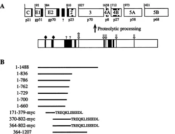
B
1 192 384 810 1027 1658 1712 1973 2421
t
tS | t |
3
|~~~~~~~~4A
mA |
BI
p21 gp3l gp7O ? p23 p70 p8 p27 p58 p68
I
Proteolytic processing
I
* ? ? t X
I
II "ii
_
iii! _
1-1488
1-836
1-786
1-762
1-729
1-700
1-660
171-379-myc
370-802-myc
364-802-myc
364-1207
-TREQKLISEEDL
TREQKLISEEDL -TREQKLISEEDL
FIG. 1. HCV genome structure andexpressionconstructs.(A) Diagramof the HCV-Hpolyproteinand itscleavage products shown as boxes. The identities of matureproteins, including putative structuralproteins (C,El, andE2)and NSproteins(NS2,NS3, NS4A,NS4B, NS5A, and
NS5B),areindicated (27).Thenumber atthe top of each cleavageproduct indicates the position of its N-terminalresiduein thepolyprotein sequence. The apparent molecular mass of each HCV protein (p) and glycoprotein (gp) is indicated under the corresponding product (in kilodaltons). Regions containing predominantly uncharged aminoacidsareindicatedasblackbars. Theregionbetween E2 and NS2 which may representanadditionalHCV-encodedproteinis indicated(?).Alsoshownareputativecleavage sites for hostsignalase(*)(31),the HCV NS2-3 proteinase
(I)
(26,32), the NS3 serineproteinase (4X)(2, 16, 25, 32, 47,68),and unknownproteinases(?). (B) HCV polyprotein expression constructs used in thisstudy. HCVpolyproteinsequencespresent ineachconstruct are indicatedbyblack lineswhich aredrawntoscaleand orientedwith respecttothediagramof the HCVpolyprotein.Also shownis the C-terminal10-residuec-mycepitopetag(EQKLISEEDL)present in some of theexpressedpolyproteins.determined as residue 810 of the HCV-H polyprotein (26), polyprotein 1-836 includesthe N-terminal 27 amino acids of NS2 while theremainingconstructsterminate upstream of this cleavage site. Lysates of[35S]methionine-labeledBHK-21cells wereimmunoprecipitated with the E2-specific antiserum and
digestedwith PNGaseF. Asdescribed previously, polyprotein
1-1488, which extends through the N-terminal two-thirds of the NS3 region, produced three E2-specific products: p62, p41, andp36(Fig. 3).Polyproteins terminatingatresidues836, 786, and 762produced onlytwoE2-specific proteinsafter PNGaseF
digestion, one of which comigrated with p36 (Fig. 3). The
secondE2-specific product generated from polyprotein 1-836 migrated slightly more slowly than p41 and, therefore, may representanuncleavedpolyprotein containingp41 followed by the N-terminal 27 residues of NS2. The apparent molecular massesof truncatedE2-specific proteins produced by polypro-teins 1-786and1-762 were 39and 38 kDa, respectively. These results suggest that the C terminus of p41 maps between residues786 and 836, most likely at residue 809.Polyproteins terminatingat residues 729, 700, and 660generated only one
E2-specificproduct of 35, 34, and 32 kDa, respectively (Fig. 3), which indicates that the C terminus of p36 is located between residues729 and 762, probablynear residue 745(see below). On the basis of these data, we refer to the small, highly hydrophobic region between E2 and NS2 as p7 (although its existence as astablecleavage product has notbeen verified).
p62,p41,andp36arehereafter calledE2-NS2, E2-p7,andE2,
respectively.
Determination ofthe E2/p7 cleavage site. Further charac-terization of the putative p7 cleavage product has proven difficult. To generatep7-specificantiserum, wesubcloned the cDNA sequence encoding HCV residues 745 to 809 into Escherichia coli expressionconstructsandattemptedtopurify p7-containing proteins in sufficient quantities to generate rabbit polyclonal antiserum. Although numerous E. coli ex-pression systems have been tried, this approach has not yet succeeded. We also screened antisera from HCV-positive patients by immunoprecipitation under either denaturing or nondenaturing conditions; noneof these sera reacted witha
distinguishable p7protein. It is possible that theputative p7
cleavage product is unstable orpoorly immunogenicbecause of thehighly hydrophobic natureof thisregion.
Because ofthe lackof antiserum specific for the p7region,
a 10-amino-acid c-myc epitope tag was fused to the p7 C terminus to allow identification of this processing product
(diagrammed in Fig. 1B). Construct HCV370-802-myc con-tains HCV residues 370to802,beginningin theputativesignal peptide sequenceatthe C terminusofEl,followedbytheE2 and p7 regions. As a control for thespecificity of the c-myc MAb, we constructed HCV171-379-myc, which includes the
putative signal peptide at the C terminus of the C protein
followed by the El region and the c-myc peptide. For both
on November 9, 2019 by guest
http://jvi.asm.org/
[image:4.612.139.496.69.342.2]A constructs, the C-terminal c-myc peptide was fused upstream of thep7/NS2 (HCV370-802-myc) and E1/E2
(HCV171-379-p41
myc) cleavage sites in a context which should discourageP removal by signalpeptidase (21, 69).
80- These two HCV-myc fusion cassettes were placed
down-70'1ETHVTGGSAGHTTAGLVG stream of the 26S
subgenomic
promoter
of a Sindbisvirus-70 E T HVT G G SAG H T TAG LVG derivedexpressionreplicon, which isauseful system forrapid generation and analysis ofexpression constructs(3, 4).
Infec-60- r --- | tious particles containing these Sindbis virus-HCV
replicons
l0
/ |wererescued(3) and used to infect BHK-21 cells, and[S]me-thionine-labeled cell lysates were immunoprecipitated with MAb
Mycl-9E10.
ForHCV171-379-myc,
at least fivec-myc-40 - l \ lspecific species, ranging from 21 to 34 kDa, were
immunopre-cipitated (Fig. 4A). Similar patterns of El-specific proteins
30- lhave been observed when the
El
protein was expressed20- l
independently
athigh
levels in insect cells and have been20- attributed to incomplete N-linked glycosylation (42, 49). For
lo
L
I.I.I_I
_III
_HCV370-802-myc,
MAb Mycl-9E10
recognized
at least twoI I 10 15. .
..higher-molecular-mass
species,
whichpresumably
represent
glycosylated andunglycosylated E2-p7-myc andaprotein of 6.5
cycle#
kDa, whose size is consistent with the p7-myc cleavage productB
(Fig. 4A).[3H]leucine-labeled
p7-mycprotein waselectroblot-ted onto Immobilon
polyvinylidene
difluoridemembranes,
p36 localized, andsequencedfor 25 cyclesof Edmandegradation.
As shown in Fig. 4B, leucine residues were recovered at
40 positions 2, 5, 8, 13, 19, and 23, whichtentatively establishes
the N terminus of p7 as HCV-H polyprotein residue 747.
ETHVTGGSAGHTTAGLVG Together with the definition of the p7/NS2 cleavage site (26),
these data suggest that p7 is 63 amino acids in length,
30 encompassing residues747 to 809of the HCV-Hpolyprotein,
.30_ 1\ witha
predicted
molecularmassof 7.0 kDa. The smallest formT 0 of the E2
glycoprotein,
whosedeglycosylated
formcorresponds
top36, spansresidues 384 to 746, withapolypeptide backbone of363 residues andapredicted molecularmassof 40.0 kDa.
: 20- Cell-free
processing
at theE2/p7 cleavage
site isdependent
upon microsomal membranes. Previous cell-free translation studies have shown that processing at the HCV C/El and
El/E2cleavage sites is dependent upon the addition of micro-somal membranes and, therefore, is likely mediated by host
10
signal peptidase
in the ER(31).
The sequences and the0 5 10 15 20 conservedhydrophobic character of the regions preceding the
cycle# N termini of p7 and NS2 (Fig. 5) are characteristic of signal
peptides (21, 69). This suggested that cleavage at the E2/p7
C
and p7/NS2 sites may also be mediatedby hostsignalpepti-dase. Thispossibilitywasinvestigated by cell-free translation of
El
twoHCVexpression constructs. HCV364-1207 initiates within100
-.70
20 the putative signal peptide at theC terminus ofEl
and extends90YQVRNSSGLYHVTNDCN .through the serine proteinase domain of NS3.
HCV364-802-90- YQVRNS S G L Y HVTNDCPN
-60
myc also begins with the signal sequence preceding E2 and80- terminates with the c-myc
epitope
fused in frame afterp7
70
t50
residue802(C
terminus
identicaltothat
ofHCV370-802-myc)
I
(Fig.
lB).
601
- \ /;t -40 Thepatterns
ofcell-freetranslationproductsfromHCV364-,,50- / \ | j ] -10 19
802-myc
areshown inFig.
6A. Inthe absence ofmicrosomal 30 .membranes,
apredominant
product
of 41kDawasrecognized
30- I_20
204_
1-
10 labeled amino acidsasdescribed in Materials and Methods.PNGaseF-10- digested E2-specific protein(p41or p36) orEl(p21)was isolatedand
0 - 0 subjected to N-terminal sequence analysis. The graphs show
uncor-0 5 10 15
20
rected countsperminute released persequencing cycle. From thesedata,the sequences ofp41, p36,andElbeginatHCV-Hresidues384, cycle# 384, and 192, respectively, and are shown at the top of the correspond-FIG. 2. N-terminal sequence analyses of the E2-specific proteins ing graph.Residues in boldface type indicatepositionsdeterminedby p41 (A)andp36(B)andofEl (C).HepG2-A16cells were coinfected N-terminal sequencing; the remaining residues are deduced from with vTF7-3 and vHCV1-1488 and labeled with the indicated 3H- HCV-H nucleotide sequence data(14,38).
on November 9, 2019 by guest
http://jvi.asm.org/
[image:5.612.60.284.74.690.2]A
- + - + - + - + - + - + - + - + PN(;aseF
200-97.4- ~~~~~~~~-E2-NS2
3-E2-p7and E2
69- E- .E2-N 2*
...
46--E2-p7*
-E2*
30- -E
[image:6.612.346.536.71.469.2]21.5- -El*
FIG. 3. C-terminal boundaries ofE2-specificproteinsp41andp36. vTF7-3-infected BHK-21 cell monolayers were mock transfected (lanesm), transfected with the indicated plasmid DNAs, or coinfected with vHCV1-1488 and were labeled with35S-proteinlabelingmixture asdescribed inMaterials and Methods.Celllysates were prepared and immunoprecipitated with anti-E2 antiserum WU 105 (27). Immuno-precipitated proteins were solubilized and digested in the absence (-) orpresence(+)of PNGaseF. Thedigestedproducts were separated by SDS-12% PAGE. HCV-specific proteins are identified at the right, and the sizes of "4C-labeled protein molecular mass markers (in kilodaltons) are indicated at the left. The positions of PNGaseF-digested forms (indicated by asterisks) are also shown. As observed before, a smallfraction of theElprotein was coimmunoprecipitated withE2-specific antiserum (27).
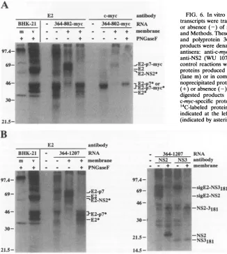
by both anti-E2 and anti-c-myc antibodies and, therefore, represented the primary translation product. Upon the addi-tion of microsomes, two glycoproteins were observed. The larger one (N-deglycosylated form,41 kDa) was immunopre-cipitated by both anti-E2 and anti-c-myc antibodies and iden-tified as the E2-p7-myc glycoprotein. The smaller species (N-deglycosylated form, 36 kDa) was present in small amounts andwas immunoprecipitated only with E2-specific antiserum, thus representing the processed E2 glycoprotein. Both E2-specific glycoproteins were fully protected against proteinaseK digestion in the absence of detergent (data not shown), indicative oftranslocation into the ER lumen. In addition, a
c-myc-specific protein of 6.5 kDa, presumably p7-myc, was observedonly in the presence of microsomal membranes (data notshown).
As shown in Fig. 6B, microsomes were also required for
processingof thelongerpolyprotein364-1207 at the E2/p7 and p7/NS2 sites, as evidenced by the appearance of the E2 and E2-p7 glycoproteins and NS2. Both glycoproteins were fully protected against proteinase K digestion in the absence of detergent (data notshown). The E2/p7 cleavage was incom-plete, in contrast to the efficient cleavage observed at the p7/NS2 site. The addition of microsomal membranes had no effecton the efficiencyofcleavage at the 2/3site in 364-1207, which isconsistent with the membrane-independent autopro-teolytic processing at this site by the HCV NS2-3 proteinase
(26, 32).
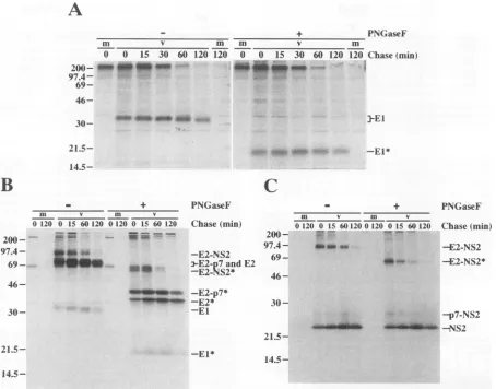
Kinetics of processing in the structural-NS2 region. To identify possible precursors involved in the structural-NS2 region processing, pulse-chase experiments were carried out in HepG2-A16 cells with a vaccinia virus-HCV recombinant, vHCV1-3011.Asshown in Fig. 7A, theEl protein was readily apparent aftera20-minpulse and was not associated with any higher-molecular-weight polyprotein precursors, which is con-sistent with theproposed cotranslational cleavage at bothC/El and
El/E2
sites by ER signal peptidase (31). No shift inC,$ C, 'CV
el
ep k5'
97.4-
.-69- ;rSE, :01
-E2-p7-myc
69-46-::
.P -E2-p7-myc*
30-21.5
12.5-6.5- -p7-myc
B
p7-myc
100
90 -
80-
t70-4
60-5040
-0 5 10 15
cycle#
20 25
FIG. 4. Identification of the p7-myc protein. (A) BHK-21 cells
weremock infected(lane m)orinfected with the indicated
SINrep5-HCVrecombinants andthen labeled with"5S-proteinlabeling mixture
asdescribed inMaterialsandMethods.Celllysateswereprepared and
immunoprecipitated with anti-c-myc MAb Mycl-9E10 (19). Immuno-precipitated proteins were solubilized and separated by SDS-14%
PAGE.c-myc-specific proteinsareidentifiedattheright, and the sizes of "4C-labeledprotein molecular mass markers (in kilodaltons) are
indicatedatthe left.Theposition of unglycosylated E2-p7-myc is also indicated by an asterisk. (B) N-terminal sequence analysis of [3H]leucine-labeled p7-myc fusion protein. The graph shows
uncor-rectedcountsperminutereleasedpersequencing cycle. From these
data, theN-terminal sequence ofp7 begins at HCV-H polyprotein residue747 and is shown atthe topof the graph. Leucine residues determinedby N-terminal sequencingareinboldfacetype;the remain-ingresiduesarededuced fromHCV-H nucleotide sequencedata(14,
38).
deglycosylated El migration was observed during the chase
period (Fig. 7A), suggestingthat El doesnotundergofurther proteolytic processing under these conditions. However, N-glycosylated forms of El did show a gradual increase in
migration over the chase period (Fig. 7A), which probably
resultsfrom trimmingof the N-linked, high-mannose glycans. Itshould be noted thatthe levelofEl increased slightly during
ALENLVILNAASLAGTHGLVSFLVF
on November 9, 2019 by guest
http://jvi.asm.org/
[image:6.612.67.304.72.214.2]E2/p7
7
p7/NS2
HCV-H RVCSCLWNMLLISQAEAAlBNLVILNAASLAGTHGLVSFLVFFCFAWYLKGRWVPGAVYAFYGMWPLLLLLLALPQRAYA LDTEV
HCV-1 --- --- ---T---
---HCV-J ---A---A---- T---- V---V--A---L---A---I---L----A--L--V---P---- M-R-M HCV-JT ---A---A---- ---V---AD-IL---A---I---L----A--L--V---P---- M-R-M HCV-BK ---A---A--- ---V--S--V--A--IL---A--I---L----T--L--V--- P---- M-R-M HCV-T ---A---A---- ---VF----V--M--TL---A---I---L----A--L--V---P---- M-R-M
HC-J6 ---A---LI-LG---- ---K--V-H---A-SCN-FLY-VI--VA---I---V--L-T-SIT-L-SFG---Q--- Y-AS-HC-J8 -I-A----LIILG---- --- I--HS--A-SAN-PLW-FI--TA---V--V-T-SVL-L-SF---V- Q--AAE
FIG. 5. Alignment of the HCVE2/p7andp7/NS2 cleavage sites. The amino acid sequences around the E2/p7 and p7/NS2 cleavage sites are aligned for the following HCV isolates: HCV-H (14, 38), HCV-1 (11), HCV-J (39), HCV-JT(67),HCV-BK(66),HCV-T(9),HCV-J6(55), and HCV-J8(54). The single-letter code for amino acids is used. A hyphen indicates a residue identical to that of the HCV-H sequence.
the chase period (Fig. 7A), in apparent conflict with the absence of anyEl-containing polyprotein precursors. This has been observed before and may be due to a delay in the formation ofthe epitope recognized by the anti-El MAb used for immunoprecipitation (15).
Processing in the E2-NS2 region was more complex. After a 20-min pulse, three E2-specific proteins, including E2, E2-p7, and E2-NS2, were apparent after PNGaseFdigestion (Fig. 7B) while the anti-NS2 antiserum recognized two products, E2-NS2 and E2-NS2(Fig. 7C). These results indicate that cleavages at the El/E2 and 2/3 sites occur rapidly, perhaps cotranslation-ally, and are followed by delayed processing within the E2-NS2
region(see Discussion). As seen in Fig. 7B and C, the level of E2-NS2decreasedconcomitantly withanincrease in the levels of E2 and NS2 during the chase. This provides evidence that E2-NS2 is a discrete precursor of E2 and NS2 (27), which has also been demonstrated recently for the HCV-BK strain (15). The precursor-product relationship between E2-p7 and E2, if any, is less clear, and the results so far do not distinguish betweena kinetic mechanism andan alternative pathway for the inefficient cleavage at the E2/p7 site. Cleavage at the p7/NS2 site relative to that at the E2/p7 sitewas efficient as evidenced by thedisappearance of E2-NS2 and the low levels of p7-NS2 detected during the chase. This observation,
to-c-myc
- 364-802-myc
_ _ + +
antibody RNA membrane
-+-- - +PNGaseF
.. E2-p7-myc
...E2-NS2*
_ z | ^^... jw .E2-p7*or _ t >J .F',. 2 .' E2-p7-myc*
-E2*
FIG. 6. Invitroprocessing ofHCVpolyproteins. Uncapped RNA transcriptswere translatedinreticulocyte lysatesin the presence (+) orabsence(-) of microsomal membranesasdescribedin Materials andMethods. Thesetranscriptsencodedpolyprotein364-802-myc(A) and polyprotein 364-1207 (B). [35S]methionine-labeled translation productswere denatured andimmunoprecipitatedwith thefollowing antisera: anti-c-myc MAb (Mycl-9E10) (19), anti-E2 (WU 105), anti-NS2 (WU 107), or anti-NS3 (WU 110) (27). Also shown are control reactions without added RNA(-) and the HCVE2-specific proteins produced in BHK-21 cells by infection with vTF7-3 alone (lanem)orin combination with vHCV1-1488 (lane v).Some immu-noprecipitated proteinsweresolubilized anddigestedin the presence (+)orabsence(-)of PNGaseF.Immunoprecipitatedand PNGaseF-digested products were separated by SDS-14% PAGE. HCV- or c-myc-specific proteins are identified at the right, and the sizes of
14C-labeled protein molecular mass markers (in kilodaltons) are indicated at the left. The positions of the PNGaseF-digested forms (indicated byasterisks)arealsoshown.
364-1207 RNA
- + + membrane
- - + PNGaseF
97.4- 69-
46-30-I
21.5-364-1207 RNA NS2 NS3 antibody
. + - + membrane
*..
I-sigE2-NS3181
-sigE2-NS2
I-NS2-3181
-NS2 -NS3181
21.5-A
E2
- 3644802-myc
_ - + +
BHK-21
m v
97.4-I
69-
46-
30-
21.5-B
E2
BHK-21
m v
_
antibody
97.4-.
69- 46-
30-
14.5-_ _
on November 9, 2019 by guest
http://jvi.asm.org/
[image:7.612.94.519.73.158.2] [image:7.612.53.379.354.718.2]A
+
m v m m v m
0 0 15 30 60 120 120 0 0 15 30 60 120 120
200- -4WI. OM
97.4- 69-46-PNGaseF Chase(min)
]-E1
30- 21.5-
14.5-B
m v m v
0120 0 15 60 120 0 120 0 15 60 120
97.4- .
C
PNGaseF Chase (min) -E2-NS2 3-E2-p7 and E2 -E2-NS2*
46-30
-_ -_ _mim -E--p7*
PIINW -2
-E2*
200- 97.4- 69- 46-
30-+
m v m v
0120 0 15 60120 0 120 0 15 60 120
21.5-21.5- -El*
14.5-FIG. 7. Processing kineticsinthe HCV structural-NS2region. HepG2-A16 cellswereinfectedwith vTF7-3alone (lanes m)orcoinfected with
vTF7-3 andvHCV1-3011 (lanes v) andpulse-labeled with35S-protein labeling mixture for 20minand then chased forthe indicated timesas
described in Materials and Methods. Celllysateswereprepared, immunoprecipitated, solubilized, digestedinthe absence(-)orpresence(+)of PNGaseF,andseparated by SDS-12%PAGE. ImmunoprecipitationwascarriedoutwithEl-specific MAb A4(A), E2-specificrabbit antiserum WU 105(B),orNS2-specificrabbit antiserum WU 107(C). HCV-specific proteins and deglycosylated forms (indicated by asterisks)areidentified
attheright,and the sizesof "'C-labeledproteinmolecularmassmarkers(inkilodaltons)from Amershamareindicated atthe left.
getherwith the decrease in the ratio ofE2-p7toE2duringthe chase, suggests that E2-p7 maybe cleaved slowly to produce E2. However, further experiments are needed tosubstantiate this conclusion, given the instability ofthese products during long chases in thepresence ofcycloheximide.
DISCUSSION
Earlier transient expression studies (25-27) and the work reported here havedefined ninecleavagesites inthe HCV-H polyprotein: C/El, E1/E2, E2/p7, p7/NS2, 2/3, 3/4A, 4A/4B, 4B/5A,and5A/5B.Thesecleavages resultintheproduction of atleast 10nonoverlapping polypeptides whose properties are
summarized in Table 2. Our studies and those utilizing the HCV-J strain (31) suggestthat the primary processingevents in theputative structural region andthe NS2regionarelikely
to be catalyzed byhost signal peptidase. Two HCV-encoded proteinases are required for processing the remainder of the
polyprotein. The 2/3 cleavage appears to be mediated by a
novelautoproteolyticactivity, perhapsaZn2'-dependent met-alloproteinase, which encompasses the NS2 region and the
NS3 serine proteinase domain (26, 32). The remaining
cleav-agesin the NSregionare dependenton anactiveNS3 serine
proteinase domain (2, 16, 25, 32, 47, 68).
Althoughwe believe it is likelythat the E2/p7andp7/NS2 cleavagesaremediatedbyahostsignal peptidase,thepresence
of a discrete E2-p7-NS2 precursor and a reasonably stable E2-p7species(reference15 and thisreport)suggeststhat these cleavagesarenotnecessarilycotranslational(incontrasttothe C/El and El/E2 cleavages). Delayed cleavage may reflect a
requirementfor additional posttranslational modifications or
interactions withother hostorviralcomponents,oritmaybe
duetothepresence ofasuboptimal signal peptidasecleavage
site. Analysis of signal peptide sequences of several HCV
strainsshowed that amongall fourputative signal
peptidase-mediatedcleavage sites, theE2/p7 site has the lowest proba-bility for signalase cleavage (21, 69). This fits with the ineffi-cientcleavageobservedfor theE2/p7 sitecomparedwith that forthep7/NS2 site in the pulse-chase analyses (Fig. 7). The relativelylowhydrophobicityinthecoreregionoftheputative
signal peptideforp7mayberesponsiblefor slow translocation
or,morelikely,for sloworinefficientprocessing bythesignal
peptidase (70). On the other hand, it is possible that an
PNGaseF Chase (min)
-E2-NS2 -E2-NS2*
-p7-NS2
-S2
'=
.Ul..'-..:;...;;.4.:;.v ..::. -:.. ..::.
-.7 ;!:--f:'l;., ..
iM:MZkl:. ..-ANW,.1!V::-. -E I*
on November 9, 2019 by guest
http://jvi.asm.org/
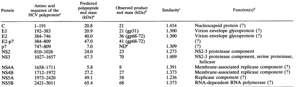
[image:8.612.87.541.69.425.2]TABLE 2. Properties of HCV proteins Aminoacid Predicted
Protein sequence of the polypeptide Observedproduct Similarity Function(s)
HVpolyprotein' molmass molmass(kDa)b iiai) Fnto()
HCV
~~~~~(kDa)a
C 1-191 20.8 21 1.454 Nucleocapsidprotein (?)
El 192-383 20.9 21 (gp31) 1.300 Virion envelopeglycoprotein (?)
E2 384-746 40.0 36(gp68-72) 1.300 Virion envelope glycoprotein (?)
E2-p7 384-809 47.0 41 (gp68-72) (?)
p7 747-809 7.0 NDe 1.309 (?)
NS2 810-1026 24.0 23 1.273 NS2-3proteinase component
NS3 1027-1657 67.3 70 1.409 NS2-3proteinasecomponent, serine proteinase,
helicase
NS4A 1658-1711 5.8 8 1.391 Membrane-associated replicasecomponent(?)
NS4B 1712-1972 27.2 27 1.373 Membrane-associated replicase component(?)
NS5A 1973-2420 49.1 58 1.236 Replicasecomponent (?)
NS5B 2421-3011 65.4 68 1.373 RNA-dependentRNApolymerase(?)
aBased on the HCV-Hstrain(14) andtheassumption that there is notrimmingat the Ctermini of theseproteins.
bForglycoproteins(El,E2,andE2-p7),themolecularweights ofN-deglycosylatedformsaregiven, with themolecularweights ofglycosylatedforms(gp)inbrackets.
cSimilarityis the arithmetic mean ofcomparisonscoresfromseveralfull-length HCVpolypeptidesequences,calculated byusingthemodified Dayhofftable(28)
andaveraged over thelengthofeachprotein. Anaverage scoreof1.5represents completeconservationwhile ascore of 0 indicates lack of similarity. Pairwise
alignmentsof 19full-lengthHCVpolypeptidesequences were analyzedwith the Genetics Computer Group suite of programs (version 7). These 19 sequences include
HCV-1(11),HCV-H (14,38),HC-J1(D10749),HC-G9(53),HC-J4/83 andJ4/91(52), HCV-J (39), HCV-JT andJT' (67), HCV-BK (66), HCV-T (9), HCV-JKI
(S18030),HCV-N (30),HCV-Unkcds (M96362), HCV-L2 (U01214), HCV-HeBei (L02836),HC-J6(55),and HC-J8 (54).Forunpublishedsequences, accession numbers aregiveninparentheses.
d(?),unknown orpredictedfunction.
'ND,notdetermined.
alternative microsomal or viral proteinase is responsible for
cleavageat this site.
Thisprocessing scheme for the HCVstructural-NS2 region hasfeatures similartoaswellasdistinct from those exhibited
byflaviviruses andpestiviruses. For members of the flavivirus
genus, the gene order is
NH2-anchC-prM-E-NS1-NS2A-NS2B-NS3-NS4A-NS4B-NS5-COOH and the mature virion proteins include C, M, andE(see reference 60 forareview).
Cleavages generating the N termini of prM, E, and NS1, a
nonstructural glycoprotein,aremediated by host signal
pepti-dase.prMis laterprocessedtopr(the N-terminalpartofprM) and M (the C-terminal fragment), presumably by a
Golgi-associated proteinase, shortly before virus release. Although earlier modelsproposedthatcleavage at aC-terminal dibasic
site of anchC(bythevirus-encoded NS2B-3 serineproteinase)
wasresponsibleforproductionofmaturevirionCprotein, this view hasrecentlybeenchallengedby evidence which indicates thatcleavageatthe dibasic sitemaybeaprerequisiteforsignal peptidase cleavage (1, 45, 72).Forpestiviruses,the firstprotein of the open reading frame, p20, is an autoproteinase (71)
responsibleforp20/C cleavageto producethe N terminus of thenucleocapsid protein, C,whichis followed bythree virion envelope glycoproteins, EO, El,and E2 (61). Cotranslational cleavageattheC/EOsite and the Cterminus of E2generatean
EO-E1-E2 precursor. Processing at the E1/E2 site, although slightly delayed, isalsothought tobemediatedbyhostsignal peptidase. TheEO/Elcleavagesite,similartotheNS1/2Asite offlaviviruses (7),hasasequencethatfulfills the(-1, -3)rule for signal peptidase cleavage sites, but it lacks an upstream
hydrophobic region. Proteinases of novelspecificityhavebeen invokedforbothcleavages (35, 60, 61), although cleavage by signal peptidasehasnotbeen excluded.Thus,while signalase-mediated cleavages appear to play an important role for
processing inthe structural regionfor all threegenerainthe flavivirus family, delayed cleavages catalyzed by proteinases other than host signal peptidase, such as the prM cleavage (flaviviruses) and perhaps the EO/E1 cleavage (pestiviruses), havenotbeenidentified for HCV.
Assuming that the C terminus of HCV E2 (polyprotein residue 746) does not undergo additional trimming, the C-terminal regionof the E2proteinischaracterizedbystretches ofhydrophobicamino acidresiduespunctuatedby occasional
charged residues which arehighlyconserved.Although short, the best candidates for potential membrane-spanning seg-ments appear to be residues 699 to 714 and 731 to 744. A similar arrangement of sequences is found nearthepredicted
Cterminus of theEl protein (residue 383),whereuncharged
residues 347to369 arefollowed byaconserved lysine residue and then asecond hydrophobic region consisting of residues 371 to 381. These elements are reminiscent of those found neartheC termini offlavivirusvirionproteinsMandEwhich appear to function as signal/anchor sequences and may also
play a role in mediating specific interactions important for virionassembly. The 63-residue p7 protein is composed mainly ofunchargedpolar or hydrophobic residues punctuated bya few conserved charged residues (Fig. 5). One model for the
topologyofp7, constrainedbylumenalsignalpeptidase cleav-ages attheNandCtermini, predicts that residues 764to778 and 782to 803constitute transmembrane segments whichare
separatedbyafewchargedresidues localizedonthe cytoplas-mic side of the membrane. Whether p7 corresponds to a structuralorNS proteinremainsto be determined.
At least forour clone ofHCV-H, inefficient processing at the E2/p7 site leads to the production of two polypeptides containingE2sequences,E2andE2-p7.Althoughitis
gener-allybelieved that the E2 glycoprotein is a structural protein,
the existence of E2-p7 raises the possibility that this protein
could also be a virion component or possibly a distinct cell-associated form of the E2 protein. However, although
E2-p7 was found to be relatively stable in our experiments (reference 15 and thisreport), preliminarystudiesusing con-structs derived from a distinct HCV subtype, HCV-BK
(66),
suggestthat
processing
at theE2/p7sitecanbe moreefficient(datanotshown).Furtherstudies,with additional
independent
cDNA constructs, arerequired to determine if
strain-specific
differencesinprocessingatthis siteexist.Inany case, roles for
on November 9, 2019 by guest
http://jvi.asm.org/
[image:9.612.55.566.89.238.2]these proteins in HCV virion assemblyor RNAreplication will remain speculative untilit becomes possible to examine virus-encodedproteinsin authentic HCV-infected cells and virions and to assesstheir importance inthe life cycle via genetic and biochemical studies.
ACKNOWLEDGMENTS
Wethank Arash Grakoui for his participation in the early stages of thisstudy; IlyaFrolov for help with the SIN replicon expression system; Henry Hsu and Harry Greenberg for HCV El-specific MAb A4; JoAnn Suzich for c-myc-specific MAb Mycl-9E10; and Sean M. Amberg, Jean Dubuisson, and Alexander Kolykhalov for critical readingof themanuscript.
This work was supported by a grant from the Public Health Service (CA57973).C.L.is apredoctoral candidate and was supported in part
by the Division of Biology and Biomedical Sciences at Washington University. B.D.L. is a graduate student in the Lucille P. Markey Special EmphasisPathway in Human Pathobiology. B.M.P. was sup-ported in part by the Ministry of Welfare (T076-ETr [90-93]), Hungary.
REFERENCES
1. Amberg, S. M., A. Nestorowicz, D. W. McCourt,and C. M. Rice. 1994.NS2B-3 proteinase-mediated processing in the yellow fever virus structural region: in vitro and in vivo studies. J. Virol. 68:3794-3802.
2. Bartenschlager, R., L.Ahlborn-Laake, J. Mous, and H. Jacobsen. 1993. Nonstructural protein 3 of the hepatitis C virus encodes a serine-type proteinase required for cleavage at the NS3/4 and NS4/5 junctions.J. Virol. 67:3835-3844.
3. Bredenbeek, P. J.,I. Frolov, C. M. Rice, and S. Schlesinger. 1993. Sindbis virusexpression vectors: packaging of RNA replicons by using defective helperRNAs. J.Virol. 67:6439-6446.
4. Bredenbeek, P. J., and C. M. Rice. 1992. Animal RNA virus expressionsystems. Semin.Virol. 3:297-310.
5. Brockman, W. W., and D. Nathans. 1974. The isolation of simian virus 40 variants with specifically altered genomes. Proc. Natl. Acad. Sci. USA 71:942-946.
6. Bukh, J., R. H. Purcell, and R. H. Miller. 1993. At least 12 genotypes ofhepatitis Cvirus predicted by sequence analysisof theputative El gene ofisolatescollected worldwide. Proc. Natl. Acad. Sci. USA 90:8234-8238.
7. Chambers,T. J., C. S. Hahn, R. Galler, and C. M. Rice. 1990. Flavivirus genome organization, expression, and replication. Annu. Rev. Microbiol. 44:649-688.
8. Chambers,T.J., D.W.McCourt, and C. M. Rice. 1989. Yellow fever virusproteins NS2A, NS2B, and NS4B: identification and partial N-terminal amino acid sequence analysis. Virology 169: 100-109.
9. Chen, P.-J.,M.-H. Lin, K.-F. Tai, P.-C. Liu, C.-J. Lin, and D.-S. Chen. 1992. The Taiwanese hepatitisC virus genome: sequence determination and mappingthe 5' termini ofviralgenomic and antigenomic RNA.Virology188:102-113.
10. Choo, Q.-L.,G. Kuo, A. J.Weiner, L. R. Overby, D. W. Bradley, and M.Houghton.1989. Isolation ofacDNA clone derived from a blood-borne non-A, non-B viral hepatitis genome. Science 244:359-362.
11. Choo, Q.-L., K. H. Richman, J. H. Han, K. Berger, C. Lee, C. Dong, C. Gallegos, D. Coit, A. Medina-Selby, P. J. Barr, A. J. Weiner, D.W.Bradley, G. Kuo, and M. Houghton. 1991.Genetic organization and diversity of the hepatitis C virus. Proc. Natl. Acad. Sci. USA 88:2451-2455.
12. Collett, M. S. 1992. Molecular genetics of pestiviruses. Comp. Immunol. Microbiol. Infect. Dis. 15:145-154.
13. Colombo, M., G. Kuo, Q.-L. Choo, M. F. Donato, E. D. Ninno, M. A.Tommasini, N.Dioguardi, and M. Houghton. 1989. Preva-lence of antibodies to hepatitis C virus in Italian patients with hepatocellularcarcinoma. Lancet ii:1006-1008.
14. Daemer, R., C. Wychowski, A. Grakoui, C. M. Rice, and S. M. Feinstone.Unpublished data.
15. Dubuisson, J., H. H. Hsu, R. C. Cheung, H. Greenberg, D. G. Russell,andC. M. Rice.Formation and intracellular localization
of hepatitis C virus envelope glycoprotein complexes expressedby recombinantvaccinia and Sindbis viruses. Submitted for publica-tion.
16. Eckart, M. R., M. Selby, F. Masiarz, C. Lee, K. Berger, K. Crawford, C. Kuo, G. Kuo, M. Houghton, and Q.-L. Choo. 1993. The hepatitis C virus encodes a serine protease involved in processing of the putative nonstructural proteins from the viral polyprotein precursor. Biochem. Biophys. Res. Commun. 192: 399-406.
17. Elder, J. H., and S. Alexander. 1982. endo-,B-N-Acetylglucosamini-dase F: endoglycosiendo-,B-N-Acetylglucosamini-dase from Flavobactenium meningosepticum that cleaves both high-mannose and complex glycoproteins. Proc. Natl. Acad. Sci. USA 79:4540-4544.
18. Ellison, M. J., and M. Hochstrasser. 1991. Epitope-tagged ubiq-uitin. J. Biol. Chem. 266:21150-21157.
19. Evan, G. I., G. K. Lewis, G. Ramsay, and J. M. Bishop. 1985. Isolation of monoclonal antibodies specific for human c-myc proto-oncogene product. Mol. Cell. Biol. 5:3610-3616.
20. Falkner, F. G., and B. Moss. 1988. Escherichia coli gpt gene provides dominant selection for vaccinia virus open reading frame expression vectors. J. Virol. 62:1849-1854.
21. Folz, R. J., and J. I. Gordon. 1987. Computer-assisted predictions of signal peptidase processing sites. Biochem. Biophys. Res. Commun. 146:870-877.
22. Francki, R. I. B., C. M. Fauquet, D. L. Knudson, and F. Brown (ed.). 1991.Classification and nomenclature of viruses: fifth report of theInternational Committee on Taxonomy of Viruses. Arch. Virol. 1991(Suppl. 2):223.
23. Frolov, I.Unpublished data.
24. Fuerst, T. R., E. G. Niles, F. W. Studier, and B. Moss. 1986. Eukaryotic transient-expression system based on recombinant vaccinia virus thatsynthesizes bacteriophage T7 RNA polymerase. Proc. Natl. Acad. Sci. USA 83:8122-8126.
25. Grakoui, A., D. W. McCourt, C. Wychowski, S. M. Feinstone, and C. M. Rice. 1993. Characterization of the hepatitis C virus-encoded serine proteinase: determination of proteinase-depen-dentpolyprotein cleavage sites. J. Virol. 67:2832-2843.
26. Grakoui, A., D. W. McCourt, C. Wychowski, S. M. Feinstone, and C. M. Rice. 1993. A second hepatitis Cvirus-encoded proteinase. Proc. Natl. Acad. Sci. USA 90:10583-10587.
27. Grakoui, A., C. Wychowski, C. Lin, S. M. Feinstone, and C. M. Rice. 1993. Expression and identification of hepatitis C virus polyprotein cleavage products. J. Virol. 67:1385-1395.
28. Gribskov, M., and R. R. Burgess. 1986. Sigma factors from E. coli, B. subtilis, phage SPOI,and phage T4 arehomologous proteins. Nucleic AcidsRes. 14:6745-6763.
29. Han, J. H., V.Shyamala, K. H. Richman, M. J. Brauer, B. Irvine, M. S. Urdea, P. Tekamp-Olson, G. Kuo, Q.-L. Choo, and M. Houghton. 1991. Characterization of the terminal regions of hepatitis C viral RNA: identification of conserved sequences in the 5' untranslated region and poly(A) tails at the 3' end. Proc.Natl. Acad. Sci. USA88:1711-1715.
30. Hayashi, N., H. Higashi, K. Kaminaka, H. Sugimoto, M. Esumi, K. Komatsu, K. Hayashi, M. Sugitani, K. Suzuki, 0. Tadao, C. Nozaki, K. Mizuno, and T. Shikata. 1993. Molecular cloning and heterogeneity of the human hepatitis C virus (HCV) genome. J. Hepatol. 17(Suppl. 3):S94-S107.
31. Hijikata, M., N. Kato, Y. Ootsuyama, M. Nakagawa, and K. Shimotohno. 1991. Gene mapping of the putative structural region of the hepatitis C virus genome by in vitro processing analysis. Proc. Natl. Acad. Sci. USA88:5547-5551.
32. Hijikata, M., H. Mizushima, T. Akagi, S. Mori, N.Kakiuchi, N. Kato, T. Tanaka, K. Kimura, and K. Shimotohno. 1993. Two distinct proteinase activities required for the processing of a putative nonstructural precursor protein of hepatitis C virus. J. Virol.67:4665-4675.
33. Hijikata, M., H. Mizushima,Y. Tanji, Y. Komoda, Y. Hirowatari, T.Akagi, N. Kato, K. Kimura, and K. Shimotohno. 1993. Proteo-lytic processing and membrane association of putative nonstruc-tural proteins of hepatitis C virus. Proc. Natl. Acad. Sci. USA 90:10773-10777.
34. Hollinger, F. B. 1990. Non-A, non-B hepatitis viruses, p. 2239-2273. In B. N.Fields (ed.), Virology. Raven Press, New York.
on November 9, 2019 by guest
http://jvi.asm.org/
35. Hori,H., andC.-J. Lai.1990.Cleavage of dengue virus NS1-NS2A requires an octapeptide sequence at the C terminus of NS1. J. Virol. 64:4573-4577.
36. Houghton, M., A. Weiner, J. Han, G. Kuo, and Q.-L. Choo. 1991. Molecular biology of the hepatitis C viruses: implications for diagnosis, development and control ofviral disease. Hepatology 14:381-388.
37. Hruby, D. E., L. A. Guarino, and J. R. Kates. 1979.Vaccinia virus replication. I. Requirement for the host-cell nucleus. J. Virol. 29:705-715.
38. Inchauspe,G., S. Zebedee, D.-H.Lee,M.Sugitani,M.Nasoff,and
A. M. Prince. 1991. Genomic structure of the human prototype
strain H of hepatitis C virus: comparison with American and Japanese isolates. Proc. Natl. Acad. Sci. USA 88:10292-10296. 39. Kato, N., M. Hijikata, Y.Ootsuyama, M. Nakagawa, S. Ohkoshi,
T.Sugimura, and K. Shimotohno. 1990. Molecular cloning ofthe human hepatitis C virus genome from Japanese patients with
non-A, non-B hepatitis. Proc.Natl.Acad. Sci. USA 87:9524-9528. 40. Kuo, G., Q.-L. Choo, H. J. Alter, G. L. Gitnick, A. G. Redeker, R. H. Purcell, T. Miyamura, J. L. Dienstag, M. J. Alter, C. E.
Stevens, G. E. Tegtmeier, F. Bonino, M. Colombo, W.-S. Lee, C. Kuo, K. Berger,J.R. Shuster,L. R.Overby,D. W. Bradley, andM. Houghton. 1989. An assay for circulating antibodies to a major
etiologic virusof human non-A, non-B hepatitis. Science 244:362-364.
41. Laemmli, U. K. 1970. Cleavage of structural proteins during the assembly of the head of bacteriophage T4. Nature (London) 227:680-685.
42. Lanford, R. E., L. Notvall, D. Chavez, R. White, G. Frenzel, C.
Simonsen, and J. Kim.1993. Analysis of hepatitis C virus capsid, El, and E2/NS1 proteins expressed in insect cells. Virology 197:225-235.
43. Laskey,R.A., and A.D.Mills. 1975. Quantitative film detection of 3H and "4C in polyacrylamide gels by fluorography. Eur. J.
Biochem. 56:335-341.
44. Lindenbach, B., S. M. Feinstone, and C. M. Rice. Unpublished
data.
45. Lobigs, M. 1993. Flavivirus premembrane protein cleavage and spike heterodimer secretion requires the function of the viral
proteinase NS3. Proc. Natl. Acad. Sci. USA 90:6218-6222. 46. Mackett, M., and G. L. Smith. 1986. Vaccinia virus expression
vectors.J. Gen. Virol. 67:2067-2082.
47. Manabe,S.,I.Fuke,0.Tanishita,C. Kaji,Y.Gomi, S.Yoshida,C. Mori, A. Takamizawa, I. Yoshida, and H. Okayama. 1994.
Pro-duction of nonstructural proteins of hepatitis Cvirus requires a
putative viralproteaseencoded by NS3.Virology 198:636-644. 48. Matsudaira, P. 1987. Sequence from picomole quantities of
proteins electroblottedontopolyvinylidene difluoridemembranes.
J. Biol.Chem. 262:10035-10038.
49. Matsuura, Y., S. Harada, R. Suzuki, Y. Watanabe, Y. Inoue, I. Saito,and T. Miyamura. 1992. Expression of processedenvelope
protein of hepatitis C virus in mammalianandinsect cells. J. Virol. 66:1425-1431.
50. Matsuura, Y., and T. Miyamura. 1993. The molecularbiology of hepatitis C virus. Semin. Virol. 4:297-304.
51. Moss, B.,0.Elroy-Stein, T. Mizukami,W. A. Alexander, and T. R.
Fuerst. 1990. New mammalian expression vectors. Nature
(Lon-don) 348:91-92.
52. Okamoto,H., M. Kojima, S.-I. Okada,H.Yoshizawa,H.lizuka,T.
Tanaka, E. E. Muchmore, D.A. Peterson,Y.Ito, and S. Mishiro.
1992.Genetic driftofhepatitisCvirus duringan8.2yearinfection
inachimpanzee: variabilityand stability. Virology 190:894-899. 53. Okamoto,H., M. Kojima, M. Sakamoto, H.Iizuka, S.
Hadiwan-dowo, S. Suwignyo, Y. Miyakawa, and M. Mayumi. 1994. The
entirenucleotidesequenceand classification ofahepatitisC virus
isolate of a novel genotype from an Indonesian patient with
chronic liverdisease.J. Gen.Virol. 75:629-635.
54. Okamoto,H., K. Kurai, S.-I. Okada, K.Yamamoto, H.lizuka,T.
Tanaka, S. Fukuda, F. Tsuda, and S. Mishiro. 1992. Full-length sequence of a hepatitis C virus genome having poor homology to reported isolates: comparative study of four distinct genotypes. Virology 188:331-341.
55. Okamoto, H., S. Okada, Y. Sugiyama, K. Kurai, H. lizuka, A. Machida, Y. Miyakawa, and M. Mayumi. 1991. Nucleotide se-quence of the genomic RNA ofhepatitis C virus isolated from a human carrier: comparison with reported isolates for conserved and divergent regions. J. Gen. Virol. 72:2697-2704.
56. Pierce, J. S., E. G. Strauss, and J. H.Strauss. 1974.Effect of ionic strength on the binding of Sindbis virus to chick cells. J. Virol. 13:1030-1036.
57. Ralston, R., K. Thudium,K. Berger, C. Kuo, B. Gervase, J. Hall, M. Selby, G. Kuo, M. Houghton, and Q.-L. Choo. 1993. Charac-terization of hepatitis C virus envelope glycoprotein complexes expressed byrecombinant vaccinia viruses. J. Virol. 67:6753-6761. 58. Rice, C. M., R. Levis, J. H. Strauss, and H. V. Huang. 1987. Production of infectious RNA transcripts from Sindbis virus cDNA clones: mapping of lethalmutations, rescue ofa tempera-ture-sensitive marker, and in vitro mutagenesis to generate de-fined mutants. J. Virol. 61:3809-3819.
59. Rice, C. M., and J. H. Strauss. 1982. Association of Sindbisvirion glycoproteins and their precursors. J. Mol. Biol. 154:325-348. 60. Rice, C. M., and J. H. Strauss. 1990. Production of flavivirus
polypeptides by proteolytic processing. Semin. Virol. 1:357-367. 61. Rumenapf, T., G. Unger, J. H. Strauss, and H.-J. Thiel. 1993.
Processing of the envelope glycoproteins of pestiviruses. J. Virol. 67:3288-3294.
62. Saito, I., T. Miyamura, A. Ohbayashi, H. Harada, T. Katayama, S. Kikuchi,Y. Watanabe, S. Koi, M. Onji, Y. Ohta, Q.-L. Choo, M. Houghton, and G. Kuo. 1990. Hepatitis C virus infection is associated with the development of hepatocellular carcinoma. Proc. Natl. Acad. Sci. USA 87:6547-6549.
63. Sambrook, J., E. F. Fritsch, and T. Maniatis. 1989. Molecular cloning: a laboratory manual, 2nd ed. Cold Spring Harbor Labo-ratory, Cold Spring Harbor, N.Y.
64. Selby, M. J., Q.-L. Choo, K. Berger, G. Kuo, E. Glazer, M. Eckart, C.Lee, D. Chien, C. Kuo, and M. Houghton. 1993. Expression, identification and subcellular localization of the proteins encoded by the hepatitis C viral genome. J. Gen. Virol. 74:1103-1113. 65. Simmonds, P., E. C. Holmes, T.-A. Cha, S.-W. Chan, F. McOmish,
B. Irvine, E. Beall, P. L. Yap, J. Kolberg, and M. S. Urdea. 1993. Classification of hepatitis C virus into six major genotypes and a series of subtypes by phylogenetic analysis of the NS5 region. J. Gen. Virol. 74:2391-2399.
66. Takamizawa, A., C. Mori,I. Fuke, S. Manabe, S. Murakami, J. Fujita, E. Onishi, T. Andoh,I. Yoshida, and H. Okayama. 1991. Structure and organization of the hepatitis C virus genome isolated from human carriers. J. Virol. 65:1105-1113.
67. Tanaka, T., N. Kato, M. Nakagawa, Y. Ootsuyama, M.-J. Cho, T. Nakazawa, M. Hijikata, Y. Ishimura, and K. Shimotohno. 1992. Molecular cloning of hepatitis C virus genome from a single Japanese carrier: sequence variation within the same individual and among infected individuals. Virus Res. 23:39-53.
68. Tomei, L., C. Failla, E. Santolini, R. De Francesco, and N. La Monica. 1993.NS3 is a serine protease required for processing of hepatitis C virus polyprotein. J. Virol. 67:4017-4026.
69. von Heijne, G. 1986. A new method for predicting signal sequence cleavage sites. Nucleic Acids Res. 14:4683-4690.
70. von Heijne, G. 1990. The signal peptide. J. Membr. Biol. 115:195-201.
71. Wiskerchen, M., S. K. Belzer, and M. S. Collett. 1991. Pestivirus gene expression: the first protein product of the bovine viral diarrhea virus large open reading frame,p20, possesses proteolytic activity. J. Virol.65:4508-4514.
72. Yamshchikov, V. F., andR. W. Compans. 1993. Regulation of the late events in flavivirus protein processing and maturation. Virol-ogy 192:38-51.