CASE REPORT
Isolated splenic tuberculosis
with subsequent paradoxical deterioration: a
case report
Frederick Wangai
1*, Loice Achieng
1, George Otieno
1, Jacqueline Njoroge
1, Tabitha Wambaire
1and Jamilla Rajab
2Abstract
Background: Isolated tuberculosis of the spleen has been described occasionally in literature, mostly in immuno-suppressed individuals with various risk factors. Sequestration in the spleen makes such Mycobacterium tuberculosis
infection difficult to diagnose. This report describes an extremely rare case of isolated splenic tuberculosis in an immu-nocompetent individual.
Case presentation: A 26 year old Kenyan male presented with pyrexia of unknown origin, with negative screening tests for bacterial, fungal and parasitic infections. Ziehl–Neelsen staining and GeneXpert tests were negative for M. tuberculosis. Diagnosis of isolated splenic tuberculosis was made on core biopsy of the spleen. The patient initially worsened upon treatment with antituberculous medication attributable to the ‘Paradoxical Reaction’ phenomenon, before making full recovery.
Conclusions: This case highlights the need to continuously be on the lookout for tuberculosis especially in unusual presentations, including subsequent paradoxical reaction which may be encountered.
Keywords: Isolated tuberculosis of spleen, Splenic tuberculosis, Tuberculosis, Spleen, Paradoxical reaction, Case report, GeneXpert, Core needle biopsy, Fine needle aspiration cytology
© The Author(s) 2017. This article is distributed under the terms of the Creative Commons Attribution 4.0 International License (http://creativecommons.org/licenses/by/4.0/), which permits unrestricted use, distribution, and reproduction in any medium, provided you give appropriate credit to the original author(s) and the source, provide a link to the Creative Commons license, and indicate if changes were made. The Creative Commons Public Domain Dedication waiver (http://creativecommons.org/ publicdomain/zero/1.0/) applies to the data made available in this article, unless otherwise stated.
Background
Isolated tuberculosis of the spleen in immunocompetent hosts is rare [1–3]. Splenic involvement is more frequently reported in cases of disseminated tuberculosis following haematogenous spread of Mycobacterium tuberculosis (MTB) and in patients with significant immune suppres-sion [4, 5]. However, there are few reports of isolated splenic tuberculosis worldwide [1, 5–9]. Risk factors associated with tuberculosis (TB) of the spleen include immunosuppression, preceding pyogenic infections, splenic abnormalities, prior trauma to the spleen, sickle cell disease and other haemopathies, and in the immune competent patient where another body site has been
infected by MTB [6]. An immune competent individual with no prevailing risk factors may prove to be a diagnos-tic conundrum, as identification of microbial aetiology sequestered in isolated sites such as the spleen is difficult and misdiagnosis is common [9]. In this article we pre-sent a previously healthy and immunocompetent young man with isolated splenic TB presenting as pyrexia of unknown origin, and subsequent paradoxical worsening of symptoms upon initiation of antituberculous (anti-TB) therapy.
Case presentation
A 26 year old Kenyan male presented with a 1-month his-tory of fever. The fever was intermittent, often at night ranging from 38.3 to 40.2 °C, with accompanying prodro-mal chills and sweats. There was reported unintentional weight loss of 3 kg over a 3 month period. He reported a transient productive cough at the beginning of his illness
Open Access
*Correspondence: [email protected]
1 Department of Clinical Medicine and Therapeutics, School of Medicine, College of Health Sciences-University of Nairobi, P.O. Box 19676, Nairobi 00202, Kenya
that subsequently resolved. His medical history did not reveal any allergies, occupational exposure to airway irri-tants, and did not include asthma, TB or human immu-nodeficiency virus (HIV) infection. He denied any history of recent significant travel to areas of endemic malaria or arboviral infections. He did not report any bites or stings, had no history of rash and had no pets. He had no sick contacts nor had he handled any animal remains or waste. He had no musculoskeletal complaints, urinary or gastrointestinal symptoms.
A month prior to his presentation at our tertiary refer-ral hospital, he had been admitted to a smaller outside facility with the same symptoms. Investigations done in that facility included a chest radiograph that was nor-mal, sputum smear that was negative for acid-fast-bacilli (AFB) and a negative HIV 4th generation enzyme linked immunosorbent assay (ELISA). He presented to our facil-ity due to worsening of his condition, with increasing fre-quency of his febrile episodes.
On admission, his physical examination revealed a young man in fair general condition, and of good nutri-tional status. He was febrile at 40 °C, had tachycardia and normal blood pressure. He had no pallor, icterus, lymph node enlargement or rash. There was no tenderness over his sinuses, and the rest of his head, ear, eye, nose and throat exam was normal. He was not in respiratory dis-tress, his chest was found to be symmetric with normal vesicular breath sounds. Cardiovascular examination did not reveal any murmurs and he had no tenderness nor organomegaly on abdominal examination. His neck was supple and he had no focal neurologic signs. There were no tender or swollen joints.
The complete blood count (CBC), showed leukope-nia of 2.53 × 10⁹ cells/L (neutrophils 1.87 × 10⁹ cells/L, lymphocytes 0.536 × 10⁹ cells/L) with a microcytic hypochromic anaemia of 9.43 g/dL (Mean Corpuscu-lar Volume 74.7 fL) and a thrombocytosis of 717 × 109 platelets/L. The C-reactive protein was elevated at 137.7 mg/L (reference <5.0), erythrocyte sedimenta-tion rate 52 mm/h (reference 1–15) and serum ferritin 911.6 ng/mL (reference 34–310). Renal and liver function tests were normal. Successive blood, urine and stool cul-tures yielded no bacterial growth. MTB was not detected in sputum Ziehl–Neelsen (ZN) smears and GeneXpert MTB/RIF tests performed in our facility. A repeat chest radiograph was normal. At this point, the patient was put on 1 g Paracetamol thrice daily for its antipyretic effect, as further tests investigating possible foci of infection were underway. However, the fevers continued to persist.
Serological tests for HIV, hepatitis B and C, Salmo-nella, Brucella, Malaria blood smears and antigen tests were all negative. Fungal blood cultures were nega-tive. He had a normal transthoracic echocardiogram.
Connective tissue screening including antinuclear anti-bodies, rheumatoid factor, extractable nuclear antigens (dsDNA, Sm, Rib-P, U1RNP, Ro, La, CENP, Scl-70, PM-Scl, Fibrillarin, RNA Pol III, Jo-1, Mi-2, PCNA) were all negative. Bone marrow aspirate and trephine biopsy revealed low normocellular marrow with trilineage dys-plasia and megakaryocytic hyperdys-plasia. There were no organisms, tumour cells or granulomata demonstrated in the aspirate. Apart from the persistent fevers, the patient while in the ward developed abdominal pain of gradual onset, localised to left upper quadrant. Abdominal ultra-sound of the patient revealed an enlarged liver with normal texture. The biliary and hepatic radicles were normal. Notably, the spleen was enlarged (15.66 cm) with a 2.04 × 1.99 cm hypoechoic mass. Computed tomography (CT) scan of the abdomen confirmed a moderately enlarged liver with homogenous echopat-tern, an enlarged spleen with multiple hypodense non-enhancing nodules, the largest being 2.4 cm in diameter (Fig. 1). A tentative diagnosis of multiple splenic micro-abscesses was made. Ultrasound-guided fine needle aspi-ration cytology (FNAC) of one of the splenic lesions was bloody in appearance, revealing scanty leucocytes, some erythrocytes and did not demonstrate any AFB or fungal elements. No bacterial or fungal growths were cultured from the aspirate.
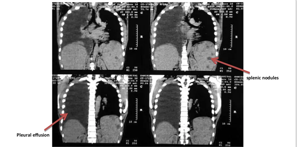
chest and abdominal pain. Examination revealed a right pleural effusion which was confirmed on chest radio-graph. Chest CT revealed massive right pleural effusion, right mid and lower zone consolidation and multiple par-atracheal and mediastinal lymph nodes (Fig. 2). He had drainage of the effusion with a sample taken for analysis.
Results showed an exudate demonstrating a heterogene-ous population of mature and immature lymphocytes within a proteinaceous haemorrhagic background, as well as normal adenine deaminase levels. Gram stain for bacteria and ZN stain for AFB were both negative. The pleural fluid had no malignant cells.
splenic nodules
Fig. 1 Computed tomography of the abdomen slices showing multiple splenic nodules
Pleural effusion
splenic nodules
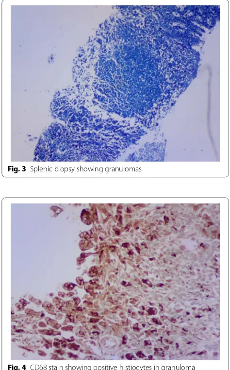
[image:3.595.58.539.87.340.2] [image:3.595.58.540.469.708.2]A percutaneous image-guided core needle biopsy (CNB) of his spleen was performed. This involved splenic sampling using a cutting needle under guidance of ultra-sound. Histopathological examination of the biopsy spec-imen revealed effaced splenic architecture with extensive multiple granulomas—mainly necrotising with multinu-cleate histiocytes, eosinophils and areas of fibrosis. There was no evidence of a malignant lymphocytic or foreign cell infiltrate. ZN stain of the biopsy processed speci-men revealed acid-fast bacilli (Figs. 3, 4, 5), confirmed by GeneXpert MTB/RIF which yielded MTB sensitive to rifampicin. With this tissue diagnosis confirmed, all the empiric antibiotics (vancomycin, ceftazidime, met-ronidazole) and antifungals (amphotericin-B) were dis-continued and the anti-TB treatment maintained with supportive management comprising analgesics, fluids and nutritional support. In view of the patient’s febrile neutropaenia, the clinicians instituted strict infection
control measures such as barrier nursing, hand washing and avoiding raw uncooked foods. These measures were taken in order to protect the patient from acquir-ing opportunistic infections from his environment, as he was severely neutropaenic thus immunologically vulner-able. The patient continued to deteriorate with persis-tent neutropaenia and fever for about 2 weeks before he registered progressive improvement and was discharged 30 days after initiation of anti-TB medication. His hos-pital stay lasted a total of 78 days. It was noted during the patient’s regular follow-up sessions as an outpatient that he remained neutropaenic for about a month’s dura-tion after discharge from the hospital. This neutropaenia resolved about 2 months into the TB treatment with a normal CBC. No adverse events were reported. He sub-sequently did well and is back to work, having completed a full 6-month course of anti-TB medication.
Discussion
According to the World Health Organization (WHO) Global Tuberculosis Report released in 2016, TB remains one of the top 10 leading causes of morbidity and death worldwide, even surpassing mortality attributable to HIV annually [12]. This report states that in the year 2015, 10.4 million people were infected with TB globally, and 1.4 million deaths were attributed to this disease. WHO surveillance data shows that TB is a major problem in the African region. Although the continent claimed 26% of the world’s new cases in the same year, it alarmingly had the most severe burden relative to population, that is 275 cases for every 100,000 persons. This figure sur-passed more than double the global average of 142 per 100,000 [12]. Our country Kenya is currently classified as one of the 30 high TB burden countries, which col-lectively account for 87% of all estimated incident cases worldwide. In the year 2015, Kenya’s estimated incidence rate of TB was 233 per 100,000 population, [12] and
Fig. 3 Splenic biopsy showing granulomas
Fig. 4 CD68 stain showing positive histiocytes in granuloma
[image:4.595.308.540.88.256.2] [image:4.595.55.292.337.718.2]this disease has been noted to rank as one of the lead-ing causes of mortality in the country [13, 14]. In view of the above, one recognises the importance and relevance of TB to clinicians and public health services in our local setting, regionally and globally at large.
This case report highlights a rare instance of primary splenic tuberculosis. The spleen is one of the organs involved in extrapulmonary TB. However, more common extrapulmonary sites include the lymph nodes, pleura, genitourinary tract, bones and joints, meninges, perito-neum and pericardium [15].
Haematogenous dissemination occurs frequently in HIV-infected individuals. Isolated splenic involvement is rare, and is usually reported in immunosuppressed indi-viduals [1–3, 8]. Coley first described TB of the spleen in 1846, referring to an “enlarged spleen secondary to tuberculosis with absent or limited involvement of other organs” [16]. Our patient was atypical in that he was a healthy immunocompetent male without any risk factors for splenic infection, and in whom MTB diagnostic tests (WHO-approved) such as ZN staining for AFB and Gen-eXpert MTB/RIF were negative. The initial presentation as pyrexia of unknown origin is characteristic of splenic TB in literature [7, 17]. Apart from fever which is present in majority of patients with splenic TB, other modes of pres-entation reported include fatigue and weight loss, spleno-megaly, [18] splenic rupture, [19] hypersplenism, portal hypertension with and without gastrointestinal bleed, and fulminant forms involving rapid progression [18]. Hemato-logic abnormalities include reduced cell counts but cases of polycythaemia have also been reported [20].
Isolated splenic tuberculosis frequently proves to be a diagnostic conundrum as demonstrated in our case report, mainly due to the vague non-specific nature of clinical presentation. In many case reports, imaging is important in initial identification of splenic pathol-ogy, and diagnosis further clinched by histopathological examination of a splenic fine needle aspirate, core needle biopsy or splenectomy specimen [4]. Abdominal ultra-sound is a cost-effective non-invasive imaging modality in such cases and may show a miliary pattern, nodular TB, tuberculous spleen abscess, calcific TB or a combination of these findings [9]. Our patient demonstrated nodular TB. On the other hand, abdominal CT scan has added value in ruling out involvement of other organs, and has higher diagnostic accuracy than ultrasound which suf-fers from operator-dependence. CT scan of the abdomen would often show multiple rounded hypodense lesions which may be present in a variety of conditions other than splenic TB [1, 21]. Differential diagnosis of solitary splenic masses that ought to be considered include cysts, haematoma, fungal infection, abscesses, infarcts, vascular tumours, lymphoma and metastatic tumours [8].
Histological confirmation via splenic biopsy provides a more accurate diagnosis of splenic pathology. Aspira-tion cytology of splenic lesions is variable. Suri et al. [22] reported up to 88% sensitivity for fine needle aspiration cytology (FNAC) for diagnosing a tuberculous pathology in the spleen. On the other hand, microscopic observa-tion of tissue secobserva-tions allows for histological typing and staging of tubercle lesions, as well as differentiation between granulomatous lesions and other radiographi-cally comparable lesions such as lymphomas. However literature suggests that fixing of tissues with formalin and xylene greatly reduces the sensitivity of acid-fast staining and could potentially lead to false negatives [23]. Pottakkat et al. [24] reported that AFB staining of splenic tissue sections was negative in over 339 patients who underwent splenectomy for indications other than trauma. Fukunaga et al. [23] reported that tubercle bacilli were frequently missed or underestimated with acid fast microscopy on formalin fixed, paraffin embedded tissues. In such cases, real-time polymerase chain reaction (PCR) of the tissue sections demonstrated markedly increased sensitivity over acid-fast staining.
CNB has a high diagnostic yield in splenic pathology [25] and has demonstrated superior diagnostic accuracy to FNAC in characterising splenic lesions [26, 27]. Tuber-cular infection is histologically confirmed by presence of typical caseation with granuloma of Langhan’s giant cells and epitheloid cells. In our case, classical granulomatous inflammation and acid fast bacilli were demonstrated upon examination of the CNB specimen and further con-firmed by GeneXpert MTB/RIF.
Therefore, in apparent “TB negative” cases, perform-ing more than one test improves yield. In our case, despite several ZN stain negative results, other diagnos-tic modalities were employed such as GeneXpert MTB/ RIF as well as histological confirmation through tissue diagnostic procedures including FNAC and CNB. All this was done in addition to the conventional ZN stain-ing technique to get the final diagnosis of TB spleen. It is important to note that culture is useful as well, as it is still considered the current reference standard for detecting MTB in spite of its drawbacks such as slow turnaround time (of up to 12 weeks) as well as dependence on well-equipped laboratories, technical expertise and resources which may be lacking in less developed localities [12]. In our resource-constrained setting, MTB culture is not performed regularly, although its contribution would have been invaluable in diagnostics.
Literature shows that anti-TB therapy alone may suf-fice in splenic TB diagnosed without splenectomy [17,
A few controlled trials recommend a 12-month course of anti-TB treatment for splenic TB [19]. However, there are few reports that show inadequate or absence of response to anti-TB therapy without splenectomy [8, 30]. In such cases, splenectomy is a viable option.
The patient’s worsening symptoms on initiation of anti-TB therapy was attributed to paradoxical reaction (PR). This is defined as the “worsening of existing lesions or presentation of new lesions during anti-TB therapy” [31], or the “worsening of clinical or radiological find-ings following the initiation of appropriate therapy” [32]. PR is typically associated with exaggerated inflamma-tory symptoms including fever, [33] lymphadenitis, [34] and pulmonary manifestations [33]—as illustrated in our case by worsening fever and pulmonary disease (pleu-ral effusion with mediastinal lymphadenopathy). It has been suggested that rapid killing of bacilli with antibiot-ics may lead to the release of large amounts of microbial components, which stimulate an exuberant inflamma-tory response, [35] and that higher baseline numbers of bacilli may potentiate this process. It has also been pos-tulated that this is essentially a hypersensitivity reaction to persistent mycobacterial antigen [34]. Ultimately, the PR phenomenon is likely to be missed, and in such cases TB may be labelled as a misdiagnosis warranting discon-tinuation of therapy. Fortunately, PR is self-limiting and does not always require steroid therapy [36]. Our patient improved with supportive management and continuous anti-TBs without steroids. In summary, the chronological course of events as pertains to this case has been repre-sented in a timeline graphic (see Additional file 1).
Conclusions
We present a rare case of splenic TB in an immune com-petent host with paradoxical worsening on TB ther-apy. Isolated splenic TB presents a profound diagnostic complexity, especially in resource-constrained settings. Splenic core needle biopsy is indispensable in scenarios where splenectomy cannot be performed, and TB-PCR techniques are useful in ZN stain negative cases. This case highlights the need to continuously be on the look-out for TB especially in unusual presentations, includ-ing subsequent paradoxical reaction which may be encountered.
Abbreviations
AFB: acid-fast-bacilli; Anti-TB: antituberculous; CBC: complete blood count; CNB: core needle biopsy; CT: computed tomography; ELISA: enzyme-linked immunosorbent assay; FNAC: fine needle aspiration cytology; HIV: human Additional file
Additional file 1. Timeline of events.
immunodeficiency virus; PCR: polymerase chain reaction; PR: paradoxical reac-tion; TB: tuberculosis; WHO: World Health Organizareac-tion; ZN: Ziehl–Neelsen.
Authors’ contributions
FW drafted the original manuscript, which was then amended with sugges-tions by LA and JR. FW, GO, TW, JN cared for the patient with LA as the consult-ant-in-charge. JR interpreted bone marrow aspirate findings and provided valuable pathological expertise during the care of the patient. All authors read and approved the final manuscript.
Author details
1 Department of Clinical Medicine and Therapeutics, School of Medicine, College of Health Sciences-University of Nairobi, P.O. Box 19676, Nairobi 00202, Kenya. 2 Haematology and Blood Transfusion Unit, Department of Human Pathology, School of Medicine, College of Health Sciences-University of Nai-robi, P.O. Box 19676, Nairobi 00202, Kenya.
Acknowledgements
The authors are grateful to the patient for his full consent to the publication of this case report. They would also like to thank Dr. Peter Chacha Magabe for his work in interventional radiology and performing the splenic biopsy.
Competing interests
The authors declare that they have no competing interests.
Availability of data and materials
Biological material deposited in Lancet Pathology laboratories, Nairobi.
Consent for publication
Written informed consent was obtained from the patient for publication of this Case Report and any accompanying images.
Publisher’s Note
Springer Nature remains neutral with regard to jurisdictional claims in pub-lished maps and institutional affiliations.
Received: 23 June 2016 Accepted: 5 April 2017
References
1. Azzam NA. Splenic tuberculosis presenting as fever of unknown origin with severe neutropenia. Ann Clin Microbiol Antimicrob. 2013;12(1):1–3. 2. Gupta PP, Fotedar S, Agarwal D, Sansanwal P. Tuberculosis of spleen pre-senting with pyrexia of unknown origin in a non-immunocompromised woman. Lung India. 2008;25(1):22–4.
3. Mishra H, Pradeep R, Rao GV, Anuradha S, Reddy DN. Isolated tubercu-losis of the spleen: a case report and review of literature. Indian J Surg. 2013;75(3):235–6.
4. Imani Fooladi AA, Hosseini MJ, Azizi T. Splenic tuberculosis: a case report. Int J Infect Dis. 2009;13(5):e273–5.
5. Raviraj S, Gogia A, Kakar A, Byotra SP. Isolated splenic tuberculosis without any radiological focal lesion. Case Rep Med. 2015;2015:2.
6. Basa JV, Singh L, Jaoude WA, Sugiyama G. A case of isolated splenic tuber-culosis. Int J Surg Case Rep. 2015;8:117–9.
7. Ho PL, Chim CS, Yuen KY. Isolated splenic tuberculosis presenting with pyrexia of unknown origin. Scand J Infect Dis. 2000;32(6):700–1. 8. Kundu PR, Mathur SK, Singh S, Duhan A, Aggarwal G, Sen R. Isolated
tuberculous splenic abscess in an immunocompetent individual. Asian Pac J Trop Med. 2011;4(1):81–2.
9. Zhan F, Wang C-J, Lin J-Z, Zhong P-J, Qiu W-Z, Lin H-H, et al. Isolated splenic tuberculosis: a case report. World J Gastrointest Pathophysiol. 2010;1(3):109–11.
• We accept pre-submission inquiries
• Our selector tool helps you to find the most relevant journal
• We provide round the clock customer support
• Convenient online submission
• Thorough peer review
• Inclusion in PubMed and all major indexing services
• Maximum visibility for your research
Submit your manuscript at www.biomedcentral.com/submit
Submit your next manuscript to BioMed Central
and we will help you at every step:
11. Klastersky J, de Naurois J, Rolston K, Rapoport B, Maschmeyer G, Aapro M, et al. Management of febrile neutropaenia: ESMO Clinical Practice Guidelines. Ann Oncol. 2016;27(suppl 5):v111–8.
12. World Health Organization. Global tuberculosis report; 2016. 13. Mbithi A, Gichangi A, Kim AA, Katana A, Weyenga H, Williamson J, et al.
Tuberculosis and HIV at the national level in Kenya: results from the second Kenya AIDS indicator survey (1999). J Acquir Immune Defic Syndr. 2014;66(Suppl 1):S106–15.
14. Mohajan HK. Improvement of health sector in Kenya. Am J Public Health Res. 2014;2(4):159–69.
15. Raviglione MC. Tuberculosis. In: Kasper DL, Fauci AS, Hauser SL, Longo DL, Jameson JL, Loscalzo J, editors. Harrison’s principles of internal medicine. 19th ed. New York: McGraw-Hill Education; 2015. p. 1102–22.
16. Meredith HC Jr, Early JQ, Becker W. Tuberculous splenomegaly with the hypersplenism syndrome: a case report. Blood. 1949;4(12):1367–73. 17. Bastounis E, Pikoulis E, Varelas P, Cirochristos D, Aessopos A.
Tuber-culoma of the spleen: a rare but important clinical entity. Am Surg. 1999;65(2):131–2.
18. Rhazal F, Lahlou MK, Benamer S, Daghri JM, Essadel E, Mohammadine E, et al. Splenomegaly and splenic pseudotumor due to tuberculosis: six new cases. Ann Chir. 2004;129(8):410–4.
19. Hamizah R, Rohana AG, Anwar SA, Ong TZ, Hamzaini AH, Zulkarnaen AN. Splenic tuberculosis presenting as pyrexia of unknown origin. Med J Malays. 2007;62(1):70–1.
20. Berady S, Rabhi M, Bahrouch L, Sair K, Benziane H, Benkirane A, et al. Isolated pseudo-tumoral tuberculosis of the spleen. A case report. La Revue de medecine interne/fondee par la Societe nationale francaise de medecine interne. 2005;26(7):588–91.
21. Denton T, Hossain J. A radiological study of abdominal tuberculosis in a Saudi population, with special reference to ultrasound and computed tomography. Clin Radiol. 1993;47:409–14.
22. Suri R, Gupta S, Gupta SK, Singh K, Suri S. Ultrasound guided fine needle aspiration cytology in abdominal tuberculosis. Br J Radiol. 1998;71(847):723–7.
23. Fukunaga H, Murakami T, Gondo T, Sugi K, Ishihara T. Sensitivity of acid-fast staining for Mycobacterium tuberculosis in formalin-fixed tissue. Am J Respir Crit Care Med. 2002;166(7):994–7.
24. Pottakkat B, Kumar A, Rastogi A, Krishnani N, Kapoor VK, Saxena R. Tuberculosis of the spleen as a cause of fever of unknown origin and splenomegaly. Gut Liver. 2010;4(1):94–7.
25. Olson MC, Atwell TD, Harmsen WS, Konrad A, King RL, Lin Y, et al. Safety and accuracy of percutaneous image-guided core biopsy of the spleen. Am J Roentgenol. 2016;206(3):655–9.
26. Sammon J, Twomey M, Crush L, Maher MM, O’Connor OJ. Image-guided percutaneous splenic biopsy and drainage. Semin Interven Radiol. 2012;29(4):301–10.
27. Singh AK, Shankar S, Gervais DA, Hahn PF, Mueller PR. Image-guided percutaneous splenic interventions. Radiographics. 2012;32(2):523–34. 28. Wilde CC, Kueh YK. Case report: tuberculous hepatic and splenic abscess.
Clin Radiol. 1991;43(3):215–6.
29. Small PM, Fujiwara PI. Management of tuberculosis in the United States. N Engl J Med. 2001;345(3):189–200.
30. Nayyar V, Ramakrishna B, Mathew G, Williams RR, Khanduri P. Response to antituberculous chemotherapy after splenectomy. J Intern Med. 1993;233(1):81–3.
31. Cheng VC, Ho PL, Lee RA, Chan KS, Chan KK, Woo PC, et al. Clinical spec-trum of paradoxical deterioration during antituberculosis therapy in non-HIV-infected patients. Eur J Clin Microbiol Infect Dis. 2002;21(11):803–9. 32. Park JA, Park SS, Park SE. A paradoxical reaction during antituberculosis therapy for congenital tuberculosis. Int J Infect Dis. 2009;13(5):e279–81. 33. Cheng SL, Wang HC, Yang PC. Paradoxical response during
anti-tubercu-losis treatment in HIV-negative patients with pulmonary tubercuanti-tubercu-losis. Int J Tuberc Lung Dis. 2007;11(12):1290–5.
34. Hawkey CR, Yap T, Pereira J, Moore DA, Davidson RN, Pasvol G, et al. Characterization and management of paradoxical upgrading reactions in HIV-uninfected patients with lymph node tuberculosis. Clin Infect Dis. 2005;40(9):1368–71.
35. Campbell IA, Dyson AJ. Lymph node tuberculosis: a comparison of vari-ous methods of treatment. Tubercle. 1977;58(4):171–9.