MODULATION OF DENDRITIC CELL
SIGNALLING AND FUNCTION BY REDOX
REGULATORS
Thesis submitted in accordance with the requirements of the University of Liverpool for the degree of Doctor in Philosophy
by
Laith Mohammed Abbas Al-Huseini
DECLARATION
This thesis is the result of my own work. The material contained within this thesis has not been presented, nor is currently being presented, either wholly or in part
for any degree or other qualification.
Laith Mohammed Abbas Al-Huseini
This research was carried out in the MRC Centre for Drug Safety Science,
TABLE OF CONTENTS
PAGE
ABSTRACT i
ACKNOWLEDGEMENTS iii
PUBLICATIONS v
ABBREVIATIONS vii
1.0 CHAPTER ONE: GENERAL INTRODUCTION
1.1 Immunity 2
1.2 Innate immune cells 3
1.3 Dendritic cells (DCs) 4
1.3.1 Origin and development of DCs 5
1.3.2 Immature versus mature DCs 7
1.3.2.1 Immature DCs 7
1.3.2.2 Mature DCs 9
1.4 Antigen acquisition by DCs 10
1.5 Dendritic cell antigen presentation – role of MHC Class I and II 11
1.5.1 MHC class I 11
1.5.2 MHC class II 12
1.6 Activation of T cells by antigen bearing mature DCs 13 1.6.1 Role for DC co-stimulatory receptors in T cell activation 13 1.6.2 Role for DC cytokines in T-cell activation and differentiation 14 1.7 Key signalling pathways involved in DC maturation 15
1.7.1 The NF-B pathway 17
1.7.2.1 Extracellular signal-regulated protein 21 kinases 1/2 (ERK1/2)
1.7.2.2 c-Jun N-terminal kinases (JNKs) 22
1.7.2.3 The p38MAPK pathway 24
1.7.2.4 The transcription factors CREB/ATF1 26 are targets of MAPKs
1.8 Redox homeostasis and immune cell function 28
1.9 Influence of redox on signalling pathways 31
1.10 Cellular defence against reactive oxygen species 31 1.10.1 Thiol-containing cellular antioxidants 32 1.10.2 Enzymes as effectors of redox homeostasis 33 1.10.3 Small non-protein antioxidant molecules 34
1.11 Nrf2 35
1.11.1 Kelch-like ECH-associated protein-1 (Keap-1) 37
1.11.2 Regulation of Nrf2 by Kelch-like ECH-associated protein-1 (Keap-1) 39
1.11.3 Nrf2 activators differentially modify Keap1 41
1.12 Role of Nrf2 in redox homeostasis and immune cell function 42
1.13 Heme oxygenases (HOs) in redox homeostasis 44
1.14 Aims of the study 49
2.0 CHAPTER TWO: MATERIALS AND METHODS
2.1 Cell culture materials and reagents 52
2.2 Antibodies 53
2.3 Cell lines 53
2.4 Mice 54
2.5 Generation of mouse bone marrow derived dendritic cells (DCs) 54
2.6 Viable cell count by Trypan Blue exclusion 55
2.9 Isolation of F5 CD8 T cells 56
2.10 F5 CD8 T cell proliferation assay 57
2.11 Measurement of reactive oxygen species (ROS) levels 57
2.12 Apoptosis assay 58
2.13 DC proliferation assay 58
2.14 ATP activity assay 58
2.15 Detergent lysis of DCs 59
2.16 Nuclear extraction of DCs 59
2.17 Protein content determination via Bradford assay 60
2.18 Western blotting analysis 60
2.19 Reprobing of blotted membranes 62
2.20 Measurement of NF-B activity 63
2.21 Histone deacetylase (HDAC) activity assay 63
2.22 Enzyme-Linked Immunosorbent Assay (ELISA) for IL-10 64
2.23 DC endocytosis assay 65
2.24 DC phagocytosis assays 65
2.25 Statistical analysis 66
3.0 CHAPTER THREE: REGULATION OF DC PHENOTYPE, FUNCTION, AND SIGNALLING PATHWAYS BY NRF2
3.1 INTRODUCTION 68
3.2 RESULTS 70
3.2.1 Loss of Nrf2 alters DC proliferative function 70 that is dependent on culture conditions
3.2.2 Loss of Nrf2 is associated with increased levels of anti-apoptotic 72 protein, Bcl-2, but does not increase DC survival
3.2.4 Reduction of cellular glutathione leads to increased ROS 78 3.2.5 Loss of Nrf2 results in altered iDCs phenotype 79 3.2.6 Loss of Nrf2 does not alter LPS responsiveness of DCs 81 3.2.7 Increased co-stimulatory receptor expression of Nrf2-/- iDCs is 82 associated with enhanced antigen-specific CD8 T cell stimulatory capacity
3.2.8 Altered immature DC function due to the loss of Nrf2 is not 84 dependent on elevated ROS
3.2.9 The NF-B pathway does not contribute to altered phenotype and 88 function observed in Nrf2-deficient DC
3.2.10 ERK1/2 MAPK is not involved in mediating Nrf2-dependent 93 modulation of DC function
3.2.11 c-Jun N-terminal kinase (JNK) is not involved in mediating Nrf2- 96 dependent modulation of DC function
3.2.12 Contribution of p38MAPK activity to Nrf2-dependent regulation 99 of DC immune function
3.2.13 Contribution of the p38MAPK-CREB/ATF1 signalling pathway to 102 Nrf2-dependent regulation of DC immune function
3.2.14 Loss of Nrf2 leads to the dysregulated IL-10 production in DCs 104 3.2.15 Increased DC co-stimulatory receptor expression due to the 106 loss of Nrf2 is not dependent on PKA activity
3.2.16 Enhanced co-stimulatory receptor expression of Nrf2-/- iDCs 107 is dependent on histone deacetylase activity
3.3 DISCUSSION 110
4.0 CHAPTER FOUR: REGULATION OF DC PHENOTYPE, FUNCTION, AND SIGNALLING PATHWAYS BY HEME OXYGENASE-1
4.1 INTRODUCTION 121
4.2.1 Loss of Nrf2 resulted in decreased expression of HO-1 122 4.2.2 HO-1 expression decreases during DC maturation 123 4.2.3 Inhibition of HO-1 activity alters immature DC phenotype 124 and function
4.2.4 HO-1 inhibition in DCs results in elevated intracellular ROS levels 129 4.2.5 Altered immature DC function by HO-1 inhibition is not 131 dependent on elevated ROS
4.2.6 HO-1 regulates DC phenotype and function through the 134 p38MAPK-CREB/ATF1 pathway
4.2.7 Up-regulation of HO-1 renders DCs refractory to LPS-induced 137 DC signalling and maturation
4.2.8 The HO-1 substrate, heme, induces DC functional maturation 140 through p38MAPK-CREB/ATF1 pathway
4.3 DISCUSSION 146 5.0 CHAPTER FIVE: FINAL DISCUSSION
5.1 INTRODUCTION 156
5.2 Loss of Nrf2 mediated dysregulation in multiple DC functions through 157 redox-sensitive p38MAPK signalling pathway
5.3 Increases heme levels and inhibition of HO-1 activity result in 162 dysregulation in multiple DC functions through redox-sensitive
p38MAPK signalling pathway
LIST OF FIGURES AND TABLES
PAGE
1.0CHAPTER ONE: GENERAL INTRODUCTIONFigure 1.1 Dendritic cells (DCs) differentiation from pluripotent 5 haematopoietic stem cells (HSCs)
Figure 1.2 Dendritic cells (DCs) have central role in the immune system by 8 generating antigen-specific T cells from naïve T cells
Table 1.1 Summary of Toll like receptor classes and ligands 16 Figure 1.3 Signalling cascades leads to NF-B activation 19 Figure 1.4 Signalling cascades leads to ERK1/2 activation 22 Figure 1.5 Signalling cascades leads JNK activation 23 Figure 1.6 Signalling cascades leads to p38MAPK activation 26 Figure 1.7 Schematic representation for the basic structure 36 of transcription factor Nrf2
Figure 1.8 Schematic presentation of Keap1 38
Figure 1.9 Schematic presentation of Nrf2 stimulation 40 Figure 1.10 Schematic diagram of heme oxygenase-1 45 Figure 1.11 Schematic representation of heme degradation 46 by heme oxygenase-1
Figure 1.12 Schematic representation of Bach1 structure 47 2.0CHAPTER TWO: MATERIALS AND METHODS
Table 2.1 Summary of Western blotting antibodies used in the study 62
3.0 CHAPTER THREE: REGULATION OF DC PHENOTYPE, FUNCTION, AND SIGNALLING PATHWAYS BY NRF2
Figure 3.1 Nrf2 modulation of DC proliferation depends on culture 71 conditions
Figure 3.3 Loss of Nrf2 results in increased DC intracellular ROS Levels 77 but is not accompanied by increased oxidative metabolism
Figure 3.4 Reduction of cellular glutathione leads to increased ROS 78 Figure 3.5 Loss of Nrf2 results in enhanced co-stimulatory 80
molecule expression in immature DCs
Figure 3.6 Responsiveness of DCs to LPS is unaffected by loss of Nrf2 82 Figure 3.7 Increased T cell stimulatory capacities of Nrf2-/- iDCs 84 Figure 3.8 Reducing ROS levels does not restore altered phenotype 87
and function of Nrf2-deficient iDCs
Figure 3.9 The NF-B pathway does not contribute to altered 92 phenotype and function of Nrf2-deficient iDCs
Figure 3.10 ERK1/2 does not contribute to the altered DC 95 phenotype and function in Nrf2-/- iDCs
Figure 3.11 Basally activated JNK is not involved in mediating 98 altered phenotype and function in Nrf2- deficient DCs
Figure 3.12 Requirement for p38MAPK activity in Nrf2-dependent 101 regulation of DC phenotype and function
Figure 3.13 Loss of Nrf2 perturbs p38MAPK-CREB/ATF1 signalling in DCs 103 Figure 3.14 Loss of Nrf2 results in elevated IL-10 production by DCs 105 Figure 3.15 PKA is not required for Nrf2-dependent regulation of 106
DC phenotype
Figure 3.16 Histone deacetylase function is required for 109 Nrf2-dependent regulation of DC phenotype
4.0 CHAPTER FOUR: REGULATION OF DC PHENOTYPE, FUNCTION, AND SIGNALLING PATHWAYS BY HEME OXYGENASE-1
Figure 4.1 Loss of Nrf2 results in reduced expression of HO-1 123 Figure 4.2 DC maturation is associated with reduction in HO-1 expression 124 Figure 4.3 Inhibiting HO-1 activity alters dendritic cell phenotype 128 and function
can be restored by vitamin treatment
Figure 4.5 Altered immature DC function by HO-1 inhibition is 133 not dependent on elevated ROS
Figure 4.6 Modulation of dendritic cell phenotype and function by 136 HO-1 is mediated through the p38MAPK-CREB/ATF1 pathway
Figure 4.7 Upregulation of HO-1 activity inhibits LPS-induced signalling, 139 phenotypic and functional changes in DCs
Figure 4.8 HO-1 substrate, heme, induces iDC signalling, 145 phenotypic and functional changes in DCs
5.0 CHAPTER FIVE: FINAL DISCUSSION
i
ABSTRACT
Dendritic cells (DCs) are antigen-presenting cells crucial for the initiation and coordination of primary adaptive immune responses. Immature DCs (iDCs) express low levels of MHC class II and co-stimulatory molecules such as CD80, CD86, and CD40, with high phagocytic capacity and limited ability to induce antigen-specific T cell activation. DC maturation is associated with up-regulation of co-stimulatory molecules and cytokine production, rendering the DCs competent in T cell activation and the elicitation of an immune response. DC function and co-stimulatory receptor gene expression are known to be regulated by intracellular redox status, NF-B and MAPKs signalling pathways.
Intracellular reactive oxygen species (ROS) levels influence DC maturation and function. The transcription factor, Nrf2, is essential for maintaining intracellular redox homeostasis. In response to oxidative stress, Nrf2 induces the transcription of a set of cytoprotective and antioxidant genes, including heme oxygenase-1 (HO-1), that are required for detoxification of xenobiotics and their reactive metabolites and the nullification of oxidative insult.
It is now emerging that Nrf2, and its gene product, HO-1, play pivotal roles in regulation of immune responses. However, the key signalling mechanisms involved in Nrf2 and HO-1-mediated altered DC function has not been fully elucidated and requires further investigation. In addition, the role of ROS in the absence of Nrf2 or HO-1 activity, in DC activation and function has not been investigated.
Using immature bone marrow-derived DCs (iDCs) from Nrf2+/+ and Nrf2-/- mice, we demonstrate in the first part of the work presented in this thesis, that Nrf2 deficiency in iDCs resulted in increased ROS levels, enhanced iDCs co-stimulatory receptor expression, and increased iDC-mediated antigen-specific CD8 T cell stimulatory capacity in response to an antigenic peptide.
ii phenotype and function did not require NF-B, ERK or JNK activity but was dependent on p38MAPK-CREB/ATF1 activity.
Based on these experimental results, we conclude that Nrf2 regulates DC maturation and function by modulating intracellular signalling pathways independent of intracellular ROS levels.
In the second part of the study, we demonstrate that inhibition of HO-1 activity in iDCs resulted in DCs with raised intracellular ROS levels, a mature phenotype, impaired phagocytic and endocytic function, and increased capacity to stimulate antigen-specific CD8 T cells. Interestingly, our results reveal that the increased ROS levels following HO-1 inhibition did not underlie the changes in phenotype and functions observed in these iDCs. Importantly, we show that the p38MAPK-CREB/ATF1 pathway was involved in the mediation of the phenotypic and functional changes arising from HO-1 inhibition. Furthermore, up-regulation of HO-1 activity rendered iDCs refractory to lipopolysaccharide-induced activation of p38MAPK-CREB/ATF1 pathway and DC maturation. Finally, we demonstrate that treatment of iDC with the HO-1 substrate, heme, recapitulated the effects that result from HO-1 inhibition. Based on these experimental results, we conclude that HO-1 regulates DC maturation and function by modulating the p38MAPK-CREB/ATF1 signalling axis.
iii
ACKNOWLEDGEMENTS
Completing this PhD degree was a very challenging task and the best and worst moments during my doctoral journey have been shared with many people.
First, I am truly indebted and thankful to my supervisor Dr Jean Sathish who has supported me throughout my thesis and without his patience, caring, guidance, and immense knowledge, this doctoral thesis would not have been completed. I would like to thank him for guiding my scientific knowledge and writing with providing priceless input. I consider it a big honour to know you and with all our scientific and philosophic discussions, I am leaving now with a huge experience and a very memorable and privilege journey.
I would like to thank all the current and past members in the “Lost in Translation Group”
Foremost Han, I cannot find words to express my gratitude for you; I would never have been able to finish my PhD without your guidance. I cannot forgetting you were always there cheering me up with your patience, motivation, and enthusiasm. I could not have imagined having a better advisor and mentor.
Junnat, your guidance helped me in all the time of research and writing of this thesis. Thanks for providing me with lab technical and theoretical assistance leading to creating enjoyable workplace.
Sammy, you were a good friend always willing to help and give his best suggestions. It was a great pleasure and pain! to work with you and having our regular Thursday’ Curry lunch at the Royals. I felt it has been a lonely lab without you.
Deepest gratitude for the members of the group: Thilipan, Yulia, Naif, and Paul for all the fun we have had in the last four years and for your continued friendship.
I want to express a big acknowledgement to the Ministry of Higher Education and Scientific Research in Iraq for choosing me to a sponsored studentship and funding my PhD.
I would like to thank University of Liverpool for accepting me in this PhD and the Institute of Translational Medicine Postgraduate Office especially Prof Andrea Varro and Miss Lisa Crimmins for all the help they provided.
iv A big thank to my wonderful son Jahfar for his understanding of my circumstances in the last four years. I am really appreciated your support my kid.
Lastly and most importantly, I wish to express my love and gratefulness to my wonderful wife Sawsan; for her support, sympathetic and endless love through the duration of my studies and stood by me through the good and bad times.
v
PUBLICATIONS
Papers
Laith M A Al-Huseini, Han Xian Aw Yeang, Junnat M Hamdam, Swaminathan Sethu, Naif Alhumeed, Wai Wong and Jean G Sathish. Heme oxygenase-1 regulates dendritic cell function through modulation of p38MAPK-CREB/ATF1 signalling. (2013, submitted)
Laith M A Al-Huseini, Han Xian Aw Yeang, Swaminathan Sethu, Naif Alhumeed, Junnat M. Hamdam, Yulia Tingle, Laiche Djouhri, Neil Kitteringham, B. Kevin Park, Christopher E. Goldring, and Jean G. Sathish. Nuclear factor-erythroid 2 (NF-E2) p45-related factor-2 (Nrf2) modulates dendritic cell immune function through regulation of p38 MAPK-cAMP-responsive element binding protein/activating transcription factor 1 signalling (2013). Journal of Biological Chemistry. 2;288(31):22281-8.
Han Xian Aw Yeang, Junnat M Hamdam, Laith M A Al-Huseini, Swaminathan Sethu, Laiche Djouhri, Joanne Walsh, Neil Kitteringham, B Kevin Park, Christopher E Goldring and Jean G Sathish. Loss of the transcription factor nuclear erythroid2 (NF-E2) p45-related factor-2 (Nrffactor-2) leads to dysregulation of immune functions, redox homeostasis and intracellular signalling in dendritic cells (2012). Journal of Biological Chemistry. 287(13):10556-64.
Reviews
vi
Abstracts for Conference Presentations
Laith M A Al-Huseini, Han Xian Aw Yeang, Junnat Hamdam, Christopher Goldring, Jean G Sathish. Cellular redox affects dendritic cells maturation and phagocytic function. 6th European Network of Immunology Institutes Summer School in Advanced Immunology, Sardinia, Italy, May, 2011. Poster presentation
Laith M A Al-Huseini, Han Xian Aw Yeang, Junnat Hamdam, Swaminathan Sethu, Thilipan Thaventhiran, Christopher Goldring, Kevin Park and Jean Sathish. Loss of Nrf2 results in perturbation of NF-B and MAPK signalling pathways in dendritic cells. Biochemical Society, Hot Topic Event - Nrf2 signalling in health and disease, London, UK, December, 2011. Poster presentation
Laith M A Al-Huseini, Han Xian Aw Yeang, Junnat Hamdam, Christopher Goldring, Kevin Park and Jean G Sathish. Loss of Nrf2 affects co-stimulatory markers expression and MAPKs signalling in dendritic cells. British Society for Immunology Congress, Liverpool, UK, December, 2011. Poster presentation
Laith M A Al-Huseini, Swaminathan Sethu, Han Xian Aw Yeang, Junnat Hamdam, Naif Alhumeed, Christopher Goldring, Kevin Park and Jean G Sathish. Altered co-stimulatory phenotype of Nrf2 deficient dendritic cells is not a result of elevated ROS levels. 3rd EUROPEAN CONGRESS OF IMMUNOLOGY, Glasgow, UK, September, 2012. Poster presentation
vii
Abbreviations
2-ME 2-Mercaptoethanol ANOVA analysis of variance AP-1 activator protein 1 APC Antigen presenting cell APL altered peptide ligand APS ammonium persulphate ARE antioxidant response element ASK1 apoptosis signal-regulating kinase 1 ATP adenosine triphosphate
ATF1 activating transcription factor 1 BMDC bone marrow– derived dendritic cell bp base pair
BSA bovine serum albumen bZip basic leucine zipper
C Celsius
cAMP cyclic adenosine monophosphate
CD cluster of differentiation (cell surface molecules expressed on various cell types) cDNA complementary deoxyribonucleic acid
CFSE carboxy fluorescein succinimidyl ester c/EBPβ
CNC
CCAAT- enhancer binding proteins cap ‘n’ collar
CIITA class II major histocompatibility complex transactivator CO2 carbon dioxide
ConA Concanavalin A COOH carboxyl
CoPPIX cobalt protoporphyrin IX cpm counts per minute CRE Cre recombinase
CREB Cyclic-AMP response element binding protein CSA Cyclosporin A
Csk Carboxy terminal Src Kinase
cSMAC central supramolecular activation cluster CTL cytotoxic T lymphocyte
Da Dalton
DAG Diacyl Glycerol DC dendritic cell DMSO dimethyl sulphoxide
DN double negative (CD4-CD8-) DNA deoxyribonucleic acid DP double positive (CD4+CD8+) DPP3 dipeptidyl-peptidase 3 dsRNA double stranded RNA
viii ECH Embedded contact homology
ECL enhanced chemiluminescence EDTA ethylenediaminetetraacetic acid ELISA Enzyme-linked immunosorbent assay ERK extracellular signal regulated protein kinase EtOH Ethanol
Fab Ig-variable region
FACS fluorescence activated cell sorting FasL Fas ligand
FBS fetal bovine serum Fc Ig-constant region FCS fetal calf serum
FITC fluorescein isothiocyante
GAPDH Glyceraldehyde 3-phosphate dehydrogenase GCL γ-glutamylcysteine ligase
GDP guanosine diphosphate
GM-CSF granulocyte-macrophage colony stimulating factor GPX GSH peroxidase
GSH Reduced glutathione GSR GSH reductase
H2O water
H2O2 hydrogen peroxide
HBSS Hanks balanced salt solution HCl Hydrochloric acid
HEPES N-2-hydroxyethylpiperazine-N-2-ethanesulfonic acid HLA Human leukocyte antigen
HO-1 heme oxygenase 1 HRP horseradish peroxidase HSF heat shock factor HSP heat shock protein IFN Interferon
Ig immunoglobulin IκB inhibitor of NF-κB IKK IκB kinase
IL interleukin IL-2 interleukin-2 IL-10 interleukin-10
iNOS inducible nitric oxide synthase IP immunoprecipitation
IS immunological synapse
ITAM immunoreceptor tyrosine-based activation motif IU international units
IκB inhibitor of κB
JNK c-Jun amino-terminal kinase kDa Kilo Dalton
ix
L liter
LPS lipopolysaccharide
µ micro
ml millilitre
M molar
mA milliamps
mAb monoclonal antibody
MAPK mitogen activated protein kinase MAPKAP
K
MAPK-activated protein kinase MgSO4 Magnesium sulfate
MHC major histocompatibility complex MHz Mega Hertz
MFI mean fluorescence intensity min minutes
MKPs MAPK phosphatases
MOPS 3-(N-morpholino)propanesulfonic acid mRNA messenger RNA
MTT (3-(4,5-Dimethylthiazol-2-yl)-2,5-Diphenyltetrazolium Bromide) NADPH nicotinamide adenine dinucleotide phosphate
Neh Nrf2-ECH homology NES nuclear export signal
NF-AT nuclear factor of activated T cells NF-E2 nuclear factor erythroid 2
NFκB Nuclear Factor Kappa Beta (nuclear factor kappa-light-chain-enhancer of activated B cells)
NH4 Ammonium
NK natural killer NP-40 nonidet P-40
Nrf2 Nuclear Factor-Erythroid 2 (NF-E2) p45-related Factor-2 OD Optical density
OD570n
m optical density at 570 nm OH hydroxyl
PBMC Peripheral blood mononuclear cell PBS phosphate buffered saline
PD-1 programmed death-1 PE phycoerythrin
PHA Phytohemagglutinin pI isoelectric point PI propidium iodide
pKa acid dissociation constant PKA Protein kinase A
PKC protein kinase C
x PS Phosphatidylserine
PTB phosphotyrosine binding PTK protein tyrosine kinase PTP protein tyrosine phosphatase RCF (g) Relative Centrifugal Force RNA ribonucleic acid
RNAi RNA interference ROS reactive oxygen species rpm revolutions per minute
SD standard deviation of the mean SDS sodium dodecyl sulphate
SDS-PAGE SDS-Polyacrylamide gel electrophesis SH Sulphydryl
siRNA short interfering RNA
STAT1 signal transducers and activators of transcription 1 Std Standard
TAP transporter associated with antigen processing TBS Tris-buffered saline
TBS-T Tris-buffered saline-Tween TCR T cell receptor
TH T-helper
TLR4 Toll-like receptor-4
TMB 3,3′,5,5′-Tetramethylbenzidine TNF Tumor necrosis factor
TNF-R TNF receptor
TNF-α tumour necrosis factor α
TRAF6 TNF receptor associated factor 6 TRX Thioredoxin
TRX-R TRX reductase Tyr Tyrosine
U Unit
UK United Kingdom
USA United States of America UV Ultraviolet
V Volts
v/v volume/volume w/v weight/volume WT Wild Type
Gamma
1
CHAPTER ONE
2 1.1 Immunity
Immunity is broadly defined as the organism’s ability to protect itself from invading pathogens. Immunity is mediated by the coordinated response of a large variety of immune cells and soluble mediators. Checks and balances also exist within the immune system that operate to prevent an attack against the body’s own constituents, a phenomenon termed tolerance. Immunity can be generally categorized into innate and adaptive (specific or acquired) immunity.
Innate immunity represents the initial rapid defensive response against encountered microbes. This was previously believed to be a non-specific immune response, characterised by the engulfment and digestion of microorganisms and foreign substances via myeloid phagocytic cells. However, recent advances in immunology showed that innate immunity has considerable specificity in discriminating between pathogens versus host components and that it can be a prerequisite for the triggering of adaptive immunity. Innate immunity is present from birth and includes physical barriers (e.g. intact skin and mucous membranes), chemical barriers (e.g. gastric acid, digestive enzymes and bacteriostatic fatty acids of the skin), phagocytic cells, antigen presenting cells and the complement system (Beutler, 2004; Kimbrell and Beutler, 2001).
3 1.2 Innate immune cells
Innate immune response is mostly dependent upon cells of myeloid origin that can engulf and destroy pathogens. While these cells have independent capabilities to deal with such infectious challenges, they have also evolved to be more effective in conjunction with cells and proteins of the adaptive immune system. For example, lymphoid cells of the adaptive immune system produce antibodies that opsonise bacteria which can then be destroyed by innate immune cells (Beutler, 2004). These myeloid cells include mononuclear and polymorphonuclear phagocytes. The mononuclear phagocytes are the macrophages and monocytes and under inflammatory conditions monocytes can also mature into a subtype of dendritic cells which are discussed in section 1.3. The mononuclear phagocytes are competent at presenting antigens to T cells of the adaptive immune system. Macrophages are widely distributed throughout the body mostly in the lining parenchyma of major organs (e.g. heart, brain, lung, and liver) where they come in contact with invasive pathogens. They are morphologically diverse with specialised tissue-resident cell types. Dendritic cells follow similar distribution and function and also exhibit a variety of morphologies (e.g. the Langerhans cell of the skin; the plasmacytoid dendritic cell of the spleen). Although they exist as a minority population among the mononuclear phagocytes, DCs are capable of initiating adaptive immune responses to most pathogens by presenting a wide range of antigens to CD4+ and CD8+ T cells (Banchereau and Steinman, 1998).
4 The innate immune system also contains cells that are of a lymphoid origin called natural killer cells (NK cells) that are morphologically similar to lymphocytes. Natural killer cells target and lyse abnormal/viral-infected cells which have low/absent expression of MHC class I cell surface molecules with no prior specific sensitization of the host (unlike T cells). They play important roles in anti-tumour and anti-viral immunity (Ljunggren and Karre, 1990).
Dendritic cells are one of the crucial components of the immune system due to their essential role in the induction and control of T cell mediated immunity, as well as in the modulation of responses elicited by B cells and NK cells.
1.3 Dendritic cells (DCs)
5 1.3.1 Origin and development of DCs
Dendritic cells develop from myeloid or lymphoid precursors (Ardavin et al., 2001). Dendritic cells were considered previously to be solely of myeloid origin; however, increasing evidences have shown that DCs can also be generated from lymphoid progenitors (Karsunky et al., 2003). Differentiation and development of haematopoietic stem cells (HSCs), during the process of haematopoiesis, gives rise to common myeloid precursors (CMPs) and common lymphoid precursors (CLPs) as shown in Figure 1.1.
[image:26.595.106.512.275.514.2]6 Macrophages, monocytes, granulocytes, megakaryocytes and erythrocytes originate from CMP, whilst NK cells, T cells and B cells originate from CLP (Akashi et al., 2000; Kondo et al., 1997). CMPs give rise to macrophage and dendritic cell precursors (MDPs) and these further differentiate into common DC-restricted progenitors (CDPs) (Cravens and Lipsky, 2002). Additional differentiation of CDPs yields either plasmacytoid DC (pDC) or pre-DC, which then migrate to various lymphoid tissues in order to differentiate into myeloid DC subsets. The differentiation into these subsets is programmed to be specific to particular tissue environments (Ardavin, 2003). Selective expression of cell surface proteins (markers) allows us to distinguish the multitude of DC subsets. For example, human pDCs express different cell markers such as CD123, BDCA-2, and BDCA-4, but do not express CD11c, while in mice pDCs are positive for CD11c, B220, PDCA-1 and Siglec-H (Kvale et al., 2006).
7 DCs possess a significant functional flexibility for either inducing immunity or tolerance. The induction of tolerance versus immunity is dependent on distinct DC development and activation states (Lutz and Schuler, 2002). Such induction is predicated on the two-signal model of T cell stimulation, in which a productive T cell immune response requires specific recognition of major histocompatibility complex (MHC)/peptide complexes by the T cell receptor (TCR) (signal 1) along with signalling through co-stimulatory molecules (signal 2). Cytokines produced by activated DCs help to shape the resultant T cell immune response (signal 3). There is cumulative evidence to indicate that immature myeloid and plasmacytoid DCs (see section 1.3.2.1) induce T cell tolerance (Jonuleit et al., 2001; Mahnke et al., 2002), whereas mature DC types (see section 1.3.2.2) have been shown to induce immunity (Cella et al., 1997b; Cerundolo et al., 2004).
1.3.2 Immature versus mature DCs
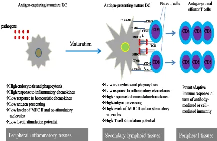
Based on their phenotypic and functional state (depending on their shape, antigen acquisition capacity, cell surface markers expression and antigen specific T cell activation; see Figure 1.2), DCs exist in two distinct forms: immature DCs (iDCs) and mature DCs (mDCs) (Banchereau et al., 2000).
1.3.2.1 Immature DCs
8 environment through phagocytosis, macropinocytosis, or endocytosis. Immature DCs express low levels of MHC class II and co-stimulatory molecules (CD40, CD80, and CD86) on their surface (as shown in Figure 1.2), hence they are considered to be unable to present antigens efficiently to T cells and initiate an effective immune response (Banchereau et al., 2000). In steady-state conditions, DCs maintain themselves in the peripheral tissue in an immature state and they express low amounts of MHC and co-stimulatory molecules. Immature DCs that migrate to lymphoid tissues and present self-antigens to T cells induce T cell anergy instead of T cell activation which forms a basis for tolerance (Hawiger et al., 2001).
[image:29.595.84.520.289.573.2]9 1.3.2.2 Mature DCs
10 1.4 Antigen acquisition by DCs
11 1.5 Dendritic cell antigen presentation – role of MHC Class I and II
Dendritic cells (DCs) initiate immune responses through activation of naive T lymphocytes. This process requires DCs to take up antigens and cleave them into peptides, which are loaded onto highly polymorphic MHC molecules and presented to antigen-specific T lymphocytes. On mDCs, antigenic peptides bound to MHC class II initiates immunogenic responses by CD4+ T lymphocytes (Banchereau and Steinman, 1998), while antigenic peptides bound to MHC class I enable mDCs to activate CD8+ T lymphocytes and generate effector cytotoxic T lymphocytes (CTLs) that possess anti-viral and anti-tumour activity (Chai et al., 1999; Kemball et al., 2006).
1.5.1 MHC class I
12 1.5.2 MHC class II
13 during DC maturation and this is due to a remarkably rapid reduction in the synthesis of class II transactivator (CIITA) mRNA (Landmann et al., 2001).
1.6 Activation of T cells by antigen bearing mature DCs
When a naïve T cell has recognised, via its T cell receptor (TCR), a specific antigen presented by the DC, a specialized area of contact between T cell and DC called the immunological synapse is formed. This synapse consists of clusters of TCRs with MHC-peptides surrounded by a ring of adhesion molecules (Grakoui et al., 1999). Signals through the TCR render the T cell fully activated. It will undergo several rounds of proliferation resulting in its distinct clonal expansion and functional differentiation into effector T cells of either CD4+ T helper cells or CD8+ cytotoxic T cells (CTLs) depending on the type of MHC molecules presented on DCs being either MHC class II or MHC class I molecules respectively (first signal). Additionally, T cells require a second signal for full activation. This second signal comes from co-stimulatory receptors expressed by DCs (Dhodapkar et al., 2000; Mellman and Steinman, 2001). The experimental study of DC induced T cell activation requires the use of transgenic systems and these are briefly described in Box 1.
1.6.1 Role for DC co-stimulatory receptors in T cell activation
14 signals, this may lead to the inappropriate activation of auto-reactive T cells resulting in an autoimmune response (Fife and Bluestone, 2008).
Co-stimulatory molecules expressed on DCs engage with their counterpart molecules on T cells and one of the dominant co-stimulatory pathways is the CD80/CD86 on DCs with its ligand CD28 on T cells (Fife and Bluestone, 2008; June et al., 1994). Binding of CD80/CD86 to CD28 will transmit co-stimulatory signals into T cells that lead to amplification of the signalling processes within T cells (Viola et al., 1999), significantly lowering the TCR activation threshold and allowing naive T cells to be readily activated (Lenschow et al., 1996).
1.6.2 Role for DC cytokines in T cell activation and differentiation
15 et al., 2002; Schakel et al., 2002; Sporri and Reis e Sousa, 2005; Trinchieri, 1995). The wide range of pathogen recognition by DCs, due to their various PRRs expression, result in the variation of cytokines produced thus linking the type of pathogen encountered to the type of T cell response elicited.
1.7 Key signalling pathways involved in DC maturation
Intracellular signalling cascades are essential for the transduction of DC receptor (e.g. PRRs, CD40 and cytokine receptors) engagement to DC maturation. The key signalling pathways involved in DC maturation are the nuclear factor kappa B
(NF-B) and the mitogen activated protein kinases (MAPKs). A major trigger for these signalling pathways is through the TLRs which are prototype PRRs expressed in
Box 1: Measurement of antigen-specific T cell responses
16 sentinel cells such as macrophages and dendritic cells. To-date, 13 different types of PRRs has been recognised. They can recognize pathogen-associated molecular patterns (PAMPs) from microorganisms or danger-associated molecular patterns (DAMPs) from damaged tissue (Akira et al., 2001). The different TLRs and their respective ligands are shown in Table 1.1.
Table 1.1 Summary of Toll like receptor classes and ligands. Adapted from Akira & Takeda 2004.
17 cytoplasmic domain, required for initiating intracellular signalling, homologous to the cytoplasmic domain of the interleukin (IL)-1 receptor (TIR) (Medzhitov et al., 1997).
Ligation of a TLR4 promotes its dimerisation and results in the recruitment of the cytosolic adaptor molecule (MyD88) which contains two domains: a C-terminal Toll homology domain that interacts with the Toll homology domain of the receptor, and an N-terminal death domain (DD), however, the efficient triggering of TLR4 response requires the expression of the secreted protein MD-2 (Re and Strominger, 2002). MyD88 also has an intermediate domain (ID) that is crucial in TLR signalling as it interacts with IL-1R associated kinase (IRAKs) (Arancibia et al., 2007). Upon ligand binding to TLR, IRAK is activated by phosphorylation and this subsequently causes its dissociation from the receptor complex and association with tumor necrosis factor (TNF) receptor-activated factor 6 (TRAF6). This event results in activation of the Rel family transcription factor NF-B and MAPK family (Akira et al., 2001).
Beside the TLR family, DC maturation can be induced by engagement of certain cell receptors with their ligands like co-stimulatory receptor CD40 with CD40 ligand (van Kooten and Banchereau, 2000) and cytokine receptors with interferon gamma
(IFN-) and tumor necrosis factor alpha (TNF-) (Bernhard et al., 2000). Signal transduction downstream of these receptors shows a complex picture involving different mediators and pathways. These pathways include MAPKs, NF-B and protein tyrosine kinases (Grammer et al., 1998; Purkerson and Parker, 1998; Rothe et al., 1995).
1.7.1 The NF-B pathway
18 inflammation, infection and stress stimuli and rapidly reprograms gene expression through activation of an exceptionally large number of genes (Rothwarf and Karin, 1999). Mature DCs express high levels of the NF-B family of transcription factors (Ouaaz et al., 2002). The NF-B family members are formed by combinations of five distinct DNA binding subunits, p65/RelA, RelB, c-Rel, p50, and p52 and exist as homo- or heterodimeric complexes. RelA, RelB and c-Rel are synthesized in their mature forms and contain a transactivation domain that interacts with the transcriptional apparatus. Both p50 and p52 lack a transactivation domain although they contain a DNA binding domain; they are synthesized in precursor forms that are proteolysed by the proteasome resulting in the production of mature proteins (Ghosh et al., 1998). In unstimulated cells, NF-B is found in an inactive form sequestered in the cytoplasm and bound to inhibitory IB proteins. When cells are stimulated, this leads to rapid phosphorylation, ubiquitination, and eventually proteolytic degradation of IB thereby freeing NF-B. NF-B then translocates to the nucleus and drives gene expression, including that of IB, which facilitates the termination of the transcriptional response by binding and retaining the NF-B dimers in the cytoplasm genes (Rothwarf and Karin, 1999) as shown in Figure 1.3. Phosphorylation of IB is induced by the multi-subunit IB kinase (IKK) which has two catalytic subunits, IKKα and IKKβ and a regulatory subunit IKK (NEMO) which is proposed to serve as a recognition site for upstream activators (Rothwarf et al., 1998). Binding of TLR with its ligand will induce IKK activity and thereby activate
19 development, maturation, and survival through transcription of a number of different genes, including those coding for chemokines (IL-8), cytokines (IL-1, IL-2, IL-12, and TNF-), and adhesion molecules (endothelial leukocyte adhesion molecule, vascular cell adhesion molecule, and intercellular adhesion molecule) (Caamano and Hunter, 2002).
Figure 1.3 Signalling cascades leads to NF-B activation. 1.7.2 The MAPK pathways
20 phosphorylated MAPKKKs subsequently phosphorylate MAPK kinases (MAPKKs or MAP2Ks or MEKs), which in turn will phosphorylate MAPKs. Different MAPKKKs exist and have large regulatory domains that interact with upstream regulators. MAPKKs typically have smaller regulatory domains and are activated by dual phosphorylation of serine and threonine residues within the activation loop of the catalytic domain. They show great specificity for their cognate MAPK but are regulated by multiple MAPKKKs (Qi and Elion, 2005). The sequential activation of the individual components of the MAPK cascade enables the final MAPK to be phosphorylated and thus modify the activity of its downstream effector molecules to generate a specific intracellular response (Lewis et al., 1998). In DCs, MAPKs pathways regulate their maturation and modulate the type of immune response in concert with NF-B and other pathways (Breckpot and Escors, 2009). Three groups of classical MAPKs have been identified: the extracellular signal-regulated protein kinases (ERK) (Boulton et al., 1991), the c-Jun N-terminal kinases (JNK) (Kyriakis et al., 1994) and the p38 stress-activated protein kinases (p38MAPK) (Lee et al., 1994). They are structurally related and their catalytic domain is blocked by a conserved activation loop. When they are activated by phosphorylation, the loop unblocks the catalytic site leading to phosphorylation of specific targets. All the MAPKs contain a Thr-X-Tyr (TXY) motif within their activation loop. The phosphorylation of both threonine and tyrosine within the activation loop is essential and sufficient for their activation (Zhang and Dong, 2007). They are activated by specific MAPKKs: MEK1/2 for ERK1/2, MEK3/6 for the p38MAPK and MEK4/7 for the JNKs (JNKK1/2) (Chang and Karin, 2001).
21 and phosphothreonine residues on activated MAPKs (Liu et al., 2007). Structurally, all MKPs have a highly conserved C-terminal catalytic domain and a less conserved N-terminal region that engages the cognate MAPKs (Liu et al., 2007). MAPK phosphatase-1 (MKP-1) is the typical member of the MKP family and has activity, along with MKP5, for p38MAPK (Franklin and Kraft, 1997; Sun et al., 1993; Tanoue et al., 1999). For ERK1/2 the key MKPs are MKP2, MKP3 and MKP4 (Guan and Butch, 1995; Muda et al., 1996; Muda et al., 1997), whereas for JNK it is MKP7 (Matsuguchi et al., 2001). However, it is pertinent to note that a single MKP can simultaneously dephosphorylate multiple MAPKs.
1.7.2.1 Extracellular signal-regulated protein kinases 1/2 (ERK1/2)
22 In DCs, the ERK signalling pathway negatively regulates maturation and IL-12 production upon LPS stimulation but positively regulates inflammatory cytokine production like TNF-, IL-1, IL-6, or IL-8 (Nakahara et al., 2006; Yanagawa et al., 2002). Additionally, the ERK signal-transduction pathway negatively regulates
[image:43.595.153.457.249.483.2]NF-B DNA- binding and IB levels on TNF- –induced maturation with enhancement of allogenic T cell response elicited by TNF- matured dendritic cells (Puig-Kroger et al., 2001)
Figure 1.4 Signalling cascades leads to ERK1/2 activation.
1.7.2.2 c-Jun N-terminal kinases (JNKs)
23 limited pattern of expression and is largely restricted to the brain, heart, and testis. They are activated by the MAPK kinases MEK4 and MEK7, via dual phosphorylation, on a specific Tyr residue by MEK4 and a specific Thr residue by MEK7 in a typical Thr–Pro–Tyr (TPY) motif within their “activation/phosphorylation loop” sequences (Davis, 2000). They have the ability to bind and phosphorylate the N-terminal transactivation domain of the transcription factor c-Jun and increase its transcriptional activity (Kyriakis et al., 1994). c-Jun is a component of the signal-responsive transcription complex activated protein-1 (AP-1), which is involved in regulation of gene expression (Leppa and Bohmann, 1999). Additionally, JNKs can also phosphorylate a variety of transcription factors such as ATF2, NFAT, Elk-1 and p53 (Buschmann et al., 2001; Chow et al., 1997; Gupta et al., 1995; Yang et al., 1998b) (Figure 1.5).
Figure 1.5 Signalling cascades leads JNK activation.
24 transactivation of a wide range of transcription factors such as JunD, activating transcription factor 2 and 3 (ATF2, ATF3), Elk-1, Elk-3, p53, Retinoid X receptor alpha and beta (RXR, RAR, androgen receptor (AR), Nuclear factor of activated T cells 4 (NFAT4), Heat shock factor protein 1 (HSF-1) and c-Myc (Dhanasekaran and Reddy, 2008; Johnson and Nakamura, 2007). In DCs, LPS is found to induce JNK activation by phosphorylation mainly by MEK7 (Yoshizawa et al., 2008) and a specific inhibitor of JNK, SP600125, inhibited the LPS-induced up-regulation of co-stimulatory molecules CD80 and CD86, but augmented the up-regulation of MHC class II (Handley et al., 2007; Nakahara et al., 2004).
1.7.2.3 The p38MAPK pathway
The p38MAPK participates in many aspects of immune cell functional responses, including respiratory burst activity, chemotaxis, cell differentiation, granular exocytosis, adherence, and apoptosis (Ono and Han, 2000; Roux and Blenis, 2004). Activation of p38MAPK is critical for DC maturation and secretion of pro-inflammatory cytokines; and it is constitutively activated at a low level in mature DCs (Fukao et al., 2000).
25 stability, mRNA processing, protein stability, nuclear export, and translation) (Raingeaud et al., 1996).
26 Figure 1.6 Signalling cascades leads to p38MAPK activation.
1.7.2.4 The transcription factors CREB/ATF1 are targets of MAPKs
The regulation of gene transcription is important for both tissue specific-gene expression and regulation of gene activity in response to specific stimuli (Latchman, 1997). Transcription factors (TFs) have crucial roles in mediating gene expression and hence in almost controlling all biological processes. TFs contain a basic domain that binds specific DNA sequence (DBDs) which influence the rate of transcription (Jakoby et al., 2002). CREB and the closely related protein ATF1 constitute a subfamily of bZIP transcription factors that binds to its DNA target sequence as a dimer and play critical roles in the regulation of gene expression as well as cell cycle, metabolism, growth, and survival (Mayr and Montminy, 2001). CREB and ATF1 represent the major transcriptional effectors of MAPKs (particularly p38MAPK but also ERK1/2). These transcription factors bind to DNA sequences termed cAMP responsive elements (CRE) and drive gene expression.
27 Ser133 (Shaywitz and Greenberg, 1999). ATF1 mediates its responses as homodimers or as heterodimers with CREB. ATF1/CREB heterodimers have a longer half-life compared with ATF1/ATF1 homodimers, when bound to the cAMP response elements (CRE) (both approximately several minutes), while CREB/CREB homodimers have the longest half-life (10–20 min). Therefore, CREB appears to be responsible for the bulk of essential functions carried out by CREB/ATF1 family members. The duration of CREB phosphorylation is of significance in determining whether transcription occurs or not (Wu et al., 2001).
Gene transcription ability of CREB/ATF1 depends on its phosphorylation state, which, in turn, regulates its recruitment to the versatile co-activators like CREB-binding protein (CBP) or p300, both responsible for trans-activation of the cell transcriptional machinery. CREB/ATF1 may compete with other transcription factors such as NF-B for recruitment and binding of CBP (Gerritsen et al., 1997; Katoh et al., 2001; Vo and Goodman, 2001). Genes that are CREB-responsive include the proinflammatory cytokine TNF- (Roach et al., 2005) and cyclooxygenase 2 (COX2) (Eliopoulos et al., 2002), anti-inflammatory cytokines such as IL-10 (Platzer et al., 1999) and the pro-survival gene, bcl-2 (Chao and Korsmeyer, 1998).
In DCs, phosphorylation of CREB is known to be associated with up-regulation of co-stimulatory molecules (CD80 and CD86) (Ardeshna et al., 2000) and increased secretion of IL-10 (Alvarez et al., 2009).
28 1.8 Redox homeostasis and immune cell function
Redox homeostasis can be generally defined as a relatively stable state of equilibrium or a tendency towards such a state between the diverse but mutually dependent oxidative and antioxidant elements or groups of elements of an organism (Buettner, 1993). Redox homeostasis is important for a variety of cellular functions such as proliferation, apoptosis, and intracellular signalling pathways including MAPK signalling (Go et al., 2004).
Disturbance in redox homeostasis leads to a condition known as oxidative stress which can be defined as a disorder in the pro-oxidant–/antioxidant balance in favour of the former, leading to potential injury. Disturbances in this normal redox state can cause toxic effects resulting in damage to components of the cell, including DNA, proteins, and lipids (Trachootham et al., 2008).
Oxidative damage is characterised as the biomolecular impairment caused by the attack of reactive species upon the constituents of living organisms.
Oxidative stress can have deleterious effects on cell function including immune cell function. It has been widely recognized that oxidative stress is involved in the aggravation of immune disorders such as pulmonary injury, inflammation and emphysema (associated with chronic obstructive pulmonary disorders, COPD) (Matthay and Zimmerman, 2005).
Oxidative stress leads to a very wide spectrum of metabolic, genetic, and cellular responses. Most oxidative stress conditions that cells might come across may stimulate cell growth, modulate gene expression, or may cause a transient adaptive response and protective temporary growth-arrest. In the most extreme outcome, which involves direct cell destruction, necrosis occurs (Davies, 2000).
29 hydrogen peroxide, singlet oxygen, ozone, hypohalous acids, and organic peroxides, and reactive nitrogen species (RNS) which comprises nitric oxide (NO•) and NO2•; hydrogen sulphide (H2S) and its anion HS−.
Reactive oxygen species are free radicals that are extremely reactive owing to the presence of unpaired electron/s in their highest atomic or molecular orbitals. Reactive oxygen species are generated as by-products of aerobic metabolism or exposure to various natural and synthetic toxicants (Davies, 2000). Furthermore, ROS can be induced by the accumulation of hemin, lipid aldehydes, and arsenic (Hsieh et al., 2006; MacKenzie et al., 2008; Reichard et al., 2007; Tjalkens et al., 1998).
Transient, low level, and controlled production of ROS is part of normal cell physiology and plays an essential role in signal transduction cascades, protein ubiquitination and degradation, as well as cytoskeleton arrangement (Yan et al., 2008). However, excessive or prolonged ROS could cause lipid peroxidation, protein amino acid side-chain oxidation, and DNA single- and double-strand breaks (D'Autreaux and Toledano, 2007). These different effects of ROS influence cell cycle progression which is dependent upon the amount and duration of ROS exposure (Boonstra and Post, 2004). ROS are found to be related to a wide variety of human disorders, such as chronic inflammation, age-related diseases, and cancers.
30 Evidence has shown that the intracellular redox status and ROS can serve as signalling mediators to initiate multiple signalling pathways in DCs. This can result in co-stimulatory molecule expression (MHC class II and CD86) up-regulation which have significant effects on DC functions with subsequent impact on the type of T cell immune response elicited (Kantengwa et al., 2003; Kim et al., 2007; Williams et al., 2008). Furthermore, ROS may induce DC chemokines and cytokines production through NF-B and MAPKs pathways so that they can act as a “danger” signal from the inflammatory micro-environment leading to DC activation, and hence an antigen-specific immune response (Rutault et al., 1999; Yan et al., 2008). Previous studies have shown that modulating redox balance in DCs leads to reduction in
IL-Box 2: Sources of ROS and mediators of their catabolism (Adopted from Nathan 2003)
Exogenous sources of ROS •Smoke
•Air pollutants •Ultraviolet radiation •γ-irradiation
•Several drugs
Endogenous sources of ROS •NADPH oxidases
•Mitochondria
•ER flavoenzyme ERO1 •Xanthine oxidase •Lipoxygenases •Cyclooxygenases
•Cytochrome P450 enzymes •Flavin-dependent demethylase •Polyamine and amino acid oxidases •Nitric oxide synthases
•Free iron or copper ions •Haem groups
•Metal storage proteins
Catabolism by antioxidant systems •Superoxide dismutases •Catalases •Glutathione peroxidases •Glutathione reductase •Thioredoxins •Thioredoxin reductases
•Methionine sulphoxide reductases
•Peroxiredoxins or peroxynitrite reductases
Catabolism by small molecules that react with ROS non-enzymatically
31 12 and TNF- cytokine production (Yamada et al., 2006) and decrease in antigen-specific T cell proliferation and IFN- synthesis (Tse et al., 2007).
1.9 Influence of redox on signalling pathways
Several reports have shown, in many cell types, that an altered redox status results in the activation of several intracellular signalling pathways like MAPKs and NF-B, suggesting a role for ROS as second messengers (Gaitanaki et al., 2003; Schaeffer et al., 2003; Wang et al., 1998). ROS may have direct protein target interactions with these signalling pathways wherein exposure of these molecules to an altered redox state alters the function of the target protein. Some recent evidence supports this notion and suggests that both certain protein-protein interactions and enzymatic functions might be regulated by cellular ROS levels (Finkel, 2000). ROS was found to induce the phosphorylation of p38MAPK, ERK, and JNK members of MAPK family and inhibition protein phosphatases (Lee and Esselman, 2002).
1.10 Cellular defence against reactive oxygen species
32 1.10.1 Thiol-containing cellular antioxidants
33 sulphydryl or disulphide moiety. The cysteine thiolate residue (Cys-S) is more readily oxidized by peroxides than is the Cys-sulfhydryl residue (Cys-SH) which serve as the immediate electron donor for the peroxidase function (Rhee et al., 2005). This leaves a reduced or dithiol target protein and a disulfide in the active site of TRX. This disulfide is reduced by TRX reductase (TRXR) using electrons from NADPH (Berndt et al., 2007). The peroxiredoxins (PRX) are a protein family which contain a conserved Cys residue that undergoes a cycle of peroxide-dependent oxidation and thiol-dependent reduction during the catalysis process which is capable of directly reducing peroxides, e.g. hydrogen peroxide and different alkyl hydroperoxides (Rhee et al., 2005).
1.10.2 Enzymes as effectors of redox homeostasis
34 1.10.3 Small non-protein antioxidant molecules
α-Tocopherol (vitamin E) is a potent lipid-soluble antioxidant that has a primary role in breaking chain reactions involving O2 and lipid peroxyl free radicals (Yu, 1994). Additionally it can stabilize and activate the redox sensitive transcription factor nuclear factor E2 p45-related factor 2 (Nrf2) (Rahman et al., 2006). Vitamin E plays a central role in disease prevention by scavenging both ROS and RNS (Zingg, 2007). Ascorbic acid (vitamin C) is a hydrophilic antioxidant that has free radical scavenging capacity and is widely distributed in mammalian tissues. In addition to this direct role, vitamin C can restore the antioxidant properties of oxidised vitamin E (Yu, 1994).
Vitamins C and E have been shown to reduce ROS levels in different cell types including neutrophils (Kanno et al., 1995), monocytes (Cachia et al., 1998; Devaraj et al., 1996), and macrophages (Sakamoto et al., 1990). Addition of vitamins C and E to DCs before challenge with stimuli that are known to induce ROS production, resulted in a reduction in ROS levels comparing to untreated DCs (Tan et al., 2005). This effect is more prominent when DCs are incubated with vitamins for longer durations before stimulation. DCs treated with these antioxidant vitamins are found to be resistant to phenotypic and functional changes subsequent to proinflammatory cytokines stimulation (Tan et al., 2005).
Other ROS scavenging molecules include: uric acid (the end product of purine metabolism), ceruloplasmin (a major copper-carrying protein), β-carotene (a metabolic precursor of retinol) and bilirubin (a breakdown product of heme catabolism) (Davies, 2000; Yu, 1994).
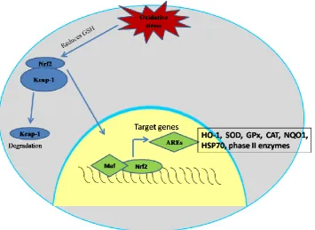
35 Glutathione and detoxifying enzymes expression (both basally and inducibly) are mainly regulated by Nrf2, a redox-responsive transcription factor which has a central role in cellular adaptive and defence systems against oxidative and electorphilic stress (Itoh et al., 1997b; Jaiswal, 2004; McMahon et al., 2001).
1.11 Nrf2
The transcription factor Nrf2 is a member of the cap‘n’collar (CNC) basic leucine zipper transcription factors and is essential for the coordinated induction of genes that encodes cytoprotective enzymes or stress-responsive proteins, such as HO-1, NQO1, SODs, GPx, GSTs, GCL, CAT and TRX (Reddy et al., 2007b).
Other members of the CNC family of transcription factors include the Nrf1, Nrf3 and the repressive factors bric-a-brac/tram-track/broad complex (BTB) and CNC homolog 1: Bach1 and Bach2 (Chan et al., 1998; Kobayashi et al., 1999), and are named as such due to structural similarities with the Drosophila protein cap ‘n’ collar (CNC). Nrf2 contains a C-terminal basic leucine zipper (bZip) structure that facilitates dimerisation and DNA binding (Moi et al., 1994). Nrf1 is less potent than Nrf2 at transactivating gene expression (Zhang et al., 2006), while Nrf3 has been proposed as a negative regulator of Nrf2 by interfering with ARE-mediated transcription (Sankaranarayanan and Jaiswal, 2004).
36 environment (skin, lung and digestive tract) (Motohashi et al., 2002). The Nrf2 structure consists of six highly conserved regions named as homology domains (Nrf2-ECH homology) Neh1 to Neh6 as shown in Figure 1.7 (Itoh et al., 1999).
Figure 1.7. Schematic representation for the basic structure of transcription factor Nrf2. Neh1 is the first conserved domain and contains the basic-leucine zipper domain and CNC homology region. The amino and carboxyl termini of the proteins are also highly conserved, and are referred to as Neh2 and Neh3, respectively. Additionally, there are two conserved acidic domains (Neh4 and Neh5) as well as a serine-rich conserved region (Neh6). Adopted from Itoh et al. 1999.
Nrf2 binds to the antioxidant response element (ARE), a DNA motif found within the promoter regions of numerous cytoprotective genes, with high affinity only as a heterodimer with its obligatory partner a small Maf protein (Itoh et al., 1997b). Small Maf proteins lack transactivation domains, and thus the ability of the Nrf2-Maf heterodimer to promote transcription is solely dependent on the transactivation ability of Nrf2 (Motohashi et al., 2002). Amongst members of the CNC family, Nrf2 is the most potent transcriptional activator from the ARE (Kobayashi et al., 1999; Papaiahgari et al., 2006).
37 transactivation domains of Nrf2, Neh4 and Neh5, which act synergistically to attain maximum activation of reporter gene expression. Indeed, specific inhibition of CBP significantly reduced Nrf2 activity (Katoh et al., 2001). CBP helps transcription through a) its intrinsic histone acetyltransferase (HAT) activity, b) interaction with other proteins possessing HAT activity, and c) bridging to components of the general transcriptional machinery (Bannister and Kouzarides, 1996; Kalkhoven, 2004). Histone acetylation (as well as DNA methylation) control the access of transcription factors to the promoter regions and thereby affect the pattern of gene transcription. Nucleosomes contain histones in their core, around which DNA is wound into a condensed structure that represses transcription, but that can be selectively unfolded to increase accessibility to general transcription factors and RNA polymerase II, and thus promote gene transcription (Grunstein, 1990; Kuo and Allis, 1998).
Transcriptional repressors Bach1 and Bach2 are basic leucine zipper (bZip) protein and forms heterodimers with one of the small Maf proteins (i.e. MafK, MafF, and MafG) that bind to the Maf recognition element (MARE) and effectively function as repressors of gene transcription over NFE2-related transcription, including suppression of a key gene in heme metabolism, heme oxygenase-1 (HO-1) (Kitamuro et al., 2003; Yoshida et al., 2007).
1.11.1 Kelch-like ECH-associated protein-1 (Keap-1)
38 1999). This interaction represses cellular Nrf2-dependent transcription activity (Zipper and Mulcahy, 2002).
The Neh2 moiety of Nrf2 is required for the repression activity by Keap1 (Itoh et al., 1999; Xue and Cooley, 1993). Keap1 resides within the cytosol where it interacts with the actin cytoskeleton and, in the absence of chemical/oxidative stress, associates with Nrf2 leading to Nrf2 proteasomal degradation. Structurally, Keap1 contains three major domains; N-terminal BTB domain, an intervening region (IVR) bridge that is rich in cysteine residues and regulates activity of Keap1 and links BTB to double glycine (DGR) domain situated on the C-terminal containing six conserved repeats also known as kelch (Figure 1.8) (Itoh et al., 1999; Kang et al., 2004). The BTB domain mediates protein dimerisation which is required for effective Nrf2 sequestration in the cytosol, whereas Kelch repeats have been implicated in binding to the actin cytoskeleton and the formation of multi-protein complexes (Albagli et al., 1995; Robinson and Cooley, 1997). Cullin3 (Cul3), a subunit of the E3 ligase complex, serves as a molecular bridge bringing together substrate adaptor protein (Keap1) and substrate (Nrf2) through Keap1 BTB and intervening-region (IVR) domains. Therefore, Keap1 would participate directly in the regulation of Nrf2 polyubiquitination and subsequent 26S proteasome-mediated degradation (Cullinan et al., 2004; Kobayashi et al., 2004).
39 1.11.2 Regulation of Nrf2 by Kelch-like ECH-associated protein-1 (Keap-1)
40 2006a). This conformational change in Keap-1 disables it from directing Nrf2 for degradation via the proteasome (Kobayashi et al., 2004). Other Nrf2 molecules then saturate the Keap-1 via binding to the newly available BTB site within the homodimer, subsequently Nrf2-binding capacity of Keap1 is saturated and even diminished due to Keap1 self-ubiquitination allowing any newly synthesised Nrf2 to translocate into the nucleus (Li and Kong, 2009). Nrf2 then associates with small maf protein forming a heterodimer which facilitates its capacity to bind to the ARE regions found in the regulatory domains of its nrf2-regulated genes (Itoh et al., 1997b). The co-activator CBP is then recruited to the Nrf2 heterodimer resulting in its transactivation (Katoh et al., 2001; Lin et al., 2006) and subsequent transcription of enzymes involved in xenobiotic detoxification ultimately resulting cellular protection and maintenance of redox homeostasis (Figure 1.9).
[image:61.595.123.479.354.613.2]41 Maf protein forming a heterodimer which facilitates its capacity to bind to the ARE regions found in the regulatory domains of its Nrf2-regulated genes. Adopted from Copple et al 2008.
Prevention of cysteine modification within the BTB and IVR regions of the Keap-1 was found to facilitate Nrf2 ubiquitination (Zhang et al., 2004b), indicating that the targeting of these residues within the Neh2 domain is critical for Keap1-mediated repression of Nrf2. There is evidence that Keap1 acts as a “sensor” of chemical or oxidative stress, through its many cysteine residues. Furthermore, several recent reports have described the phosphorylation of Nrf2 as an event that is required for the nuclear export of the transcription factor (Jain and Jaiswal, 2006; Kaspar et al., 2009). Protein kinase C has been shown to phosphorylate Nrf2 in its Neh2 domain at Ser-40, disrupting the association between Nrf2 and Keap1 thus promoting the translocation of Nrf2 into the nucleus (Huang et al., 2002).
1.11.3 Nrf2 activators differentially modify Keap1
42 heme oxygenase-1 and Nrf2/ARE signalling as well as up-regulating Nrf2 nuclear translocation (Yates et al., 2006). These triterpenoid derivatives provide an effective tool for investigating the role of Nrf2 in the maintenance of oxidative stress. Pre-treatment with CDDO-Me or CDDO-Im attenuates LPS-induced pro-inflammatory cytokine production and ROS generation in human PBMCs and neutrophils, respectively (Thimmulappa et al., 2007).
Because Nrf2–regulated cytoprotective genes protect from oxidative damage as well as suppress inflammation by modulating immune cell function, it is possible that the molecules which are able in activation of the Nrf2 pathway might prove to be suitable therapeutic strategy for intervention in sepsis as well as in systemic inflammatory response and other inflammatory disorders. (Thimmulappa et al., 2002).
1. 12 Role of Nrf2 in redox homeostasis and immune cell function
43 Nrf2 deficiency in mice have been shown to result in enhanced expression of several innate immune response proteins including specific cytokines, chemokines, and cell surface adhesion molecules and receptors. The lungs of Nrf2-deficient mice exhibited persistent cellular injury, pro-inflammatory IL-6 and TNF- production, impaired endothelial and alveolar cell regeneration, and continual cellular infiltration by macrophages and lymphocytes which might impair lung innate immunity and promotes susceptibility to bacterial infection (Reddy et al., 2009). Disruption of Nrf2 causes regenerative immune-mediated hemolytic anemia (Lee et al., 2004). Furthermore, it was demonstrated that Nrf2-deficient mice exhibit prolonged inflammation during cutaneous wound healing (Braun et al., 2002). Through its influence on macrophages and neutrophil function, Nrf2 is implicated in the inflammatory response to bacterial infection, and protection against sepsis. Hence, Nrf2 has been described as a gene modifier for mounting appropriate innate immune responses (Thimmulappa et al., 2006).
44 antibodies, multi-organ inflammation, immune complex deposition in blood vessels and glomerular nephritis (Ma et al., 2006).
However, the molecular and mechanistic basis for these altered co-stimulatory receptor expression of Nrf2-/- DCs are unknown and require investigation.
1.13 Heme oxygenases (HOs) in redox homeostasis
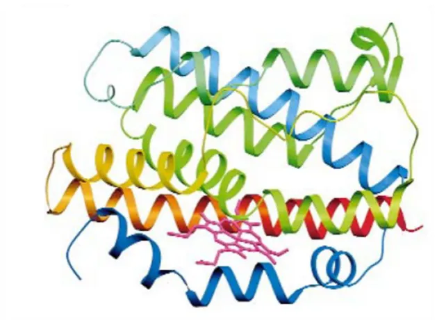
45 heme in addition to providing flexibility for substrate binding and product release (Schuller et al., 1999).
[image:66.595.134.449.135.365.2]
Figure 1.10. Schematic diagram of heme oxygenase-1. The N-terminus is blue and the C-terminus is red, with green in the middle. Adopted from Schuller et al. 1999.