181 CK2 = casein kinase 2; CypB = cyclophilin B; EGF = = epidermal growth factor; EGFr = epidermal growth factor receptor; ER = endoplasmic reticulum; FGF = fibroblast growth factor; FGFr = fibroblast growth factor receptor; GHBP = growth hormone binding protein; GHr = growth hormone receptor; IFN = interferon; IL = interleukin; MHC = major histocompatibility complex; PIAS3 = peptide inhibitor of activated Stat3; PRL = prolactin; PRLr = prolactin receptor; PPI = peptidyl prolyl isomerase.
Introduction
Peptide hormones and their cognate receptors are required for the development and differentiation of the mammary gland. The traditional paradigm of peptide hormone/receptor action in mammary tissues is best summarized as one of ‘action-at-a-distance’. In short, a temporal and spatial activation of receptor-associated transduction/transcription factors is thought to mediate a specific signal triggered by ligand binding. These net-works undoubtedly induce the necessary post-transla-tional modifications (phosphorylation/dephosphorylation, prolyl isomerization, proteolytic cleavage, etc.) required for transcription factor/co-activator function. On careful exam-ination of the signaling networks emerging from function-ally disparate peptide hormone receptors, however, a molecular basis for specificity remains unclear, as the transduction cascades utilized by these receptors are widely shared.
A potential solution to the conundrum of polypeptide hormone specificity has recently emerged in a series of articles detailing the intranuclear translocation and action
of peptide ligands and their receptors, or fragments thereof. As contrasted later, striking parallels in the intranuclear actions of peptide hormone/receptors exist with steroid and Notch receptor complexes. These find-ings taken together lead to the novel hypothesis that peptide hormone specificity is generated by the conjoint activation of receptor-associated signaling networks (i.e. ‘nongenomic action’) and by the direct intranuclear function of ligand and/or receptor complexes (i.e. ‘genomic action’).
Translocation and function of polypeptide
receptors within the nucleus
Several transmembrane polypeptide receptors have been reported within the nucleus, including epidermal growth factor receptor (EGFr) [1], erbB-3 [2], erbB-4 [3], fibroblast growth factor receptor (FGFr) [4,5], nerve growth factor receptor [6], IL-5 [7], prolactin receptor (PRLr) [8], and growth hormone receptor (GHr)/growth hormone receptor binding protein [9,10]. Aside from delineating the nuclear localization of these peptides, recent studies have begun to elucidate their potential intranuclear functions and mecha-nisms of nuclear internalization (also referred to as
‘retrotrans-Review
Nuclear localization and function of polypeptide ligands and
their receptors: a new paradigm for hormone specificity within
the mammary gland?
Charles V Clevenger
Department of Pathology and Laboratory Medicine, University of Pennsylvania, Philadelphia, Pennsylvania, USA Correspondence: Charles V Clevenger (e-mail: [email protected])
Published: 11 April 2003
Breast Cancer Res2003, 5:181-187 (DOI 10.1186/bcr601)
© 2003 BioMed Central Ltd (Print ISSN 1465-5411; Online ISSN 1465-542X)
Abstract
The specific effects triggered by polypeptide hormone/growth factor stimulation of mammary cells were considered mediated solely by receptor-associated signaling networks. A compelling body of new data, however, clearly indicates that polypeptide ligands and/or their receptors are transported into the nucleus, where they function directly to regulate the expression of specific transcription factors and gene loci. The intranuclear function of these complexes may contribute to the explicit functions associated with a given ligand, and may serve as new targets for pharmacologic intervention.
182
port’). The present review will focus on those receptors with acknowledged function within the mammary gland.
Epidermal growth factor receptor
While immunohistochemical and biochemical studies have localized the EGFr within the nucleus, the potential func-tion of this receptor at this site has only been recently elaborated [1]. Through the use of cross-linked 125 I-epi-dermal growth factor (EGF), Kwong and colleagues revealed that phosphorylated, full-length EGFr is rapidly transported into the nucleus within 1 min of ligand stimula-tion. They also showed that the C-terminal (intracytoplas-mic) domain of the EGFr potently activated the expression of a luciferase reporter construct when fused to a GAL DNA binding domain. Using the approach of cyclic amplifi-cation and selection of targets (‘CASTing’), Kwong and colleagues demonstrated that the EGFr preferentially bound to an AT-rich sequence. Stimulation of cells trans-fected with a luciferase reporter construct containing this AT-rich sequence resulted in EGF-induced expression of the reporter. Furthermore, when a similar sequence was identified in the cyclin D1 (an EGF-inducible gene) pro-moter region and subsequently mutated in a reporter con-struct, EGF-induced expression was lost. Additional in vivo confirmation of a physical association of the EGFr with the endogenous promoter region of cyclin D1 was demonstrated by chromatin immunoprecipitation analysis.
These findings taken together suggest that the nuclear retrotransport and promoter binding of the EGFr following ligand stimulation may contribute to the expression of spe-cific EGF-induced genes. While this study represents a groundbreaking analysis of intranuclear EGFr function [1], several points remain to be addressed: how does the cell surface EGFr extricate itself from the plasma membrane? How does the EGFr cross the nuclear membrane? How does the intranuclear EGFr modulate endogenous gene expression from a mechanistic perspective? What gene loci are transactivated by the intranuclear EGFr? Indeed, the lack of a defined mechanism for EGFr extrication from the cell membrane and retrotransport to the nucleus has been challenged [11]. The facts remain, however, that multiple independent studies have now demonstrated EGFr within the nucleus [1,12]. The inability of some groups to reproduce these findings [11] may be related to the difficulty of some anti-EGFr antibodies to recognize the EGF in certain intracellular environments.
erbB-3
A recent study [2] has elegantly demonstrated, by confo-cal immunomicroscopy, immunogold electron microscopy, and biochemistry, the existence of full-length erbB-3 within the nucleus of several cultured malignant human breast cancer lines. Unlike the functional studies already detailed for the EGFr, Offterdinger and colleagues [2] examined the basis for the nuclear transport of erbB-3.
Structure/function mutagenesis revealed a sequence in erbB-3 (termed NLS-2; RRRRHSP) that functioned as a nuclear internalization signal. As such, their study is note-worthy for its direct demonstration of this motif; however, the study needs independent confirmation and does not address any potential intranuclear function of erbB-3, nor has intranuclear erbB-3 been demonstrated in malignant or normal tissues. In addition, the mechanisms through which erbB-3 can remove itself from the cell membrane prior to nuclear translocation remain unresolved.
erbB-4
The presence of a soluble fragment of erbB-4 within the nucleus has been recently reported [3], following reports of the erbB-4 receptor within the nuclei of primary human breast cancers [13]. This fragment spanning the intracyto-plasmic domain of this receptor appears to be the result of a sequential cleavage of erbB-4; first by an unidentified metalloprotease (resulting in cleavage of a soluble extra-cellular domain and a membrane-bound intracytoplasmic domain), followed by cleavage of the intracytoplasmic domain proper (into a cytoplasmic-soluble form), by a γ-secretase-like activity. This was shown through the use of pharmacologic inhibitors or dominant-negative precursors of γ-secretase that effectively blocked the formation of the soluble erbB-4 fragment. Use of immunofluorescence-based studies revealed that this fragment was capable of nuclear retrotransport, and that the motif mediating this event was present in the C-terminus of the intracytoplas-mic domain. Fusion of the erbB-4 intracytoplasintracytoplas-mic domain to the GAL4 DNA binding domain revealed a modest transactivation of a GAL4 reporter construct, suggesting that the erbB-4 fragment may be capable of modulating endogenous gene expression.
The study by Ni and colleagues [3] is remarkable because it demonstrated parallels between erbB-4 and Notch sig-naling. However, whether the erbB-4 fragment is associ-ated with active chromatin and which gene loci this fragment activates remain to be determined. In addition, formal biochemical proof of the existence of this fragment within the nucleus remains to be demonstrated, as well as the precise motifs involved in the nuclear retrotransport and transactivation.
Fibroblast growth factor receptor
183 nucleus (by replacement of its endoplasmic reticulum [ER]
leader signal with a nuclear localization signal sequence from SV40 T antigen) resulted in enhanced c-Jun and cyclin D1expression.
As with the members of the EGFr/erbB family, the mecha-nisms of transport of the FGFr from the cell membrane remain to be determined. In addition, the motifs required for receptor-induced gene expression have not been iden-tified, and the physical association of the FGFr with the transcriptional apparatus on the endogenous c-Jun and cyclin D1loci requires demonstration.
Growth hormone receptor
The presence of both growth hormone and the GHr within the nucleus has been previously demonstrated [15]. Recent data from Lobie and colleagues have suggested that the extracellular domain of the GHr, also known as the growth hormone binding protein (GHBP), exists and func-tions within the nucleus in a manner independent of the ligand [9]. These studies have demonstrated that a modest potentiation of the expression of a Spi2.1 reporter construct occurred upon transfection of the GHBP. The potentiation of the expression of this reporter construct was increased if a nuclear localization signal was added to the GHBP via a recombinant technique [9]. These studies suggest that the extracellular domain of the GHBP may stimulate Spi2.1 transcription. These studies stand in con-trast to other reports that suggest the GHBP acts in a dominant negative manner [16]. In addition, the manner of nuclear transport of the GHBP from either the ER or extra-cellular space, the effect of GHBP on endogenous gene expression, the gene loci activated by the GHBP, and the mechanisms utilized during transcriptional activation remain to be determined.
Prolactin receptor
The nuclear retrotransport of the PRLr has been noted [8,17] and associated with the activation of nuclear protein kinase C. Little else is know of the mechanisms of its nuclear internalization or intranuclear function. Indeed, whether the intranuclear PRLr represents a full-length receptor or a fragment thereof has been debated.
Translocation and function of polypeptide
ligands within the nucleus
The nuclear retrotransport of numerous polypeptide ligands, including prolactin (PRL) [18–21], growth hormone [15], EGF [22], IFN-γ [23], nerve growth factor [6,22], platelet-derived growth factor [22], FGF [24–26], IL-5 [7], and insulin [27–29], has been reported within the nucleus following their addition into the extracellular medium. While the intranuclear transport and action of many of these ligands are not known, a considerable understanding of such mechanisms for FGF and PRL has been newly forthcoming, and is now detailed.
Fibroblast growth factor
A growing body of evidence has indicated a function for FGF internalized within the cytoplasm, and possibly within the nucleus. Precedent reports from Olsnes and col-leagues have demonstrated that FGF is retrotransported into the cytoplasm and nucleus [26]. This transport required several hours and was dependent on the cyto-plasmic domain of the FGFr, but not on its kinase activity [25]. Stimulation of NIH3T3 cells by the external applica-tion of a mutant FGF ligand containing a CAAX motif resulted in the demonstrable intracellular and intranuclear farnesylation of this mutant after several hours [30], revealing that the intracellular FGF was capable of cross-ing cellular membranes. Followcross-ing receptor-mediated endocytosis, FGF could be found in the Golgi/ER. Trans-port from this vesicular compartment required the pres-ence of an active proton pump; when blocked with the specific inhibitor bafilomycin A1, entry into the cytoplasm was blocked [31]. In the absence of FGFr, a fusion protein of FGF and diphtheria toxin was capable of stimu-lating the proliferation of NIH3T3 cells expressing diph-theria toxin receptor [26]. These data suggest that the primary function of the FGFr, with respect to FGF-induced mitogenesis, was for the endocytosis of FGF. Phosphorylation of FGF appeared to contribute to its mitogenicity, as a specific mutation (K132E) to FGF resulted in a ligand demonstrating comparable levels of receptor binding and activation, but demonstrating decreased phosphorylation and ligand-induced prolifera-tion [32].
What is the function of intracellular FGF? Several proteins have been found to interact with FGF intracellularly, including FGF intracellular binding protein, mortalin, synaptotagmin, and S100A13. A recent study has also demonstrated an interaction between FGF and casein kinase 2 (CK2) [33]. The interaction between these pro-teins, as determined by surface plasmon resonance, was of moderate affinity (in the order of 1µM). Despite this, in vitro FGF was phosphorylated by CK2 and, in turn, the presence of FGF stimulated the autophosphorylation of CK2 [33]. CK2, like FGF, exists in both the cytoplasm and the nucleus. The ability of FGF mutants to bind CK2 was found to correlate with their mitogenic potential. Additional studies have indicated that intranuclear FGF2 can bind to the CK2 and can stimulate its activity towards the nucleo-lar substrate nucleolin [34]. These findings suggest, but obviously do not prove, a potential function for CK2 in the intracellular actions of FGF.
184
cell/animal models where CK2 is either knocked down or knocked out (i.e. one would predict a decrease in FGF action)? What are the relevant substrates for CK2 in both the cytoplasm and nucleus? These points are discussed at length in two recent reviews on the intranuclear function of FGF [35,36].
Prolactin
PRL has been reported in the nucleus of T cells and human breast cancer cell lines following co-mitogenic stimulation [19,37]. The nuclear uptake of PRL appears to be widespread, as previous biochemical and/or immunohistochemical studies have demonstrated this ligand in tissues of the breast, the immune system, the liver, the ovary, and the adrenal [38–40]. Nuclear retro-transport occurs within 1–2 hours of extracellular ligand stimulation and is dependent upon the PRLr. Immunogold electron microscopy studies using anti-PRL antibodies and colloidal gold-labeled PRL have revealed that, follow-ing endocytosis, the ligand is retrotransported from the endosome into the multivesicular body, where the ligand is either targeted into the lysosome (presumably for degradation) or into the Golgi/ER [20]. Shortly after this, ligand can be noted within the nucleus. When transfected into T-cell lines neither the secreted wild-type PRL (con-taining an ER leader sequence) nor a mutant of PRL lacking its leader (which accumulated in the cytoplasm) had a significant effect on T-cell growth. However, mutant PRL containing the SV40 large T nuclear signal sequence in place of the ER leader induced robust T-cell proliferation [21].
The mechanisms for PRL nuclear retrotransport and action were not fully appreciated until followup studies were performed to identify the binding partners of inter-nalized PRL capable of mediating such functions. A yeast two-hybrid analysis was performed with PRL as bait to identify PRL-binding partners. This analysis, confirmed by in vitro and in vivo binding studies, revealed that a signifi-cant binding partner for PRL was the peptidyl prolyl iso-merase (PPI) cyclophilin B (CypB). A ubiquitously expressed 22 kDa protein secreted from most cells, CypB is found in serum and milk, and also within cell nuclei. Exogenously applied CypB was found to dramati-cally potentiate PRL and growth hormone-driven prolifera-tion and to potentiate gene expression (i.e. β-casein or Spi2.1), but only in cell types expressing either the PRLr or the GHr [37].
The observation of an interaction between CypB and PRL has enabled further insights into the mechanisms of PRL retrotransport and intranuclear function. Following endo-cytosis, PRL is presumably released from its receptor as a consequence of vesicular acidification, thus enabling transport from the multivesicular body to the Golgi/ER. Retrotransport of many bacterial toxins and MHC-targeted
peptides has been demonstrated to occur via the Sec61 apparatus [41,42]. Of note, the PRL/CypB complex can also be co-immunoprecipitated with the Sec61 pore following endocytosis of this complex, an event that paral-lels the activation of PRLr-affiliated signaling pathways [19,20]. Nuclear transport of the PRL/CypB complex is facilitated by a newly recognized nuclear translocation signal in the N-terminus of CypB [37]. CypB mutants lacking this signal, while enzymatically active and fully capable of binding PRL, did not potentiate PRL-driven proliferation or the intranuclear accumulation of PRL. Taken together, these findings have revealed that CypB serves as a ‘reverse’ chaperone, facilitating the transport of PRL from the vesicular compartment into the nucleus.
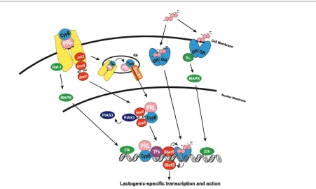
Within the nucleus, the PRL/CypB complex has been found to interact with Stat5 in a PPI-dependent manner and to potentiate the activity of this transcription factor. Stat5 is a well-recognized mediator of PRL action within the mammary gland, as Stat5a knockout mice fail to lactate [43]. Following tyrosine phosphorylation by recep-tor-activated Jak2, Stat5 is known to dimerize, to trans-locate into the nucleus, and to bind to cognate DNA binding sites. Electrophoretic mobility shift analysis revealed that the PRL/CypB complex facilitated Stat5 DNA binding, in a manner that indicated the PRL/CypB complex was removing a repressor of Stat5 function. Co-immunoprecipitation analysis demonstrated this repressor to be the peptide inhibitor of activated Stat3 [44]. The exogenous addition of PRL/CypB released the peptide inhibitor of activated Stat3 from intranuclear Stat5, thereby enhancing Stat5 DNA binding [45]. The PRL/CypB complex directly interacts with the N-terminus of Stat5 and appears to induce a conformational change in this transcription factor, enabling the release of peptide inhibitor of activated Stat3 (PIAS3), thereby facilitating its association with additional elements of the transcriptional apparatus with Stat5 [45].
The functions of the PRL/CypB complex are dependent upon the PPI activity of CypB. While perfectly capable of binding PRL and undergoing nuclear retrotransport, CypB mutants deficient in this enzymatic activity are incapable of stimulating Stat5-mediated gene expression or prolifera-tion. Indeed, the PPI-deficient CypB mutants are able to block the proliferation of breast cancer cell lines at con-centrations two to three orders of magnitude less than other described mutant PRL antagonists [45].
185
Conclusions and perspective
The intranuclear transport and function of polypeptide ligands and their receptors have been a matter of consid-erable scientific debate over the past decade. Some of this controversy was due to a lack of mechanistic ground-ing inherent in the initial descriptions of intranuclear ligand and/or receptor. As outlined in the present review, however, the recent demonstration of pathways for the nuclear translocation of peptide ligands and receptors (i.e. receptor cleavage, ligand association with binding pro-teins/chaperones, etc.) from the cell surface/vesicular compartment and bona fidetargets for intranuclear action (i.e. DNA, transcription factor, or kinase binding) should lay to rest many of these mechanistic concerns. Indeed, these studies provide an intelligent basis for speculation regarding how a ligand/receptor complex may make its way into the nucleus and exert functional activity at this site [46].
Acceptance of an intranuclear function for polypeptide ligands and their receptors, however, is not universal. This
[image:5.612.96.547.97.368.2]may be due to a lack of a larger perspective on the intranuclear action of all ligand/receptor complexes. Many of the concerns regarding the function of intranuclear polypeptide ligand/receptors can be addressed by exam-ining the parallels that exist with other acknowledged sig-naling pathways. For instance, the ability of polypeptide receptors and, in particular, receptor fragments (such as the soluble erbB-4 intracytoplasmic domain) to function within the nucleus as a transcription factor/co-activator is highly analogous to Notch-associated signaling within the mammary gland [47]. Indeed, cleavage of the erbB-4 intra-cytoplasmic domain may use similar, if not identical, pro-teases required for the cleavage of the transcriptionally active Notch intracellular domain. Additional mechanistic parallels can also be drawn between the PRL/CypB complex and the actions of steroid/steroid receptor com-plexes (see Fig. 1). While the intranuclear actions of steroid receptor complexes have long been appreciated, the function of these complexes at the cell surface has only been recently recognized [48]. Indeed, the rapid ‘nongenomic’ functions of steroid receptor complexes
Figure 1
186
appear to utilize many of the signaling pathways associ-ated with polypeptide receptors.
Many questions remain regarding the transport and actions of intranuclear polypeptide ligands and receptors. First, the mechanisms surrounding the extrication of mem-brane-inserted, full-length receptors need to be estab-lished. Second, the molecular basis for retrotransport of ligand into the cytoplasm requires further detailing. Third, the targets of intranuclear ligands and receptors (i.e. gene loci, transcription factors, kinases, etc.) are largely unidentified. Finally, detailing of the larger in vivo context for the actions of intranuclear ligand/receptor complexes at the tissue and organismal level will provide a final level of proof necessary to establish the biologic relevance of this process.
Several theories currently exist regarding how signaling specificity is achieved in a given cell by ligand/receptor complexes that share several common transduction path-ways [49,50]. While some data support the notion that rare signaling pathways may be uniquely associated with a given receptor, most evidence now supports the notion that a combination of signals emanating from a given receptor results in signaling specificity. These signals may be modified in unique ways by the duration of recep-tor action, by compartmentalization of the receprecep-tor or its associated signaling networks, and by the proteome of a given cell. However, a rigorous demonstration of how combinatorial signaling alone works to achieve speci-ficity in the context of peptide hormones remains to be demonstrated.
The identification of the intranuclear transport and action of polypeptide ligands and receptors may therefore provide an additional key to the conundrum of hormone/growth factor specificity. As such, the hypothe-sis emerging from these data would suggest that it is the function of ligand/receptor complexes in the nucleus that provides an additional level of direct regulation requisite for the specificity. Given this, the advances made in our understanding of the intranuclear actions of polypeptide ligands and receptors hold considerable pharmacologic promise. As evinced by the PPI-deficient CypB mutants, the development of pharmacophores capable of interrupt-ing the intranuclear transport and/or function of these polypeptides may provide highly effective agents capable of inhibiting the hormone/growth factor-driven proliferation and progression of breast malignancies.
Competing interests
None declared.Acknowledgement
Support for some of the studies discussed was obtained from a grant from the American Cancer Society RPG-00-307-01-TBE.
References
1. Lin SY, Makino K, Xia W, Matin A, Wen Y, Kwong KY, Bour-guignon L, Hung M-C: Nuclear localization of EGF receptor and its potential new role as a transcription factor. Nat Cell Biol2001, 3:802-808.
2. Offterdinger M, Schofer C, Weipoltshammer K, Grunt TW: c-erbB-3: a nuclear protein in mammary cells. J Cell Biol2002, 157:929-939.
3. Ni C-Y, Murphy MP, Golde TE, Carpenter G: γγ-secretase cleav-age and nuclear localization of erbB-4 receptor tyrosine kinase.Science2001, 294:2179-2181.
4. Maher PA: Nuclear translocation of fibroblast growth factor (FGF) receptors in response to FGF-2. J Cell Physiol 1996, 134:529-536.
5. Reilly JF, Maher PA: Importin ββ-mediated nuclear import of fibroblast growth factor receptor: role in cell proliferation. J Cell Biol2001, 152:1307-1312.
6. Rakowicz-Szulczynska EM, Herlyn M, Koprowski H: Nerve growth factor receptors in chromatin of melanoma cells, proliferating melanocytes, and colorectal carcinoma cells in vitro. Cancer Res1988, 48:7200-7206.
7. Jans DA, Briggs LJ, Gustin SE, Jans P, Ford S, Young IG: The cytokine interleukin-5 (IL-5) effects cotransport of its receptor subunits to the nucleus in vitro.FEBS Lett1997, 410:368-372. 8. Rao Y-P, Buckley DJ, Buckley AR: The nuclear prolactin recep-tor: a 62 kDa chromatin associated protein in rat Nb2 lym-phoma cells.Arch Biochem Biophys1995, 322:506-515. 9. Graichen R, Sandstedt J, Goh ELK, Isaksson OGP, Tornell J,
Lobie PE: The growth hormone binding protein is a location dependent cytokine receptor transcriptional enhancer.J Biol Chem2003, 278:6346-6354.
10. Lobie PE, Barnard R, Waters MJ: The nuclear growth hormone receptor binding protein. J Biol Chem 1991, 266 :22645-22652.
11. Oksvold M, Huitfeldt H, Stang E, Madshus I: Localizing the EGF receptor [letter].Nat Cell Biol2002, 4:E22.
12. Bourguignon L, Lan K-H, Singleton P, Lin S-Y, Yu D, Hung M-C: Localizing the EGF receptor [reply]. Nat Cell Biol2002, 4:E22. 13. Srinivasan R, Gillet CE, Barnes DM, Gullick WJ: Nuclear
expres-sion of the c-erbB-4/HER-4 growth factor receptor in invasive breast cancers. Cancer Res2000, 60:1483-1487.
14. Maher PA: Nuclear translocation of fibroblast growth factor (FGF) receptor in response to FGF-2. J Cell Biol1996, 134: 529-536.
15. Lobie PE, Mertani H, Morel G, Marales-Bustos O, Norstedt G, Waters MJ: Receptor-mediated nuclear translocation of growth hormone.J Biol Chem1994, 269:21330-21339. 16. Barnard R, Waters MJ: The serum growth hormone binding
protein: pregnant with possibilities. J Endocrinol1997, 153 :1-14.
17. Buckley AR, Crowe PD, Russell DH: Rapid activation of protein kinase C in isolated rat liver nuclei by prolactin, a known hepatic mitogen.Proc Natl Acad Sci USA1988, 85:8649-8653. 18. Rao Y-P, Buckley DJ, Olson MD, Buckley AR: Nuclear transloca-tion of prolactin: collaboratransloca-tion of tyrosine kinase and protein kinase C activation in rat Nb2 node lymphoma cells. J Cell Physiol1995, 163:266-276.
19. Clevenger CV, Russell DH, Appasamy PM, Prystowsky MB: Reg-ulation of IL2-driven T-lymphocyte proliferation by prolactin.
Proc Natl Acad Sci USA1990, 87:6460-6464.
20. Clevenger CV, Sillman AL, Prystowsky MB: Interleukin-2 driven nuclear translocation of prolactin in cloned T-lymphocytes.
Endocrinology1990, 127:3151-3159.
21. Clevenger CV, Altmann SW, Prystowsky MB: Requirement of nuclear prolactin for interleukin-2-stimulated proliferation of T lymphocytes.Science1991, 253:77-79.
22. Rakowicz-Szulczynska EM, Rodeck U, Herlyn M, Koprowski H: Chromatin binding of epidermal growth factor, nerve growth factor, and platelet-derived growth factor in cells bearing the appropriate surface receptors.Proc Natl Acad Sci USA1986, 83:3728-3732.
23. Bader T, Witzerbin J: Nuclear accumulation of interferon γγ.Proc Natl Acad Sci USA1994, 91:11831-11835.
187 25. Munoz R, Kligenberg O, Wiedlocha A, Rapak A, Falnes PO,
Olsnes S: Effect of mutation of cytoplasmic receptor domain and of genistein on transport of acidic fibroblast growth factor into cells.Oncogene1997, 15:525-536.
26. Wiedlocha A, Falnes PO, Madshus IH, Sandvig K, Olsnes S: Dual mode a signal transduction by externally added acidic fibro-blast growth factor.Cell1994, 76:1039-1051.
27. Smith RM, Goldberg RI, Jarett L: Preparation and characteriza-tion of colloidal gold–insulin complex with binding and biolog-ical activities identbiolog-ical to native insulin.J Histochem Cytochem
1988, 36:359-365.
28. Smith RM, Jarett L: Ultrastructural evidence for the accumula-tion of insulin in nuclei of intact 3T3-L1 adipocytes by an insulin-receptor mediated process.Proc Natl Acad Sci USA
1987, 84:459-463.
29. Soler AP, Thompson KA, Smith RM, Jarett L: Immunological demonstration of the accumulation of insulin, but not insulin receptors, in nuclei of insulin-treated cells.Proc Natl Acad Sci USA1989, 86:6640-6644.
30. Wiedlocha A, Falnes PO, Rapak A, Klingenberg O, Munoz R, Olsnes S: Translocation to cytosol of exogenous, CAAX-tagged acidic fibroblast growth factor. J Biol Chem1995, 270: 30680-30685.
31. Malecki J, Wiedlocha A, Wesche J, Olsnes S: Vesicle transmem-brane potential is required for translocation to the cytosol of externally added FGF-1. EMBO J2002, 21:4480-4490. 32. Klingenberg O, Wiedlocha A, Rapak A, Munoz R, Falnes PO,
Olsnes S: Inability of the acidic fibroblast growth factor mutant K132E to stimulate DNA synthesis after translocation into cells.J Biol Chem1998, 273:11164-11172.
33. Skjerpen CS, Nilsen T, Wesche J, Olsnes S: Binding of FGF-1 variants to protein kinase CK2 correlates with mitogenicity.
EMBO J2002, 15:4058-4069.
34. Bonnet H, Filho O, Truchet I, Brethenou P, Cochet C, Amalric F, Bouche G: Fibroblast growth factor-2 binds to the regulatory b subunit of CK2 and directly stimulates dk2 activity toward nucleolin.J Biol Chem1996, 271:24781-24787.
35. Keresztes M, Boonstra J: Import(ance) of growth factor in(to) the nucleus.J Cell Biol1999, 3:421-424.
36. Pederson T: Growth factors in the nucleolus.J Cell Biol1998, 143:279-281.
37. Rycyzyn MA, Reilly SC, O’Malley K, Clevenger CV: Role of cyclophilin B in PRL signal transduction and nuclear retro-translocation.Mol Endocrinol2000, 14:1175-1186.
38. Clevenger CV, Furth PA, Hankinson SE, Schuler LA: The role of prolactin in mammary carcinoma. Endocrine Rev2003, 24:1-27. 39. Nolin JM: Incorporation of regulatory peptide hormones by individual cells of the adrenal cortex: prolactin–adrenocorti-cotropin differences.Peptides1980, 1:249-255.
40. Nolin JM: Intracellular prolactin in rat corpus luteum and adrenal cortex.Endocrinology1978, 102:402-406.
41. Hazes B, Read RJ: Accumulating evidence suggests that several AB toxins subvert the endoplasmic reticulum-associ-ated protein degradation pathway to enter target cells. Bio-chemistry1997, 36:11051-11054.
42. Johannes L, Goud B: Surfing on a retrograde wave: how does Shiga toxin reach the endoplasmic reticulum.Trends Cell Biol
1998, 8:158-162.
43. Liu X, Robinson GW, Wagner K-U, Garrett L, Wyhshaw-Boris A, Henninghausen L: Stat5a is mandatory for adult mammary gland development and lactogenesis. Genes Dev 1997, 11: 179-186.
44. Rodel B, Tavassoli K, Karsunky H, Schmidt T, Bachmann M, Schaper F, Heinrich P, Shuai K, Elsasser HP, Moroy T: The zinc finger protein Gfi-1 can enhance STAT3 signaling by interact-ing with the STAT3 inhibitor PIAS3.EMBO J2000, 19 :5845-5855.
45. Rycyzyn MA, Clevenger CV: The intranuclear prolactin/ cyclophilin B complex as a transcriptional inducer. Proc Natl Acad Sci USA2002, 99:6790-6795.
46. Wells A, Marti U: Signalling shortcuts: cell-surface receptors in the nucleus?Nat Rev Mol Cell Biol2003, 3:1-6.
47. Allman D, Aster JC, Pear WS: Notch signaling in hematopoiesis and early lymphocyte development. Immunol Rev2002, 187: 75-86.
48. Kelly MJ, Levin ER: Rapid actions of plasma membrane estro-gen receptors.Trends Endocrinol Metab2001, 12:152-156.
49. Saltiel AR, Pessin JE: Insulin signaling pathways in time and space. Trends Cell Biol2002, 12:65-71.
50. Madhani HD: Accounting for specificity in receptor tyrosine kinase signaling.Cell2001, 106:9-11.