This is a repository copy of
Atomically resolved chemical ordering at the nm-thick TiO
precipitate/matrix interface in V-4Ti-4Cr alloy
.
White Rose Research Online URL for this paper:
http://eprints.whiterose.ac.uk/129265/
Version: Accepted Version
Article:
Impagnatiello, A, Hernandez-Maldonado, D, Bertali, G et al. (5 more authors) (2017)
Atomically resolved chemical ordering at the nm-thick TiO precipitate/matrix interface in
V-4Ti-4Cr alloy. Scripta Materialia, 126. pp. 50-54. ISSN 1359-6462
https://doi.org/10.1016/j.scriptamat.2016.08.016
(c) 2016, Acta Materialia Inc. Published by Elsevier Ltd. This manuscript version is made
available under the CC BY-NC-ND 4.0 license
https://creativecommons.org/licenses/by-nc-nd/4.0/
[email protected] https://eprints.whiterose.ac.uk/
Reuse
This article is distributed under the terms of the Creative Commons Attribution-NonCommercial-NoDerivs (CC BY-NC-ND) licence. This licence only allows you to download this work and share it with others as long as you credit the authors, but you can’t change the article in any way or use it commercially. More
information and the full terms of the licence here: https://creativecommons.org/licenses/
Takedown
If you consider content in White Rose Research Online to be in breach of UK law, please notify us by
59 60 4 10 15 44
*Manuscript (Text only)
Click here to view linked References
Atomically resolved chemical ordering at the nm-thick TiO
12 3
precipitate/matrix interface in V-4Ti-4Cr alloy
56
7 A. Impagnatielloa,b
, D. Hernandez-Maldonadoc, G. Bertallib, E. Prestatb, 8
9
D. Kepaptsoglouc, Q. Ramassec, S.J. Haighb, E. Jimenez-Melero,a,b 11
12 13
14 a
Dalton Cumbrian Facility, The University of Manchester, Moor Row CA24 3HA, UK 16
17 bSchool of Materials, The University of Manchester, Manchester M13 9PL, UK 18
19 c
SuperSTEM Laboratory, STFC Daresbury Campus, Keckwick Lane, Daresbury WA4 4AD, UK 20 21 22 23 24 25 26 27 28 29 30 31 32 33 34 35 36 37 38
39 Corresponding author (*):
40
41 Dalton Cumbrian Facility
42 43
University of Manchester
45
46 Westlakes Science & Technology Park 47
48
Moor Row 49
50
51 CA24 3HA
52
53 United Kingdom
54 55
56 Tel.: +44 1946 508860
57
59 60 10
15
Abstract 1
2 We have used advanced analytical electron microscopy to characterise the local structure and
3 4
5 chemistry at the interface between nm-thick TiO precipitates and the V-based matrix in a 6
7 V-4Ti-4Cr alloy. Our results reveal the presence of an intergrowth between the fcc TiO and
8 9
bcc vanadium structures, with a repeat lattice distance that equals 2.5 times the vanadium 11
12 lattice parameter along the c-axis. Our atomic resolution analysis of the interface will impact
13 14
the mechanistic understanding of its interaction with interstitials and radiation-induced lattice
16
17 defects, and consequently trigger the development of improved alloy structures with 18
19
interfaces engineered for enhanced radiation tolerance. 20
21 22
23 Keywords: refractory metal, crystalline oxides, lattice defects, high-resolution electron
24
25 microscopy, nuclear fusion reactor 26
27 28 29 30 31 32 33 34 35 36 37 38 39 40 41 42 43 44 45 46 47
48 .
Vanadium-based alloys constitute advanced structural material candidates for the first wall of 1
2 3 4
future magnetically-confined fusion reactors, due to their relatively low cross section for
60 10 20 42 47 52
5 neutron activation [1,2]. Consequently, the targeted tritium breeding ratio will be achieved 6
7 without the need of an additional neutron-multiplier material such as beryllium. The high
8 9
strength and creep resistance of V-based alloys, enhanced by the addition of Cr, will allow 11
12 these materials to withstand temperatures up to 750°C without compromising reactor 13
14
15 operability and safety [3]. In addition, the body-centred cubic (bcc) nature of the V matrix, 16
17 with additions of Ti, provides these materials with good resistance to radiation-induced void
18 19
swelling [4,5]. These considerations have led to the V-4Cr-4Ti alloy being identified as the
21
22 prime V-based candidate material for fusion reactor applications [2,6,7]. 23
24
However, the presence of H, C, O and N as ‘free interstitials’ in the V-matrix causes 25
26
27 the detrimental shift of the ductile-to-brittle transition temperature from -200°C to values 28
29
well above room temperature [8-10]. Ti was identified as an effective scavenger for 30
31
32 interstitials [11], by forming plate-like oxycarbo-nitride precipitates [12-15]. The additional 33
34 benefit is that the precipitate/matrix interface could potentially act as an effective sink for
35 36
37 radiation-induced lattice defects, such as vacancies, vacancy clusters or dislocation loops, or 38
39 for transmutant helium atoms [16,17]. Unfortunately, a unified view about the local structure
40 41
and chemistry of these nano-precipitates is still lacking. In this paper, we have addressed this 43
44 by characterizing, with atomic resolution, the chemical distribution and local structure inside
45 46
the precipitates and within the precipitate-matrix interface region.
48
49 In this study we annealed V-4Cr-4Ti (wt.%) sheet material for 2h at 1200°C in an 50
51
inert atmosphere, followed by water-quenching to room temperature. For transmission
53
54 electron microscope (TEM) imaging and analysis discs were prepared by mechanical pre- 55
56
thinning, followed by electro-polishing using an electrolyte of 60vol.% methanol – 35vol.% 57
58
scanning transmission electron microscope (STEM) imaging, coupled with Energy- 1
2 3 4
Dispersive X-ray Spectroscopy (EDS) and Electron Energy Loss Spectroscopy (EELS)
60 10 15 20 37 42 57
5 measurements performed in a STEM microscope, was used to obtain structural and chemical 6
7 information with atomic resolution. A High Angle Annular Dark Field (HAADF) detector
8 9
was used to collect the Rutherford-like scattering signal while the subnanometer-sized 11
12 electron probe was scanning the sample. The intensity in the HAADF images is proportional
13 14
to Zν, where Z denotes the atomic number and
ν
= 1.6-1.9 [18]. The HAADF data therefore16
17 yielded information about the atomic positions and local arrangements inside the Ti-rich 18
19
precipitates present in the V-4Cr-4Ti alloy. For the chemical identification we used
21
22 (1) STEM-EDS to detect elements with high atomic number, and (2) STEM-EELS suitable to 23
24
detect low-Z elements, and also to determine the chemical environment of a specific element. 25
26
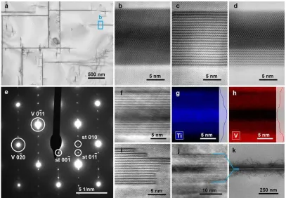
27 Fig. 1a shows a TEM bright-field (BF) image of the V-4Cr-4Ti specimen after the 28
29 annealing treatment. A significant number of plate-like precipitates, with lengths of up to a
30 31
32 few microns and only a few tens of nanometers in width, were observed in this material. The 33
34 selected area electron diffraction pattern (SADP) of Fig. 1e shows the [001] zone axis of the
35 36
V matrix with additional reflections due to the presence of one of the plate-like precipitates.
38
39 The lattice parameter of bcc V (av) is ∼3.02Å. The pattern clearly shows that the plates are 40
41
lying on the V{100} family of planes. Additionally, we have observed the presence of 43
44 diffraction spots corresponding to a superstructure with a spacing of ∼7.56Å, which 45
46
47 corresponds to ∼2.5 times the lattice parameter of the matrix. 48
49 High resolution STEM imaging of the precipitates (Fig. 1) revealed a range of atomic
50 51
52 structures within different plate-like precipitates: uniform (b), showing a superstructure 53
54 through the thickness (c), or showing a superstructure localised on one (d) or both (f) long
55 56
sides of the precipitates. The spacing determined in the high resolution STEM data for the
58
60
related background-subtracted EDS spectrum images for the Kα Ti and Kα V lines, 1
2 3 4
respectively. The EDS data show that the uniform area of the precipitate mainly contains Ti,
10
15
32
2 3
49
5 whereas both Ti and V are present in the superstructure regions. Cr has not been detected in 6
7 significant amounts inside any of the precipitates. Two additional cases are also reported in
8 9
Fig. 1: superstructure at the short edge of the precipitates (i), and surrounding a region of 11
12 uniform atomic structure (j). Interestingly, the interface between the precipitate superstructure 13
14
and the matrix acts as an effective trap for dislocations present in the matrix (k).
16
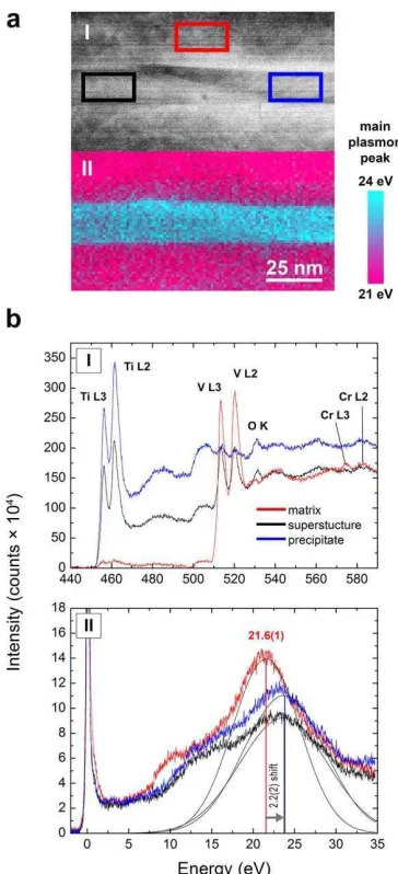
17 In order to obtain further information about the local chemical distribution and the 18
19
origin of the interfacial superstructure phase, we recorded the EELS core loss and low loss 20
21
22 spectra [19] at representative locations of the matrix, the superstructure and the uniform 23
24 structure of the precipitate (Fig. 2). The EELS core loss spectrum of the matrix is dominated
25 26
27 by the L2,3 edges of vanadium (in the region of 440-590eV). A relatively weak L2,3 edge from 28
29 the substitutional Cr atoms can also be observed close to 580eV. When profiling from the
30 31
matrix to the superstructure of the precipitate, the vanadium edge is still visible but decreases
33
34 in intensity, whereas the L2,3 edge of Ti starts to appear at characteristic peak energies of 35
36
457 and 462eV. The edge consists of L and L ‘white lines’ which originate from electron 37
38
39 transitions from the inner 2p3/2 and 2p1/2 orbitals respectively to empty 3d orbitals of Ti [20]. 40
41 Those characteristic Ti lines constitute the main feature of the EELS spectrum in the uniform
42 43
44 structure of the precipitate. Additionally, we can also observe the appearance of the K-edge 45
46 of oxygen at 530-550eV, with the most distinctive maximum located just above the edge
47 48
onset at 532eV. The lower-energy features of this peak are known to originate from 50
51 transitions between oxygen 1s and 2p σ* states that are hybridized with empty Ti 3d orbitals 52
53
[20]. The intensity of the 532eV peak increases when moving the beam from the matrix into 54
55
56 the superstructure and further into the uniform structure of the precipitate. We have not 57
58
60
suggests the absence of C or N within the precipitate. These results would point to the 1
2 3 4
precipitates consisting of a titanium oxide phase. To confirm this, we have also examined the
10
15
20
37
v
5 EELS low loss or valence spectrum below 35eV (Fig. 2c), which is dominated by plasmon 6
7 excitations. The main plasmon peak of the matrix at 21.6(1) eV can be attributed to metallic
8 9
vanadium [19]. The position of the maximum consistently shifts to a value of 23.8(1)eV 11
12 inside the precipitate. An equivalent peak shift is observed for the superstructure and the 13
14
uniform structure in the precipitate, see Fig. 2a-II. The main plasmon peak of metallic Ti
16
17 would be located at 18 eV [19], and is expected to shift to ∼20 eV when forming Ti hydrides 18
19
[21] and to 22-26 eV when Ti forms compounds with C, N or O [19, 20]. Our combined
21
22 EELS core loss and low loss spectral data are therefore consistent with a titanium oxide phase 23
24
as precipitate. 25
26
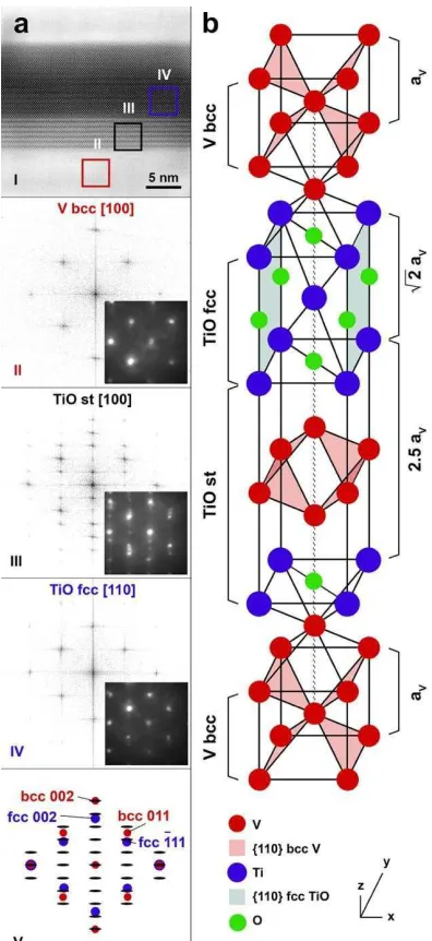
27 An HAADF image of one of the precipitates and surrounding matrix is shown in 28
29 Fig. 3a, together with its Fast Fourier Transform (FFT) and the measured electron diffraction
30 31
32 pattern taken at selected locations in the precipitate and the matrix. The diffraction pattern of 33
34 the matrix corresponds to the vanadium bcc structure acquired along the [100] zone axis. The
35 36
pattern of the uniform structure in the precipitate has been indexed based on a face-centred
38
39 cubic TiO unit cell (S.G. Fm-3m) along the [110] zone axis. This titanium mono-oxide phase 40
41
presents a NaCl-type fcc structure, with a reported value of its lattice parameter of 4.184Å. Its 42
43
44 structure can host up to approx. 15% of vacancies [22, 23], and also small amounts of C and 45
46 N, since both TiN and TiC are isostructural with TiO [24]. We have determined the lattice
47 48
49 parameter of the TiO-type precipitates in V-4Cr-4Ti as 4.28Å. This value corresponds to 50
51
, where a is the lattice parameter of V matrix. Furthermore, the TiO precipitates must 52
53
54 be related to the V matrix by the Baker–Nutting orientation relationship: [001]TiO // [001]V, 55
56 (110)
TiO // (100)V. From these results, it is then possible to construct a proposed model for the 57
58
the ‘V bcc matrix’ and the ‘TiO fcc uniform structure, see Fig. 3b. The unit cell contains one 1
2 3 4
vanadium octahedron from the V bcc structure, located between two ‘TiO layers’. The unit
60 20
25
47
52
5 cell becomes elongated along the c-axis with a lattice parameter of c = 2.5 × aV ∼ 7.56Å. The 6
7 orientation relationship of the superstructure with the V matrix is thus: [001]
st // [001]V, 8
9
10 (100)st // (100)V. 11
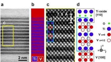
12 Fig. 4 contains a plan view representation of the proposed model along the
13 14
15 [100] direction, together with an experimental atomic resolution HAADF image of the 16
17 superstructure, and the spatial distribution of the V and Ti derived from the EELS spectra 18
19
using the L2,3 edges of both elements. The combined interpretation of the chemical and 21
22 structural data strongly supports the validity of the proposed superstructure model. The repeat 23
24
distance of ∼7.56Å corresponds to the c-axis of the simple tetragonal unit cell. The dark and
26
27 bright lines observed in the HAADF images can therefore be linked to TiO and V layers 28
29
respectively. Careful examination and comparison of the images and chemical information 30
31
32 from the EELS data also suggest that it is possible to identify individual V atoms at two 33
34 positions in the unit cell, i.e. x=0 and x=1/2 from the V octahedron in the intergrowth, and
35 36
37 also interleaving Ti atoms. 38
39 Some simple diffusion considerations can be put forward to explain how the
40 41
42 superstructure forms. When the sample is annealed at 1200°C, both Ti and O diffuse in the 43
44 V bcc matrix to form the TiO precipitates. The diffusion of O interstitials is relatively fast;
45 46
with a reported value of the activation energy of 119.6-122.5 kJ mol-1 [25-27]. Early trace 48
49 diffusion experiments in binary V-Ti alloys yielded a value for the Ti diffusion coefficient of 50
51
∼1.5 × 10⁻³ µm2/s at 1200°C [28]. If we assume a random walk approach for the diffusion of 53
54 Ti in the V matrix, we obtain a Ti diffusion length of √Dt ∼ 3.3µm. This value is in good 55
56
57 agreement with our experimental value for the average precipitate length in this material 58
species, Ti in this case, to the diffusion coefficient calculated assuming randomly oriented 1
2 3 4
jump vectors [29]. The reported value of this correlation value for Ti diffusion, assuming that
60 20
30
52
5 the Ti atoms produce only a weak perturbation of the V lattice and also a vacancy-mediated 6
7 mechanism, takes a value lower than but close to 1, i.e. f
Ti = 0.75-0.80 in the temperature 8
9
10 range of 1100-1550°C [28]. In general, the greater the freedom of movement of the vacancy, 11
12 the less important the correlation effects become, and therefore the smaller 1-f is [30]. This
13 14
15 means that the binding energy for a Ti-vacancy is relatively low, and the random walk 16
17 treatment is a suitable approach for the Ti diffusion in the V matrix. The V self-diffusion is
18 19
slower than the Ti diffusion, but the V diffusivity is affected by the rate at which Ti-vacancy 21
22 complexes break up. The V diffusion coefficient takes a value of ∼6.0 × 10⁻⁴ µm2/s at 1200°C 23
24
25 [28, 31], which yields a V diffusion length of √Dt ∼ 2.1µm. V can therefore form the 26
27 superstructure phase together with Ti along the precipitate-matrix interface during the
28 29
annealing treatment.
31
32 The local structure and chemical distribution at the precipitate interface with the 33
34
V matrix will influence the strength of the precipitates as sinks and recombination sites for 35
36
37 radiation-induced lattice defects and He atoms [32]. Helium has a relatively low solubility in 38
39
metals [33, 34], and hence the diffusion and accumulation of He at interfaces and grain 40
41
42 boundaries can potentially form bubbles. The presence of He has also been proposed to 43
44 accelerate the radiation-induced swelling, both by stabilising the void nuclei formed by
45 46
47 clustering of the vacancy defects, and by enhancing the void growth that may lead to 48
49 percolating networks [35]. The leading approach to mitigating void swelling in He-containing
50 51
materials is to delay the bubble transformation into voids by nano-structuring [36, 37].
53
54 Recent work reports the role of semi-coherent fcc-bcc heterophase interfaces in delaying 55
56
bubble growth in nano-layered composites, materials in which helium seems to accumulate at 57
58
60
could therefore be optimised to influence the effectiveness of the interface as point defect 1
2 3 4
sinks so that enhanced damage tolerance is achieved [35, 38]. In the case of the TiO(fcc)-
10
15
32
49
5 V(bcc) system, the observed atomic ordering at the interface could effectively delay the He 6
7 bubble growth, and also accommodate significant amounts of lattice defects and interstitial
8 9
atoms at the interface, so that low-temperature embrittlement is minimised or delayed. 11
12 In conclusion, our atomic-resolution STEM imaging and analysis results have 13
14
revealed the presence of an intergrowth of the TiO fcc and V bcc structures at the
16
17 precipitate/matrix interface in the V-4Ti-4Cr alloy. The O atoms are primarily concentrated 18
19
inside the nm-thick precipitates, where they seem to be homogeneously distributed, while the 20
21
22 V/Ti superstructure atomic ordering can in some cases extend through the full thickness of 23
24 the plate-like precipitates. This atomic-scale characterization of the local structure and
25 26
27 chemistry of the interface and precipitate structure will assist the mechanistic understanding 28
29 of the interaction of interstitials and radiation-induced lattice defects with the precipitate
30 31
interface, and hopefully trigger the development of novel alloy structures with enhanced
33
34 radiation tolerance.
35 36
We acknowledge the Engineering and Physical Sciences Research Council (EPRSC) 37
38
39 for providing funding for this project via the Centre for Doctoral Training in the Science and 40
41 Technology of Fusion Energy (http://www.fusion-cdt.ac.uk/), and also for providing access to
42 43
44 the SuperSTEM Laboratory, the U.K. National Facility for Aberration-Corrected STEM 45
46 (http://www.superstem.com/). S.J.H. would like to acknowledge EPSRC grant
47 48
EP/M010619/1 as well as the defence threat reduction agency grant number HDTRA1-12-1- 50
51 0013. We would also like to thank Matthew Smith for his valuable help with the FEI Titan 52
53
microscope in Manchester. 54
55
56 References
57 58
[2] T. Muroga, J.M. Chen, V.M. Chernov, R.J. Kurtz, M. Le Flem, J. Nucl. Mater. 455 (2014) 263. 1 2 3 61 15 35 46 57
4 [3] S.J. Zinkle, N.M. Ghoniem, Fusion Eng. Des. 51–52 (2000) 55.
5 [4] B.A. Loomis, D.L. Smith, F.A. Garner, J. Nucl. Mater. 179-181 (1991) 771. 6
7 [5] H.M. Chung, B.A Loomis, D.L. Smith, J. Nucl. Mater. 212-215 (1994) 804.
8
9 [6] D.L. Smith, H.M. Chung, H. Matsui, A.F. Rowcliffe, Fusion Eng. Des. 41 (1998) 7. 10
11 [7] D.L. Smith, M.C. Billone, K. Natesan, Int. J. Refract. Met. Hard Mat. 18 (2000) 213. 12
13 [8] N. Baluc, Phys. Scr. T138 (2009) 014004. 14
[9] T. Nagasaka, N.J. Heo, T. Muroga, M. Imamura, Fusion Eng. Des. 61/62 (2002) 757. 16
[10] D.L. Smith, H.M. Chung, B.A. Loomis, H.C. Tsai, J. Nucl. Mater. 233-237 (1996) 356. 17
18 [11] D.R. Diercks, B.A. Loomis. J. Nucl. Mater. 141-143 (1986) 1117. 19
20 [12] B. Zhu, S. Yang, M. Zhang, J. Ding, Y. Long, F. Wan, Mater. Charact. 111 (2016) 60. 21
22 [13] M. Hatakeyama, T. Muroga, S. Tamura, I. Yamagata. J. Nucl. Mater. 417 (2011) 303. 23
24 [14] J.M. Chen, T. Muroga, T. Nagasaka, Y. Xu, C. Li, S.Y. Qiu, Y. Chen, J. Nucl. Mater. 25
334 (2004) 159. 26
27 [15] T. Muroga, T. Nagasaka, P.F. Zheng, J.M. Chen, Adv. Sci. Tech. 73 (2010) 22.
28
29 [16] M.S. Staltsov, I.I. Chernov, B.A. Kalin, K.Z. Oo, A.A. Polyansky, O.S. Staltsova, et al., 30
31 J. Nucl. Mater. 461 (2015) 56.
32
33 [17] A. van Veen, A.V. Fedorov, A.I. Ryazanov, J. Nucl. Mater. 258-263 (1998) 1400.
34 [18] P. Hartel, H. Rose, C. Dignes, Ultramicroscopy 63 (1996) 93. 36
[19] R.F. Egerton, Electron Energy-Loss Spectroscopy in the Electron Microscope. 37
38 Springer, 2011.
39
40 [20] E. Stoyanov, F. Langenhorst, G. Steinle-Neumann, Am. Miner. 92 (2007) 577. 41
42 [21] N. G. Alexandropoulos, G. Bambakidis, T. Sparrow, B. Williams, J. Phys. F: Met. Phys. 43
44 16 (1986) L24S.
45
[22] M. D. Banus, T. B. Reed, A. J. Strauss, Phys. Rev. B 5 (1972) 2775. 47
[23] A. I. Gusev, J. Exp. Theor. Phys. 117 (2013) 293. 48
49 [24] H.O. Pierson, Handbook of Refractory Carbides and Nitrides: Properties,
50
51 Characteristics, Processing, and Applications. Noyes Publications, 1996. 52
53 [25] R.W. Powers, M.V. Doyle. Acta Metall. 6 (1958) 643. 54
55 [26] R.W. Powers, M.V. Doyle. J. Appl. Phys. 30 (1959) 514. 56
[27] H. Nakajima, S. Nagata, H. Matsui, S. Yamaguchi. Phil. Mag. 67 (1993) 557. 58
[28] J.F. Murdock, C.J. McHargue. Acta Metall. 16 (1968) 493. 59
[30] R.E. Howard, A.B. Lidiard. Rep. Prog. Phys. 27 (1964) 161.
[31] Y. Liu, Y. Ge, T. Pan, L. Zhang. J. Alloys Comp. 470 (2009) 176. 1
2 3
15
4 [32] M.J. Demkowicz, P. Bellon, B.D. Wirth. MRS Bull. 35 (2010) 992.
5 [33] J. Laakmann, P. Jung, W. Uelhoff. Acta Metall. 35 (1987) 2063. 6
7 [34] F. Gao, H. Heinisch, R.J. Kurtz. J. Nucl. Mater. 351 (2006) 133.
8
9 [35] M.J. Demkowicz, A. Misra, A. Caro. Curr. Opin. Solid State Mat. Sci. 16 (2012) 101. 10
11 [36] P.D. Edmonson, C.M. Parish, Y. Zhang, A. Hallén, M.K. Miller. Scripta Mater. 65 12
13 (2011) 731.
14
[37] Y. Wu, J. Ciston, S. Kräemer, N. Bailey, G.R. Odette, P. Hosemann. Acta Mater. 111 16
(2016) 108. 17
18 [38] E.G. Fu, A. Misra, H. Wang, L. Shao, X. Zhang. J. Nucl. Mater. 407 (2010) 178.
Figure 1
Fig. 1. (a) TEM BF image of the V-4Ti-4Cr alloy microstructure with the plate-like precipitates viewed along the [001] zone axis. (b-d) HAADF images showing precipitates with (b) a uniform atomic structure, (c) a superstructure and (d) both uniform and superstructure regions. (e) The corresponding SADP of (a) with the V matrix and simple tetragonal (st) superstructure reflections highlighted. Precipitates with different proportions of superstructure fringes: (f) with superstructure at both long sides with the relative EDS spectrum images for (g) Kα Ti and (h) Kα V, (i) with
Figure 2
Figure 3
Fig. 3. (a-I) HAADF image showing a TiO precipitate with regions of both superstructure and uniform
atomic structure. (a-II) to (a-IV) show the FFT and diffraction patterns taken from the regions outlined by the coloured squares in (a-I). (a-V) is the superimposed pattern containing the position of the spots in (a-II) to (a-IV). (b) Proposed crystal structure model relating the V bcc matrix structure to the TiO fcc (NaCl-type) precipitate and the superstructure (simple tetragonal ‘st’ of TiO with V). The lattice parameter of both TiO structures is indicated taking the lattice parameter (aV) of the V
Figure 4
Fig. 4. (a) HAADF image of the superstructure within the precipitate, (b) composite image constructed from the EELS maps generated by integrating the L2,3 edge intensity of V (red) and
Supplementary Material